Paeds · gastroenterology-hepatology-and-nutrition
Chronic liver disease, cirrhosis and portal hypertension
Also known as Paediatric cirrhosis · Portal hypertension in children · Oesophageal varices in children · Hepatopulmonary syndrome · Paediatric chronic liver disease
Fellowship guide to chronic liver disease, cirrhosis and portal hypertension in children: the prehepatic, intrahepatic and posthepatic classification, biliary atresia as the paradigmatic paediatric cause, the pathophysiology of the hyperdynamic splanchnic circulation, variceal bleeding prophylaxis and acute management, ascites and spontaneous bacterial peritonitis with the 250 PMN threshold, hepatopulmonary syndrome and portopulmonary hypertension, and the PELD score for transplant listing.
On this page & tools
Your progress
Saved locally on this device.
Practise this topic
Target exams
Red flags
Life stages
Care settings
Clinical exam formats
Board mappings
Overview & Definition
A child who presents with a massively enlarged spleen, unexplained thrombocytopenia, or an episode of haematemesis may be revealing chronic liver disease that has progressed to portal hypertension long before jaundice or other signs of hepatic failure appear. This is the central clinical challenge: portal hypertension can declare itself through a life-threatening complication while the underlying liver disease has been smouldering silently for years. Understanding the cascade from hepatic fibrosis to the complications of variceal bleeding, ascites, spontaneous bacterial peritonitis, and hepatopulmonary syndrome is essential for every paediatric trainee. [1]
Cirrhosis is the advanced, generally irreversible stage of chronic liver injury in which fibrous septa and regenerative nodules distort the hepatic architecture and disrupt blood flow. Portal hypertension is the pathological rise in pressure within the portal venous system that results from this distortion, and it is the engine driving nearly all the life-threatening complications of chronic liver disease. The pressure gradient that defines it is the hepatic venous pressure gradient, the difference between the wedged hepatic venous pressure and the free hepatic venous pressure. A gradient of 5 mmHg or more defines portal hypertension, while a gradient of 10 mmHg or more defines clinically significant portal hypertension, which is the threshold at which varices form and the risk of bleeding and ascites becomes real. [1]
In children, the causes of cirrhosis and portal hypertension differ fundamentally from those in adults. Biliary atresia is the single most common cause, followed by inherited metabolic diseases, autoimmune hepatitis, cystic fibrosis liver disease, and chronic viral hepatitis. Portal vein thrombosis with cavernous transformation is a uniquely important prehepatic cause in children because it produces severe portal hypertension with well-preserved synthetic liver function. Recognising this distinction is critical because it fundamentally changes the management approach and the prognosis. [9]
Classification
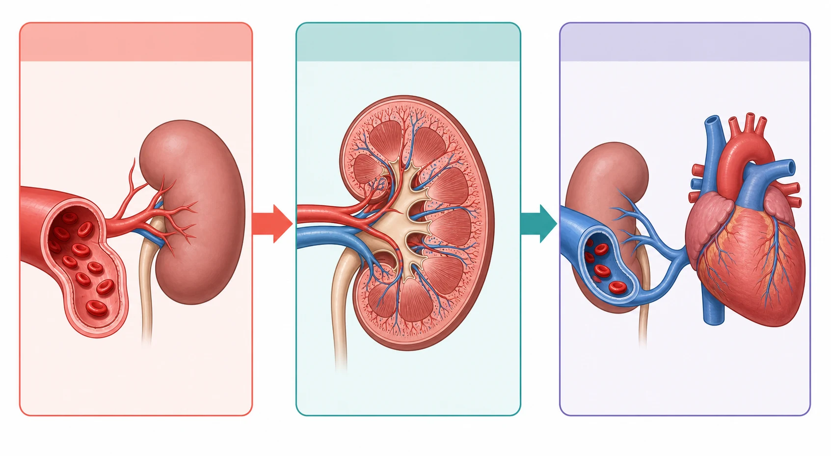
Portal hypertension is classified by the anatomical site of the obstruction to portal blood flow. This classification is not merely an academic exercise because the site determines the synthetic function, the management, and the prognosis. A child with prehepatic portal hypertension may have catastrophic variceal bleeding but entirely normal liver function, while a child with advanced intrahepatic cirrhosis may bleed less but face hepatic failure. [1]
[1]Within the intrahepatic category, the obstruction can be further localised. Presinusoidal causes include congenital hepatic fibrosis and schistosomiasis, where the portal tracts are affected but the sinusoids and hepatocytes remain relatively intact. Sinusoidal causes are the classic cirrhoses, where fibrosis and nodular regeneration distort the hepatic sinusoids themselves. Postsinusoidal causes include veno-occlusive disease, now called sinusoidal obstruction syndrome, where the hepatic venules are occluded. Biliary atresia is the archetype of the paediatric intrahepatic cause because it produces both cholestasis and progressive fibrosis that culminates in cirrhosis and portal hypertension in a substantial proportion of children surviving with their native liver after Kasai portoenterostomy. [9]
Gastric varices also warrant classification because they carry a different bleeding risk and require different management. The Sarin classification divides gastric varices into gastro-oesophageal varices, which extend along the lesser or greater curvature from oesophageal varices, and isolated gastric varices, which occur in the fundus or elsewhere without oesophageal varices. Isolated gastric fundal varices carry a particularly high bleeding risk and are treated with cyanoacrylate injection rather than band ligation. [11]
Epidemiology & Risk Factors
Biliary atresia remains the paradigmatic paediatric liver disease and the single most common indication for liver transplantation in children. It occurs in approximately 1 in 8,000 to 1 in 18,000 live births worldwide, and despite the Kasai portoenterostomy performed in the first weeks of life, a substantial proportion of affected children develop progressive fibrosis leading to cirrhosis and portal hypertension. The French national series of biliary atresia from 1986 to 2009 demonstrated improving overall survival but persistent challenges, with portal hypertension remaining the dominant long-term complication in children surviving with their native liver. [9]
Beyond biliary atresia, the major paediatric causes of chronic liver disease and portal hypertension include alpha-1-antitrypsin deficiency, Wilson disease, tyrosinaemia type 1, glycogen storage diseases, progressive familial intrahepatic cholestasis, autoimmune hepatitis, cystic fibrosis liver disease, and chronic viral hepatitis. Each of these diseases damages the liver through a different mechanism but converges on the common pathway of fibrosis, architectural distortion, and portal hypertension. [9]
Portal vein thrombosis with cavernous transformation deserves special mention as the most common prehepatic cause in children. It is often idiopathic but is associated with a history of umbilical vein catheterisation, neonatal sepsis, omphalitis, or inherited thrombophilia. Children with this condition have normal synthetic liver function but severe portal hypertension, and they may present with dramatic variceal bleeding at a young age. [1]
Pathophysiology
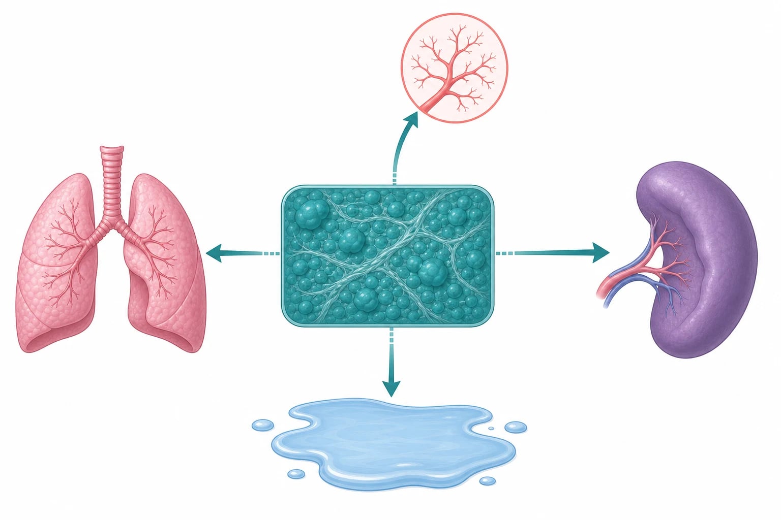
Portal hypertension is driven by two mechanisms that work together to raise portal pressure. The first is increased resistance to portal blood flow through the liver. In cirrhosis, this resistance has a mechanical component, the physical obstruction from fibrous septa and regenerative nodules, and a dynamic component, increased vascular tone from reduced nitric oxide bioavailability within the hepatic sinusoids. The dynamic component is important because it is the target of pharmacological therapy with non-selective beta-blockers. [1]
The second mechanism is the hyperdynamic splanchnic circulation. As portal pressure rises, the body responds with splanchnic vasodilation driven by increased local nitric oxide and other vasodilators. This vasodilation increases blood flow into the portal system, which paradoxically worsens portal hypertension by increasing portal inflow against the fixed intrahepatic resistance. The vasodilation also reduces effective arterial blood volume, activating the renin-angiotensin-aldosterone system and causing sodium and water retention. This is the mechanism behind ascites formation: the combination of sinusoidal hypertension driving fluid into the peritoneal space and the activated neurohormonal cascade retaining sodium and water. [1]
The body attempts to decompress the portal system by forming portosystemic collateral vessels. The most clinically important of these collaterals are the oesophageal and gastric varices, which develop from the dilatation of the left gastric and short gastric veins. Other collaterals include retroperitoneal veins, haemorrhoidal veins, and the recanalised umbilical vein producing the caput medusae. The problem is that these collaterals are fragile, particularly the submucosal oesophageal varices, which are poorly supported by surrounding tissue and can rupture under the pressure of the portal system, producing catastrophic bleeding. [1]
In hepatopulmonary syndrome, the same nitric oxide-mediated vasodilation that occurs in the splanchnic circulation also affects the pulmonary vasculature. Precapillary and capillary pulmonary vessels dilate, producing intrapulmonary shunting and ventilation-perfusion mismatch that cause hypoxaemia. The characteristic positional component arises because the basal lung segments are the most affected, so standing worsens the shunt and the hypoxaemia, producing platypnoea and orthodeoxia. [7]
Clinical Presentation
The presentation of portal hypertension in children ranges from an incidental finding of splenomegaly on a routine examination to a life-threatening variceal bleed. The first and most common sign is splenomegaly, often detected incidentally by a parent who feels a mass in the left upper quadrant, or by a clinician performing a routine abdominal examination. Splenomegaly is frequently accompanied by hypersplenism, with thrombocytopenia and leucopenia resulting from splenic sequestration. A platelet count under 150,000 per cubic millimetre in a child with liver disease is a strong clue to clinically significant portal hypertension. [1]
Acute variceal bleeding presents as sudden, painless, and often massive haematemesis with or without melaena. This is the most dramatic and feared presentation of portal hypertension. The bleed may stop spontaneously but can recur, and the child may present in hypovolaemic shock. It is essential to recognise that not all upper gastrointestinal bleeding in a child with liver disease is from varices; peptic ulcer disease, portal hypertensive gastropathy, and Mallory-Weiss tears are also possible, but varices must be assumed until proven otherwise. [1]
Ascites presents as progressive abdominal distension, which the parents may describe as weight gain or a swollen belly. In severe cases, the child may develop an umbilical hernia, scrotal swelling, or respiratory embarrassment from the diaphragmatic splinting. The presence of ascites in a child with chronic liver disease signals decompensated cirrhosis and warrants urgent investigation for spontaneous bacterial peritonitis, particularly if there is fever, abdominal pain, or altered mental status. [6]
Hepatopulmonary syndrome presents with dyspnoea that is characteristically worse on standing (platypnoea) and improves on lying down. Digital clubbing and cyanosis are common physical findings. A child with chronic liver disease who develops unexplained hypoxaemia, particularly with a positional component, should be evaluated for hepatopulmonary syndrome. Portopulmonary hypertension, in contrast, presents with dyspnoea, fatigue, chest pain, and syncope from pulmonary arterial hypertension, and carries a very different prognosis and management. [7]
Differential Diagnosis
The differential for splenomegaly in a child is broad, and portal hypertension is the most common cause in the child with known liver disease. However, the general paediatric differential includes infection (Epstein-Barr virus, cytomegalovirus, malaria), haematological malignancy (leukaemia, lymphoma), storage disorders (Gaucher disease, Niemann-Pick disease), and haemolytic anaemia (hereditary spherocytosis, sickle cell disease). The key distinguishing features are the presence of other signs of liver disease and the pattern of blood count abnormalities, with the disproportionate thrombocytopenia of hypersplenism pointing toward portal hypertension. [1]
The differential for upper gastrointestinal bleeding in a child includes oesophageal and gastric varices, portal hypertensive gastropathy, peptic ulcer disease, gastritis (including Helicobacter pylori), Mallory-Weiss tear, oesophagitis, Dieulafoy lesion, and, in infants, a bleeding diathesis or swallowed maternal blood. The clinical context is critical: massive, painless haematemesis in a child with known liver disease, splenomegaly, or thrombocytopenia is variceal bleeding until proven otherwise. [1]
The differential for ascites in a child includes cirrhotic ascites (the most common in the hepatology setting), nephrotic syndrome, congestive heart failure, peritoneal infection (tuberculosis), malignancy, and protein-losing enteropathy. The serum-ascites albumin gradient, calculated by subtracting the ascitic fluid albumin from the serum albumin, helps distinguish cirrhotic ascites (gradient of 11 g per litre or more) from other causes. [6]
[6]Clinical & Bedside Assessment
The focused examination of a child with suspected chronic liver disease and portal hypertension must look for both the stigmata of chronic liver disease and the specific signs of portal hypertension. The hands may reveal palmar erythema and digital clubbing, the latter suggesting hepatopulmonary syndrome in this context. The skin may show spider naevi, typically in the distribution of the superior vena cava, and easy bruising from coagulopathy. The face may show parotid enlargement and facial wasting. [1]
Abdominal examination is the cornerstone. The clinician should palpate for splenomegaly, the single most important physical sign of portal hypertension in children, and for hepatomegaly or a shrunken, hard, nodular liver edge suggesting cirrhosis. Ascites is assessed by shifting dullness and a fluid thrill. The presence of a caput medusae, dilated periumbilical veins from a recanalised umbilical vein, is pathognomonic of portal hypertension. Testicular enlargement from an inguinal hernia or hydrocele may accompany tense ascites. [1]
Neurological examination should assess for hepatic encephalopathy, which in children may present subtly as declining school performance, irritability, or sleep disturbance before progressing to drowsiness, asterixis (the flapping tremor), and coma. The grading of hepatic encephalopathy in children follows a similar framework to adults, from grade 0 (subclinical, detectable only on neuropsychometric testing) through grade 4 (coma). [1]
Respiratory examination should specifically look for the signs of hepatopulmonary syndrome: digital clubbing, central or peripheral cyanosis, and the demonstration of platypnoea (dyspnoea on standing) and orthodeoxia (a fall in oxygen saturation on standing). These positional signs, though not universal, are highly characteristic when present. [7]
Investigations
The investigation of a child with chronic liver disease and portal hypertension serves three purposes: to define the underlying cause, to assess the severity of portal hypertension, and to screen for its complications. Blood tests include a full blood count (revealing the thrombocytopenia of hypersplenism), liver function tests (with a low albumin and prolonged INR indicating impaired synthetic function), and a comprehensive metabolic and infectious workup to identify the aetiology. [1]
Upper gastrointestinal endoscopy is the gold standard for diagnosing and characterising oesophageal and gastric varices. The size, location, and presence of red wale signs (longitudinal dilated venules on the varix surface) or cherry-red spots guide the decision about primary prophylaxis. Endoscopy also identifies portal hypertensive gastropathy, which appears as a mosaic-like or snakeskin mucosal pattern in the stomach and may contribute to chronic anaemia. [1]
Abdominal ultrasound with Doppler is the first-line imaging modality. It assesses liver size and echotexture, portal vein patency and flow direction (hepatopetal versus hepatofugal), splenomegaly, ascites, and the presence of collaterals. In portal vein thrombosis, the portal vein is replaced by a tangle of collateral vessels known as cavernous transformation or cavernoma. Elastography may be added to assess liver stiffness as a non-invasive marker of fibrosis severity. [1]
Diagnostic paracentesis is mandatory in any child with cirrhotic ascites who develops fever, abdominal pain, altered mental status, or unexplained clinical deterioration. The ascitic fluid is sent for cell count, culture, albumin, and total protein. The diagnostic criterion for spontaneous bacterial peritonitis is a polymorphonuclear neutrophil count of 250 or more per cubic millimetre. The serum-ascites albumin gradient, calculated by subtracting ascitic albumin from serum albumin, distinguishes cirrhotic from non-cirrhotic ascites. [6]
The Paediatric End-Stage Liver Disease (PELD) score is a validated scoring system that predicts three-month mortality in children with chronic liver disease and is used to prioritise liver transplant allocation. It incorporates total bilirubin, INR, serum albumin, age at listing (with younger age carrying worse prognosis), and growth failure (height and weight deficits). The score transformed paediatric liver allocation toward an evidence-based system that prioritises the sickest children first, replacing the previous system that was based on waiting time. [8]
Management — Resuscitation
Acute variceal bleeding is managed with a systematic approach that begins with airway, breathing, and circulation. The airway must be protected, as massive haematemesis can cause aspiration, particularly in an encephalopathic child. Two large-bore intravenous cannulae are established, and fluid resuscitation begins with crystalloid followed by blood transfusion. The transfusion target is a haemoglobin of approximately 7 to 8 g per decilitre, deliberately avoiding overtransfusion because excessive blood volume raises portal pressure and may worsen or precipitate further bleeding. [1]
Vasoactive therapy should be started as early as possible, ideally at the time of presentation. Octreotide is the most commonly used agent in children, given as an initial bolus of 1 to 2 micrograms per kg followed by a continuous infusion of 1 to 5 micrograms per kg per hour. Terlipressin, a vasopressin analogue, is an alternative supported by a systematic review and meta-analysis in adults showing a survival benefit for acute variceal bleeding, and is used in some paediatric centres at a dose of 0.04 to 0.08 mg per kg every 4 to 6 hours. These vasoactive drugs reduce splanchnic blood flow and portal pressure, helping to control active bleeding and reduce early rebleeding. [12]
Octreotide for acute variceal bleeding
Dose
1 to 2 micrograms per kg bolus, then 1 to 5 micrograms per kg per hour infusion
Prophylactic intravenous antibiotics are mandatory in any child with cirrhosis and acute variceal bleeding. A third-generation cephalosporin such as ceftriaxone at 50 to 100 mg per kg per day should be given for 5 to 7 days. This practice, established in adult guidelines and extrapolated to children, reduces bacterial infections including spontaneous bacterial peritonitis and improves survival. [6]
Emergency upper gastrointestinal endoscopy is performed once the child is haemodynamically stabilised, ideally within 12 to 24 hours. Endoscopic variceal band ligation is the first-line endoscopic therapy for oesophageal variceal bleeding, as it mechanically ligates the bleeding varix. For gastric variceal bleeding, endoscopic injection of N-butyl-2-cyanoacrylate tissue glue is preferred over band ligation. If bleeding cannot be controlled endoscopically, a balloon tamponade tube (Sengstaken-Blakemore or Minnesota tube) can provide temporary haemostasis as a bridge to definitive therapy, and a transjugular intrahepatic portosystemic shunt (TIPS) or surgical shunt may be required for refractory bleeding. [1]
Management — Definitive & Stepwise
The definitive management of portal hypertension in children has four pillars: primary prophylaxis to prevent the first variceal bleed, secondary prophylaxis to prevent rebleeding, management of ascites and spontaneous bacterial peritonitis, and liver transplantation as the ultimate treatment for decompensated cirrhosis and hepatopulmonary syndrome. [1]
Primary prophylaxis is offered to children with high-risk varices, defined as large varices or varices with red wale signs. The two options are non-selective beta-blockers and endoscopic variceal ligation. Propranolol is the most commonly used beta-blocker, at a starting dose of 1 mg per kg per day in divided doses, titrated to reduce the resting heart rate by approximately 25 per cent without producing symptomatic bradycardia or hypotension. Endoscopic variceal ligation is performed under general anaesthesia, with repeat sessions every 2 to 4 weeks until variceal eradication. Crucially, the Cochrane reviews by Cifuentes and Gana found insufficient evidence from randomised trials to determine which is superior in children, and current paediatric practice is largely extrapolated from adult data and guided by individual patient factors. [3]
[3]Secondary prophylaxis after a first variceal bleed combines non-selective beta-blockers with serial endoscopic variceal ligation, as this combination is more effective than either alone. The goal is complete variceal eradication, typically requiring 3 to 6 endoscopy sessions, with ongoing beta-blocker therapy to reduce portal pressure and prevent the development of new varices. [4]
Ascites is managed with a stepwise approach. The first step is dietary sodium restriction, typically 1 to 2 mmol per kg per day, combined with fluid restriction only if hyponatraemia develops. The first-line diuretic is spironolactone at 1 to 3 mg per kg per day, with furosemide at 0.5 to 1 mg per kg per day added if needed to maintain a balanced potassium and a daily weight loss of no more than 0.5 kg. For tense or refractory ascites, large-volume paracentesis is performed with concomitant albumin replacement at 6 to 8 g per litre of fluid removed to prevent circulatory dysfunction. Refractory ascites is an ominous sign indicating decompensated cirrhosis and warrants assessment for liver transplantation. [6]
Spontaneous bacterial peritonitis is treated empirically with a third-generation cephalosporin (cefotaxime or ceftriaxone at 50 to 100 mg per kg per day) for 5 to 7 days, guided by culture and sensitivity results when available. Albumin is given at 1.5 g per kg on day 1 and 1 g per kg on day 3 to reduce the risk of hepatorenal syndrome. After a first episode of SBP, secondary prophylaxis with oral norfloxacin or trimethoprim-sulfamethoxazole is continued indefinitely until liver transplantation. SBP carries a one-year mortality risk of 50 to 70 per cent and is an indication for transplant assessment. [6]
Acute variceal bleeding management sequence
Airway protection, two large-bore IV cannulae, crystalloid resuscitation
Transfuse to haemoglobin 7 to 8 g per decilitre; avoid overtransfusion
Start octreotide 1 to 2 micrograms per kg bolus then 1 to 5 micrograms per kg per hour
Start prophylactic ceftriaxone 50 to 100 mg per kg per day
Urgent endoscopy within 12 to 24 hours for band ligation
Balloon tamponade or TIPS for refractory bleeding
Liver transplantation is the definitive treatment for decompensated cirrhosis. Listing is based on the PELD score for children under 12 years and the MELD score for those 12 years and older. Hepatopulmonary syndrome with significant hypoxaemia is an independent indication for transplantation, and in some allocation systems a PaO2 under 60 mmHg confers additional priority because it predicts worse survival without transplant. Portopulmonary hypertension, in contrast, is a relative contraindication to transplantation unless the pulmonary pressure is controlled, as it carries a high perioperative mortality. [7]
Specific Subtypes & Scenarios
Biliary atresia is the paradigmatic paediatric cause of cirrhosis and portal hypertension. Despite the Kasai portoenterostomy performed in the first weeks of life, many children develop progressive fibrosis, and portal hypertension remains the dominant long-term complication in those surviving with their native liver. These children require systematic surveillance with serial endoscopy to detect varices before they bleed, and timely initiation of primary prophylaxis. The French national series demonstrated improving overall survival with advances in surgical technique and post-operative care, but the burden of portal hypertension persists. [9]
Extrahepatic portal vein obstruction with cavernous transformation represents a distinct clinical scenario. These children have severe portal hypertension with variceal bleeding but entirely normal synthetic liver function. The management challenge is controlling the bleeding risk without liver transplantation, as the liver itself is healthy. Primary and secondary prophylaxis with beta-blockers and endoscopic therapy is the mainstay, and in selected cases a surgical portosystemic shunt, such as the mesenterico-left portal (Rex) bypass, can restore physiological portal flow by connecting the superior mesenteric vein to the left portal vein through a jugular vein graft, effectively curing the portal hypertension. [1]
Hepatopulmonary syndrome is defined by the triad of chronic liver disease, arterial oxygen deoxygenation with a PaO2 under 80 mmHg or an alveolar-arterial gradient over 15 mmHg, and intrapulmonary vascular dilatation demonstrated by contrast echocardiography (agitated saline appearing in the left atrium within 3 to 6 cardiac cycles) or a positive technetium-99m macroaggregated albumin brain uptake scan. The severity is graded by the degree of hypoxaemia, with a PaO2 under 60 mmHg defining severe disease that confers additional transplant priority. The key teaching point is that hepatopulmonary syndrome can resolve completely after liver transplantation, making it one of the few indications where transplantation reverses the pulmonary complication. [7]
Portopulmonary hypertension is a distinct entity that must not be confused with hepatopulmonary syndrome. It is pulmonary arterial hypertension associated with portal hypertension, defined by a mean pulmonary artery pressure of 20 mmHg or more with a pulmonary vascular resistance above 240 dynes per second per centimetre to the fifth power. Unlike hepatopulmonary syndrome, portopulmonary hypertension may worsen after transplantation and is a major risk factor for perioperative death. Management involves pulmonary vasodilator therapy and careful transplant evaluation. [7]
Complications & Pitfalls
The complications of portal hypertension span the vascular, infectious, renal, pulmonary, and neurological systems. Variceal haemorrhage remains the most dramatic and life-threatening complication, with mortality from a first bleed that is lower in children than in adults but still significant. Ascites and spontaneous bacterial peritonitis are the infectious complications that signal decompensated cirrhosis. Hepatorenal syndrome, a form of functional kidney injury from intense renal vasoconstriction in the setting of splanchnic vasodilation, is a feared complication with a poor prognosis. Hepatic encephalopathy, from the accumulation of neurotoxins normally cleared by the liver, ranges from subtle cognitive changes to coma. [1]
Several classic pitfalls deserve emphasis. The first is overtransfusion during acute variceal bleeding: transfusing to a normal haemoglobin raises portal pressure and may worsen bleeding. The target is a haemoglobin of 7 to 8 g per decilitre, not normalisation. The second is failing to perform a diagnostic paracentesis in a child with cirrhotic ascites who has fever, abdominal pain, or altered mental status. These symptoms may be subtle in children, and SBP is easily missed without paracentesis. The third is confusing hepatopulmonary syndrome with portopulmonary hypertension; the former improves with transplantation, the latter may worsen. [7]
Endoscopic therapy has its own complications. Band ligation can produce oesophageal ulceration, which may bleed, and strictures with repeated sessions. Sclerotherapy, largely replaced by band ligation, carries risks of mediastinitis, stricture, and bacterial peritonitis. General anaesthesia for endoscopy carries additional risks in a child with decompensated cirrhosis and coagulopathy, requiring careful anaesthetic planning. [5]
Prognosis & Disposition
The prognosis of paediatric chronic liver disease depends on three factors: the underlying cause, the degree of synthetic dysfunction, and the timeliness of liver transplantation. Children with biliary atresia who achieve successful Kasai portoenterostomy and avoid transplantation may live for years with their native liver but require ongoing surveillance for portal hypertension and its complications. The French national series showed improving overall and native liver survival over the 23-year study period, reflecting advances in surgical technique, nutritional support, and complication management. [9]
Children with extrahepatic portal vein obstruction have a generally favourable prognosis, as their synthetic function is preserved. The risk of variceal bleeding is highest in the first decade and tends to decrease with age, as the portosystemic collaterals mature and stabilise. Many of these children outgrow their bleeding risk, though they require ongoing monitoring for portal biliopathy and hypersplenism. [1]
The PELD score predicts three-month mortality and is used to prioritise transplant listing. A higher score reflects worse synthetic function, younger age, and growth failure, and correlates with a higher risk of death without transplantation. The introduction of the PELD score in 2002 transformed paediatric liver allocation toward an evidence-based system that prioritises the sickest children first, replacing a system based on waiting time that disadvantaged those who deteriorated rapidly. [8]
All children with decompensated cirrhosis are managed by a specialist paediatric hepatology service with multidisciplinary input from dietitians, specialist nurses, psychologists, social workers, and transplant surgeons. Transition to adult care is a structured process that begins in early adolescence, building the young person's knowledge of their disease, self-management skills, and independence so that continuity of care is preserved into adulthood. [9]
Special Populations
Children with biliary atresia surviving with their native liver are the largest single group requiring long-term portal hypertension management in paediatric hepatology. These children require systematic surveillance endoscopy, typically starting from the age of diagnosis or when splenomegaly or thrombocytopenia develop, with primary prophylaxis initiated when high-risk varices are found. Nutritional support is critical, as cholestasis impairs fat absorption and fat-soluble vitamin status, and growth failure itself worsens the PELD score and transplant priority. [9]
Adolescents with chronic liver disease approaching transition to adult care face unique challenges. Contraception counselling is essential for adolescent girls, as pregnancy in decompensated cirrhosis carries significant risks. Alcohol avoidance is critical for all adolescents with chronic liver disease, as even small amounts can accelerate fibrosis. Vaccination against hepatitis A and B, pneumococcus, and influenza is recommended, and the young person should be empowered to understand their condition, recognise the signs of decompensation, and know when to seek emergency care. [1]
Children with cystic fibrosis liver disease represent a special population in which significant portal hypertension can coexist with relatively preserved synthetic function. These children may present with variceal bleeding or splenomegaly while their liver function tests remain near-normal, making the portal hypertension easy to underestimate. Management focuses on endoscopic surveillance and prophylaxis, with transplant reserved for those with progressive synthetic dysfunction or refractory complications. [1]
Children with portal vein thrombosis and cavernous transformation are candidates for surgical shunting, particularly the Rex shunt (mesenterico-left portal bypass), which restores physiological portal venous flow to the liver. This procedure is unique among portosystemic shunts because it relieves portal hypertension without creating a shunt that bypasses the liver, preserving hepatic function and eliminating the risk of encephalopathy. Careful patient selection and specialised surgical expertise are required. [1]
[9]Evidence, Guidelines & Regional Differences
The Baveno VII consensus, published in 2022, provides the current international framework for the management of portal hypertension. While it is primarily adult-focused, its principles are widely applied to children, including the concept of clinically significant portal hypertension defined by a hepatic venous pressure gradient of 10 mmHg or more, the role of non-selective beta-blockers and endoscopic therapy in primary prophylaxis, and the importance of rescue therapy in acute variceal bleeding. The Baveno process has progressively refined the risk stratification and management recommendations through seven iterations since 1990. [1]
The AASLD practice guidance, published in 2024, provides the most recent North American recommendations on risk stratification and management of portal hypertension and varices in cirrhosis. It reinforces the Baveno principles and adds detail on non-invasive assessment with elastography and the role of TIPS in specific scenarios. While adult-focused, it informs paediatric practice through extrapolation. [2]
The Cochrane reviews by Cifuentes and Gana are the definitive paediatric evidence base for primary prophylaxis of variceal bleeding. They systematically reviewed beta-blockers versus placebo, band ligation versus no intervention, and band ligation versus sclerotherapy, finding in each case that the evidence from randomised trials in children is insufficient to recommend one strategy over another with confidence. This is the honest answer to a common exam question: the choice between beta-blockers and endoscopic band ligation in children is currently guided by extrapolation from adult data, individual patient factors, and centre preference, not by robust paediatric evidence. [3]
[1]The PELD score, introduced into United States liver allocation in 2002, transformed paediatric transplant prioritisation. It was developed and validated using data from thousands of children on the transplant waiting list and demonstrated that bilirubin, INR, albumin, age, and growth failure independently predicted three-month mortality. Its introduction moved paediatric liver allocation from a waiting-time-based to a severity-based system, prioritising the sickest children first and reducing waiting-list mortality. [10]
Exam Pearls
H-P-S for hepatopulmonary syndrome triad
SPLEEN for the complications of portal hypertension
References
- [1]de Franchis R, Bosch J, Garcia-Tsao G, Reiberger T, et al Baveno VII - Renewing consensus in portal hypertension. J Hepatol, 2022.PMID 35120736
- [2]Kaplan DE, Ripoll C, Thiele M, Fortune BE, et al AASLD Practice Guidance on risk stratification and management of portal hypertension and varices in cirrhosis. Hepatology, 2024.PMID 37870298
- [3]Cifuentes LI, Gattini D, Torres-Robles R, Gana JC Beta-blockers versus placebo or no intervention for primary prophylaxis of oesophageal variceal bleeding in children with chronic liver disease or portal vein thrombosis. Cochrane Database Syst Rev, 2021.PMID 33498095
- [4]Cifuentes LI, Gattini D, Torres-Robles R, Gana JC Band ligation versus sham or no intervention for primary prophylaxis of oesophageal variceal bleeding in children and adolescents with chronic liver disease or portal vein thrombosis. Cochrane Database Syst Rev, 2021.PMID 33522602
- [5]Gana JC, Cifuentes LI, Gattini D, Torres-Robles R Band ligation versus sclerotherapy for primary prophylaxis of oesophageal variceal bleeding in children with chronic liver disease or portal vein thrombosis. Cochrane Database Syst Rev, 2020.PMID 33164205
- [6]Runyon BA, AASLD Introduction to the revised American Association for the Study of Liver Diseases Practice Guideline management of adult patients with ascites due to cirrhosis 2012. Hepatology, 2013.PMID 23463403
- [7]Krowka MJ, Fallon MB, Kawut SM, Fuhrmann V, et al International Liver Transplant Society Practice Guidelines: Diagnosis and Management of Hepatopulmonary Syndrome and Portopulmonary Hypertension. Transplantation, 2016.PMID 27326810
- [8]McDiarmid SV, Merion RM, Dykstra DM, Harper AM Use of a pediatric end-stage liver disease score for deceased donor allocation: the United States experience. Indian J Pediatr, 2007.PMID 17476086
- [9]Chardot C, Buet C, Serinet MO, Golmard JL, et al Improving outcomes of biliary atresia: French national series 1986-2009. J Hepatol, 2013.PMID 23402746
- [10]Freeman RB Jr, Wiesner RH, Harper A, McDiarmid SV, et al The new liver allocation system: moving toward evidence-based transplantation policy. Liver Transpl, 2002.PMID 12200791
- [11]Sarin SK, Lahoti D, Saxena SP, Murthy NS, et al Prevalence, classification and natural history of gastric varices: a long-term follow-up study in 568 portal hypertension patients. Hepatology, 1992.PMID 1446890
- [12]Zhou X, Tripathi D, Song T, Shao L, et al Terlipressin for the treatment of acute variceal bleeding: A systematic review and meta-analysis of randomized controlled trials. Medicine (Baltimore), 2018.PMID 30508958