Paeds · gastroenterology-hepatology-and-nutrition
Micronutrient deficiencies
Also known as Micronutrient deficiencies · Vitamin and mineral deficiencies · Iron deficiency · Iron-deficiency anaemia · Vitamin D deficiency · Rickets · Vitamin A deficiency · Zinc deficiency · Vitamin B12 deficiency · Hidden hunger
Fellowship guide to micronutrient deficiencies in children, the so-called hidden hunger: the iron-deficient toddler with pallor, pica and faltering growth screened at twelve months, the exclusively breastfed infant of a vegan mother with vitamin B12 deficiency and developmental regression, the dark-skinned or covered adolescent with vitamin D deficiency and rickets, the malnourished or malabsorbing child with zinc and vitamin A deficiency and impaired immunity, the framework of inadequate intake, malabsorption, increased losses and increased needs, the laboratory confirmation with haemoglobin, ferritin, 25-hydroxyvitamin D and vitamin B12, and the region-aware prevention with supplementation, fortification and dietary diversification.
On this page & tools
Your progress
Saved locally on this device.
Practise this topic
Target exams
Red flags
Life stages
Care settings
Clinical exam formats
Board mappings
Overview & Definition
Micronutrients are the vitamins and minerals the body needs in small amounts, and their deficiency is the hidden face of undernutrition. A child can look well-fed and still be depleted, because iron, vitamin D, vitamin A, zinc, vitamin B12, iodine and vitamin C do their work inside cells, blood, bone and nerve long before the deficiency is visible at the bedside. The whole task of the clinician is to recognise the pale toddler with pica, the rachitic infant with bowed legs, the apathetic breastfed baby of a vegan mother, and the malnourished child with recurrent infections, confirm the deficiency with targeted blood tests, and replace what is missing while treating the cause. [11]
The term hidden hunger captures why micronutrient deficiency is so easily missed. Stunting, recurrent infection, faltering growth, developmental delay and poor school performance may be the only clues, and they overlap with poverty, restricted diets and chronic disease. Iron deficiency alone affects a large fraction of the world's infants and young children, vitamin D deficiency is re-emerging in high-income countries, and vitamin A and zinc deficiency remain major causes of preventable blindness, infection and death in low-resource settings. The clinical face changes with the nutrient, but the principle is one: deficiency harms the growing child, and replacement reverses it when it is caught in time. [11] [6]
Classification
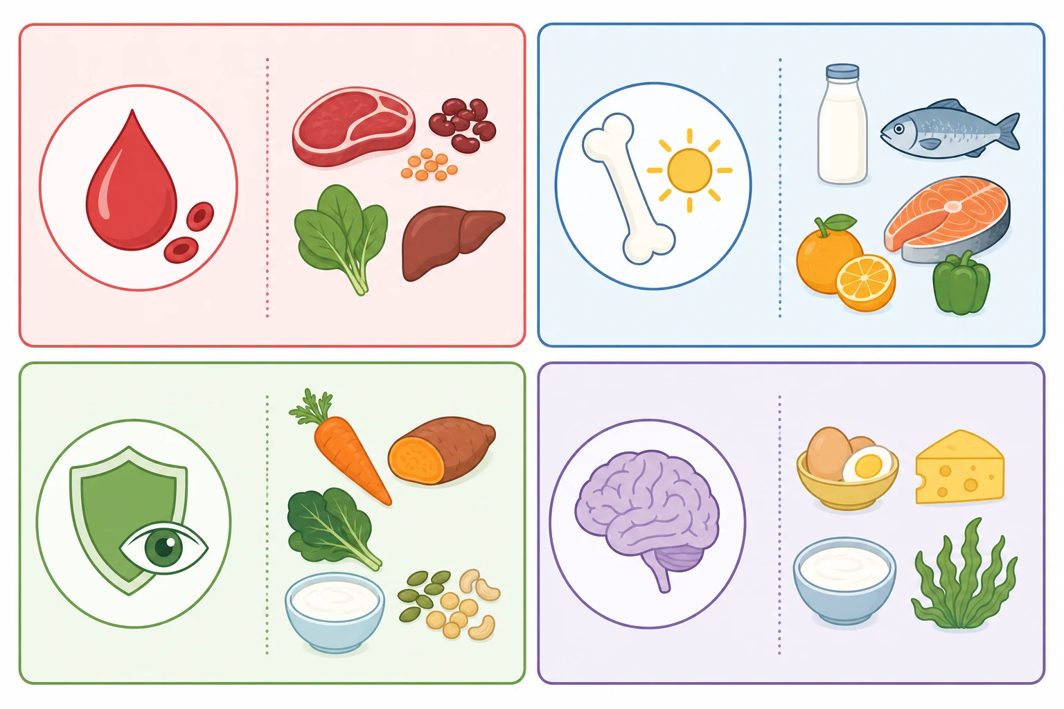
The most useful clinical way to hold micronutrient deficiency is by the system the nutrient serves, because that is what drives the presenting complaint. The haematological nutrients, iron and vitamin B12, present with anaemia: iron causes a microcytic hypochromic picture, while B12 causes a macrocytic megaloblastic one and adds neurological injury. Recognising the cell size on the film is the first fork in the workup of the anaemic child, because it directs the confirmatory tests. [1] [7]
The musculoskeletal nutrients, vitamin D and vitamin C, present with bone. Vitamin D deficiency softens the growing skeleton into rickets in the infant and toddler and osteomalacia in the adolescent, with bowing, bone pain, muscle weakness and delayed motor milestones. Vitamin C deficiency, scurvy, presents with bleeding gums, perifollicular haemorrhage, petechiae and leg pain from subperiosteal bleeding, and it remains a diagnosis of the malnourished or restricted-diet child. [6]
The immune and epithelial nutrients, vitamin A and zinc, present with infection and skin. Vitamin A deficiency impairs the conjunctival and corneal epithelium, causing night blindness, Bitot spots, corneal xerosis and ulceration, and it suppresses mucosal immunity so that measles, diarrhoea and respiratory infections become severe. Zinc deficiency presents with stunting, chronic diarrhoea, an acral or periorificial dermatitis, impaired wound healing and recurrent infection, and in its inherited form, acrodermatitis enteropathica, it is a severe blistering eruption of infancy. [9] [10]
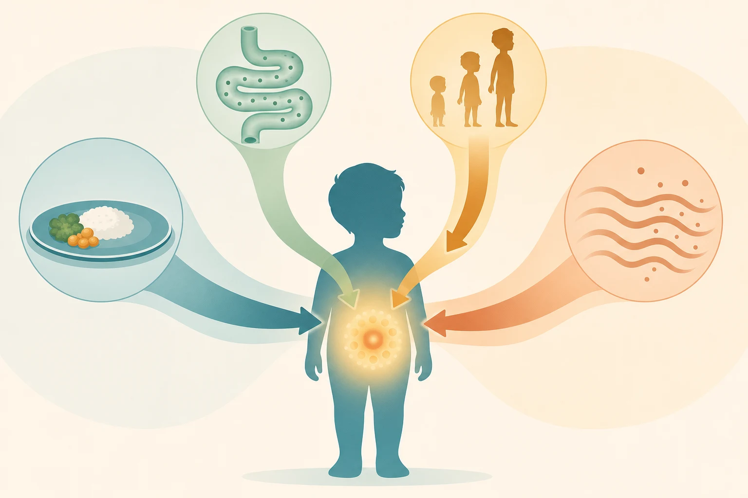
A second axis is whether the deficiency is dietary, absorptive or loss-driven, because this decides whether treatment is replacement alone or replacement plus an underlying cause. Dietary deficiency is the commonest, seen in the exclusively breastfed infant, the picky toddler, the vegan or plant-based family, the food-insecure household and the adolescent on a restricted diet. Absorptive deficiency follows coeliac disease, inflammatory bowel disease, short bowel syndrome and pancreatic insufficiency, and loss-driven deficiency follows chronic diarrhoea, malabsorption and the increased turnover of severe illness. [1] [3]
Epidemiology & Risk Factors
Iron deficiency is the commonest micronutrient deficiency of childhood worldwide. Infants and toddlers are the peak group because rapid growth expands the blood volume and the iron store at the same time as the diet is often low in absorbable iron, so universal screening at around twelve months with a haemoglobin and a dietary risk assessment is recommended. The risk is highest in preterm and low-birth-weight infants, who are born with smaller iron stores, in exclusively breastfed infants beyond six months without iron-rich complementary foods, in children with early or excessive cow's milk intake, and in any child with chronic blood loss or malabsorption. [1] [11]
Vitamin D deficiency is re-emerging in high-income countries and is near-universal in some low-resource settings. The risk is highest in dark-skinned, fully covered, exclusively breastfed infants who get little ultraviolet exposure, in preterm infants, in obese children who sequester the vitamin in adipose tissue, in children with malabsorption, liver or renal disease, and in those on anticonvulsants. Nutritional rickets remains a disease of infancy and early childhood, with a seasonal and latitudinal pattern that tracks sunlight. [5] [6]
Vitamin A and zinc deficiency are diseases of poverty and of the malabsorbing child. In low- and middle-income countries they are leading causes of preventable blindness, growth failure and death from measles, diarrhoea and pneumonia, and they cluster with stunting and recurrent infection. In high-income countries they hide in restricted diets, food insecurity, fat malabsorption, chronic diarrhoea and the technology-dependent child on long-term unsupplemented nutrition. Vitamin B12 deficiency clusters in exclusively breastfed infants of vegan, vegetarian or deficient mothers, and in children with pernicious anaemia, ileal disease or gastric surgery. [9] [7]
Pathophysiology
Four mechanisms converge to deplete the growing child, and naming them guides both the workup and the prevention. Inadequate intake is the first, whether from a monotonous or restricted diet, exclusive breastfeeding beyond the age of sufficiency, food insecurity, or the adolescent who restricts for weight, belief or eating disorder. Malabsorption is the second, when coeliac disease, inflammatory bowel disease, short bowel syndrome, pancreatic insufficiency or mucosal injury prevents the nutrient crossing the gut. Increased losses are the third, driven by chronic diarrhoea, blood loss, nephrotic syndrome or dialysis. Increased needs are the fourth, set by the rapid growth of infancy and adolescence, pregnancy, and the demands of recovery from illness. [1] [3]
Iron sits at the centre of haemoglobin and of the developing brain. When intake or absorption falls behind the demands of growth, the storage iron in ferritin is spent first, then the transport iron falls, and only then does the haemoglobin drop, so iron deficiency without anaemia is the common and easily missed early stage. The same depletion harms dopamine metabolism and myelination in the developing brain, which is why iron deficiency has been linked to deficits in attention, cognition and behaviour that may not fully reverse. Hepcidin, the liver hormone, governs intestinal iron absorption and iron release from macrophages, and it is raised in inflammation so that chronic disease traps iron and produces a functional iron deficiency. [1] [3]
Vitamin D is made in the skin by ultraviolet light and consumed in the diet, then hydroxylated in the liver to 25-hydroxyvitamin D and again in the kidney to the active 1,25-dihydroxyvitamin D, which governs calcium and phosphate absorption from the gut. When vitamin D falls, calcium and phosphate absorption falls, the mineralisation of growing bone fails, and the soft osteoid accumulates to produce the widened, cupped metaphyses and bowed legs of rickets. A secondary rise in parathyroid hormone may partially correct the calcium at the cost of bone resorption and a low phosphate, which is the classic biochemical signature of nutritional rickets. [6] [4]
Vitamin B12 is needed for DNA synthesis and for the maintenance of myelin, and its deficiency produces two injuries at once. In the bone marrow, the failure of DNA synthesis blocks red-cell maturation and produces the large, fragile megaloblastic red cells of macrocytic anaemia. In the nervous system, demyelination of the dorsal and lateral columns produces peripheral neuropathy, subacute combined degeneration of the cord, and in the infant, severe developmental regression, seizures and brain atrophy. Because the infant's store is small and entirely dependent on the mother, a deficient or vegan mother can deplete her exclusively breastfed infant within months of birth. [7] [8]
Clinical Presentation
The iron-deficient child is the classic presentation and the one examiners expect you to own. A toddler between one and three years presents with pallor, fatigue, irritability and a fallen growth centile, and the history often reveals excessive cow's milk intake, a diet low in meat or iron-rich solids, or pica, the craving to eat non-food items such as dirt, ice or starch. The cardiovascular signs of severe anaemia, a flow murmur, tachycardia and, in the extreme, high-output cardiac failure, belong to the profoundly depleted child, and iron deficiency is the commonest cause. [1] [2]
The vitamin D deficient child presents with bone. The infant may show craniotabes, a soft skull, widened wrists and ribs, and delayed fontanelle closure and motor milestones, while the weight-bearing toddler or older child shows genu varum or valgum, bone pain, muscle weakness and a waddling gait. Adolescents may present with bone pain or a stress fracture. The biochemical signature of a low calcium, low phosphate and high alkaline phosphatase with a low 25-hydroxyvitamin D makes the diagnosis, and hypocalcaemic seizures or tetany are the dramatic, occasionally first, sign in the young infant. [6] [5]
The vitamin B12 deficient infant is the neurological emergency that must not be missed. An exclusively breastfed infant of a vegan, vegetarian or deficient mother presents between four and twelve months with failure to thrive, lethargy, apathy, feeding difficulty, hypotonia, tremor, regression of motor and social skills, and sometimes seizures. The blood film may show a macrocytic anaemia, but the neurological signs can precede the anaemia, so a normal haemoglobin does not exclude the diagnosis. The adolescent or adult presentation adds glossitis, paraesthesia and a dorsal-column sensory loss. [7] [8]
The vitamin A and zinc deficient child presents with eye and skin and the company of recurrent infection. Vitamin A deficiency moves through night blindness, then conjunctival xerosis with Bitot spots, then corneal xerosis and ulceration that can perforate and blind, all set against a background of severe infection, measles and malnutrition. Zinc deficiency presents with a periorificial and acral dermatitis, chronic diarrhoea, alopecia, impaired wound healing, recurrent infection and stunting, and the inherited acrodermatitis enteropathica declares itself in the weaning infant when breast milk zinc falls. [9] [10]
Differential Diagnosis
The differential of the anaemic child turns on the mean cell volume. A microcytic anaemia is iron deficiency until proven otherwise, but the competitors are thalassaemia trait, which gives a microcytosis with a normal or near-normal haemoglobin and a family history, anaemia of chronic disease, which traps iron through hepcidin, lead poisoning, sideroblastic anaemia and the rare congenital sideroblastic causes. The serum ferritin and transferrin saturation, read alongside the C-reactive protein to allow for inflammation, separate iron deficiency from its mimics, and a haemoglobin electrophoresis excludes thalassaemia when the ferritin is normal. [1] [3]
The differential of the macrocytic anaemia turns on the serum B12 and folate. Vitamin B12 deficiency is distinguished from folate deficiency by the serum levels and the methylmalonic acid, which is raised in B12 deficiency but normal in folate deficiency, while homocysteine is raised in both. Drug causes, hypothyroidism, reticulocytosis, liver disease, alcohol and the bone-marrow failure syndromes all raise the mean cell volume, so the neurological signs and the maternal and dietary history are what anchor the diagnosis of B12 deficiency in the infant. [7] [8]
The differential of the bowing and bone pain turns on the biochemistry. Nutritional rickets from vitamin D deficiency shows a low calcium, low phosphate and high alkaline phosphatase with a low 25-hydroxyvitamin D, and it is separated from the hypophosphatasia of a low alkaline phosphatase, the calcium-deficiency rickets of a low calcium with a normal vitamin D, and the hereditary hypophosphataemic rickets of a low phosphate with a normal calcium and vitamin D. The bone pain and bleeding of scurvy mimics infection, malignancy and bruising, and a dietary history of a near-zero vitamin C intake makes the diagnosis. [6] [5]
Causes of microcytic anaemia — 'TAILS'
Clinical & Bedside Assessment
Assessment begins, as in all of paediatrics, with the growth chart and the diet. Plot the weight, the height and the head circumference, read the trend across the centiles, and look for the faltering that signals an organic deficiency, because stunting from zinc deficiency and the growth failure of iron and B12 deficiency may be the only sign. A focused diet history asks about breastfeeding duration, the timing and content of complementary feeding, the volume of cow's milk, the intake of iron-rich foods, meat, eggs and dairy, the use of a vegan or plant-based diet, food security, and any pica or restricted eating in the adolescent. [1] [11]
The examination then hunts the system the nutrient serves. Look for pallor, a flow murmur and the koilonychia of severe iron deficiency; the craniotabes, widened wrists, rachitic rosary and bowing of vitamin D deficiency; the glossitis, jaundice and neurological signs of B12 deficiency; the night blindness, Bitot spots and corneal xerosis of vitamin A deficiency; and the acral dermatitis, alopecia and periorificial rash of zinc deficiency. Developmental assessment is essential, because developmental regression in the breastfed infant is the red flag for B12 deficiency and the clue to iodine deficiency. [6] [7]
The company a deficiency keeps narrows the cause. Ask about prematurity and low birth weight for iron, about sunlight, skin colour, clothing and latitude for vitamin D, about maternal diet and pernicious anaemia for B12, about chronic diarrhoea, malabsorption and pancreatic disease for the fat-soluble vitamins and zinc, and about food security, household poverty and remote or Indigenous status for the cluster of deficiencies that travel with undernutrition. The child with a single deficiency in a privileged setting points to diet or malabsorption, while the child with several deficiencies points to poverty or chronic disease. [11] [3]
Investigations
The iron workup is built on the full blood count and the iron studies. A microcytic, hypochromic anaemia with a low mean cell volume and a wide red-cell distribution width is the screening finding, and a low serum ferritin confirms empty stores, with a ferritin below roughly fifteen micrograms per litre in children beyond infancy supporting deficiency. Because ferritin rises with inflammation, a concurrent C-reactive protein is needed, and a low transferrin saturation with a raised transferrin and a low reticulocyte count complete the iron-deficient picture. When the ferritin is normal but deficiency is still suspected, the soluble transferrin receptor, which is not affected by inflammation, can help. [1] [3]
The vitamin D workup measures the 25-hydroxyvitamin D, the stable circulating form, alongside the calcium, phosphate, alkaline phosphatase and parathyroid hormone. A 25-hydroxyvitamin D below fifty nanomoles per litre supports deficiency, with values below twenty-five nanomoles per litre indicating severe deficiency, and the characteristic biochemical pattern of rickets is a low or low-normal calcium, a low phosphate, a raised alkaline phosphatase and a secondary rise in parathyroid hormone. A wrist or knee radiograph shows the widened, cupped, frayed metaphyses of active rickets, and it confirms the skeletal injury. [6] [4]
The vitamin B12 workup measures the serum B12 and confirms with the methylmalonic acid, which is raised when cellular B12 is deficient. A low or borderline serum B12 with a raised methylmalonic acid and homocysteine confirms deficiency, and the full blood count shows a macrocytic anaemia with hypersegmented neutrophils, though the haemoglobin may be normal when the neurological signs lead. In the breastfed infant, the maternal serum B12 and dietary history are part of the workup, and the blood film and the bone-marrow megaloblastic change complete the picture when the diagnosis is unclear. [7] [8]
Management — Resuscitation
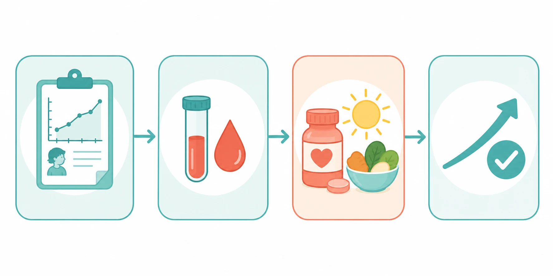
Most micronutrient deficiency is managed electively, but three presentations need urgent action. A profoundly anaemic child with a haemoglobin far below normal, tachycardia, breathlessness and signs of cardiac failure needs careful assessment, because transfusion in a chronically anaemic child can precipitate volume overload; a cautious, slow transfusion of packed red cells with close monitoring, or an exchange transfusion in the most severe, is reserved for the haemodynamically unstable. Severe iron deficiency is then corrected with oral or intravenous iron once the child is stable. [2] [1]
The hypocalcaemic infant with vitamin D deficiency can seize, and this is a metabolic emergency. Intravenous calcium gluconate under cardiac monitoring stops the seizure, after which the vitamin D deficiency is treated with cholecalciferol and adequate calcium, because rapid mineralisation of osteoid on replacement can deepen the hypocalcaemia if calcium intake is inadequate. The infant with severe, sight-threatening vitamin A deficiency needs immediate high-dose vitamin A to prevent corneal ulceration and blindness, and the B12-deficient infant with neurological signs needs prompt parenteral hydroxocobalamin to arrest and reverse the neurological injury. [6] [9]
Oral iron for iron-deficiency anaemia
Loading dose
Elemental iron 3 mg/kg once daily for most infants and young children, increased to a divided dose up to 6 mg/kg per day if tolerated and if the response is inadequate
Maintenance dose
Continue for around three months after the haemoglobin normalises, to replete the iron stores, then reassess the diet and the cause
Immediate management of the severely deficient child
Assess the airway, breathing and circulation, and the haemodynamic and neurological status
Transfuse a profoundly anaemic child cautiously and only if haemodynamically unstable, watching for volume overload
Give intravenous calcium gluconate under cardiac monitoring to the hypocalcaemic, seizing infant with rickets
Start parenteral hydroxocobalamin urgently in the breastfed infant with suspected B12 deficiency and neurological signs
Give immediate high-dose vitamin A to the child with corneal xerosis or ulceration to prevent blindness
Once stable, confirm the deficiency with biochemistry, replace the missing nutrient and treat the underlying cause
Management — Definitive & Stepwise
The definitive treatment is replacement of the missing nutrient and treatment of the cause, delivered together. Iron deficiency is treated with oral elemental iron at around three milligrams per kilogram per day, continued for about three months after the haemoglobin normalises to replete the stores, and the cause, whether a milk-heavy diet, malabsorption or blood loss, is corrected at the same time. Dietary advice introduces iron-rich solids, limits cow's milk to around five hundred millilitres a day in the toddler, and pairs iron with vitamin C to aid absorption, and intravenous iron is reserved for the severe, non-responsive or malabsorbing child. [1] [2]
Vitamin D deficiency is treated with cholecalciferol in a loading regimen followed by a maintenance dose, with adequate dietary calcium throughout. The loading regimen varies by region and age, but the principle is to replenish the store over several weeks and then continue a maintenance dose of four hundred international units a day in infants and six hundred international units a day in older children and adolescents. Nutritional rickets resolves radiographically within months, and the bowing often corrects with growth, so orthopaedic intervention for the deformity itself is rarely needed. [5] [6]
Vitamin B12 deficiency is treated with parenteral hydroxocobalamin, because the injury is neurological and time-critical. The regimen varies with the cause and the age, but the principle is daily parenteral replacement for the first weeks followed by regular maintenance, and the breastfed infant of a deficient or vegan mother needs the infant treated and the mother supplemented. Vitamin A deficiency is treated with high-dose vitamin A, and zinc deficiency with oral zinc, continued until the cause, whether dietary, malabsorptive or inherited, is controlled. Each deficiency is monitored by the clinical response, the growth and the repeat blood indices. [7] [9]
The region-aware prevention doses examiners probe
Prevention rests on supplementation, fortification and dietary diversification, and the doses are worth memorising. Vitamin D supplementation of four hundred international units a day is recommended for all infants from the first days of life, especially the exclusively or partially breastfed, and six hundred international units a day for children and adolescents. Iron is provided through iron-fortified formula and complementary foods, with preterm and low-birth-weight infants needing around two milligrams per kilogram per day from one month of age. In vitamin-A-deficient regions, the World Health Organization recommends high-dose supplementation every four to six months from six months to fifty-nine months, at one hundred thousand international units for infants six to eleven months and two hundred thousand international units for children twelve to fifty-nine months. Universal salt iodisation prevents iodine deficiency. [5] [9]
Specific Subtypes & Scenarios
The exclusively breastfed infant of a vegan mother is the B12-deficiency scenario that examiners reward for owning. Because the infant's vitamin B12 store is laid down in pregnancy and replenished only by breast milk, a mother who is herself deficient through a strict vegan or vegetarian diet, pernicious anaemia, gastric surgery or ileal disease depletes her infant within months. The infant presents with failure to thrive, apathy, hypotonia, tremor and developmental regression, sometimes with a macrocytic anaemia and sometimes before it, and the serum B12 and methylmalonic acid confirm the diagnosis. Prompt parenteral hydroxocobalamin and lifelong supplementation of mother and child, or a reliable B12-fortified or supplemented diet, prevent recurrence. [7] [8]
The iron-deficient toddler with excessive cow's milk intake is the classic dietary scenario. A young child who drinks more than about five hundred millilitres of cow's milk a day displaces iron-rich solids, and the milk both irritates the gut to cause microscopic blood loss and binds dietary iron through its high calcium and casein, so the child becomes iron deficient despite an apparently adequate calorie intake. The management is to limit the milk, introduce iron-rich solids, treat the deficiency with oral iron, and recheck the haemoglobin to confirm a reticulocyte rise within one to two weeks and a normal haemoglobin within two to three months. [1] [2]
The child with malabsorption is the cluster-deficiency scenario. Coeliac disease, inflammatory bowel disease, short bowel syndrome, cystic fibrosis and chronic liver disease impair the absorption of iron, the fat-soluble vitamins A and D, zinc and B12 in varying combinations, so the malabsorbing child often presents with more than one deficiency at once. The workup screens the panel of nutrients at risk, the treatment replaces each deficiency and treats the underlying disease, and the child on long-term parenteral or enteral nutrition needs a structured micronutrient review to prevent deficiency in the technology-dependent setting. [3] [11]
Across Australia, New Zealand and the United Kingdom, universal iron screening at around twelve months is common, vitamin D supplementation of four hundred international units a day is advised for breastfed infants, and food fortification with iron, folate and iodine is the backbone of prevention. In many low- and middle-income countries, the World Health Organization programmes of vitamin A supplementation every four to six months, zinc for diarrhoea, iron and folic acid for pregnancy, and universal salt iodisation prevent the blindness, stunting and brain injury of deficiency. The principles are the same across regions; the difference is in access to diverse diets, supplements and fortified foods, which is why deficiency clusters in remote, Indigenous, migrant and food-insecure communities even within high-income countries. [11] [12]
Complications & Pitfalls
The complications flow from the system the nutrient serves and from the duration of the deficiency. Untreated iron deficiency in infancy and early childhood has been associated with deficits in attention, cognition and behaviour that may not fully reverse even after treatment, which is the neurodevelopmental argument for early screening. Severe nutritional rickets leaves residual skeletal deformity, and profound vitamin D deficiency in the infant can cause hypocalcaemic seizures, cardiomyopathy and, rarely, death. Untreated vitamin B12 deficiency in the infant causes irreversible neurological injury, and vitamin A deficiency blinds and kills through severe infection. [1] [7]
The treatment pitfalls are the failures the examiner rewards for naming. The first is missing iron deficiency by testing the haemoglobin alone, because the ferritin and the neurodevelopmental injury fall before the anaemia. The second is misreading the ferritin in inflammation, because ferritin rises as an acute-phase reactant and can mask iron deficiency unless the C-reactive protein is read alongside it. The third is treating vitamin D deficiency without adequate calcium, because rapid osteoid mineralisation on replacement can deepen the hypocalcaemia and precipitate seizures. The fourth is attributing the breastfed infant's developmental regression to cerebral palsy or a metabolic disease and missing the treatable B12 deficiency. [3] [6]
Prognosis & Disposition
The prognosis is excellent for the deficiency caught and treated early, and poor for the one missed. Iron-deficiency anaemia corrects within two to three months of oral iron, the reticulocytes rising within the first one to two weeks as the first sign of response, and nutritional rickets heals radiographically within months, the deformity correcting with growth. Vitamin A and zinc deficiency respond within days to weeks to replacement, and the acute signs resolve quickly. The caveat is the developing brain: the cognitive and behavioural effects of early iron deficiency and the neurological injury of infant B12 deficiency may not fully reverse, which is why early recognition is the whole game. [1] [7]
Disposition follows the cause and the severity. The straightforward dietary deficiency is managed in primary care or general paediatrics with a dietitian, a supplement and a follow-up blood test. The child with malabsorption, multiple deficiencies, severe anaemia, hypocalcaemic rickets or neurological B12 deficiency is managed in a tertiary centre with paediatric gastroenterology, haematology or neurology as needed, and the technology-dependent child on long-term nutrition needs a structured micronutrient surveillance plan. Adolescents with restrictive eating are supported with a mental-health pathway alongside the nutritional correction. [2] [11]
Special Populations
The preterm and low-birth-weight infant is the first special population, because the iron, vitamin D and calcium stores laid down in the third trimester are missed. Preterm infants need around two milligrams per kilogram per day of elemental iron from one month of age, and a higher dose of vitamin D, with careful attention to the minerals in those on human-milk fortifier or special formula, and they are at risk of osteopenia of prematurity as well as iron deficiency. The dietitian-led surveillance of the ex-preterm infant in the first two years prevents the deficiencies that complicate catch-up growth. [1] [5]
The child on a restricted or plant-based diet is the second. A well-planned vegan or vegetarian diet can meet a child's needs, but it requires reliable sources of vitamin B12, iron, zinc, calcium and vitamin D, and the unsupplemented vegan infant or the adolescent on a self-restricted diet is at high risk of B12, iron and vitamin D deficiency. The clinician's role is to support the chosen diet with evidence-based supplementation, to screen for deficiency, and to recognise the eating disorder that hides behind a restrictive label in the adolescent. [7] [8]
The remote, Indigenous, migrant and food-insecure child is the third. In these communities the deficiencies cluster, because a monotonous or poverty-limited diet, limited access to diverse foods and supplements, and a higher burden of chronic infection combine to deplete iron, vitamin A, zinc and vitamin D together. Culturally safe shared-care pathways, telehealth support for the local team, school-based supplementation and food programmes, and attention to the cost and availability of nutrient-rich foods are what make prevention achievable for families far from a specialist centre. [11] [12]
Evidence, Guidelines & Regional Differences
The evidence base is anchored on the American Academy of Pediatrics iron guideline and the Institute of Medicine vitamin D report. The Baker and Greer guideline set the framework for universal iron screening at around twelve months and the prevention of iron deficiency in infants and young children, grounding the case for screening in the neurodevelopmental harm of deficiency. The Institute of Medicine report fixed the dietary reference intakes for calcium and vitamin D, establishing the sufficiency threshold and the recommended intakes that underpin supplementation and fortification worldwide. [1] [4]
The management and clinical-practice evidence spans the nutrients. The Benson consensus on iron management brought together the evidence for oral and intravenous iron across children, adults and pregnancy, while the Pasricha clinical update framed the diagnosis and management of iron deficiency for the Australasian reader. The Wagner and Greer guideline set the prevention of rickets and vitamin D deficiency, and the Elder and Bishop review of rickets integrated the nutritional and hereditary forms. The Green and Stabler reviews anchored the modern understanding of vitamin B12 deficiency, and the Brown zinc meta-analysis quantified the growth response to supplementation. [2] [3] [5] [10]
The global burden and the regional differences complete the picture. The Black maternal and child undernutrition series quantified the toll of hidden hunger on growth, immunity and neurodevelopment across low- and middle-income countries, and the Song review framed the recognition of vitamin A deficiency even in low-prevalence areas. The Zimmermann review of iodine fixed the worldwide status of iodine deficiency and excess in children, and regional practice differs chiefly in access to diverse diets, supplements and fortified foods rather than in the principles of prevention. [11] [9] [12]
Exam Pearls
The key micronutrients and their deficiency states — 'IRON-VAD-ZB12'
References
- [1]Baker RD; Greer FR Diagnosis and prevention of iron deficiency and iron-deficiency anemia in infants and young children (0-3 years of age). Pediatrics, 2010.PMID 20923825
- [2]Benson AE; Lo JO; Achebe MO; Aslan JS; Auerbach M; Bannow BTS Management of iron deficiency in children, adults, and pregnant individuals: evidence-based and expert consensus recommendations. Lancet Haematol, 2025.PMID 40306833
- [3]Pasricha SR; Flecknoe-Brown SC; Allen KJ; Gibson PR; McMahon LP; Olynyk JK Diagnosis and management of iron deficiency anaemia: a clinical update. Med J Aust, 2010.PMID 21034387
- [4]Ross AC; Manson JE; Abrams SA; Aloia JF; Brannon PM; Clinton SK The 2011 report on dietary reference intakes for calcium and vitamin D from the Institute of Medicine: what clinicians need to know. J Clin Endocrinol Metab, 2011.PMID 21118827
- [5]Wagner CL; Greer FR Prevention of rickets and vitamin D deficiency in infants, children, and adolescents. Pediatrics, 2008.PMID 18977996
- [6]Elder CJ; Bishop NJ Rickets. Lancet, 2014.PMID 24412049
- [7]Green R; Allen LH; Bjørke-Monsen AL; Brito A; Guéant JL; Miller JW Vitamin B(12) deficiency. Nat Rev Dis Primers, 2017.PMID 28660890
- [8]Stabler SP Clinical practice. Vitamin B12 deficiency. N Engl J Med, 2013.PMID 23301732
- [9]Song A; Mousa HM; Soifer M; Perez VL Recognizing vitamin A deficiency: special considerations in low-prevalence areas. Curr Opin Pediatr, 2022.PMID 35125379
- [10]Brown KH; Peerson JM; Rivera J; Allen LH Effect of supplemental zinc on the growth and serum zinc concentrations of prepubertal children: a meta-analysis of randomized controlled trials. Am J Clin Nutr, 2002.PMID 12036814
- [11]Black RE; Victora CG; Walker SP; Bhutta ZA; Christian P; de Onis M Maternal and child undernutrition and overweight in low-income and middle-income countries. Lancet, 2013.PMID 23746772
- [12]Zimmermann MB Iodine deficiency and excess in children: worldwide status in 2013. Endocr Pract, 2013.PMID 23757630