Paeds · haematology-oncology-and-transfusion
Pancytopenia and marrow infiltration
Also known as Pancytopenia · Marrow infiltration · Myelophthisis · Bone marrow failure · Leukoerythroblastic anaemia · Marrow replacement
Fellowship guide to pancytopenia and marrow infiltration in children. Covers the definition of a fall in all three blood lineages, the three pathogenetic mechanisms of reduced marrow production, marrow replacement by leukaemia, neuroblastoma, Langerhans cell histiocytosis and rhabdomyosarcoma, and peripheral consumption, the leucoerythroblastic blood film with nucleated red cells and teardrop poikilocytes that signals myelophthisis, the urgent diagnostic pathway from full blood count and film to bone marrow aspirate, trephine biopsy, flow cytometry, cytogenetics and molecular testing, the stabilisation of the unstable child with transfusion of irradiated leucodepleted red cells and platelets, the prevention of tumour lysis syndrome with hyperhydration and rasburicase, and the cause-specific definitive therapy for acute lymphoblastic and myeloid leukaemia, acquired and inherited marrow failure, Down syndrome transient myeloproliferative disorder, neuroblastoma, Langerhans cell histiocytosis and parvovirus B19 pure red cell aplasia.
On this page & tools
Your progress
Saved locally on this device.
Practise this topic
Target exams
Red flags
Life stages
Care settings
Clinical exam formats
Board mappings
Overview & Definition
A child is brought in pale, covered in bruises, with a fever, and the blood count shows that every cell line has fallen at once. This is pancytopenia, and the first job at the bedside is not to name the disease but to decide which of two very different things is happening to the bone marrow. Either the factory is empty and unable to make cells, or the factory is full but the cells inside it are the wrong ones. That single distinction, between an empty marrow and a full one, drives every decision that follows, from how fast the child must be worked up to whether the family is about to hear the word leukaemia. [1]
Pancytopenia is defined by a fall in all three circulating lineages below the age-appropriate reference range: the haemoglobin, the neutrophil count, and the platelet count. A practical working threshold is a neutrophil count under one point five times ten to the nine per litre and a platelet count under one hundred and fifty times ten to the nine per litre, alongside the anaemia. The word is a laboratory finding, never a diagnosis, and the value of recognising it quickly is that it forces the clinician away from thinking about each line in isolation and towards the marrow itself as the seat of the problem. [1]
Marrow infiltration, also called myelophthisis, is the specific mechanism in which the haematopoietic tissue of the bone marrow is crowded out and replaced by something else: malignant blasts, metastatic tumour cells, fibrous tissue, or a granulomatous process. The normal blood-forming cells are squeezed out, the marrow architecture is distorted, and immature cells leak into the blood. Acute leukaemia is the diagnosis every paediatrician fears when pancytopenia appears, because it is the commonest marrow malignancy of childhood and because a delay of days can change the outcome. [3]
The gravity of the finding is why the topic sits at the heart of the fellowship examination. A candidate who can look at a pale, bruising child, read the count and the film, decide whether the marrow is empty or full, and chart a safe path to diagnosis and stabilisation is demonstrating exactly the reasoning the boards test. The landmark evidence on childhood leukaemia, marrow failure and the oncologic emergencies frames every part of the pathway that follows. [3][12]
Classification
The single most useful way to classify pancytopenia at the bedside is by what the marrow is doing, because that one observation splits the differential in two. In a reduced-production marrow the space where cells should be made is empty or hypocellular, and the causes are the marrow failure syndromes, the inherited and the acquired, plus the transient injuries from drugs, viruses and radiation. In a replaced marrow the space is full, but it is occupied by blasts, tumour cells, fibrosis or granulomata, and the causes are the marrow malignancies and the infiltrative processes. A third, smaller group sits outside the marrow, where the cells are made but lost peripherally through sequestration, haemolysis, or overwhelming consumption. [1]
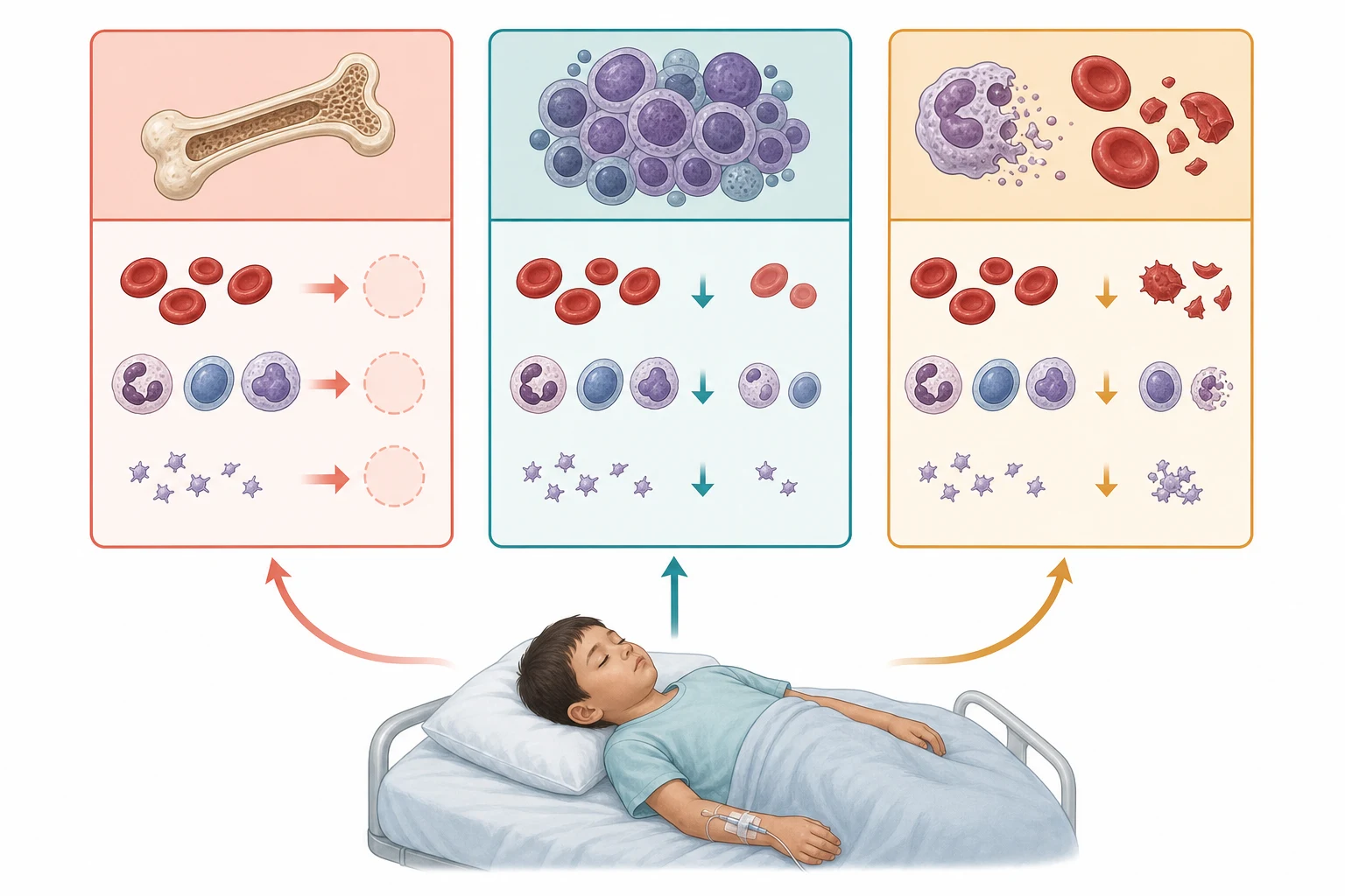
The infiltrative causes are the heart of this topic, and they sort themselves by the kind of cell that has taken over the marrow. The haematological malignancies are the acute leukaemias, lymphoblastic and myeloid, in which sheets of uniform blasts replace the normal precursors. The solid tumours that seed the marrow are the small-round-blue-cell family, led by neuroblastoma and including rhabdomyosarcoma and the Ewing family. The histiocytic disorders, above all Langerhans cell histiocytosis, occupy a category of their own. The non-malignant infiltrates are the granulomatous diseases, chiefly disseminated tuberculosis, and the parasites such as visceral leishmaniasis in endemic regions. [3][7]
A parallel classification runs through the marrow failure group and matters because it changes surveillance and the family advice. Acquired severe aplastic anaemia is an immune-mediated destruction of the haematopoietic stem cell, often apparently idiopathic but sometimes triggered by a drug or a virus. The inherited bone marrow failure syndromes are the germline disorders of which Fanconi anaemia is the prototype, and they carry a lifelong risk of clonal evolution into myelodysplasia and leukaemia. The transient marrow failures, dominated by parvovirus B19 pure red cell aplasia, are reversible once the cause is recognised and treated. [9][10]
Reduced production (empty marrow)
marrow failure
- Aplastic anemia, acquired or inherited
- Inherited syndromes: Fanconi, dyskeratosis congenita, Shwachman-Diamond
- Drug, toxin or radiation injury
- Parvovirus B19 pure red cell aplasia
Marrow replacement (full marrow)
infiltration, myelophthisis
- Acute lymphoblastic and myeloid leukaemia
- Neuroblastoma and the small-round-blue-cell tumours
- Langerhans cell histiocytosis
- Bone marrow fibrosis, necrosis or granulomata
Peripheral consumption
made but lost
- Hypersplenic sequestration
- Overwhelming sepsis and disseminated intravascular coagulation
- Haemophagocytic lymphohistiocytosis
- Megaloblastic crisis of folate or B12 deficiency
Epidemiology & Risk Factors
The epidemiology of pancytopenia in a child is the epidemiology of its causes, and in almost every series from every region the marrow malignancies dominate. Acute lymphoblastic leukaemia alone accounts for the largest share of new cases of pancytopenia referred to a paediatric haematology service, with a peak incidence between two and five years of age. Acute myeloid leukaemia is less common and is more evenly spread across the paediatric age range, including a distinctive peak in the neonate with Down syndrome. The acquired and inherited marrow failure syndromes together make up a smaller but important minority, and the transient and infectious causes fill out the remainder. [3][4]
The risk factors cluster around the conditions that predispose to marrow malignancy and failure. Down syndrome is the strongest single predisposition, carrying a ten to twenty-fold raised lifetime risk of leukaemia and a unique neonatal presentation, the transient myeloproliferative disorder. The inherited marrow failure syndromes carry their own malignancy risk: Fanconi anaemia, dyskeratosis congenita, and Shwachman-Diamond syndrome all predispose to myelodysplasia and acute myeloid leukaemia across childhood and young adulthood. Prior chemotherapy or radiation, the inherited bone marrow failure genotypes, and the acquired immune dysregulation syndromes each raise the background risk on which pancytopenia may declare a malignancy. [5][6]
The infectious and environmental risk factors shape the differential by region. In endemic areas disseminated tuberculosis and visceral leishmaniasis are genuine causes of pancytopenia through granulomatous and parasitic infiltration of the marrow, and parvovirus B19 is a worldwide cause of transient red cell aplasia, especially in the child with a chronic haemolysis whose red cell lifespan is already short. Exposure to marrow-toxic drugs, to pesticides and industrial solvents, and to radiation are the acquired environmental contributors, and they matter most when they sit on top of an inherited susceptibility. [9]
Pathophysiology
The pathophysiology of marrow infiltration is best understood as a contest for space inside the bone marrow cavity. The healthy marrow is a tightly organised tissue in which a small pool of self-renewing haematopoietic stem cells gives rise to every blood cell, with each lineage maturing along a scaffolding of stromal cells, fat, and blood vessels. When blasts or tumour cells pour in, they do not merely crowd the normal precursors; they actively distort the niche, disturb the signalling that controls maturation, and trigger a fibrotic and inflammatory response that further throttles normal blood formation. The result is a trilineage failure in which red cells, white cells and platelets all fall together. [3]
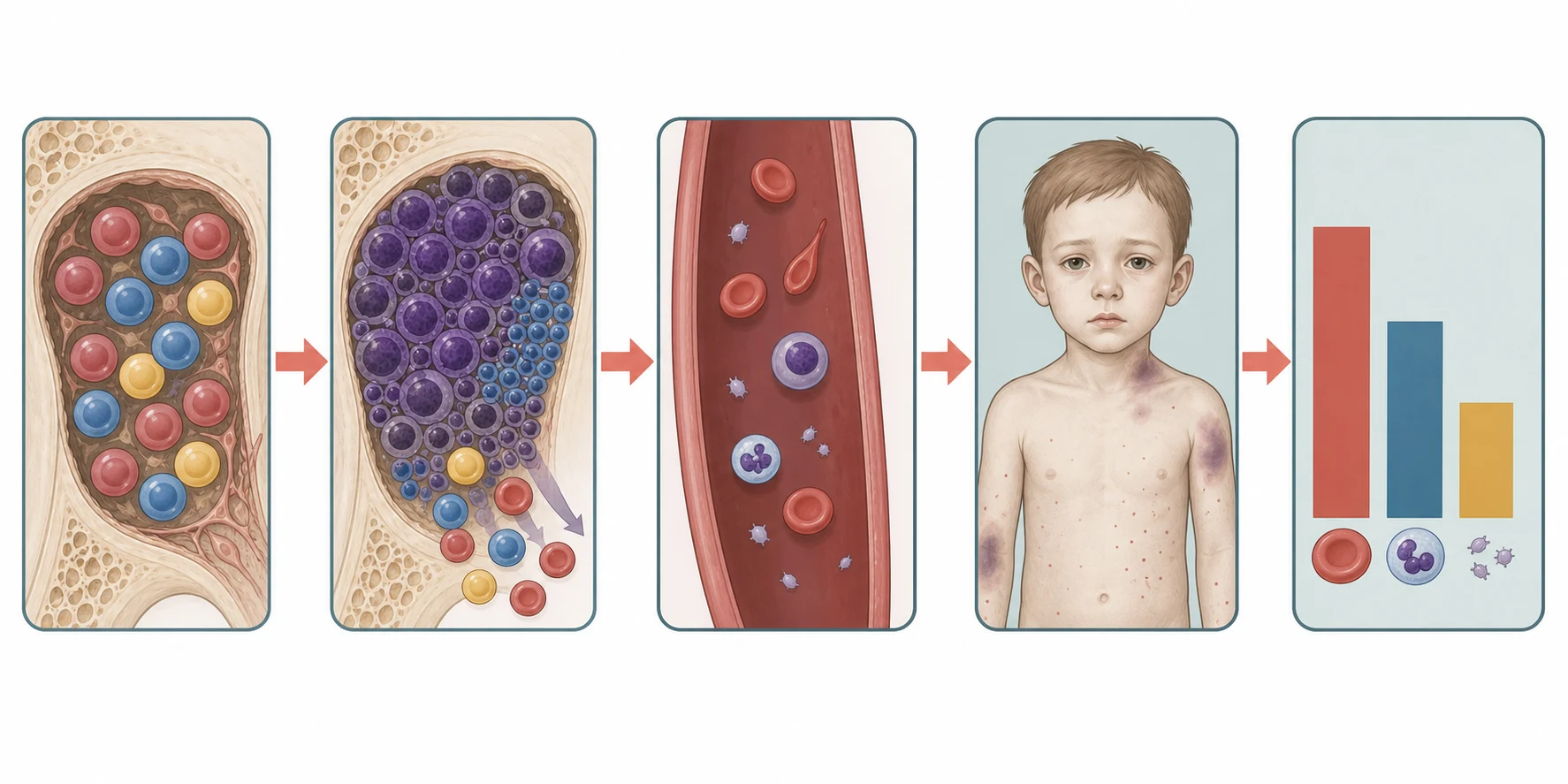
A second, distinctive mechanism explains why the blood film of an infiltrated marrow looks the way it does. When the marrow architecture is distorted and the sinusoids are disrupted, the normal barriers that keep immature cells inside the marrow break down, and both nucleated red blood cells and immature myeloid precursors escape into the peripheral blood. This is the leucoerythroblastic picture, and it is the hallmark of myelophthisis. Teardrop-shaped red cells, the dacrocytes, appear because the cells are mechanically deformed as they squeeze past fibrous tissue and tumour, and they are a tell-tale sign of marrow fibrosis and infiltration. [11]
The pathophysiology of the marrow failure syndromes is altogether different, and understanding it prevents the error of treating a failure as if it were an infiltration. In acquired aplastic anaemia, an aberrant T-cell attack destroys the haematopoietic stem cells, leaving the marrow empty and fatty and the count falling in all three lines with a reticulocyte count that is inappropriately low. In parvovirus B19 pure red cell aplasia, the virus is tropic for the red cell precursor through the blood group P antigen, and it shuts down red cell production entirely, producing a near-zero reticulocyte count and giant proerythroblasts in the marrow; the other lineages are usually spared because the virus targets the red cell line specifically. [9][10]
The oncologic emergencies that accompany a newly presenting marrow malignancy are pure pathophysiology and are the reason the first hours matter. A high blast count, with a white cell count over one hundred times ten to the nine per litre, makes the blood too viscous for the microcirculation and risks leukostasis in the brain and the lungs. The rapid turnover of a bulky tumour releases potassium, phosphate and urate faster than the kidneys can clear them, producing the tumour lysis syndrome. The compromised marrow leaves the child defenceless against bacteria, so fever becomes an emergency. Recognising these mechanisms at the bedside is what allows the stabilisation that precedes diagnosis. [12]
Clinical Presentation
The child with pancytopenia walks in carrying the symptoms of all three failing lineages at once, and the history usually tells the story before the count is back. The anaemia declares itself as pallor, tiredness, breathlessness on exertion, and in the young child as a new reluctance to walk or to feed. The thrombocytopenia declares itself as bruising that is out of proportion to the injury, petechiae over the lower limbs and pressure points, epistaxis, gum bleeding, and heavy or prolonged menstrual bleeding in the adolescent girl. The neutropenia declares itself as fever, mouth ulcers, perianal infection, and bacterial infections that fail to settle. The tempo is often only a few weeks, and a parent will describe a child who was entirely well a month before. [2]
The physical signs refine the differential, and the single most useful finding is the size of the liver, the spleen and the lymph nodes. An enlarged spleen and liver alongside lymphadenopathy point strongly towards a marrow malignancy with extramedullary disease, because the infiltrating cells populate the reticuloendothelial system as they spread. A mediastinal mass detected as stridor, facial swelling, or superior vena cava obstruction raises T-cell acute lymphoblastic leukaemia and is an anaesthetic emergency. Bone pain, limping and refusal to weight-bear are under-recognised presentations of leukaemia and are often misattributed at first to trauma or to a transient synovitis. [2]
The atypical and subtle presentations are the ones that catch the unwary, and the boards test them deliberately. A school-age child with a rheumatic-like limp, a normal or near-normal count, and a high sedimentation rate may have leukaemia with a low blast burden and is the classic mimic of juvenile idiopathic arthritis. An isolated thrombocytopenia labelled immune thrombocytopenia can be the first glimpse of an evolving aplastic anaemia or a marrow malignancy, which is why any child with a low platelet count has a full blood count and film examined for the other lineages. A teenager with a family history of early hair greying, nail dystrophy and pulmonary fibrosis may carry a telomere biology disorder that is about to declare marrow failure. [10]
The stigmata of the inherited marrow failure syndromes are sought in every child with an unexplained pancytopenia, because they change the counselling, the transplant donor choice and the long-term surveillance. Fanconi anaemia shows short stature, café-au-lait patches, and radial ray anomalies of the thumb and the radius. Dyskeratosis congenita shows the triad of abnormal nails, reticular skin pigmentation and oral leukoplakia, and may carry pulmonary fibrosis and liver disease. Shwachman-Diamond syndrome pairs marrow failure with pancreatic insufficiency and skeletal dysplasia. Diamond-Blackfan anaemia presents in infancy with a pure red cell aplasia and physical anomalies. These features are easy to miss unless they are actively sought. [10]
Differential Diagnosis
The differential of pancytopenia is built around the empty-versus-full marrow distinction, and the blood film and the reticulocyte count are the two tests that point the clinician in the right direction before the marrow is sampled. A low reticulocyte count in the face of anaemia is the single most important clue, because it means the marrow is failing to respond, and it points towards a production failure or an infiltration rather than a haemolysis or a blood loss. A high reticulocyte count, by contrast, suggests the marrow is trying and the cells are being lost peripherally, which redirects the search to the spleen, to haemolysis, and to sequestration. [1]
Acute leukaemia
full marrow, blasts
- Sheets of uniform blasts on the marrow aspirate
- Flow cytometry defines the lineage, lymphoid or myeloid
- Hepatosplenomegaly, lymphadenopathy, mediastinal mass
- Cytogenetics and molecular testing drive risk stratification
Acquired aplastic anaemia
empty marrow
- Hypocellular marrow with fat replacement, no abnormal clone
- No organomegaly and no lymphadenopathy
- Severe disease defined by Camitta criteria
- Treated by immunosuppression or matched-sibling transplant
Neuroblastoma marrow disease
small round blue cell
- Clumps and rosettes of small round blue cells in the marrow
- Primary adrenal or paraspinal mass on imaging
- Raised urinary catecholamines, VMA and HVA
- Young child, often under five years
Langerhans cell histiocytosis
clonal histiocyte
- CD1a and CD207 positive Langerhans cells in the lesion
- Multisystem disease may involve liver, spleen and marrow
- Bone lesions, diabetes insipidus, skin rash
- Risk-organ involvement defines high-risk disease
The benign mimics of pancytopenia must be excluded, because treating them as malignancy is as harmful as missing a malignancy. A severe megaloblastic anaemia from folate or vitamin B12 deficiency can drop all three lines and shows macrocytosis and hypersegmented neutrophils on the film, and it reverses with replacement. An overwhelming infection, whether bacterial sepsis, disseminated intravascular coagulation, or a viral bone marrow suppressor, can produce a transient pancytopenia that resolves as the child recovers. Hypersplenic sequestration from any cause of a large spleen, including portal hypertension and the haemoglobinopathies, traps cells peripherally and produces a pancytopenia with a marrow that is working hard. [1]
The chief diagnostic pitfalls for the fellow are the cases that sit on the border between categories. The child with evolving aplastic anaemia may first present with an isolated thrombocytopenia that is labelled immune thrombocytopenia; a careful blood film that shows no other line affected and a marrow biopsy weeks later settle the question, but the lesson is to recheck the count and the film in any apparent immune thrombocytopenia that does not behave as expected. The child with parvovirus B19 pure red cell aplasia may show only a profound anaemia with a near-zero reticulocyte count, and the other lines may fall later if the marrow is already stressed. The message for the exam is that the marrow aspirate and trephine biopsy, read with the film and the reticulocyte count, are what separate these entities, and that a single normal count never closes the question in a child with suspicious clinical features. [9]
Clinical & Bedside Assessment
The bedside assessment of the child with pancytopenia is a search for the clues that separate malignancy from failure and that flag the unstable child who needs resuscitation before investigation. The assessment begins with the airway, the breathing and the circulation, because a child with a mediastinal mass, a severe anaemia, or an active bleed is in danger before any diagnosis is reached. Once the child is safe, the focused history turns to the tempo of the symptoms, the bleeding and the fever, the drug and toxin exposures, the family history of marrow failure or early cancer, and the developmental history that might reveal an inherited syndrome. [2]
The examination is systematic and takes only a few minutes, but each finding carries weight. The skin is inspected for pallor, bruising, petechiae, and the café-au-lait and pigmentary changes of the inherited marrow failure syndromes. The mouth is inspected for mucosal bleeding, ulceration and leukoplakia, and the nails for the dystrophy of dyskeratosis congenita. The lymph node chains are palpated for size, consistency and distribution, the abdomen for the liver and the spleen, and the chest for the signs of a mediastinal mass, an effusion or a consolidation. The thumbs and the forearms are checked for radial ray anomalies, and the growth is plotted for the short stature of Fanconi anaemia. [2][10]
The focused examination of the child with pancytopenia
Assess the airway, breathing and circulation first, looking for stridor, distress, and the pallor and tachycardia of severe anaemia
Inspect the skin for pallor, bruising, petechiae, café-au-lait patches and abnormal pigmentation
Inspect the mouth for mucosal bleeding, ulceration and leukoplakia, and the nails for dystrophy
Palpate all lymph node chains and examine the abdomen for hepatosplenomegaly
Auscultate the chest for a mediastinal mass, effusion or consolidation, and assess for superior vena cava obstruction
Examine the thumbs, radii and stature for the radial ray anomalies and short stature of Fanconi anaemia
Plan the urgent blood tests and arrange the bone marrow examination with the haematology team
The severity of the bleeding and the haemodynamic consequences of the anaemia are judged at the bedside and decide how fast the transfusion must be given. Active mucosal bleeding, a rapidly falling haemoglobin, or signs of shock override any plan for an elective workup and demand immediate stabilisation. The teaching of the family is part of the examination, because a child sent home while awaiting the marrow result must be told to return at once with fever or new bleeding, and the neutropenic precautions, the avoidance of rectal temperatures and the need for urgent presentation with any fever, are explained before the family leaves. [12]
Investigations
The investigation of pancytopenia moves in two steps, the bedside blood tests that point to the mechanism, and the bone marrow examination that delivers the diagnosis. The first-line panel is the full blood count with the differential, the reticulocyte count, and the peripheral blood film, together with the blood group and screen, the coagulation screen, the electrolytes and renal function, the lactate dehydrogenase, the urate, the calcium and phosphate, and the liver function tests. The film is read personally by the clinician with the haematologist, because the presence of blasts, a leucoerythroblastic picture, or teardrop poikilocytes can change the working diagnosis in seconds. [1]
The reticulocyte count is the cornerstone of the first step, and it is read against the degree of anaemia. In a production failure or an infiltration, the reticulocyte count is inappropriately low for the anaemia, because the marrow cannot or will not make new cells. In a haemolysis or a sequestration, the reticulocyte count is high, because the marrow is responding to the loss. The absolute reticulocyte count and the corrected reticulocyte percentage are the formal expressions of this, and a corrected reticulocyte count under one percent in a moderately anaemic child is a strong signal that the marrow is the problem. [10]
The bone marrow aspirate and trephine biopsy together are the diagnostic standard, and they are indicated in any child with an unexplained or persistent pancytopenia, with blasts on the peripheral film, or with a leucoerythroblastic picture. The aspirate provides the cells for morphology, for flow cytometry to define the lineage of a leukaemia, and for the molecular studies that drive the modern risk stratification. The trephine biopsy provides the architecture, the cellularity that separates an empty from a full marrow, and the pattern of infiltration that identifies the small-round-blue-cell tumours and the fibrotic processes. The two are complementary and are always sent together. [3][11]
Across Australia, Aotearoa New Zealand, the United Kingdom, the United States and Canada, the bone marrow aspirate and trephine biopsy with flow cytometry and conventional cytogenetics is the standard diagnostic procedure for an unexplained paediatric pancytopenia. The molecular panel sent on the marrow, and the specific risk-stratification genes, differ between the international ALL and AML study groups, and the fellow should know the local protocol. In endemic regions, marrow culture for tuberculosis and the parasitological examination for leishmania are added where the travel and exposure history demands it.
[3][4]The targeted second-line tests are requested once the marrow has pointed to a specific cause, and they confirm the named diagnosis. Urinary catecholamines, the vanillylmandelic and homovanillic acids, are sent when the marrow shows a small-round-blue-cell tumour and the imaging suggests neuroblastoma. Parvovirus B19 polymerase chain reaction and immunoglobulin M confirm the transient pure red cell aplasia. The chromosome breakage test with diepoxybutane or mitomycin C confirms Fanconi anaemia, and the telomere length assay supports dyskeratosis congenita. The cytogenetics and the molecular panel on the marrow, including the GATA1 mutation in the neonate with Down syndrome, deliver the subclassification that drives the treatment. [5][6]
Management — Resuscitation
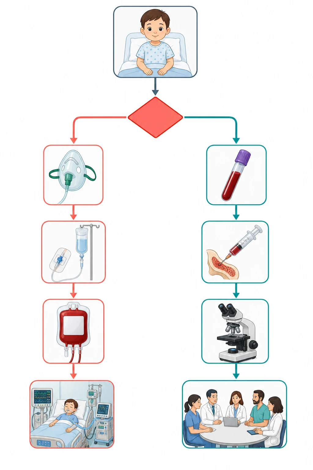
The unstable child with pancytopenia is resuscitated before the diagnosis is pursued, and the resuscitation rests on three legs: transfusion of red cells and platelets, prevention and treatment of the oncologic emergencies, and empiric cover for the febrile neutropenia. Red cell transfusion is given for symptomatic anaemia or a haemoglobin that is falling fast, and it is given slowly and cautiously in the child who may be volume-overloaded or who has a long-standing severe anaemia in which a rapid rise can precipitate heart failure. [12]
The blood products given to a child with a suspected or confirmed marrow malignancy must be irradiated, leucodepleted, and, where the child is a candidate for a haematopoietic stem cell transplant, cytomegalovirus-safe. Irradiation prevents transfusion-associated graft-versus-host disease, a fatal complication in which donor T-lymphocytes engraft in the immunocompromised recipient, and it is required for all cellular products in the child with a known or suspected immunodeficiency, a malignancy, or a stem cell transplant. Leucodepletion reduces the febrile and the cytomegalovirus-transmission risk. Platelet transfusion is held for a platelet count under ten times ten to the nine per litre in the stable child, under twenty in the febrile or bleeding child, and at a higher threshold in the child with active bleeding or before a procedure. [12]
Tumour lysis syndrome is the metabolic emergency of a rapidly turning-over tumour, and it is anticipated and prevented from the moment a high-risk marrow malignancy is suspected, before the first dose of chemotherapy. Hyperhydration with an isotonic fluid without potassium, started early and run to maintain a high urine output, is the foundation. Rasburicase, a recombinant urate oxidase, is given to the high-risk child to break down the urate already formed, and it is far more effective than allopurinol, which only blocks new urate formation. The potassium, phosphate, calcium and creatinine are measured every four to six hours, and the urine output is maintained with careful fluid balance and the avoidance of potassium in the fluids. [12]
The child with fever and a neutrophil count under zero point five times ten to the nine per litre has febrile neutropenia, and the empiric antibiotic is given within one hour of presentation. An antipseudomonal beta-lactam, such as piperacillin-tazobactam, ceftazidime, or meropenem, is the standard monotherapy, and an aminoglycoside and a glycopeptide are added for the haemodynamically unstable child or when a resistant organism is suspected. Blood cultures are drawn before the first dose, and the child is monitored closely for the first twenty-four hours. For the child with hyperleukocytosis and signs of leukostasis, a white cell count over one hundred times ten to the nine per litre with respiratory or neurological compromise, cytoreduction with leukapheresis or hydroxyurea and the rapid initiation of definitive therapy are the measures that reduce the viscosity. [12]
Management — Definitive & Stepwise
The definitive management of pancytopenia is cause-specific, and it begins the moment the bone marrow delivers the named diagnosis. For the acute leukaemias, the treatment is risk-stratified multi-agent chemotherapy built around the lineage, the cytogenetics and the early response, and it runs through the phases of remission induction, consolidation, and maintenance or intensification over two to three years for acute lymphoblastic leukaemia and through intensive blocks for acute myeloid leukaemia. The contemporary survival of childhood acute lymphoblastic leukaemia exceeds ninety percent in the best protocols, and the treatment is delivered in a specialist paediatric oncology centre. [3]
[3] [12]Acquired severe aplastic anaemia is treated by haematopoietic stem cell transplant when a matched sibling donor is available, which offers the best chance of cure, and by immunosuppressive therapy with antithymocyte globulin and ciclosporin when no donor is available. Eltrombopag, a thrombopoietin receptor agonist, has a role in the refractory disease and as a bridge to transplant, although its place in children is still being defined. The inherited marrow failure syndromes are managed with the same transplant and supportive approach, but with a modified conditioning regimen to avoid the excess toxicity of standard regimens, and with lifelong surveillance for the clonal evolution that these genotypes invite. [10]
Immunosuppression for acquired aplastic anaemia
Dose
Horse antithymocyte globulin 40 mg per kg per day for four days with ciclosporin from day one at 5 mg per kg per day in two divided doses, adjusted to trough levels
The infiltrative solid tumours are treated by their own disease-specific protocols. Neuroblastoma is risk-stratified by age, stage and biology, and ranges from observation for the low-risk localised disease to intensive multi-agent chemotherapy, surgery, radiotherapy, immunotherapy with dinutuximab, and autologous stem cell transplant for the high-risk disease. Langerhans cell histiocytosis with multisystem risk-organ involvement is treated with vinblastine and prednisone, with cytarabine and cladribine increasingly used for refractory disease, and with the BRAF and MAPK pathway inhibitors for the clonal disease that carries the mutation. Parvovirus B19 pure red cell aplasia in the immunocompromised child is treated with intravenous immunoglobulin and the reduction of immunosuppression where possible. [7][9]
Intravenous immunoglobulin for parvovirus B19 pure red cell aplasia
Dose
Immunoglobulin 400 mg per kg per day for five to ten days, or 1 g per kg per day for two to three days, titrated to the reticulocyte recovery
Specific Subtypes & Scenarios
Down syndrome and the transient myeloproliferative disorder
The neonate with Down syndrome holds a unique position in this topic, because the extra chromosome 21 carries a set of mutations in the GATA1 gene that uniquely predispose to a self-limiting neonatal leukaemoid reaction and to a true leukaemia. The transient myeloproliferative disorder affects roughly one in ten newborns with Down syndrome and presents in the first weeks of life with blasts in the blood, often with jaundice, hepatosplenomegaly, and a leukocytosis, and it resolves spontaneously in most. A minority, however, develop a life-threatening complication of hepatic fibrosis, and roughly ten to twenty percent of the survivors go on to develop acute megakaryoblastic leukaemia within the first few years of life, which is why every neonate with the transient disorder enters a surveillance programme. [5][6]
WATCH
The acute megakaryoblastic leukaemia of Down syndrome is treated with a tailored protocol that exploits its remarkable sensitivity to cytarabine, and the outcomes are now among the best in paediatric acute myeloid leukaemia when the diagnosis is made correctly and the treatment is given on protocol. The pitfall for the fellow is to mistake the transient disorder for a true leukaemia and to over-treat the neonate, or to miss the later leukaemia by lapsing the surveillance; the message is that every Down syndrome neonate with blasts is referred to paediatric haematology for the monitoring that distinguishes the two. [5]
Neuroblastoma in the marrow
Neuroblastoma is the commonest extracranial solid tumour of childhood, and it has a striking predilection for the bone and the bone marrow, so that a marrow examination is both diagnostic and a measure of the stage. The marrow aspirate shows the clumps and rosettes of a uniform small-round-blue-cell cell, and the trephine biopsy confirms the infiltration; the primary tumour is most often an adrenal or a paraspinal mass on imaging, and the urinary catecholamines, the vanillylmandelic and homovanillic acids, are raised. The child is typically under five years and may also show the periorbital ecchymoses of orbital metastasis, the raccoon eyes, and an abdominal mass. [8]
Langerhans cell histiocytosis with multisystem disease
Langerhans cell histiocytosis spans a spectrum from a single lytic bone lesion to a life-threatening multisystem disease, and the marrow is one of the risk organs whose involvement defines the high-risk category alongside the liver, the spleen and the lung. The diagnosis rests on the demonstration of the clonal Langerhans cell, the CD1a and CD207 positive cell with the Birbeck granule on electron microscopy, in a tissue biopsy. The multisystem disease with risk-organ involvement in a young child is treated with vinblastine and prednisone, and the contemporary introduction of the MAPK pathway inhibitors, reflecting the BRAF mutation in many cases, has transformed the refractory disease. [7]
Infections of the marrow: tuberculosis and leishmaniasis
In the endemic setting, the granulomatous and parasitic infiltration of the marrow is a genuine cause of pancytopenia, and the fellow in a region with tuberculosis or leishmaniasis holds the diagnosis actively in mind. Disseminated tuberculosis produces a granulomatous marrow infiltration, sometimes with marrow necrosis, and a haemophagocytic picture, and it sits on the differential of any unexplained pancytopenia in the at-risk child. Visceral leishmaniasis produces the parasite-laden macrophages in the marrow and the spleen, with fever, massive splenomegaly and pancytopenia, and it is confirmed by the demonstration of the amastigote in the marrow or the spleen aspirate. The lesson is that the differential is shaped by the epidemiology of the place. [1]
Complications & Pitfalls
The complications of pancytopenia divide into the disease-related and the treatment-related, and the fellow must hold both in mind because the iatrogenic harm can rival the disease. The disease-related complications are bleeding from the thrombocytopenia, infection from the neutropenia, the anaemic failure of the heart and the brain, and the organ infiltration that produces the superior mediastinal syndrome, the leukostasis, and the tumour lysis. These are the complications that drive the resuscitation, and each is anticipated from the moment the count is seen. [12]
The treatment-related complications are the costs of the therapy that saves the child. Transfusional iron overload accumulates across years of red cell transfusion in the chronic marrow failure syndromes and demands iron chelation to protect the liver, the heart and the endocrine pancreas. Transfusion-associated graft-versus-host disease is the fatal consequence of a non-irradiated cellular product in the immunocompromised child, which is why irradiation is mandatory in this population. The mucositis, the neutropenic sepsis, the cardiotoxicity of the anthracyclines, and the second malignancies of the alkylating agents and the topoisomerase inhibitors are the late costs of the leukaemia chemotherapy, and they shape the long-term survivorship plan. [3]
The procedural complications of the marrow sampling itself are few but real, and the fellow is expected to know them. The bone marrow aspirate and trephine biopsy are safe procedures in trained hands, with the posterior iliac crest the standard site, and the chief risks are bleeding in the thrombocytopenic child, infection in the neutropenic child, and the rare anaesthetic complication in the child with a mediastinal mass. The procedures are planned with the haematology and the anaesthesia teams together, and the clotting and the platelet count are corrected before the biopsy where indicated. [3]
Prognosis & Disposition
The prognosis of a child presenting with pancytopenia is the prognosis of the underlying cause, and it spans the full range from the complete recovery of a transient process to the curative therapy of a leukaemia to the lifelong management of an inherited marrow failure syndrome. The contemporary survival of childhood acute lymphoblastic leukaemia exceeds ninety percent on the best modern protocols, the survival of acute myeloid leukaemia sits around sixty-five to seventy percent, and the survival of the high-risk neuroblastoma remains the chief challenge in paediatric oncology despite the modern immunotherapy. These numbers are the background against which the family is counselled and the long-term plan is built. [3][4]
The disposition of the child is determined by the diagnosis and the stability. The unstable child and the child with a new diagnosis of acute leukaemia or severe aplastic anaemia are managed in a specialist paediatric haematology-oncology centre, with the paediatric intensive care available for the oncologic emergencies. The child with a transient or a benign cause is followed in the outpatient clinic until the count recovers, and the child with an inherited marrow failure syndrome enters a lifelong surveillance programme that runs alongside the specialist centre and the primary care team. The safety-net is the family taught to return at once with fever or new bleeding. [12]
In Australia and Aotearoa New Zealand, the child with a new diagnosis of acute leukaemia or severe marrow failure is managed in a tertiary paediatric oncology centre, with the paediatric retrieval services transferring the unstable child from the regional or the rural hospital. The family is supported by the paediatric oncology group, the social work and the educational liaison, and the long-term survivor is followed in the late-effects clinic. The regional differences are chiefly in the distance and the retrieval time, which is why the early recognition and the stabilisation in the referring hospital are so heavily weighted in the exam.
[3]The long-term surveillance of the child with an inherited marrow failure syndrome is one of the most rewarding parts of the specialty, because the clonal evolution into myelodysplasia and leukaemia can be caught early. The child with Fanconi anaemia, dyskeratosis congenita, or Shwachman-Diamond syndrome has an annual blood count and a periodic marrow examination, and the development of a clone or a falling count triggers the transplant evaluation. The adolescent is counselled and transitioned into the adult inherited marrow failure service, and the family is offered the genetic counselling that informs the reproductive choices and the sibling testing. [10]
Special Populations
The neonate with Down syndrome is the special population that sits at the heart of this topic, and the screening for the transient myeloproliferative disorder is built into the newborn care of these infants. A full blood count with a differential is performed in the first weeks of life, and any blast excess is referred to paediatric haematology for the GATA1 testing and the surveillance that follows. The family is counselled on the self-limiting nature of the transient disorder in most, on the risk of the later leukaemia, and on the surveillance plan, and the primary care team is kept in the loop for the long-term follow-up. [6]
The immunocompromised child, whether on chemotherapy, after a stem cell transplant, or with a primary immunodeficiency, holds a special position because the marrow failure and the opportunistic infection are intertwined. The parvovirus B19 pure red cell aplasia in the transplant recipient, the cytomegalovirus marrow suppression, and the drug-induced marrow toxicity of the immunosuppression are all in the differential of a falling count, and the management rests on the careful distinction between them. The blood products are irradiated and cytomegalovirus-safe throughout, and the child is monitored for the viral reactivations that can suppress the marrow. [9]
Socioeconomic disadvantage, remoteness, and the migrant or refugee status shape the access to the diagnosis and the treatment, and they are the reason the early recognition in the primary care and the regional hospital is so heavily emphasised. A child far from a specialist centre may first present to a clinician who sees few such cases, and the count and the film that flag the marrow malignancy are the bridge to the retrieval and the specialist care. The endemic infections of the country of origin, including tuberculosis and leishmaniasis, are held in the differential of the migrant or the refugee child with an unexplained pancytopenia. [1]
The adolescent with an inherited marrow failure syndrome or a long-term cancer survivorship plan is prepared for the transition to the adult service with the counselling and the documentation that make it safe. The reproductive and the genetic counselling, the iron overload and the endocrine late effects, the pulmonary fibrosis of dyskeratosis congenita, and the second malignancy risk are addressed before the handover, and the young person leaves the paediatric service with a survivorship plan and a named adult provider. The transition is a clinical act as important as the diagnosis. [10]
Evidence, Guidelines & Regional Differences
The landmark evidence that underpins the modern treatment of childhood acute leukaemia is the product of half a century of successive international collaborative trials, and it is the reason survival has risen from a uniformly fatal disease to a ninety percent cure in the lymphoblastic form. The risk stratification by age, the initial white cell count, the cytogenetics and the early response, the backbone of the therapy with the glucocorticoids, the vincristine, the asparaginase and the anthracyclines, and the central nervous system-directed therapy, were all refined through these trials. The molecular classification, including the rearrangements and the mutations that drive the modern minimal residual disease monitoring, is the contemporary frontier. [3]
The treatment of acute myeloid leukaemia in children rests on the intensive multi-agent chemotherapy built around the anthracycline and the cytarabine, with the stem cell transplant reserved for the high-risk and the refractory disease, and the supportive care that manages the profound marrow suppression of each cycle. The distinctive biology of the Down syndrome myeloid leukaemia, with its GATA1 mutation and its cytarabine sensitivity, is treated on a tailored protocol that exploits its favourable profile. The contemporary refractory and relapsed disease is increasingly targeted by the immunotherapies, including the CD123 and the FLT3 directed agents. [4][5]
The transfusion thresholds and the blood product selection in the child with a marrow malignancy are broadly consistent across Australia, Aotearoa New Zealand, the United Kingdom, the United States and Canada, with the irradiated and leucodepleted products the standard for the immunocompromised child. The stem cell transplant donor choice in the acquired aplastic anaemia differs, with some regions favouring the matched sibling transplant in the first complete remission and others holding the unrelated donor transplant in reserve for the immunosuppressive non-responder. The fellow should know the local protocol and the regional blood service guidance on the irradiation and the cytomegalovirus-safe products.
[10][12]The controversies in the field are the live ones. The place of eltrombopag in the child with newly diagnosed acquired aplastic anaemia, whether as a front-line immunosuppressive combination or a bridge to transplant, is still being defined by the trials. The choice between rasburicase and allopurinol for the tumour lysis prophylaxis is settled by the risk stratification, with rasburicase reserved for the high-risk and the established syndrome, but the cost and the availability vary by region. The optimal surveillance of the clonal evolution in the inherited marrow failure syndromes, and the timing of the transplant, are the questions that occupy the inherited marrow failure clinics. The fellow holds these as open questions, and cites the trials and the guidelines that frame them. [10][12]
Exam Pearls
The high-yield facts for the exam are the ones that change a decision at the bedside, and they are worth carrying in the memory as sharp statements. Pancytopenia is a reduction in all three lineages, and the first decision is whether the marrow is empty (failure) or full (infiltration). A leucoerythroblastic film with nucleated red cells and teardrop poikilocytes signals marrow infiltration, fibrosis or myelophthisis, and it demands a marrow examination. A low reticulocyte count in the face of anaemia means the marrow is not responding, pointing to a failure or an infiltration rather than a haemolysis. [1][11]
Down syndrome carries a markedly raised risk of the transient myeloproliferative disorder and the acute megakaryoblastic leukaemia through the GATA1 mutation, and the transient disorder resolves in most but evolves into leukaemia in roughly ten to twenty percent of the survivors. Parvovirus B19 produces a transient pure red cell aplasia with a near-zero reticulocyte count, treated with intravenous immunoglobulin in the immunocompromised child. Severe acquired aplastic anaemia is defined by the Camitta criteria, a neutrophil under zero point five, platelets under twenty, and a corrected reticulocyte count under one percent, and it is treated by a matched sibling transplant or by the antithymocyte globulin and ciclosporin immunosuppression. [5][9][10]
The final pearls are the ones that catch the candidate who has learned the headline and forgotten the corner. The bone marrow aspirate is taken from the posterior iliac crest in the child, not the sternum. The platelet transfusion threshold is higher in the bleeding or the pre-procedure child than in the stable child. Rasburicase is contraindicated in the glucose-6-phosphate dehydrogenase deficiency because it causes haemolysis and methaemoglobinaemia. The first sign of a parvovirus B19 aplastic crisis in a child with a chronic haemolysis is a sudden fall in haemoglobin with a reticulocyte count near zero. The message for the exam is that the corners are where the marks are won, and the reasoning that holds the empty-versus-full distinction at the centre is what carries the candidate through. [9][12]
References
- [1]Bhatnagar SK, Chandra J, Narayan S Pancytopenia in children: etiological profile J Trop Pediatr, 2005.PMID 16014764
- [2]Fragkandrea I, Nixon JA, Panagopoulou P Signs and symptoms of childhood cancer: a guide for early recognition Am Fam Physician, 2013.PMID 23939697
- [3]Hunger SP, Mullighan CG Acute Lymphoblastic Leukemia in Children N Engl J Med, 2015.PMID 26465987
- [4]Rubnitz JE, Kaspers GJL How I treat pediatric acute myeloid leukemia Blood, 2021.PMID 34115839
- [5]Verma A, Lupo PJ, Shah NN Management of Down Syndrome-Associated Leukemias: A Review JAMA Oncol, 2023.PMID 37440251
- [6]Sas V, Blag C, Zaharie G Transient leukemia of Down syndrome Crit Rev Clin Lab Sci, 2019.PMID 31043105
- [7]Rodriguez-Galindo C, Allen CE Langerhans cell histiocytosis Blood, 2020.PMID 32106306
- [8]Rastogi P, Naseem S, Varma N Bone Marrow Involvement in Neuroblastoma: A Study of Hemato-morphological Features Indian J Hematol Blood Transfus, 2015.PMID 25548446
- [9]Means RT Jr Pure red cell aplasia Blood, 2016.PMID 27881371
- [10]Yoshida N Recent advances in the diagnosis and treatment of pediatric acquired aplastic anemia Int J Hematol, 2024.PMID 36867357
- [11]Janssens AM, Offner FC, Van Hove WZ Bone marrow necrosis Cancer, 2000.PMID 10760751
- [12]Prusakowski MK, Cannone D Pediatric Oncologic Emergencies Hematol Oncol Clin North Am, 2017.PMID 29078932