Paeds · neurology-neurodisability-and-neuromuscular
Brain death, death by neurological criteria and organ donation
Also known as Death by neurologic criteria · Brain death determination · Brainstem death · Neurological determination of death · Donation after brain death
Fellowship guide to brain death, death by neurological criteria and organ donation in children. Covers the irreversible cessation of all function of the entire brain including the brainstem, the difference between circulatory and neurological death and the whole-brain versus brainstem concept, the catastrophic prerequisites of a known irreversible cause with confounders corrected including normothermia of a core temperature at least 36 degrees Celsius and a blood pressure normal for age, the bedside examination of coma with absent brainstem reflexes, the apnoea test positive when there is no respiratory effort and the PaCO2 reaches at least 60 mmHg or rises by at least 20 mmHg from baseline, the two clinical evaluations performed by independent senior clinicians separated by an observation period of 48 hours for term neonates up to 30 days and 24 hours for infants and children, the ancillary tests of cerebral angiography and nuclear brain scan and transcranial Doppler and EEG, the declaration of death and the donation after brain death pathway with donor management, and the ANZICS, AAN and World Brain Death Project evidence on regional practice.
On this page & tools
Your progress
Saved locally on this device.
Practise this topic
Target exams
Red flags
Life stages
Care settings
Clinical exam formats
Board mappings
Overview & Definition
A child who has suffered a catastrophic and irreversible brain injury may reach a point where the entire brain, including the brainstem, has permanently ceased to function. At that point the law regards the child as dead, even though a ventilator keeps the heart beating. Brain death, or death by neurological criteria, is the irreversible cessation of all function of the entire brain including the brainstem. It is one of the two legal definitions of human death. The other is the irreversible cessation of the circulatory and the respiratory function. [4]
The bedside question is never whether the heart is beating, because the ventilated brain-dead child often has a strong pulse. The question is whether every trace of brain and brainstem function has been lost forever. That loss is the legal line at which death is declared and at which organ donation can begin. The determination rests on a deliberate and reproducible sequence, designed to prevent the one error that must never occur. That error is the declaration of death in a child whose brain is not truly dead. [4]
Before any testing begins, the clinician must establish that a known and irreversible catastrophic cause has produced a deep coma. Every reversible factor that can mimic brain death must be sought and corrected. Only then is the bedside examination performed, looking for the total absence of consciousness and of every brainstem reflex. The apnoea test follows, asking whether any drive to breathe survives when the carbon dioxide is allowed to rise. The American Academy of Neurology set out the adult criteria in its guideline reported by Wijdicks and colleagues. Nakagawa and colleagues extended the framework to infants and children, and the 2023 consensus guideline of Greer and colleagues harmonised the two. [1][2][3]
Three ideas make this topic central to the paediatric fellowship examination. The determination of brain death is a legal declaration of death, and it carries the same finality as a circulatory death, so the prerequisites and the examination must be performed without error. The apnoea test, with its carbon dioxide thresholds, is the most testable single step and the one that converts a suspicion into a determination. The organ donation that follows is the one pathway by which the death of one child can save several other lives, and it demands a rigorous separation of the treating team from the transplant team so that the trust of the family is never broken. The World Brain Death Project, reported by Greer and colleagues, set out the global common ground on all of this. [4]
Classification
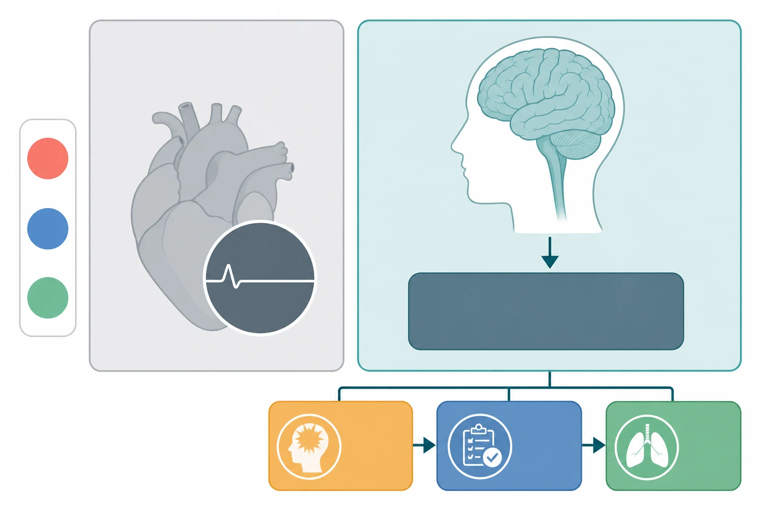
Death is recognised in law by two distinct routes, and brain death is one of them. The first route is circulatory death, the irreversible cessation of the circulation and the breathing that follows the stopping of the heart. This is the older and more familiar definition. The second route is death by neurological criteria, the irreversible cessation of all function of the entire brain including the brainstem. This was added to the law in most countries from the late 1960s onward, once mechanical ventilation made it possible for a dead brain to be kept inside a beating body. The two definitions are legally equivalent, so a child declared dead by neurological criteria is dead in every legal sense, and the certificate records that death. [10]
The conceptual basis of the neurological definition has two formulations that the fellowship candidate must distinguish. The whole-brain formulation, used in the United States and in the consensus documents of the American Academy of Neurology, requires the irreversible loss of all function of the entire brain including the brainstem. The brainstem formulation, used in the United Kingdom and in parts of the Commonwealth, requires the irreversible loss of the functions of the brainstem alone. Its reasoning is that the brainstem is the integrating centre whose loss in a ventilated patient means the whole brain has failed. In practice the bedside determination is identical across the two formulations, because the examination tests the brainstem reflexes and the apnoea centre. The World Brain Death Project harmonised these concepts across jurisdictions. [4][5]
The determination is also classified by the age of the child, because the developing brain and the newborn carry a caution that the older child does not. The 2023 consensus guideline and the pediatric guideline of Nakagawa and colleagues apply the determination to the term neonate of at least 37 weeks gestational age and to the infant, the child and the adolescent. The determination is not made in the neonate born before 37 weeks gestational age. The reason is that the immaturity of the brainstem reflexes in the very preterm infant makes the examination unreliable. It is applied with extra caution in the term neonate in the first days of life. The age band matters because it sets the observation period that separates the two examinations, which is longer for the neonate than for the older child. [2][3]
Epidemiology & Risk Factors
Brain death in children is the end point of a small number of catastrophic and irreversible brain insults. It is most often reached in the paediatric intensive care unit in a child who has been placed on a ventilator for a severe neurological injury. The causes fall into a recognisable set. Severe traumatic brain injury from a road traffic crash, a serious fall, or abusive head trauma is a leading cause in the infant and the older child. Hypoxic ischaemic encephalopathy from a drowning, a strangulation, a cardiac arrest, or a severe asphyxial event is the other dominant cause, and it is especially common in the toddler and the preschool child. Intracranial haemorrhage, central nervous system infection with brain swelling, and the catastrophic end of a metabolic or a neoplastic process account for the remainder. [2]
The risk of reaching brain death is shaped by the severity and the irreversibility of the initial injury, by the quality of the early resuscitation, and by whether the brain swelling and the rising pressure inside the skull have been controlled. A child whose intracranial pressure has run out of control, whose brainstem has been compressed by herniation, or whose perfusion has collapsed under hypotension and hypoxia is the child who progresses to the loss of all brain function. The epidemiology of organ donation is tied directly to this, because the brain-dead child on a ventilator is the ideal donor for the heart, the lungs, the liver, the kidneys and the pancreas. Most paediatric organs transplanted in Australia and New Zealand come from donation after brain death or from the controlled donation after circulatory determination of death pathway set out by Weiss and colleagues. [9]
The variability of practice around the world is itself a feature of the epidemiology, because it shapes how often the determination is made and how the organs are used. Lewis and colleagues surveyed the determination of death by neurological criteria around the world. They found wide variation in the number of examinations, the observation period, and the use of ancillary tests between countries. Braksick and colleagues showed that even within a single country the reported physician practices for the determination varied in the prerequisites and the apnoea test. This variation is the reason that the consensus documents, and the local framework such as the ANZICS statement, exist. The determination must be uniform and defensible wherever it is performed. [5][6]
Pathophysiology
The path to brain death is the path of an intracranial catastrophe that overwhelms the rigid skull and destroys the brainstem. It follows the same physics that govern raised intracranial pressure after any severe injury. The skull is a rigid box that holds the brain tissue, the blood and the cerebrospinal fluid in a fixed volume. When a haematoma expands or the brain swells after an impact or an asphyxia, the pressure inside the box rises steeply once the small compensatory reserve is exhausted. The cerebral perfusion pressure, which is the mean arterial pressure minus the intracranial pressure, collapses as the intracranial pressure climbs. The cerebral blood flow ceases, and the brain tissue becomes ischaemic. [4]
The end of the cascade is herniation, when the swollen brain shifts within the skull and compresses the brainstem against the tentorial edge and the foramen magnum. The brainstem houses the centres that drive the breathing, the pupillary responses, the eye movements, the corneal and the gag reflexes, and the consciousness. As the brainstem is compressed and then infarcted these functions are lost in sequence. When the ischaemia and the compression become complete and sustained, the loss of all brain and brainstem function becomes irreversible, and this is the physiological substrate of brain death. The cardiorespiratory machine continues for a time because the ventilator oxygenates the blood and the heart, with its own pacemaker, keeps beating. This is why the brain-dead child is warm and perfused rather than cold and pulseless. [4]
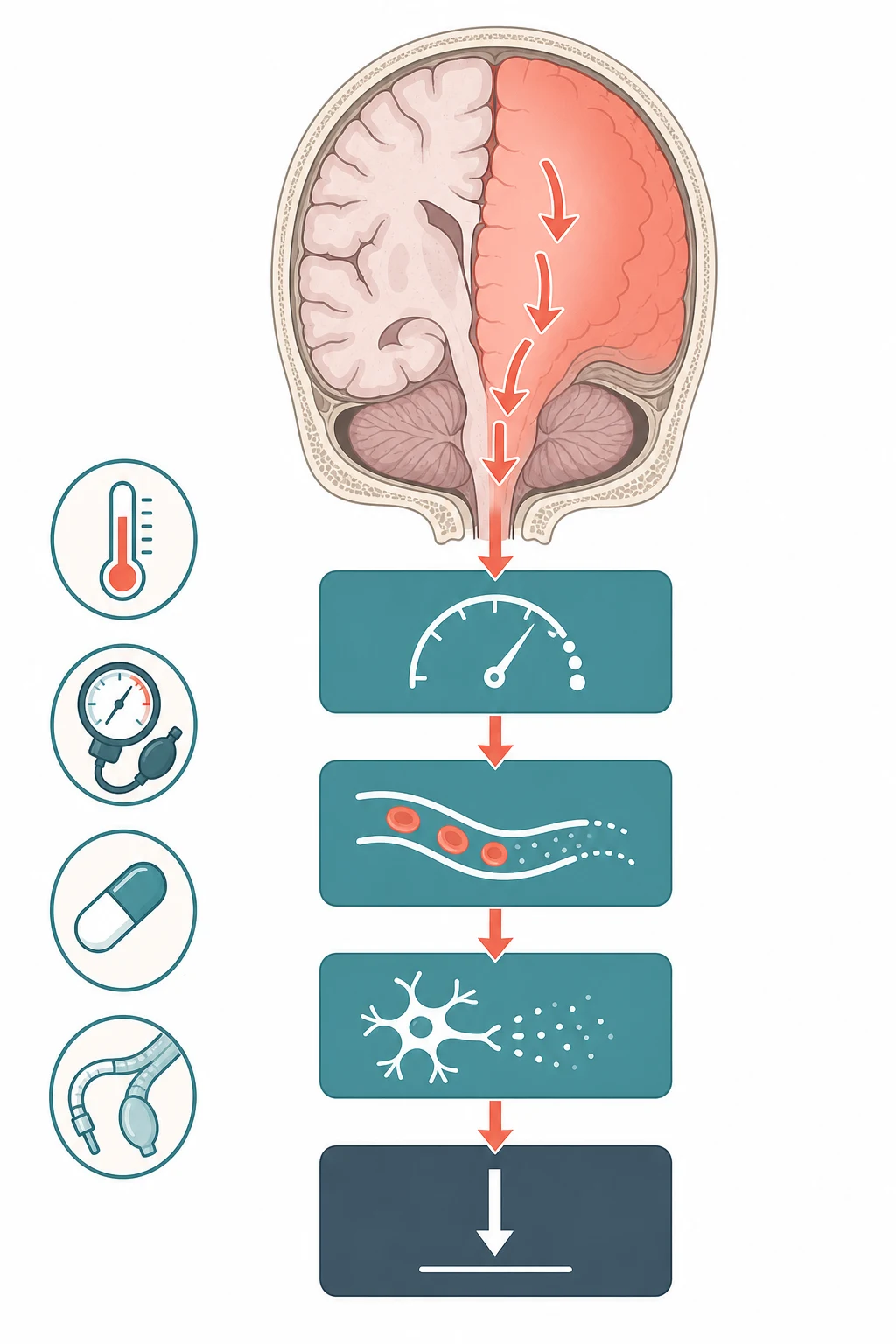
The irreversibility is the property that separates brain death from every reversible mimic, and it is built into the physiology and into the determination. Once the intracranial pressure has exceeded the mean arterial pressure for long enough to infarct the whole brain, including the brainstem, no recovery is possible. This is why the determination requires a sustained and known catastrophic cause. The distinction matters because a child in a barbiturate coma, a child with a profound metabolic derangement, or a child who is cold can lose the brainstem reflexes reversibly. None of these is brain death until the factor is removed and the loss proves permanent. The whole determination, from the prerequisite checks onward, is designed to prove that the loss is both total and irreversible. [1][4]
Clinical Presentation
The child who is being considered for a brain death determination is already deep in the paediatric intensive care unit, intubated and ventilated. The presentation is the profound and unresponsive coma of a known catastrophic brain injury. The child does not open the eyes, makes no sound, and shows no motor response to any stimulus, giving a Glasgow Coma Scale of 3. The pupils are often already fixed and dilated from the brainstem compression. The vital signs show a heart that is still beating and a blood pressure that the vasopressors and the fluids are holding in the normal range, and the ventilator is doing all of the breathing. The clinical picture is the backdrop against which the determination is made, and it is never the determination itself, because the picture alone does not prove irreversibility. [1]
How the child who reaches brain death presents across the age spectrum
A term neonate after a severe perinatal asphyxia who remains in deep coma and apnoeic on the ventilator beyond the first days of life, considered for the determination only after 37 weeks gestational age
A toddler who drowned and suffered a cardiac arrest, now in fixed coma with absent brainstem reflexes after the hypoxic ischaemic encephalopathy has run its course
A school-age child with severe traumatic brain injury from a road traffic crash whose intracranial pressure could not be controlled and whose pupils have become fixed and dilated
An adolescent with a ruptured arteriovenous malformation and an intracranial haemorrhage whose brainstem reflexes have been lost as the brainstem compressed
A child with a metabolic or a neoplastic catastrophe whose brain swelling has progressed to the loss of all brain function on the ventilator
The focused examination looks for the total absence of every brainstem reflex, and it is performed only after the prerequisites are met. The pupils are checked for size and reactivity to a bright light. The brain-dead child has pupils that are fixed in the midposition, often four to nine millimetres, with no constriction. The corneal reflex is absent to a touch of the cornea. The oculocephalic reflex, the doll's eye manoeuvre, is absent and is omitted if the cervical spine is not cleared. The oculovestibular reflex is absent to the cold caloric stimulus. The gag and the cough reflexes are absent to pharyngeal and tracheal suction, and the facial motor response to a deep pain is absent. The child makes no respiratory effort at all, which is confirmed separately in the apnoea test. [1][4]
Two presentations deserve a deliberate word because they catch the unwary. The first is the child with a few residual spinal reflexes, the Lazarus movements. In such a child a limb may twitch or the shoulders may shrug after the brainstem reflexes are gone. These spinal-cord-mediated movements can persist in the truly brain-dead child and do not exclude the determination. The second is the child on a high-dose barbiturate infusion for refractory intracranial pressure. In this child the brainstem reflexes may be abolished by the drug rather than by the death of the brainstem. The barbiturate must be cleared and confirmed at a therapeutic-zero level before the clinical examination is trusted, or an ancillary test must be used instead. [4][6]
Differential Diagnosis
The deep coma with absent brainstem reflexes has reversible causes that must be excluded before the determination is even contemplated, because a reversible encephalopathy must never be mistaken for brain death. These are the confounders, and excluding them is the second prerequisite of the determination. The clinician asks whether any factor remains that could itself abolish the brainstem reflexes. If such a factor is present and uncorrected, the clinical examination cannot be interpreted and the determination cannot proceed on it. [1][4]
Hypothermia
core temp under 36 C
- Suppresses brainstem reflexes and the respiratory drive
- Mimics brain death at a low core temperature
- Core temperature must be at least 36 degrees Celsius before testing
- Rewarm fully and confirm before any examination
Metabolic and endocrine
correct first
- Severe metabolic, acid-base, endocrine derangement abolishes reflexes
- Sodium, glucose, calcium, magnesium, ammonia and the blood gas are checked
- Each derangement is corrected to within the normal range
- Determination proceeds only once the biochemistry is normal
Drug and toxin
clear first
- Sedatives, opioids, barbiturates, anticonvulsants suppress the brainstem
- A drug screen is sent and at least five half-lives are allowed
- Barbiturate level must be therapeutic-zero, confirmed by EEG if needed
- An ancillary test is used if clearance cannot be assured
Neuromuscular blockade
train of four
- A residual neuromuscular blocker abolishes reflexes and breathing
- A train of four confirms full neuromuscular recovery
- The apnoea test alone would be falsely positive
- Confirm recovery before any testing
The reversible causes overlap with the structural causes, and the task is to separate the child whose brainstem is dead from the child whose brainstem is merely suppressed. A severe metabolic encephalopathy, a drug overdose, a hypothermic exposure, and a profound endocrine crisis can each present as a deep coma with absent reflexes in a ventilated child. Each is fully reversible with the correction of the factor. The structural causes of a brain-dead picture, the traumatic and the hypoxic and the haemorrhagic catastrophes, are distinguished by the known catastrophic event, the imaging, and the course in the intensive care unit. The determination is made only in the child whose cause is known, irreversible, and structurally devastating. [2][5]
Clinical & Bedside Assessment
The bedside assessment is the structured prerequisite check, run before any neurological testing, and it exists to guarantee that the examination will be interpretable. The first step is to confirm the catastrophic and irreversible cause. The clinician reads the imaging and the clinical course to establish that a known disaster has destroyed the brain, such as a massive traumatic injury, a devastating hypoxic ischaemic encephalopathy, or a large intracranial haemorrhage. The second step is to work through the confounders in turn, correcting each before the examination is attempted. [1]
The core temperature must be at least 36 degrees Celsius before the examination, because hypothermia suppresses the brainstem reflexes and the respiratory drive and is the classic mimic of brain death. The child is rewarmed fully if the temperature is low. The blood pressure must be normal for the age, sustained with fluids and vasopressors as needed, because hypotension alone can abolish the brainstem reflexes. The adult guideline of Wijdicks and colleagues sets a systolic blood pressure of at least 100 mmHg. The sedatives, the opioids and the anticonvulsants must be cleared, allowing at least five half-lives. A barbiturate level must be therapeutic-zero, confirmed by an electroencephalogram if there is any doubt, because a barbiturate coma is the commonest drug confounder in the neurointensive care unit. The metabolic, endocrine, electrolyte and acid-base disturbance must be corrected to the normal range, and the neuromuscular blockade must be excluded with a train of four. [1][3]
The neurological examination is then performed and documented by two independent senior clinicians, and it tests the consciousness and every brainstem reflex in turn. The child is in a deep coma with a Glasgow Coma Scale of 3. The pupils are fixed and dilated with no reaction to a bright light. The corneal reflex is absent. The oculocephalic reflex is absent and is omitted if the cervical spine is not cleared. The oculovestibular reflex is absent to the cold caloric stimulus, and the gag and the cough reflexes are absent to suction. The examination is recorded with the time and the names of the two clinicians, because the documentation is the legal record of the determination. [4][8]
Investigations
The investigations support the prerequisite checks rather than prove the determination, because brain death is a clinical diagnosis made at the bedside. The blood is sent for the electrolytes, the glucose, the calcium, the magnesium, the ammonia and the blood gas to exclude and correct a metabolic or an acid-base confounder. A drug and a toxin screen is sent to exclude a depressant, and a barbiturate level is checked when a barbiturate infusion has been used. The imaging, the computed tomography and the magnetic resonance imaging, has already established the catastrophic cause. It is reviewed to confirm that a known and irreversible structural disaster underlies the coma. [1][4]
The ancillary tests, sometimes called confirmatory tests, are not required when the prerequisites are met and the clinical examination and the apnoea test are complete and concordant. The clinical determination alone is sufficient in that case. The ancillary test is required when a part of the examination cannot be performed or interpreted, when the apnoea test cannot be completed safely or is inconclusive, or when a confounder such as a barbiturate cannot be fully cleared. The options are the cerebral angiography, which is the historical gold standard, the nuclear brain scan or brain scintigraphy, the transcranial Doppler ultrasonography showing the absent cerebral blood flow, the electroencephalogram showing electrocerebral silence, and the computed tomography or the magnetic resonance angiography. Each tests for the absence of the cerebral blood flow or the cortical electrical activity, and the choice is governed by the local protocol and the availability. [1][2]
A common pitfall is to reach for an ancillary test too early, in the belief that it makes the determination more certain. The 2011 pediatric guideline of Nakagawa and colleagues was clear that an ancillary test is not a substitute for the clinical examination when the prerequisites are met. The ancillary tests have their own pitfalls, such as a false signal in a child with a skull defect or an open fontanelle, or a residual flow on an early study. The ancillary test resolves the determination only in the child in whom the clinical examination cannot be completed. In every other child the clinical examination and the apnoea test are the determination. [2][8]
Management — Resuscitation
The resuscitation of the child who is on the path to brain death is the prevention of the secondary brain injury and the maintenance of the organ perfusion. It is the same intensive care that protects the injured brain while it is still salvageable. The airway is secured and the cervical spine immobilised in the trauma case. The oxygenation and the ventilation are controlled, the blood pressure is maintained with fluids and vasopressors, and the intracranial pressure is managed with the stepwise ladder of sedation, hyperosmolar therapy and surgery. The goal at this stage is the survival of the child. The brain death determination is contemplated only when this resuscitation has failed and the brain is irretrievably lost. [4]
Once the catastrophic loss is established and the brain is irretrievable, the resuscitation shifts to the preservation of the organs for the donation. The brain-dead child is now a potential donor whose heart, lungs, liver, kidneys and pancreas may save several other lives. The physiological derangement that follows brain death is real and damaging to the organs. It includes the loss of the vasomotor tone with a falling blood pressure, the diabetes insipidus from the posterior pituitary failure, the hypothermia from the hypothalamic failure, and the hormonal derangement. The intensive care continues, now with the aim of keeping the organs perfused and oxygenated. The blood pressure, the temperature, the electrolytes and the glucose are all kept in the normal range until the retrieval. [9]
The team that manages this donor resuscitation is the treating intensive care team, and it remains separate from the transplant team throughout. The dead donor rule and the trust of the family require that the team caring for the child is not the team that will receive the organs. The donation specialist is engaged early, the family is approached by a clinician trained in the donation conversation, and the consent is sought and recorded according to the local registry and the law. The somatic support continues until the organs are retrieved in the operating theatre or until the family declines the donation and the ventilator is withdrawn. [9][10]
Management — Definitive & Stepwise
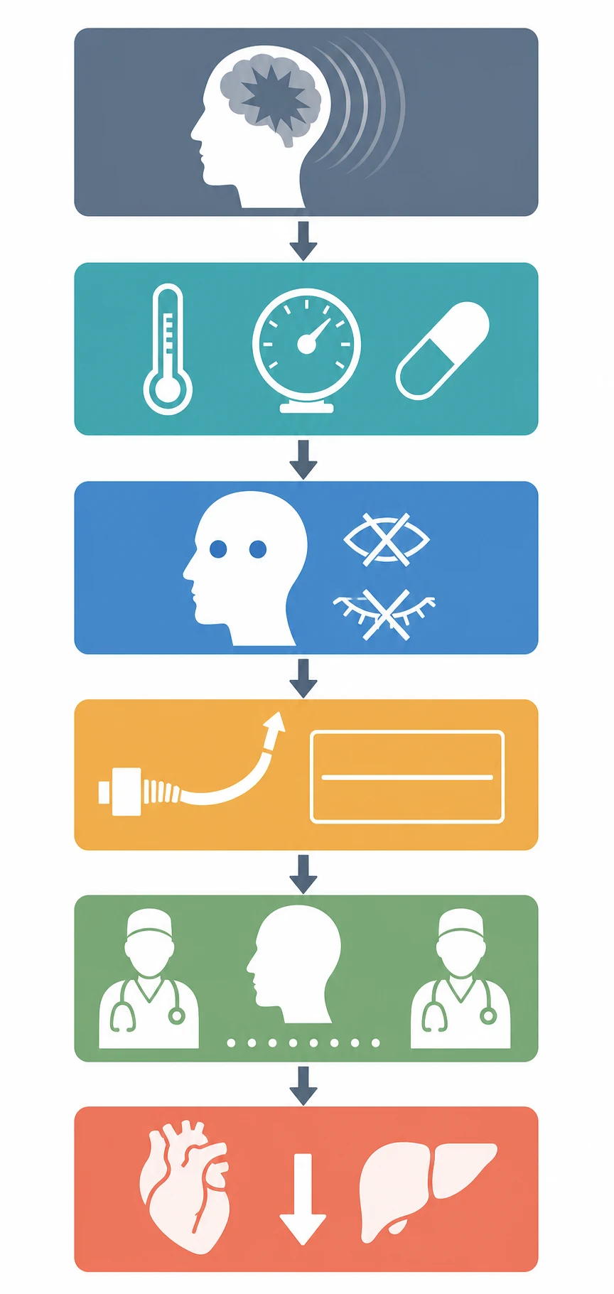
The definitive step is the determination itself, performed as a structured sequence that converts the suspicion of brain death into a legal declaration of death. The sequence begins with the prerequisite check. It runs through the bedside examination of the coma and the absent brainstem reflexes, then the apnoea test, and then the second examination separated by the observation period, and it ends with the declaration and the donation. Each step is documented with the time and the two clinicians, because the record is the legal proof of the determination. [1][3]
[1] [3]The apnoea test is the step that tests the last surviving brainstem function, the drive to breathe, and it is the most testable single element of the determination. The child is first preoxygenated with pure oxygen for at least ten minutes to denitrogenate the lungs and protect against the hypoxia. The arterial carbon dioxide is normalised to a baseline in the normal range. The ventilator is then disconnected, and the oxygen is delivered to the trachea while the carbon dioxide is allowed to rise. The clinician watches the chest and the abdomen for any respiratory effort. An arterial blood gas is taken at the end, and the test is positive when there is no respiratory effort and the PaCO2 reaches at least 60 mmHg or rises by at least 20 mmHg from the baseline. The test is negative, and brain death is excluded, the moment any respiratory effort appears, and the ventilator is reconnected at once. [1][3]
The apnoea test for the determination of brain death
Dose
Preoxygenate with pure oxygen for at least ten minutes, normalise the PaCO2 to the baseline, disconnect the ventilator, and deliver oxygen to the trachea while the PaCO2 rises
The determination requires two clinical evaluations, each including the apnoea test, performed by two independent senior clinicians. The two are separated by an observation period whose length depends on the age. The 2011 pediatric guideline of Nakagawa and colleagues set the observation period at 48 hours for the term neonate up to 30 days of age and at 24 hours for the infant and the child from 30 days to 18 years. The two clinicians are senior practitioners who are not part of the transplant team. The 2023 consensus allows a single evaluation in the adult in many jurisdictions, but the pediatric standard, and the ANZICS framework in Australia and New Zealand, retains the two-examination standard for the child. The candidate should state the local requirement. [2][8]
The stepwise pathway of the brain death determination
Confirm a known and irreversible catastrophic brain injury from the imaging and the clinical course
Correct and exclude every confounder, with a core temperature at least 36 degrees Celsius and a blood pressure normal for age
Perform the bedside examination of the coma and the absent brainstem reflexes with two independent senior clinicians
Run the apnoea test, positive when there is no respiratory effort and the PaCO2 reaches at least 60 mmHg or rises by at least 20 mmHg
Repeat the full evaluation after the observation period of 48 hours for the neonate up to 30 days and 24 hours for the infant and child
Declare the death, record it with the time and the two clinicians, and offer the donation after brain death pathway
When the two examinations are complete and concordant, death is declared and recorded with the time, the date, the findings and the names of the two clinicians. This is the legal record of the death. The donation after brain death pathway is then offered to the family, in partnership with the donation specialist, and the donor resuscitation continues until the organ retrieval or the withdrawal of the ventilator. When the apnoea test cannot be completed or a confounder persists, an ancillary test confirms the absence of the cerebral blood flow or the cortical activity. The determination then proceeds on the combination of the clinical examination and the ancillary test. [9][10]
Specific Subtypes & Scenarios
The term neonate is the scenario in which the determination carries the most caution, because the immature brainstem and the open fontanelle change the reliability of the examination. The determination is not made in the neonate born before 37 weeks gestational age. The brainstem reflexes are not reliably assessable in the very preterm infant. It is applied with care in the term neonate in the first days of life. The observation period for the term neonate up to 30 days is the longest, at 48 hours, because the guideline retains the caution that the newborn deserves, and the two clinicians perform the two examinations across that interval. The cause is most often a severe hypoxic ischaemic encephalopathy or a large intracranial haemorrhage in this age group. [2][3]
The child on the extracorporeal membrane oxygenation is the scenario in which the apnoea test and the ancillary tests must be adapted, because the circuit complicates the assessment of the circulation and the respiration. The apnoea test can be performed with the adjustment of the circuit sweep gas to allow the carbon dioxide to rise, but it is technically demanding. Many units use an ancillary test such as the cerebral angiography or the nuclear brain scan to confirm the absence of the cerebral blood flow in this setting. The determination is otherwise the same, with the prerequisites met and the brainstem reflexes absent. The candidate who faces this scenario should state that the ancillary test is preferred when the apnoea test is not feasible. [4]
The donation after circulatory determination of death is the separate pathway that runs alongside the donation after brain death, and the candidate must distinguish the two. In the donation after brain death, the child is declared dead by the neurological criteria while the heart is still beating, and the organs are retrieved from a perfused donor. In the controlled donation after circulatory determination of death, the withdrawal of the ventilator is planned in a child whose death by neurological criteria is not established and whose prognosis is futile. The death is then declared by the circulatory criteria after the heart stops, and the organs are retrieved after a defined no-touch interval. The Canadian pediatric guideline of Weiss and colleagues set out the controlled pathway, and the two pathways share the principles of consent, the separation of the teams, and the donor care. [9]
The child with a family objection to the determination or the donation is the scenario that tests the communication as much as the medicine. The determination of brain death is the declaration of a death that has already occurred, and it is not the withdrawal of life support. The law in most jurisdictions allows the determination to proceed even over a reservation, though the practice is to engage the family with great care and time. The organ donation, by contrast, requires the consent of the family and the respect of the registered decision. The conversation is held by a trained donation specialist, with the treating team providing the clinical information. The candidate should state that the determination and the donation are separable, and that the family is supported throughout. [5][10]
Complications & Pitfalls
The central complication of the determination is the error that the whole sequence is designed to prevent, the declaration of brain death in a child whose brain is not truly dead. The classic pitfalls are the failure to correct a confounder, the failure to clear a drug, and the failure to complete the apnoea test. Each can produce a falsely absent set of brainstem reflexes. The candidate who declares brain death in a cold, hypotensive, or barbiturate-loaded child has made the cardinal error, and the prerequisite check exists to make it impossible. The second pitfall is the misreading of the apnoea test, either by stopping it too early before the carbon dioxide has risen, or by ignoring a small respiratory effort that excludes the determination. [1][6]
The physiological instability that follows brain death is the complication that threatens the donation, and it must be managed actively from the moment of the loss. The loss of the vasomotor tone produces a falling blood pressure that must be supported with fluids and vasopressors. The diabetes insipidus produces a massive urine output that must be matched with vasopressin and fluid. The hypothalamic failure produces a hypothermia that must be corrected, and the hormonal derangement may need a hormonal resuscitation with vasopressin, thyroid hormone, insulin and corticosteroid. The arrhythmias and the coagulopathy are common, and the donor management is an active intensive care that continues until the retrieval, because a poorly managed donor yields poor organs. [9]
A subtler pitfall is the spinal reflex, the movement of a limb in a truly brain-dead child that alarms the bedside nurse and the family. The Lazarus signs, the arm flexion, the head turning, and the stepping movements, are generated by the intact spinal cord below the dead brainstem. They do not exclude the determination, provided the brainstem reflexes are absent and the apnoea test is positive. The clinician who abandons the determination on the strength of a spinal movement has mistaken a cord reflex for a brainstem recovery. The candidate should be ready to explain the distinction in an examination. [4][6]
Prognosis & Disposition
The prognosis of brain death is, by definition, the prognosis of death, because the determination is the legal declaration that the child has died. There is no recovery from a correctly made determination. The long series of cases that have been reported to recover after a brain death determination have, on review, been cases in which the determination was incorrectly made, usually because a confounder was not corrected. The irreversibility is the core of the definition, and the consensus documents reaffirm it. This is why the determination is made with such rigor and with the two examinations and the observation period. [3][4]
The disposition is the declaration of the death, the recording of it with the time and the two clinicians, and the transition to the donation after brain death pathway. The child remains in the paediatric intensive care unit, the donor resuscitation continues, and the donation specialist and the family manage the consent and the conversation. When the family consents to the donation, the child is transferred to the operating theatre for the organ retrieval, and the organs are allocated to the recipients. When the family declines the donation, or when the child is not a suitable donor, the ventilator is withdrawn with dignity, and the care shifts to the bereavement support of the family. [9][10]
The bereavement is a part of the disposition that the fellowship candidate must not omit. The family of a brain-dead child has suffered a sudden and a catastrophic loss, and the donation conversation, if it is to be offered, must be handled with sensitivity and without pressure. The family must be given the time and the information to decide. The follow-up includes the bereavement support, the return of the personal effects, and, where the family wishes, the feedback on the recipients of the organs. All of these are part of the care of the family after the death of the child. [9]
Special Populations
The term neonate is the population in whom the determination carries the most caution and the longest observation period, and in whom the cause is most often the hypoxic ischaemic encephalopathy. The determination is not made before 37 weeks gestational age. The observation period is 48 hours up to 30 days of age, and the two clinicians perform the examinations across that interval. The immaturity of the brainstem reflexes and the presence of the open fontanelle are the reasons for the caution. The ancillary test is used more often in this age group when the examination is uncertain. [2][3]
The Aboriginal and Torres Strait Islander child and the child from a remote setting is the population in whom the access and the cultural respect shape the determination and the donation. The retrieval to a paediatric intensive care unit, the long distance from the family, and the cultural obligations around the death all require an early and a respectful engagement with the family and the community. The consent for the donation is sought with the cultural context in mind, the interpreter is engaged when the language is a barrier, and the return of the child to the community is facilitated when it is the wish of the family. The determination itself is the same, but the communication and the cultural care are central. [9]
The child with a pre-existing neurodisability or a complex chronic condition is the population in whom the baseline neurological state complicates the interpretation. The determination must be made with extra care in this group. The baseline brainstem reflexes may already be altered by the condition or the medication, the family is involved early in the decisions, and the distinction between the chronic baseline and the acute catastrophe is made carefully from the imaging and the course. The adolescent is at risk from the road traffic crash, the drowning, and the suicide, and the transition to the adult services and the consent of the young person are part of the determination and the donation. [4][5]
The child from a refugee or a migrant family is the population in whom the communication and the continuity are the challenges. The language barrier can hide the prior state and the values of the family. An interpreter is engaged early, and the family is given the time and the information to understand the determination and the donation. The legal framework of the host country applies to the determination, but the cultural and the religious context of the family is respected throughout the conversation, and the family is supported to a decision that is right for them. [5][10]
Evidence, Guidelines & Regional Differences
The evidence base is anchored by the consensus guidelines, and the candidate must know the lineage of them. The American Academy of Neurology guideline reported by Wijdicks and colleagues in 2010 set out the criteria for the determination of brain death in the adult. It covered the prerequisites, the bedside examination, and the apnoea test, and it remains the standard for the adult. The pediatric guideline reported by Nakagawa and colleagues in 2011 extended the framework to the infant and the child. It added the two examinations and the observation period of 48 hours for the neonate up to 30 days and 24 hours for the older child. The 2023 consensus guideline of Greer and colleagues, from the American Academy of Neurology, the American Academy of Pediatrics, the Child Neurology Society and the Society of Critical Care Medicine, harmonised the adult and the pediatric pathways into a single document. [1][2][3]
AAN 2010 Wijdicks
adult guideline
- Prerequisites, brainstem reflexes, and the apnoea test for the adult
- PaCO2 at least 60 mmHg or a rise of at least 20 mmHg
- Core temperature at least 36 C and systolic BP at least 100 mmHg
- The standard for the adult determination
Pediatric 2011 Nakagawa
infant and child
- Two examinations with the apnoea test by two clinicians
- Observation of 48 hours for the neonate up to 30 days and 24 hours for the child
- Applied from 37 weeks gestational age
- Ancillary test not required when the examination is complete
Consensus 2023 Greer
adult and child
- Harmonised the adult and the pediatric pathways
- Single evaluation may suffice in the adult in some jurisdictions
- Two evaluations retained for the pediatric standard
- The current AAN, AAP, CNS and SCCM consensus
World Brain Death Project 2020
global
- Greer and colleagues, reported in JAMA
- Mapped the determination and the variability across the world
- Set out the global common ground
- The reference for the international comparison
The World Brain Death Project, reported by Greer and colleagues in JAMA in 2020, is the global reference, and it set out the determination and the variability across the world. Lewis and colleagues documented the variation in the number of examinations, the observation period, and the ancillary tests between countries. Braksick and colleagues showed the variation in the physician practices within a country. Wang and colleagues showed that the uniformity of the policies has improved over time but that the variation remains. The ANZICS statement on death and organ donation is the framework that governs the practice in Australia and New Zealand, and it requires the two examinations by two independent senior clinicians, neither of whom is part of the transplant team. [4][5][6][7]
The regional differences that the candidate should state are the number of examinations, the observation period, and the use of the ancillary tests. The United States adult practice, under the 2023 consensus, may use a single evaluation, while the pediatric and the ANZICS practice retain the two examinations separated by the observation period. The United Kingdom applies the brainstem formulation with the two doctors. Canada applies the neurological determination from the Canadian forum recommendations reported by Shemie and colleagues, with the controlled donation after circulatory determination of death alongside the donation after brain death from the guideline of Weiss and colleagues. The controversies that remain are the role of the ancillary tests, the number of examinations in the adult, and the management of the donation after circulatory determination of death. [5][9][10]
Exam Pearls
The brain death determination as the D E A D steps
References
- [1]Wijdicks EF, Varelas PN, Gronseth GS, et al Evidence-based guideline update: determining brain death in adults: report of the Quality Standards Subcommittee of the American Academy of Neurology. Neurology, 2010.PMID 20530327
- [2]Nakagawa TA, Ashwal S, Mathur M, et al Clinical report—Guidelines for the determination of brain death in infants and children: an update of the 1987 task force recommendations. Pediatrics, 2011.PMID 21873704
- [3]Greer DM, Kirschen MP, Lewis A, et al Pediatric and Adult Brain Death/Death by Neurologic Criteria Consensus Guideline. Neurology, 2023.PMID 37821233
- [4]Greer DM, Shemie SD, Lewis A, et al Determination of Brain Death/Death by Neurologic Criteria: The World Brain Death Project. JAMA, 2020.PMID 32761206
- [5]Lewis A, Bakkar A, Kreiger-Benson E, et al Determination of death by neurologic criteria around the world. Neurology, 2020.PMID 32576632
- [6]Braksick SA, Robinson CP, Gronseth GS, et al Variability in reported physician practices for brain death determination. Neurology, 2019.PMID 30804063
- [7]Wang HH, Varelas PN, Henderson GV, et al Improving uniformity in brain death determination policies over time. Neurology, 2017.PMID 28077490
- [8]Nakagawa TA, Ashwal S, Mathur M, et al Guidelines for the determination of brain death in infants and children: an update of the 1987 Task Force recommendations. Crit Care Med, 2011.PMID 21849823
- [9]Weiss MJ, Hornby L, Rochwerg B, et al Canadian Guidelines for Controlled Pediatric Donation After Circulatory Determination of Death-Summary Report. Pediatr Crit Care Med, 2017.PMID 28925929
- [10]Shemie SD, Doig C, Dickens B, et al Severe brain injury to neurological determination of death: Canadian forum recommendations. CMAJ, 2006.PMID 16534069