Phys · cardiovascular
Cardiac Tumours AND Systemic Manifestations OF Cardiac Disease
Also known as Cardiac Tumours AND Systemic Manifestations OF Cardiac Disease · cardiac tumours and systemic manifestations of cardiac disease
Consultant-physician depth guide to Cardiac Tumours AND Systemic Manifestations OF Cardiac Disease for FRACP DWE/DCE preparation — presentation, differentials, investigations, management, complications and exam angles.
On this page & tools
Your progress
Saved locally on this device.
Practise this topic
Target exams
Red flags
The answer first
Cardiac tumours are primary or secondary neoplasms of the heart. Primary cardiac tumours are rare (autopsy incidence 0.001-0.3 per cent) and are mostly benign (75 per cent); metastatic involvement of the heart is 20-40 times more common. The FRACP candidate must defend three syndromes: the myxoma triad (intracardiac obstruction, embolisation, systemic constitutional features mimicking vasculitis or infection), the right atrial myxoma mimicking right-heart failure or pulmonary embolism, and the carcinoid heart disease of right-sided valvular fibrosis. The imaging modality of choice is transthoracic echocardiography for initial detection, with cardiac MRI for tissue characterisation and assessment of infiltration. [1] [2]
The answer-first synthesis: recognise the syndrome (obstructive, embolic, constitutional), image with echocardiography, characterise with MRI/CT, refer for surgical resection (the treatment of curable primary cardiac tumours), and screen for associated syndromes (Carney complex, Li-Fraumeni). [1] [9]

Definition and overview
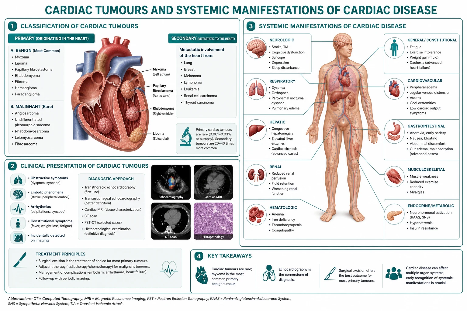
Cardiac tumours are classified by histology (benign vs malignant), by origin (primary vs metastatic), and by location (chamber, valve, pericardium). The most common primary cardiac tumour in adults is the atrial myxoma (50 per cent), a benign tumour of mesenchymal origin typically arising from the interatrial septum at the fossa ovalis, three to four times more often in the left atrium than the right. The next most common benign tumours are papillary fibroelastoma (on valves), lipoma, and fibroma. Malignant primary tumours are predominantly sarcomas (angiosarcoma, rhabdomyosarcoma, leiomyosarcoma). Metastatic cardiac involvement most commonly arises from lung, breast, melanoma, renal, and lymphoproliferative malignancies, and may manifest as pericardial effusion, myocardial infiltration, or intracavitary extension via the vena cava (renal cell carcinoma is the classic "tumour thrombus" extending up the IVC). [1] [5]
Epidemiology
Primary cardiac tumours have an autopsy incidence of 0.001-0.3 per cent. Of these, 75 per cent are benign and 25 per cent malignant. Atrial myxoma is the commonest primary tumour (50 per cent of all primary cardiac tumours), with a female predominance (65-75 per cent) and a mean age at presentation of 50 years. Papillary fibroelastoma is the commonest valvular tumour. Sarcomas (angiosarcoma commonest) have a slight male predominance and a mean age of 40-50 years. Metastatic cardiac involvement is found at autopsy in 10-12 per cent of patients with malignancy, far exceeding primary cardiac malignancy. [1] [7]
Pathophysiology linked to bedside decisions
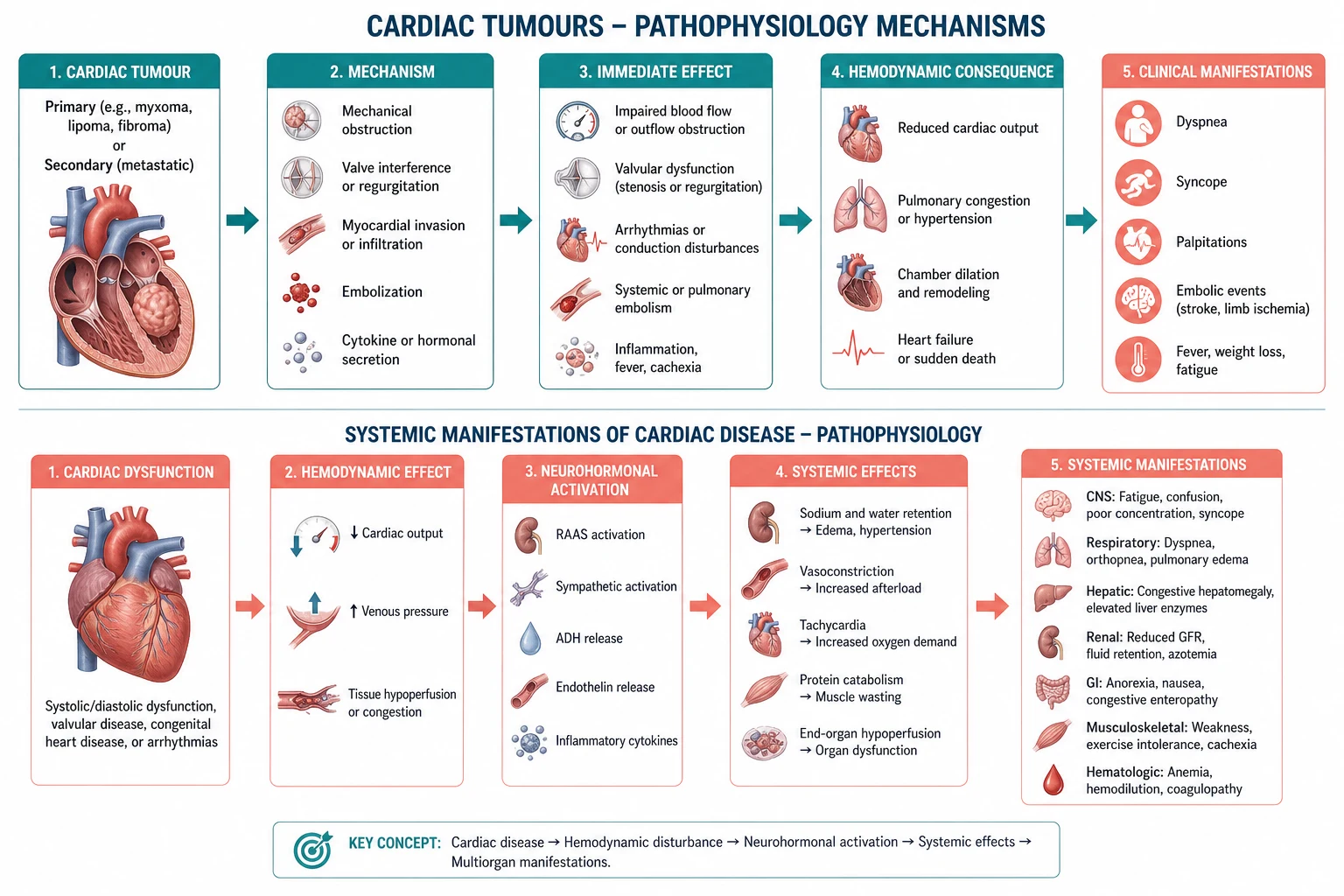
The pathophysiology of cardiac tumours is mechanical, embolic, and constitutional. [2]
- Mechanical obstruction: myxomas grow on a stalk and prolapse through the mitral (or tricuspid) valve during diastole, mimicking rheumatic mitral stenosis with a tumour "plop" on auscultation, diastolic murmur, and signs of left atrial hypertension (dyspnoea, orthopnoea, pulmonary oedema). Position-dependent symptoms (worse when upright, better when supine) are the classic clue.
- Embolisation: tumour fragments or surface thrombus embolise systemically (left-sided tumours to brain, retina, limbs, viscera; right-sided tumours to lungs causing pulmonary hypertension or infarction). Cerebral embolisation may mimic stroke; retinal embolisation may cause visual loss; peripheral embolisation may cause limb ischaemia. Histology of an excised embolus may be the first clue to an occult myxoma. [3] [6]
- Constitutional features: myxomas secrete interleukin-6 (IL-6), driving fever, weight loss, fatigue, myalgia, arthralgia, and a raised erythrocyte sedimentation rate and C-reactive protein — the syndrome mimics infective endocarditis, autoimmune vasculitis, or lymphoma. The correct recognition is "myxoma — the great mimicker." [4] [2]
- Malignant tumours infiltrate the myocardium causing arrhythmia, heart failure, and pericardial effusion with tamponade; angiosarcoma typically arises in the right atrium and causes haemorrhagic pericardial effusion. [8]
Classification that changes management
The classification that changes action separates benign from malignant, primary from metastatic, and sporadic from familial (Carney complex). [1] [9]
| Tumour | Location | Behaviour | Management |
|---|---|---|---|
| Atrial myxoma (sporadic) | LA, fossa ovalis (75 per cent); RA (20 per cent) | Benign but mechanically and embolically dangerous | Surgical resection (curative); screen for Carney complex |
| Myxoma in Carney complex | Multiple, atypical sites, recurrent | AD, PRKAR1A mutation; associated pigmentation, endocrine tumours, schwannomas | Resect; lifelong surveillance; family screening |
| Papillary fibroelastoma | Valves (aortic, mitral) | Benign; embolic risk | Resect if symptomatic or greater than 10 mm and mobile |
| Lipoma, fibroma, rhabdomyoma | Variable | Benign; rhabdomyoma associated with tuberous sclerosis | Resect if symptomatic |
| Angiosarcoma, rhabdomyosarcoma, leiomyosarcoma | RA, pericardium (angiosarcoma); variable | Malignant; poor prognosis | Multimodal: surgery, chemotherapy, radiotherapy; often palliative |
| Metastatic involvement | Pericardium commonest; myocardium; intracavitary extension | Reflects advanced malignancy | Treat the primary; pericardiocentesis for tamponade |
Clinical features
The classic triad of atrial myxoma is intracardiac obstruction, systemic embolisation, and constitutional features. [2]
- Obstructive: position-dependent dyspnoea, orthopnoea, pulmonary oedema (left atrial myxoma); peripheral oedema, hepatomegaly, syncope (right atrial myxoma). Auscultation: tumour "plop" in early diastole (low-pitched third sound as the tumour strikes the ventricular wall or valve), a diastolic rumble resembling mitral stenosis (without the opening snap of rheumatic MS), and a systolic murmur if mitral regurgitation results from valve trauma. Symptoms change with position — a clue to suspect on the ward round.
- Embolic: stroke, transient ischaemic attack, retinal artery occlusion, limb or visceral ischaemia; multiple systemic emboli in a young patient with sinus rhythm is a red flag for myxoma or endocarditis. Right-sided myxoma causes pulmonary embolism, pulmonary hypertension, and cor pulmonale. [3] [10]
- Constitutional: fever, weight loss, fatigue, myalgia, arthralgia, Raynaud phenomenon, clubbing; laboratory features include raised ESR, CRP, anaemia, leukocytosis, and hypergammaglobulinaemia — the picture mimics infective endocarditis, vasculitis, or lymphoma. IL-6 elevation correlates with tumour size and constitutional severity. [4]
DCE trap: A young woman with fever, weight loss, raised inflammatory markers, multiple embolic events, and a mitral diastolic murmur but no history of rheumatic fever — suspect atrial myxoma, not endocarditis or SLE. Echocardiography resolves the question in minutes. [2] [4]
Differentials and discrimination
The differential for an intracardiac mass includes myxoma, papillary fibroelastoma, thrombus (especially in atrial fibrillation or within the left atrial appendage), vegetation (endocarditis), lipoma, fibroma, rhabdomyoma, sarcoma, metastatic deposit, and tumour thrombus (renal cell extending up the IVC). Discriminating features: myxoma is attached to the fossa ovalis by a stalk; thrombus is in the left atrial appendage or akinetic ventricular segment; vegetation is on the upstream side of a valve; fibroelastoma is a frond-like lesion on a valve; tumour thrombus extends from the IVC into the right atrium (think renal cell). Cardiac MRI with late gadolinium enhancement and tissue characterisation helps discriminate. [1] [5]
Investigations
Transthoracic echocardiography (TTE) is the first-line modality for any suspected cardiac mass, with sensitivity above 95 per cent for myxoma. A pedunculated mass arising from the interatrial septum is characteristic. [1]
Transoesophageal echocardiography (TOE) better defines attachment, mobility, and the relationship to valves, especially for small or right-sided lesions, and is the modality of choice intra-operatively. [1]
Cardiac MRI is the gold standard for tissue characterisation: T2-weighted imaging for oedema, late gadolinium enhancement for fibrosis, and perfusion sequences for vascularity. Lipoma is hyperintense on T1 and suppresses on fat-saturation; fibroma shows marked late enhancement; angiosarcoma has a "cauliflower" appearance with heterogeneous enhancement and pericardial effusion. [1] [8]
CT is useful for assessing calcification (fibroma, hamartoma) and for staging when malignancy is suspected. Coronary angiography before surgery identifies tumour vascularity (myxomas have a characteristic tumour blush) and rules out concomitant coronary disease requiring bypass. [1]
Baseline bloods: FBC (anaemia, leukocytosis), ESR and CRP (elevated in myxoma), renal and hepatic function, and IL-6 if available. Blood cultures if endocarditis is in the differential. ECG may show non-specific changes, atrial fibrillation, or signs of chamber enlargement. [7]
Management — immediate then definitive
- Surgical resection is the treatment of choice for primary cardiac tumours, ideally before embolisation or haemodynamic compromise. Myxoma resection is curative; recurrence rate is 1-3 per cent for sporadic myxoma, 20-40 per cent for familial or Carney complex myxoma. Median sternotomy with cardiopulmonary bypass and tumour excision with a margin of normal atrial septum, with reconstruction of the septum as needed. [1]
- Papillary fibroelastoma — resect if symptomatic (embolic event), greater than 10 mm in size, or highly mobile; small non-mobile lesions may be observed with serial imaging. [1]
- Sarcoma — multimodal therapy with surgical debulking where feasible, chemotherapy (e.g., doxorubicin-based for soft-tissue sarcoma), and radiotherapy where the histology permits; prognosis is poor (median survival less than 1 year for angiosarcoma). [8]
- Metastatic involvement — treat the underlying malignancy; pericardiocentesis or pericardial window for tamponade; palliative care in advanced disease. [8]
- Carney complex screening — for any young patient or patient with multiple myxomas, atypical locations, or family history: skin pigmentation (lentigines, blue naevi), endocrine tumours (pituitary, adrenal, testicular), and PRKAR1A genetic testing with family counselling. [9]
Complications and prognosis
Untreated atrial myxoma may cause sudden death (acute mitral valve obstruction), embolic stroke, or chronic heart failure. Post-resection complications include recurrence (1-3 per cent sporadic, 20-40 per cent Carney complex), atrial fibrillation, and surgical wound complications. Sarcoma has a median survival of 6-12 months for angiosarcoma and 12-24 months for other subtypes; metastatic cardiac involvement portends survival measured in weeks to months. [1] [8]
Special populations and multimorbidity
In pregnancy, atrial myxoma may cause position-dependent syncope or pulmonary oedema — surgical resection is safe and indicated. In patients with renal cell carcinoma extending up the IVC into the right atrium, combined nephrectomy and tumour thrombectomy with cardiopulmonary bypass is the standard. In frail or elderly patients, the risk-benefit of surgery for a slow-growing benign tumour must weigh peri-operative mortality (1-5 per cent for elective myxoma resection) against life expectancy. [1] [6]
DCE long-case angles
Open with the synthesis ("Mr X is a 47-year-old man presenting with a 3-month history of fever, weight loss, myalgia, and a single episode of right-arm weakness; transthoracic echocardiography demonstrated a 3 cm pedunculated left atrial mass arising from the fossa ovalis, consistent with an atrial myxoma"). Present the problem list and integrated plan covering resection, IL-6 monitoring, exclusion of Carney complex (skin exam, endocrine screening, PRKAR1A testing), family counselling, and post-operative echocardiographic surveillance. [1] [9]
DCE short-case angles
Be prepared to recognise the tumour plop on auscultation, to interpret an echocardiographic image of a left atrial mass, to defend the indication for surgical resection, and to counsel a patient on the risk of recurrence and the importance of surveillance echocardiography. [2]
Exam traps
- Diagnosing "mitral stenosis" without echocardiography in a young patient with position-dependent symptoms — the diagnosis may be myxoma. [2]
- Treating "endocarditis" or "vasculitis" with empirical antibiotics or steroids without echocardiography in a patient with constitutional features and emboli — myxoma is missed. [4]
- Forgetting to screen for Carney complex in a young patient or in recurrent myxoma — the familial form recurs and has endocrine associations. [9]
- Missing a right atrial myxoma in a patient with pulmonary hypertension, recurrent pulmonary embolism, and right-heart failure. [7]
- Treating a sarcoma as if curable by resection alone — multimodal therapy is required, and prognosis is poor. [8]
Exam pearls
- Myxoma triad: obstruction, embolisation, constitutional.
- Tumour "plop" + diastolic rumble + position-dependent symptoms = myxoma until proven otherwise.
- IL-6 elevation causes the constitutional syndrome.
- Multiple systemic emboli in sinus rhythm in a young patient = myxoma or endocarditis.
- Cardiac MRI characterises tissue; TTE detects; TOE defines.
- Carney complex: AD, PRKAR1A, lentigines, endocrine tumours, schwannomas — screen the family. [1] [9]
Systemic manifestations of cardiac disease
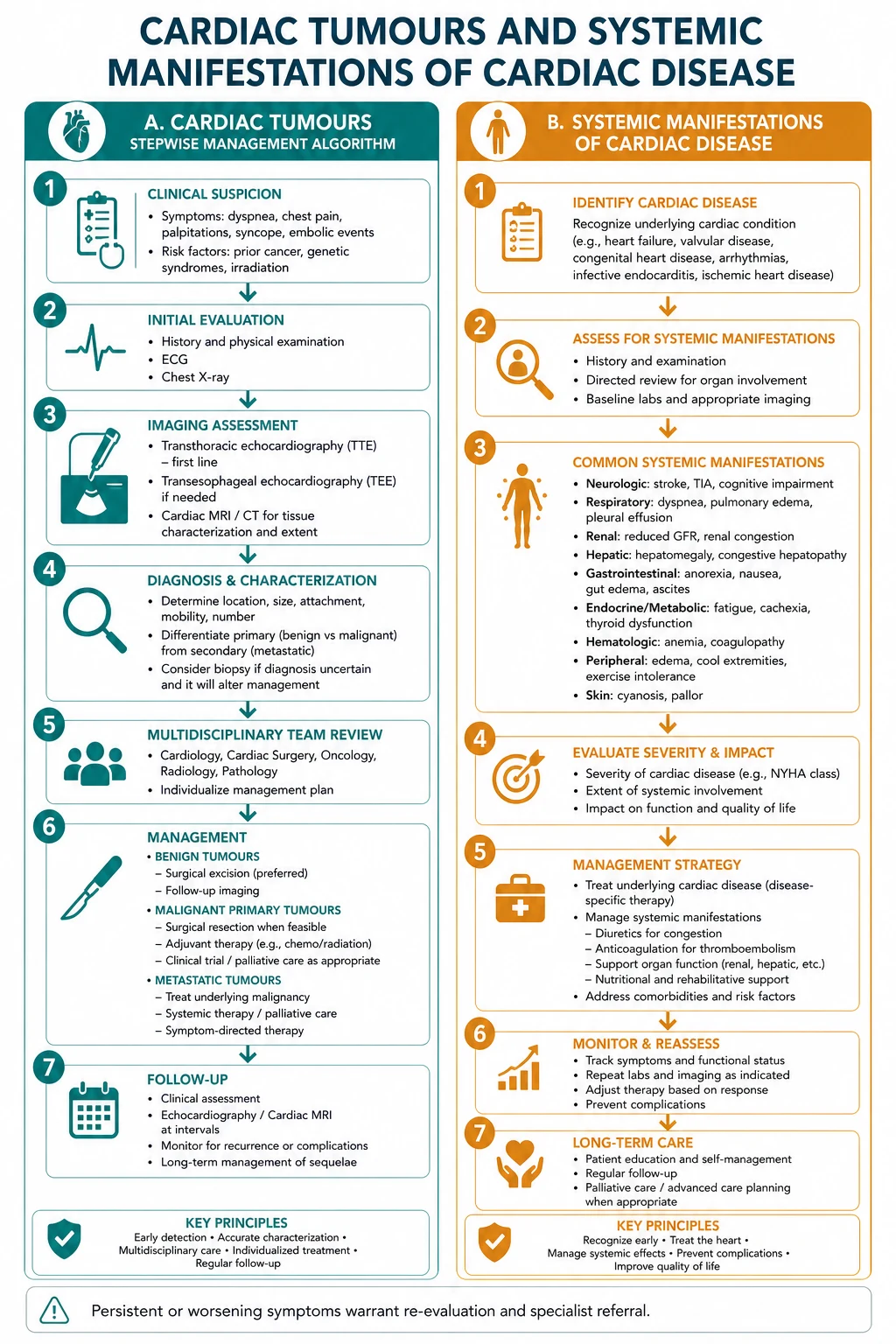
The DCE examiner may also test recognition of systemic manifestations of cardiac disease — the heart as a window to multisystem pathology. Echogenic or structural cardiac findings may herald systemic disease: marantic (non-bacterial thrombotic) endocarditis in malignancy or autoimmune disease (sterile vegetations causing emboli), Libman-Sacks endocarditis in SLE, carcinoid heart disease (right-sided valvular fibrosis from serotonin and bradykinin in metastatic neuroendocrine tumour — tricuspid regurgitation and pulmonary stenosis), restrictive cardiomyopathy in amyloidosis (low voltages on ECG with thick walls on echo; wipe-out on cardiac MRI), dilated cardiomyopathy in chemotherapy (anthracycline) and haemochromatosis (and chagas disease in endemic regions), and coronary embolisation in atrial fibrillation or endocarditis. [1] [2]
In the long case, the candidate who links the cardiac finding to its systemic cause demonstrates integrated physician thinking. Examples: thick-walled ventricles with low ECG voltages and carpal tunnel syndrome — amyloidosis (request serum free light chains, cardiac MRI, and consider tafamidis for ATTR); new diastolic rumble with positional symptoms and constitutional features — myxoma (echo); right-sided regurgitation with flushing and diarrhoea — carcinoid syndrome (24-hour urinary 5-HIAA); marantic vegetations with mucin-producing adenocarcinoma — paraneoplastic endocarditis (treat the underlying malignancy plus anticoagulation). [4] [8]
Approach to the intracardiac mass — a unifying algorithm
When confronted with an intracardiac mass on imaging, the candidate should ask four questions in sequence. (1) Is the mass mobile or sessile? (Mobile masses are higher embolic risk.) (2) Where is it attached? (Interatrial septum suggests myxoma; valve upstream surface suggests vegetation; left atrial appendage suggests thrombus; akinetic ventricular apex suggests thrombus; IVC extension suggests tumour thrombus.) (3) What is the tissue characterisation on MRI? (Fat suppression for lipoma; marked enhancement for fibroma; heterogeneous enhancement for sarcoma.) (4) Is there a systemic clue? (Constitutional features suggest myxoma or malignancy; embolic events suggest myxoma, fibroelastoma, or thrombus; endocrine features suggest Carney complex.) This four-question framework replaces a long differential with a directed evaluation. [1] [9]
Post-resection surveillance and follow-up
After resection of a sporadic atrial myxoma, transthoracic echocardiography at 6 months and 1 year is standard to detect recurrence; thereafter clinical follow-up is sufficient. After resection of a Carney complex myxoma, surveillance is lifelong with annual echocardiography because recurrence approaches 20-40 per cent and additional cardiac and extracardiac tumours may develop. After sarcoma resection, surveillance imaging (chest CT) every 3-6 months for recurrence and metastases is standard, alongside oncology review for adjuvant therapy. Patients with embolic complications prior to resection may have residual neurological or visceral deficits requiring rehabilitation. [1] [8]
References
- [1]Okongwu CC, Olaofe OO Cardiac myxoma: a comprehensive review Journal of cardiothoracic surgery, 2025.PMID 40082903
- [2]Thyagarajan B, Kumar MP, Patel S et al. Extracardiac manifestations of atrial myxomas Journal of the Saudi Heart Association, 2017.PMID 28127217
- [3]Lee VH, Connolly HM, Brown RD Jr Central nervous system manifestations of cardiac myxoma Archives of neurology, 2007.PMID 17698701
- [4]Moreno-Ariño M, Ortiz-Santamaria V, Deudero Infante A et al. A classic mimicker of systemic vasculitis Reumatologia clinica, 2016.PMID 25935820
- [5]Markel ML, Waller BF, Armstrong WF Cardiac myxoma. A review Medicine, 1987.PMID 3547010
- [6]Bhatia V, Jain C, Ray S et al. Mechanical Thrombectomy in Embolic Cardiac Myxoma: Case Report and Literature Review Neurology India, 2021.PMID 34169873
- [7]Rabiee Rad M, Ghasempour Dabaghi G, Darouei B et al. Clinical and laboratory manifestations, ECG findings, and outcomes of right atrial myxoma: a systematic review of cases reported worldwide The Egyptian heart journal : (EHJ) : official bulletin of the Egyptian Society of Cardiology, 2024.PMID 39266935
- [8]Marrone S, Gueli IA, Lo Coco R et al. Brain Metastases from Primary Cardiac Tumors: A Systematic Review of Diagnosis, Treatment, and Prognosis Cancers, 2025.PMID 40427120
- [9]Adam MP, Bick S, Mirzaa GM et al. Carney Complex 1993.PMID 20301463
- [10]Salman RA, Northridge D, Graham ANJ et al. Stroke due to a cardiac myxoma BMJ case reports, 2025.PMID 40783193