Phys · cardiovascular
Syncope
Also known as fainting · vasovagal syncope · neurocardiogenic syncope · carotid sinus hypersensitivity · carotid sinus syndrome · orthostatic hypotension · postural hypotension · cardiogenic syncope · arrhythmic syncope · situational syncope · micturition syncope · cough syncope
Consultant-physician-depth guide to syncope — the transient loss of consciousness from global cerebral hypoperfusion — using the 2018 ESC classification of reflex, orthostatic, and cardiac causes, with a directed investigation pathway from ECG to implantable loop recorder, risk stratification, and evidence-based management including physical counter-pressure manoeuvres, fludrocortisone, midodrine, and pacemaker therapy for selected patients.
On this page & tools
Your progress
Saved locally on this device.
Practise this topic
Target exams
Red flags
Syncope

The answer first
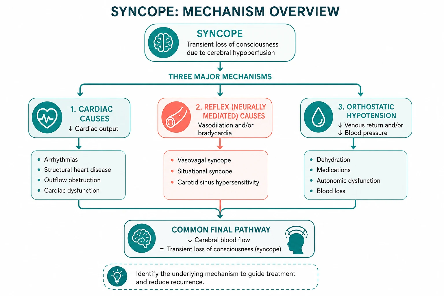
Syncope is a transient loss of consciousness caused by global cerebral hypoperfusion, characterised by rapid onset, short duration, and spontaneous complete recovery [1]. The mechanism is always the same: cerebral blood flow falls below the minimum needed to sustain consciousness. What varies is why.
The 2018 ESC guidelines classify syncope into three categories, and this classification governs every decision below [1]:
- Is it reflex (neurocardiogenic) syncope? The most common cause. A triggered autonomic reflex produces vasodilation and/or bradycardia — vasovagal, situational, or carotid sinus hypersensitivity. The heart and vasculature are structurally normal; the problem is the reflex.
- Is it orthostatic hypotension? The autonomic system or medications fail to maintain blood pressure on standing. The mechanism is a failure of vasoconstriction — not a reflex, but an inability to compensate for gravity.
- Is it cardiac syncope? The most dangerous. An arrhythmia or structural lesion prevents the heart from maintaining cardiac output. This category carries the highest mortality and demands urgent investigation. [1]
The two pivotal questions at the bedside:
- Was it truly syncope? Exclude seizure, concussion, psychogenic pseudosyncope, and metabolic causes of transient loss of consciousness. A careful history alone establishes the diagnosis in 23 to 50 per cent of cases [1].
- Is there a high-risk feature? Exertional syncope, no prodrome, structural heart disease, abnormal ECG, or family history of sudden death demand admission and expedited workup. Everything else can be investigated as an outpatient.
DWE high-yield: Every patient with syncope gets a 12-lead ECG. It is non-negotiable, cheap, and identifies the highest-risk causes. If you remember one rule, remember this — the ECG is mandatory for all syncope, regardless of how typical the story sounds. [1]
ESC 2018 classification — the three categories
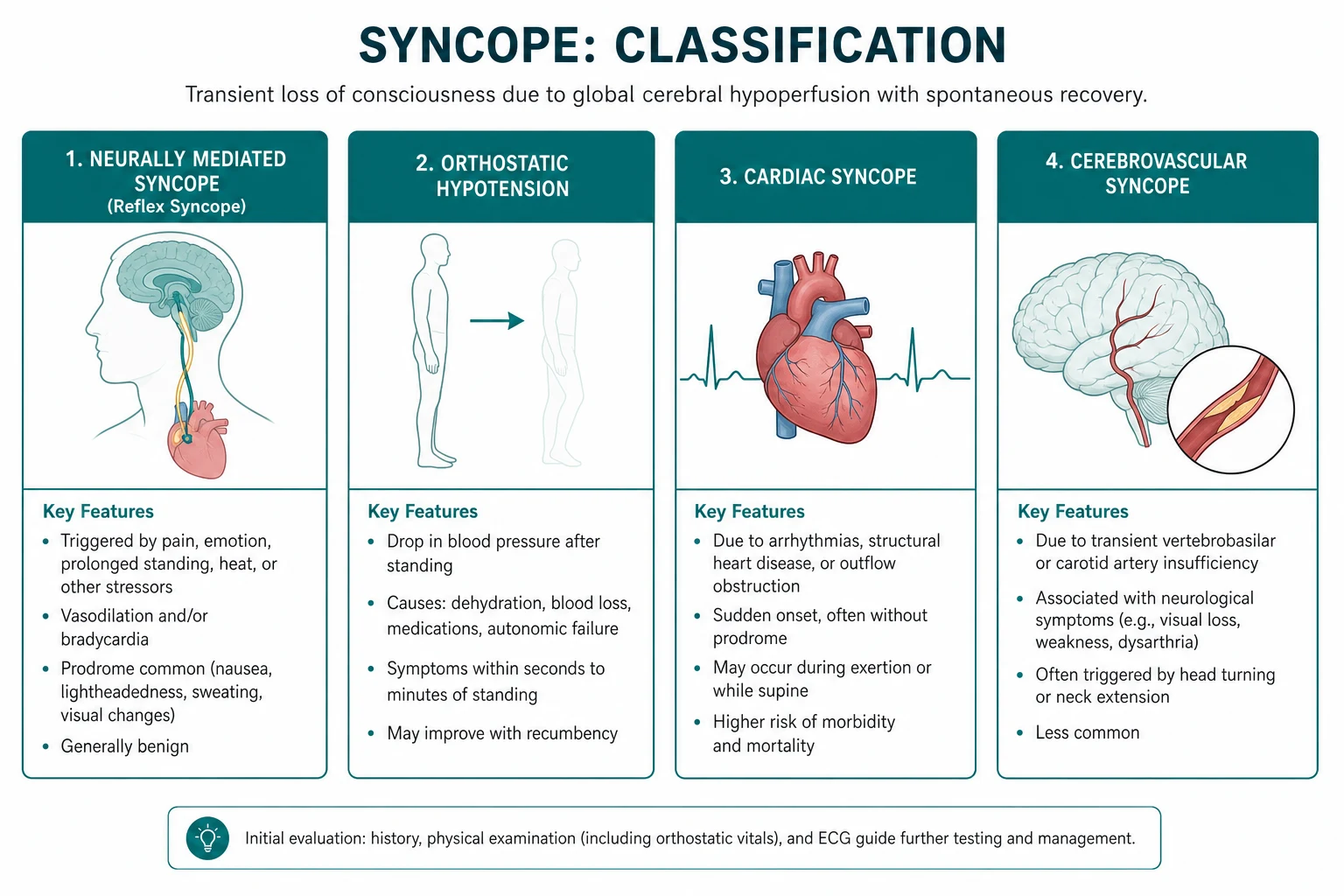

| Category | Mechanism | Typical features | Prognosis |
|---|---|---|---|
| Reflex (neurocardiogenic) | Autonomic reflex causes vasodilation and/or bradycardia | Prodrome (nausea, warmth, pallor), trigger (standing, pain, emotion), rapid full recovery | Benign — excellent prognosis |
| Orthostatic hypotension | Failure of vasoconstriction on standing | Lightheadedness on standing, progressive, worse in morning or after meals | Variable — depends on cause |
| Cardiac | Arrhythmia or structural lesion prevents adequate cardiac output | No prodrome, during exertion, while supine, with palpitations; structural heart disease or abnormal ECG | High — one-year mortality up to 30 per cent |
The classification is not merely academic — it determines the investigation pathway. Reflex syncope is diagnosed clinically and confirmed with tilt testing. Orthostatic hypotension is diagnosed with orthostatic blood pressure measurement. Cardiac syncope requires ECG, echocardiography, and rhythm monitoring. A reflexive battery of tests applied without classification wastes resources and delays diagnosis [1].
Reflex (neurocardiogenic) syncope
Vasovagal syncope — the most common cause
Vasovagal syncope is mediated by the Bezold-Jarisch reflex. Venous pooling (from prolonged standing, warmth, or dehydration) reduces venous return. The underfilled but vigorously contracting ventricle activates vagal afferent mechanoreceptors, which trigger paradoxical vasodilation and bradycardia. The result is a critical fall in blood pressure and cerebral perfusion. [1]
Triggers: prolonged standing, pain, strong emotion, fear, the sight of blood, venepuncture, warm crowded environments, dehydration, recent illness. [1]
Prodrome: the hallmark. Nausea, warmth, flushing, pallor, diaphoresis, lightheadedness, visual dimming, yawning, and a feeling of impending collapse. The prodrome typically lasts seconds to a few minutes and is the single most useful feature distinguishing vasovagal from cardiac syncope. [1]
Recovery: rapid and complete. The patient regains consciousness within seconds to a minute once supine. There is no postictal confusion — this is a key discriminator from seizure. [1]
DWE trap — convulsive syncope. Anoxic cerebral hypoperfusion can cause myoclonic jerks that mimic a seizure. The distinction: syncope is brief, preceded by pallor and a prodrome, recovery is rapid without postictal confusion, tongue biting and lateral injury are rare, and the jerks are brief and irregular rather than rhythmic and sustained. Misdiagnosing syncope as epilepsy delays correct treatment and exposes the patient to unnecessary antiepileptic drugs. [1]
Carotid sinus hypersensitivity
Carotid sinus hypersensitivity is an exaggerated response to carotid sinus stimulation, defined as a ventricular pause of 3 seconds or more, or a systolic blood pressure drop of 50 mmHg or more, in response to carotid sinus massage [1]. It becomes carotid sinus syndrome when the response reproduces the patient's spontaneous symptoms.
Typical presentation: an older man (male-to-female ratio 4:1) with syncope precipitated by head turning, tight collars, shaving, or neck tumours. Many patients have amnesia for the loss of consciousness and present with unexplained falls [8].
Three response types:
- Cardioinhibitory: ventricular asystole of 3 seconds or more. This type responds to pacing.
- Vasodepressor: systolic BP drop of 50 mmHg or more without significant bradycardia. Pacing does NOT help — the problem is vasodilation, not bradycardia.
- Mixed: both cardioinhibitory and vasodepressor components. [1]
Carotid sinus massage technique: the patient is supine and monitored with continuous ECG and non-invasive blood pressure. Firm pressure is applied to the carotid bifurcation (at the level of the cricoid cartilage) for 5 to 10 seconds, first on the right then on the left after a 30-second rest. It is contraindicated in patients with a carotid bruit, recent TIA or stroke, or known significant carotid stenosis. [1]
Situational syncope
Situational syncope is reflex syncope triggered by a specific action. The mechanism is a vagal reflex producing bradycardia and vasodilation in response to a mechanical or physiological stimulus: [1]
- Cough syncope: paroxysmal coughing raises intrathoracic pressure, reducing venous return and cardiac output. Typically occurs in middle-aged men with chronic lung disease.
- Micturition syncope: typically nocturnal, in older men with prostatism who stand to void after waking from sleep. The combination of decompression of the bladder, orthostatic stress, and vagal tone causes syncope.
- Defecation syncope: straining (Valsalva) reduces venous return; seen in older patients, often with cardiovascular comorbidities.
- Post-prandial syncope: mesenteric vasodilation after a meal causes relative hypovolaemia; common in older adults and those with autonomic failure.
- Swallow syncope: in patients with oesophageal disease (tumour, achalasia, spasm), swallowing triggers a vagal reflex causing profound bradycardia or AV block — a pacemaker may be indicated. [1]
Orthostatic hypotension
Orthostatic hypotension is a reduction in blood pressure on standing that is sufficient to cause cerebral hypoperfusion. The 2018 ESC guidelines define three temporal patterns [1]:
Definitions
| Type | Definition | Mechanism |
|---|---|---|
| Classical OH | Drop of 20 mmHg systolic or 10 mmHg diastolic within 3 minutes of standing | Failure of baroreflex-mediated vasoconstriction |
| Initial OH | Drop of 40 mmHg systolic within 15 seconds of standing, with rapid spontaneous recovery | Transient mismatch between venous return and cardiac output |
| Delayed OH | Onset after 3 minutes of standing (often 10 to 30 minutes) | Gradual failure of neurohumoral compensation |
Delayed OH is frequently missed because standard orthostatic observations end at 3 minutes. If the history suggests symptoms after prolonged standing, extend the observation period. [1]
Causes
Medication-induced (most common and most reversible):
- Vasodilators: nitrates, calcium channel blockers, ACE inhibitors, ARBs
- Diuretics: especially loop diuretics
- Alpha-blockers: tamsulosin, prazosin — the most potent cause of OH
- Dopaminergic agents: levodopa, pramipexole
- Antipsychotics and antidepressants: tricyclics, phenothiazines, quetiapine
- Others: opioids, sildenafil, alcohol [1]
Autonomic failure — primary neurodegenerative:
- Parkinson disease (late-stage)
- Multiple system atrophy (MSA) — OH is often severe and early
- Pure autonomic failure
- Dementia with Lewy bodies [1]
Autonomic failure — secondary:
- Diabetes mellitus (the most common cause of autonomic neuropathy)
- Amyloidosis (particularly AL amyloid)
- Autoimmune autonomic ganglionopathy
- Paraneoplastic syndromes
- Alcohol-induced neuropathy
- Vitamin B12 deficiency [1]
Volume depletion:
- Bleeding (gastrointestinal, occult)
- Dehydration (vomiting, diarrhoea, inadequate intake)
- Adrenal insufficiency (Addison disease) [1]
Deconditioning:
- Prolonged bed rest, prolonged immobility, post-intensive care syndrome
- Significant weight loss, anorexia [1]
DCE long-case trap. In the elderly patient with syncope, the medication list is the single most fruitful target. Medication review alone resolves up to 20 per cent of cases. Start by asking: which of these drugs could lower blood pressure on standing? Then ask: can it be stopped, reduced, or switched? [1]
Cardiac syncope
Cardiac syncope carries the highest mortality — up to 30 per cent at one year in some series [1]. It is caused by either an arrhythmia or a structural cardiac lesion that prevents the heart from maintaining adequate cardiac output.
Arrhythmic causes
Bradyarrhythmias:
- Sinus node dysfunction (sick sinus syndrome, tachy-brady syndrome) — sinus pauses, chronotropic incompetence
- Atrioventricular block: second-degree Mobitz II, high-grade AV block (2:1 or worse), complete heart block
- Drug-induced bradycardia: beta-blockers, non-dihydropyridine calcium channel blockers, digoxin, amiodarone [1]
Tachyarrhythmias:
- Ventricular tachycardia — the most concerning; especially in patients with structural heart disease, previous MI, or cardiomyopathy
- Supraventricular tachycardia with rapid ventricular response
- Atrial fibrillation with rapid ventricular response — syncope occurs at onset when the rate is fastest
- Polymorphic VT / torsades de pointes — in long QT syndrome or drug-induced QT prolongation [1]
Channelopathies (inherited):
- Long QT syndrome: QTc over 470 ms in males, over 480 ms in females; torsades de pointes triggered by exertion (LQT1), emotion (LQT2), or rest/sleep (LQT3)
- Brugada syndrome: characteristic coved ST elevation in V1 to V3, syncope or sudden death from polymorphic VT
- Catecholaminergic polymorphic VT (CPVT): bidirectional VT triggered by exercise; structurally normal heart, normal ECG at rest [1]
Structural causes
| Condition | Mechanism | Examination clue |
|---|---|---|
| Aortic stenosis | Fixed cardiac output cannot increase on exertion | Slow-rising carotid pulse, ejection systolic murmur radiating to carotids, delayed carotid upstroke |
| Hypertrophic cardiomyopathy | LVOT obstruction limits output | Jerky bisferiens pulse, ejection systolic murmur at LLSE, increases with Valsalva |
| Pulmonary hypertension | Right heart failure, low cardiac output | Raised JVP, RV heave, loud P2, tricuspid regurgitation murmur |
| Pulmonary embolism (massive) | Acute right ventricular failure | Tachycardia, hypoxia, raised JVP, RV strain on ECG |
| Aortic dissection | Tamponade, acute aortic regurgitation, or neural compromise | Tearing chest/back pain, blood pressure differential, new murmur |
| Cardiac tamponade | Impaired ventricular filling | Beck triad (hypotension, raised JVP, muffled heart sounds), pulsus paradoxus |
| Severe mitral stenosis | Fixed obstruction to LV filling | Loud S1, opening snap, diastolic murmur at apex |
Device malfunction
Always ask about implanted devices (pacemaker, ICD, CRT). Device malfunction — generator end-of-life, lead fracture, lead displacement, electromagnetic interference, or sensing/pacing failure — can cause syncope. The device should be interrogated as part of the workup in any patient with a device who presents with syncope. [1]
DWE trap — exertional syncope is never vasovagal. Syncope during exertion demands exclusion of aortic stenosis, HCM, VT, and pulmonary hypertension. The heart cannot increase output to meet demand, or an exercise-induced arrhythmia occurs. The diagnosis of exertional vasovagal syncope is one of exclusion, made only after complete cardiac workup. [1]
Initial evaluation — history, examination, ECG
History
The history is the single most powerful diagnostic tool in syncope. The 2018 ESC guidelines estimate that a careful history and physical examination alone can establish the diagnosis in 23 to 50 per cent of cases [1].
What to ask: [1]
| Question | Why it matters |
|---|---|
| Circumstances: posture, activity, position | Standing suggests orthostatic or vasovagal; exertion suggests cardiac; supine suggests arrhythmia |
| Prodromal symptoms | Nausea, warmth, pallor = vasovagal; none or palpitations = arrhythmic |
| Onset | Sudden without warning favours arrhythmia; gradual with prodrome favours reflex |
| Duration of unconsciousness | Seconds = syncope; minutes to hours = seizure or other TLOC |
| Recovery | Rapid and complete = syncope; slow with confusion = seizure |
| Associated symptoms | Chest pain, dyspnoea, palpitations = cardiac; neurological focal signs = TIA/stroke |
| Frequency and pattern | Increasing frequency or changing pattern warrants investigation |
| Medications | Vasodilators, diuretics, rate-limiting drugs — the reversible causes |
| Family history | Sudden death under 50 = inherited channelopathy or cardiomyopathy |
| Witnessed features | Pallor, seizure-like activity, incontinence, tongue biting, duration |
Physical examination
- Orthostatic blood pressure: measure lying and standing at 1 and 3 minutes. Document the systolic, diastolic, and heart rate response. In autonomic failure, the heart rate does not rise appropriately (less than 15 beats per minute increase) despite a significant blood pressure drop.
- Cardiac auscultation: listen for the murmurs of aortic stenosis (ejection systolic, radiating to carotids), HCM (ejection systolic at the lower left sternal edge, dynamic), and mitral stenosis (diastolic at the apex).
- JVP: elevated in heart failure, pulmonary hypertension, and tamponade; low in volume depletion.
- Neurological examination: focal deficits suggest a cerebrovascular cause rather than syncope.
- Carotid sinus massage: in patients over 40 with unexplained syncope suggestive of a reflex mechanism, after excluding carotid bruit and cerebrovascular disease. [1]
The 12-lead ECG — mandatory for all syncope
The ECG is the single highest-yield investigation. Every patient with syncope gets one — no exceptions. The yield of an abnormal ECG finding that contributes to the diagnosis is approximately 5 per cent, but the conditions it identifies (high-grade AV block, VT, long QT, Brugada) are the highest-risk causes [1].
ECG patterns to look for in syncope: [1]
| Pattern | Diagnosis | Significance |
|---|---|---|
| Bifascicular block (RBBB + LAFB or LPFB) | Risk of progression to complete heart block | High-risk — requires monitoring |
| Mobitz II second-degree AV block | Intermittent complete heart block | High-risk — pacing indicated |
| Complete heart block | Ventricular asystole | High-risk — urgent pacing |
| Sinus pause over 3 seconds | Sinus node dysfunction | High-risk — pacing if symptomatic |
| Long QTc (over 470 ms male, 480 ms female) | Long QT syndrome | Risk of torsades de pointes |
| Coved ST elevation V1 to V3 | Brugada syndrome | Risk of polymorphic VT |
| Epsilon waves, T-wave inversion V1 to V3 | ARVC | Risk of ventricular arrhythmia |
| Pre-excitation (delta wave) | WPW syndrome | Risk of rapid AF degenerating to VF |
| Pathological Q waves, ST changes | Previous MI, ischaemia | Substrate for VT |
| LVH with strain | HCM, aortic stenosis, hypertensive heart | Structural cause |
Risk stratification — who needs admission?
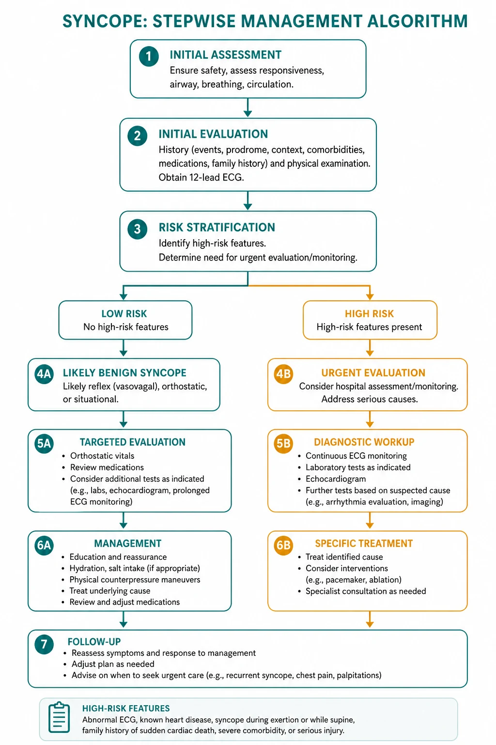
The 2018 ESC guidelines define high-risk features that warrant admission for expedited investigation and monitoring [1]. The purpose of risk stratification is to identify patients at risk of death or serious injury from a cardiac cause, who need urgent intervention.
High-risk features (admit and investigate urgently)
- Severe structural or coronary artery disease: heart failure, low LVEF (below 35 per cent), previous MI
- Clinical features suggesting arrhythmic syncope:
- During exertion
- While supine
- Palpitations at the time of syncope
- Absence of prodrome (sudden onset)
- Abnormal ECG: bifascicular block, Mobitz II or high-grade AV block, complete heart block, sinus pauses over 3 seconds, pre-excitation, long QT, Brugada pattern, epsilon waves, sustained or non-sustained VT
- Family history of sudden cardiac death at a young age (under 50 years)
- Major injury from syncope: fracture, head injury, motor vehicle accident
- High-risk occupation: pilot, commercial driver, heavy machinery operator, working at heights [1]
Low-risk features (manage as outpatient)
- Typical vasovagal syncope with a clear trigger and prodrome
- Orthostatic hypotension explained by medications or volume depletion
- No structural heart disease
- Normal ECG
- No family history of sudden death [1]
DWE decision point. A patient with low-risk features and a normal ECG can be investigated as an outpatient. A patient with any high-risk feature is admitted for continuous monitoring and expedited cardiac workup. The threshold for admission is low when structural heart disease or an abnormal ECG is present. [1]
Investigation approach — directed, not reflexive
The 2018 ESC guidelines emphasise a directed approach: the choice of investigation is guided by the initial evaluation and risk stratification, not applied as a blanket battery [1].
Echocardiography
Indicated when structural heart disease is suspected or known. Assesses:
- Valve function (aortic stenosis severity, mitral stenosis)
- Left ventricular ejection fraction and wall motion abnormalities
- Wall thickness (HCM, amyloid)
- Right ventricular size and function (pulmonary hypertension, ARVC)
- Pericardial effusion (tamponade) [1]
Tilt table testing
Used for reflex syncope when the diagnosis is uncertain after initial evaluation. The patient is strapped to a motorised table and tilted head-up to 60 to 70 degrees for 20 to 45 minutes, with or without provocation (nitroglycerin, isoproterenol). The test reproduces the reflex pathway and classifies the response:
- Type 1 (mixed): heart rate falls but remains above 40 bpm, or falls below 40 bpm for less than 10 seconds, without asystole; blood pressure falls before heart rate.
- Type 2A (cardioinhibitory without asystole): heart rate falls below 40 bpm for more than 10 seconds, without asystole over 3 seconds.
- Type 2B (cardioinhibitory with asystole): asystole over 3 seconds; blood pressure falls before or simultaneously with heart rate.
- Type 3 (vasodepressor): heart rate does not fall more than 10 per cent from peak; blood pressure falls. [1]
The sensitivity of tilt testing is approximately 60 per cent and specificity 80 per cent. It is most useful in patients with suspected reflex syncope where the history is not diagnostic. [1]
Carotid sinus massage
Performed in patients over 40 with unexplained syncope suggestive of a reflex mechanism, after excluding carotid bruit and cerebrovascular disease. The test is positive if carotid sinus massage produces a ventricular pause of 3 seconds or more (cardioinhibitory), a systolic blood pressure drop of 50 mmHg or more (vasodepressor), or both (mixed), AND reproduces the patient's symptoms. [1]
Ambulatory ECG monitoring
The choice of monitoring duration depends on the frequency of episodes: [1]
| Modality | Duration | Indication |
|---|---|---|
| Holter monitor | 24 to 48 hours | Frequent episodes (daily) |
| Patch monitor | Up to 14 days | Intermediate frequency |
| Implantable loop recorder (ILR) | Up to 3 years | Infrequent unexplained syncope with suspected arrhythmic cause |
The implantable loop recorder is the gold standard for infrequent unexplained syncope. It is inserted subcutaneously and monitors the ECG continuously for up to 3 years, with automatic and patient-activated recording. The ISSUE-3 trial demonstrated the importance of ILR documentation before committing to pacemaker therapy in suspected neurally mediated syncope [7].
Electrophysiology study
Indicated in patients with syncope and structural heart disease or an abnormal ECG where an arrhythmic cause is suspected but not documented by non-invasive monitoring. EP testing can identify:
- His-Purkinje conduction disease (prolonged HV interval, inducible infra-His block)
- Inducible ventricular tachycardia
- Bypass tract conduction (in WPW) [1]
The diagnostic yield is highest in patients with structural heart disease and abnormal ECG, and low in patients with a normal heart and normal ECG. [1]
Exercise testing
Indicated when syncope occurs during or shortly after exertion. It can unmask:
- Exercise-induced arrhythmias (VT, CPVT)
- Chronotropic incompetence (failure to increase heart rate with exercise)
- Exertional AV block (typically infranodal, high-grade)
- Ischaemia-driven arrhythmia [1]
Management — treat the underlying cause
Reflex syncope
First-line — education and lifestyle (all patients):
- Explain the benign nature and the mechanism
- Recognise and avoid triggers (prolonged standing, warm environments, dehydration)
- Ensure adequate hydration (2 to 3 litres per day) and salt intake (10 g per day if not hypertensive)
- Recognise prodromal symptoms and lie down immediately [1]
Physical counter-pressure manoeuvres (PCM): At the onset of prodromal symptoms, the patient performs:
- Leg crossing with muscle tensing
- Arm tensing (gripping one hand with the other and pulling apart)
- Hand gripping (squeezing a ball) [1]
The PC-Trial (PMID 17045903) randomised 223 patients with recurrent vasovagal syncope and a recognisable prodrome to PCM plus conventional therapy versus conventional therapy alone. PCM produced a 39 per cent relative risk reduction in recurrent syncope [4]. It is first-line, risk-free, and low-cost.
Fludrocortisone: A mineralocorticoid that expands plasma volume and sensitises peripheral alpha-receptors. The POST2 trial (PMID 27364043) randomised 210 patients to fludrocortisone 0.1 to 0.2 mg daily versus placebo. The primary endpoint was not met (HR 0.69, p=0.069), but post-hoc analysis after dose stabilisation showed significant benefit (HR 0.51, p=0.019) [5]. It is considered second-line in recurrent VVS.
Midodrine: An alpha-1 agonist that increases venous return and blood pressure. The POST4 trial (PMID 34339231) randomised 133 patients to midodrine 5 mg three times daily versus placebo. Midodrine significantly reduced syncope recurrence (42 per cent vs 61 per cent, relative risk 0.69, p=0.035) [6]. Particularly useful in patients with a prominent vasodepressor component.
Beta-blockers are NOT recommended. Multiple randomised trials have shown no benefit in vasovagal syncope. The theoretical rationale (blunting the initial sympathetic surge) has not translated into clinical benefit. [1]
Pacemaker therapy for reflex syncope: Indicated only in a highly selected group: patients over 40 with recurrent severe reflex syncope and documented asystole on implantable loop recorder. The ISSUE-3 trial (PMID 22565936) randomised such patients to dual-chamber pacing ON versus OFF. The 2-year syncope recurrence was 25 per cent (pacing ON) versus 57 per cent (pacing OFF), a 57 per cent relative risk reduction [7]. Pacemaker therapy is NOT indicated for:
- Typical vasovagal syncope with a prodrome
- Young patients (under 40)
- The vasodepressor type of carotid sinus syndrome
- Patients without documented asystole
Pacing for carotid sinus hypersensitivity: Indicated for the cardioinhibitory or mixed type with recurrent syncope confirmed by carotid sinus massage. The evidence is stronger for patients presenting with syncope than for those presenting with unexplained falls — the SAFE PACE 2 trial (PMID 20228148) found no significant difference between pacing and ILR in fallers [9]. Pacing is NOT indicated for the vasodepressor type, as it does not prevent reflex-mediated vasodilation.
Orthostatic hypotension
Step 1 — Medication review (the most impactful intervention): Identify and discontinue or reduce offending agents. The medication list is the single most fruitful target. In older patients with polypharmacy, deprescribing vasodilators, diuretics, and alpha-blockers can resolve OH entirely. [1]
Step 2 — Non-pharmacological measures:
- Adequate hydration: 2 to 3 litres per day
- Salt supplementation: 10 g per day (if not hypertensive) [1]- Compression stockings: waist-high preferred (20 to 30 mmHg), to reduce venous pooling
- Raised head of bed by 10 to 20 cm: reduces nocturnal diuresis and morning orthostatic hypotension
- Slow sequential standing: sit on the edge of the bed for 1 to 2 minutes before standing
- Physical counter-pressure manoeuvres: leg crossing and tensing on standing
- Avoid large meals (post-prandial hypotension) and alcohol [1]
Step 3 — Pharmacological:
- Fludrocortisone: mineralocorticoid that expands intravascular volume; start at 0.1 mg daily, titrate to 0.2 to 0.3 mg; monitor for hypertension, hypokalaemia, and ankle oedema
- Midodrine: alpha-1 agonist; 2.5 to 5 mg three times daily; last dose in early afternoon to avoid supine hypertension; monitor for urinary retention (prostatism) and piloerection
- Droxidopa: synthetic amino acid converted to norepinephrine; particularly useful in neurogenic OH (Parkinson disease, MSA); approved by the FDA for neurogenic OH [1]
DCE long-case trap — supine hypertension. In autonomic failure, treating orthostatic hypotension with fludrocortisone or midodrine worsens supine hypertension. The solution: raise the head of the bed (which also helps OH), give pressor agents only during daytime hours, and use a short-acting antihypertensive at night (e.g., amlodipine, hydralazine). This is a balancing act that examiners expect you to acknowledge. [1]
Cardiac syncope
The principle is simple: treat the underlying cause. [1]
Arrhythmic:
- Pacemaker for sinus node dysfunction and high-grade AV block
- Catheter ablation for SVT and selected VT
- Antiarrhythmic drugs or ICD for sustained VT
- ICD for channelopathies with high arrhythmic risk (long QT, Brugada, CPVT)
- Correct electrolyte abnormalities (potassium, magnesium)
- Review and adjust proarrhythmic medications [1]
Structural:
- Aortic valve replacement for severe aortic stenosis (TAVR or surgical)
- Septal reduction therapy (myectomy or alcohol septal ablation) for HCM with LVOT obstruction
- Anticoagulation for AF with rapid ventricular response
- Pulmonary thromboendarterectomy or riociguat for CTEPH
- Pericardiocentesis for tamponade
- Surgical repair for aortic dissection [1]
Device malfunction:
- Interrogate the device
- Reprogram if sensing or pacing thresholds are suboptimal
- Replace generator if at end-of-life
- Revise lead system if fracture or displacement is detected [1]
Driving and occupational advice
Driving advice is a safety-critical decision that examiners expect to hear explicitly. For private driving, a single uncomplicated vasovagal syncope requires no restriction; recurrent or unexplained syncope requires a symptom-free period of typically 4 weeks (Austroads guidelines); treated cardiac syncope (e.g., pacemaker implanted) allows driving after the device is confirmed functional, usually 1 week. For commercial driving, any unexplained syncope warrants permanent or prolonged restriction until a definitive treatable cause is identified and successfully treated; vasovagal syncope with a clear trigger and prodrome requires case-by-case assessment; cardiac syncope requires permanent restriction unless the cause is definitively treated (e.g., successful aortic valve replacement for AS). Patients in high-risk occupations (pilot, commercial driver, heavy machinery, working at heights) require full investigation and definitive treatment before return to work, with a lower threshold for restriction than private driving.
Key trials — what the evidence shows
| Trial | PMID | Intervention | Key finding |
|---|---|---|---|
| PC-Trial (van Dijk, 2006) | 17045903 | Physical counter-pressure manoeuvres vs conventional therapy | 39 per cent relative risk reduction in recurrent VVS [4] |
| POST2 (Sheldon, 2016) | 27364043 | Fludrocortisone vs placebo | Primary endpoint not met (HR 0.69, p=0.069); post-hoc benefit after dose stabilisation (HR 0.51, p=0.019) [5] |
| POST4 (Sheldon, 2021) | 34339231 | Midodrine vs placebo | Significant reduction in syncope recurrence (42 per cent vs 61 per cent, RR 0.69, p=0.035) [6] |
| ISSUE-3 (Brignole, 2012) | 22565936 | Dual-chamber pacing ON vs OFF in asystolic NMS | 57 per cent relative risk reduction (25 per cent vs 57 per cent at 2 years) — only in ILR-documented asystole [7] |
| SAFE PACE 2 (Ryan, 2010) | 20228148 | Pacing vs ILR in cardioinhibitory CSH fallers | No significant difference between groups [9] |
| Parry (2009) | 19124530 | Pacing ON vs OFF in CSH with falls | No significant effect on falls; amnesia for LOC common [8] |
Exam traps and high-yield discriminators
1. Exertional syncope is never vasovagal. Until proven otherwise, exertional syncope is aortic stenosis, HCM, arrhythmia, or pulmonary hypertension. The heart cannot increase output to meet demand. The diagnosis of exertional vasovagal syncope is one of exclusion. [1]
2. No prodrome equals arrhythmia. The absence of a prodrome is the single most useful bedside discriminator against reflex syncope. Reflex syncope (vasovagal) almost always has a prodrome; arrhythmic syncope often does not — the patient drops without warning. [1]
3. A normal ECG does not exclude arrhythmic syncope. Intermittent arrhythmias are common. A single normal ECG is reassuring but not definitive. If the index of suspicion is high, ambulatory monitoring or an implantable loop recorder is warranted. [1]
4. Do not pacemaker a young patient with typical vasovagal syncope. The ISSUE-3 trial showed benefit only in patients over 40 with documented asystole on ILR. Pacemaker therapy for reflex syncope is a last resort for a highly selected population — not a first-line treatment. [1]
5. Carotid sinus massage is contraindicated with a carotid bruit. Performing carotid sinus massage in a patient with significant carotid stenosis can precipitate a stroke. Always auscultate for a carotid bruit first and exclude recent TIA or stroke. [1]
6. Medication review is the most impactful intervention in the elderly. In older patients with polypharmacy, syncope is often multifactorial and medication-induced. Deprescribing vasodilators, diuretics, and alpha-blockers alone resolves up to 20 per cent of cases. [1]
7. Convulsive syncope is not epilepsy. Anoxic cerebral hypoperfusion can cause myoclonic jerks. The keys: syncope is brief, preceded by pallor and a prodrome, recovery is rapid without postictal confusion, and tongue biting or lateral injury is rare. Misdiagnosis exposes the patient to unnecessary antiepileptic drugs. [1]
8. Always check the device. In a patient with a pacemaker or ICD presenting with syncope, interrogate the device. Generator end-of-life, lead fracture, lead displacement, and sensing failure are all reversible causes that non-invasive testing will miss. [1]
Summary — the syncope framework for exams
Confirm it was syncope (TLOC from cerebral hypoperfusion), classify using ESC 2018 (reflex, orthostatic, or cardiac), risk-stratify for high-risk features (exertion, no prodrome, structural disease, abnormal ECG, family history of SCD), investigate directedly (ECG for all; echo if structural disease; tilt for reflex; orthostatic BP for OH; ILR for arrhythmia), treat the cause (education and PCM for vasovagal; medication review and fludrocortisone/midodrine for OH; specific therapy for cardiac causes), and address driving and occupation explicitly. [1]
2018 ESC Guidelines for the diagnosis and management of syncope (Brignole, Eur Heart J 2018); 2017 ACC/AHA/HRS Guideline for the Evaluation and Management of Patients With Syncope (Shen, Circulation 2017); PC-Trial (van Dijk, JACC 2006); POST2 (Sheldon, JACC 2016); POST4 (Sheldon, Ann Intern Med 2021); ISSUE-3 (Brignole, Circulation 2012); National Heart Foundation of Australia guidance. [1]
References
- [1]Brignole M, Moya A, de Lange FJ, et al. 'Ten Commandments' of ESC Syncope Guidelines 2018: The new European Society of Cardiology (ESC) Clinical Practice Guidelines for the diagnosis and management of syncope were launched 19 March 2018 at EHRA 2018 in Barcelona Eur Heart J, 2018.PMID 29860370
- [2]Brignole M, Moya A, de Lange FJ, et al. Practical Instructions for the 2018 ESC Guidelines for the diagnosis and management of syncope Eur Heart J, 2018.PMID 29562291
- [3]Shen WK, Sheldon RS, Benditt DG, et al. 2017 ACC/AHA/HRS Guideline for the Evaluation and Management of Patients With Syncope: A Report of the American College of Cardiology/American Heart Association Task Force on Clinical Practice Guidelines and the Heart Rhythm Society Circulation, 2017.PMID 28280231
- [4]van Dijk N, Quartieri F, Blanc JJ, et al. Effectiveness of physical counterpressure maneuvers in preventing vasovagal syncope: the Physical Counterpressure Manoeuvres Trial (PC-Trial) J Am Coll Cardiol, 2006.PMID 17045903
- [5]Sheldon R, Raj SR, Rose MS, et al. Fludrocortisone for the Prevention of Vasovagal Syncope: A Randomized, Placebo-Controlled Trial J Am Coll Cardiol, 2016.PMID 27364043
- [6]Sheldon R, Faris P, Tang A, et al. Midodrine for the Prevention of Vasovagal Syncope : A Randomized Clinical Trial Ann Intern Med, 2021.PMID 34339231
- [7]Brignole M, Menozzi C, Moya A, et al. Pacemaker therapy in patients with neurally mediated syncope and documented asystole: Third International Study on Syncope of Uncertain Etiology (ISSUE-3): a randomized trial Circulation, 2012.PMID 22565936
- [8]Parry SW, Steen N, Bexton RS, Tynan M, Kenny RA. Pacing in elderly recurrent fallers with carotid sinus hypersensitivity: a randomised, double-blind, placebo controlled crossover trial Heart, 2009.PMID 19124530
- [9]Kenny RA, Richardson DA, Steen N, et al. SAFE PACE 2: Syncope and Falls in the Elderly--Pacing and Carotid Sinus Evaluation: a randomized controlled trial of cardiac pacing in older patients with falls and carotid sinus hypersensitivity Europace, 1999.PMID 11220545