Psych · Foundations — neuroimaging in psychiatry
Neuroimaging in psychiatry
Also known as Brain imaging psychiatry · MRI in psychiatry · fMRI psychiatry · Structural neuroimaging psychosis · PET occupancy psychiatry · CT brain psychiatric work-up · ENIGMA psychiatry imaging
Exam-exhaustive fellowship foundation on neuroimaging in psychiatry: modality map (CT/MRI/fMRI/PET-SPECT), red-flag indications, group-level structural findings versus individual diagnosis, BOLD physiology and reverse-inference traps, PET occupancy teaching, incidental findings, and regional practice framing for FRANZCP, MRCPsych, ABPN, and MD/DNB.
On this page & tools
Your progress
Saved locally on this device.
Target exams
Red flags
Overview and definition
Neuroimaging for the psychiatrist is a methods discipline: choose the right modality for the clinical question, interpret results with Bayesian humility, and translate research maps without reverse-inference fantasy. Fellowship exams (FRANZCP theory and clinical reasoning; MRCPsych Paper A/B and CASC communication; ABPN blueprint; MD/DNB viva) test both the organic rule-out algorithm and the critique of research imaging. [15][16]
| Purpose | Exam framing | Typical tools |
|---|---|---|
| Clinical organic exclusion | Red flags change pre-test probability | MRI (elective), CT (emergency pathways) |
| Mechanism teaching | Pathways and occupancy models | PET/SPECT literature; structural group meta-analyses |
| Network research | Psychopathology models | Task fMRI; resting-state connectivity |
| Experimental targeting | Highly selected research settings | Circuit-level neuromodulation imaging anchors |
| Purpose map for viva answers. [16][14] |
What this topic is for. Indications, modality logic, landmark group findings, and communication traps. What it is not. A radiology atlas, a substitute for neurological examination, or permission to order research-style fMRI as a personal diagnostic test. [16]
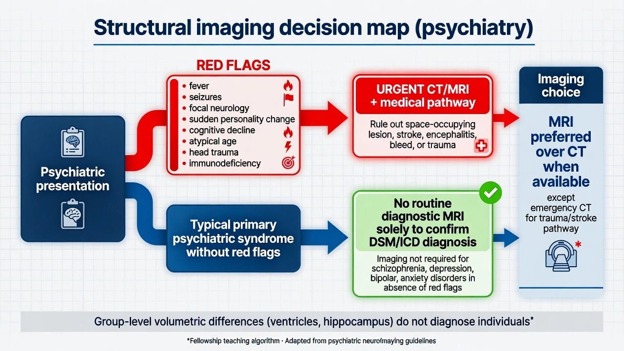
Classification of modalities
Structural CT and MRI
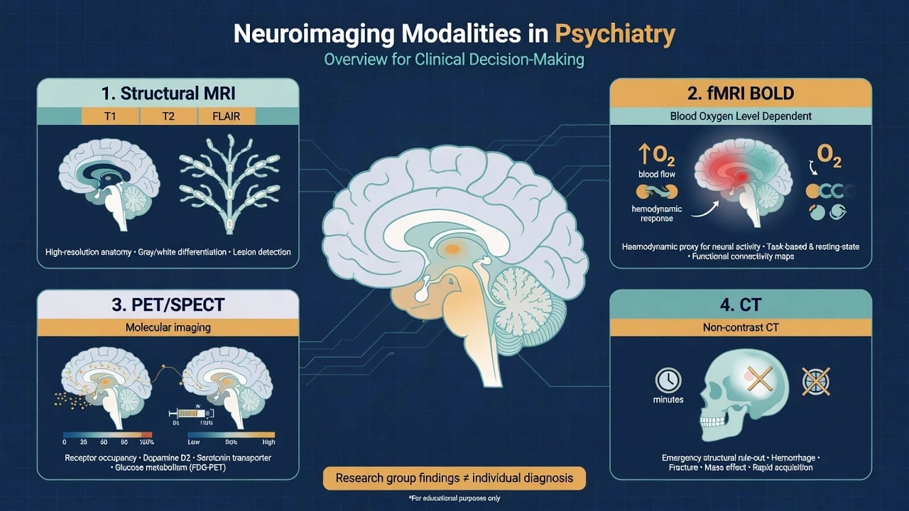
CT is fast and widely available for trauma, haemorrhage, and acute stroke pathways when MRI would delay care. MRI is preferred for most elective psychiatric organic work-ups because soft-tissue contrast better evaluates mass lesions, white-matter disease, and many inflammatory or developmental patterns at clinical radiology depth. Conceptual sequences examiners expect you to name: T1 anatomy, T2/FLAIR for white-matter and oedema patterns, and DWI for acute ischaemia when that pathway is relevant. [15][16]
Functional MRI (BOLD)
Blood-oxygen-level-dependent (BOLD) fMRI is a haemodynamic proxy related to local neural activity, not a direct spike counter. Logothetis and colleagues demonstrated the complex neurophysiological basis of the fMRI signal and later summarised what fMRI can and cannot claim — the classic viva caution against naive reverse inference. [9][10]
Resting-state and large-scale networks
Spontaneous BOLD fluctuations organise into large-scale systems, including a default mode of brain function when people are not focused on external tasks. [11] Menon's triple-network model (default mode, salience, frontoparietal/central executive) is the standard teaching scaffold for psychopathology network language — again as group research framing, not a radiology report template. [12]
PET and SPECT
Radioligand imaging underpins receptor occupancy teaching. Increased baseline D2 receptor occupancy by dopamine in schizophrenia samples supports excess dopaminergic tone models that feed Howes and Kapur's dopamine hypothesis version III (final common pathway / aberrant salience). [13][14] These methods justify mechanism language in exams; they are not required before every antipsychotic prescription.
Epidemiology, yield, and risk framing
Most patients with typical primary psychiatric syndromes do not need imaging to confirm diagnosis. Yield of structural imaging for changing diagnosis is higher when pre-test probability of organic disease rises (atypical age, neurological signs, cognitive decline, systemic features). [15] Population neuroimaging consortia demonstrate small-to-moderate group differences in brain structure across major disorders; those differences do not become cut-points for individual diagnosis. [6][7][8]
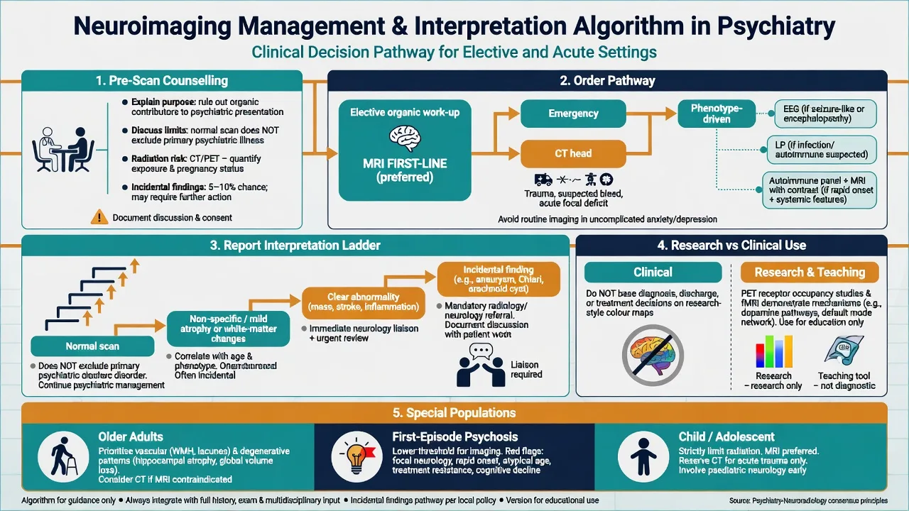
Incidental findings are frequent enough that pre-scan counselling (purpose, limits, possibility of unexpected results, follow-up pathways) is part of good practice. [16]
Pathophysiology and landmark group findings
Structural era: from CT ventricles to MRI synthesis
Johnstone and colleagues' CT work linking cerebral ventricular size to chronic schizophrenia samples opened the modern structural imaging era in psychiatry. [1] Subsequent MRI systematic reviews and meta-analyses documented smaller whole-brain and regional volumes with relatively enlarged ventricles in schizophrenia cohorts, with regional emphases that vary by method (ROI vs VBM). [2][3][4] First-episode samples already show volumetric differences in meta-analysis — arguing against purely chronic medication or institutional artefact explanations for all findings, while still forbidding individual diagnostic use. [2][4]
High-risk and longitudinal MRI
Pantelis and colleagues showed neuroanatomical abnormalities before and after onset of psychosis in high-risk longitudinal MRI comparisons — foundational for neurodevelopmental staging models and for the viva line that risk-biology is not clinic prediction for one person. [5]
ENIGMA-scale patterns
Large multi-site consortia refine effect sizes: subcortical volume abnormalities in schizophrenia and bipolar disorder, and cortical thickness/surface patterns in major depression, are reproducible at population scale yet remain non-diagnostic at the bedside. [6][7][8]
Mood circuits and mechanism bridges
Subgenual prefrontal abnormalities are repeatedly implicated in mood disorder imaging literature, and experimental deep brain stimulation of subcallosal cingulate targets in highly treatment-resistant depression illustrates circuit-level intervention concepts — research proof more than routine psychiatry. (See linked neuromodulation topics for clinical DBS status.) Network-level mood and psychosis formulations still rest on clinical diagnosis. [12][14]
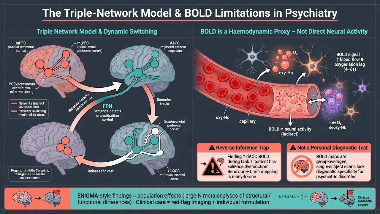
Clinical presentation: when the phenotype is the indication
Order or escalate neuroimaging when history or MSE raises organic probability — atypical age or tempo, focal neurology, seizures, fever, head trauma, fluctuating attention, rapid cognitive decline, progressive language or praxis failure, subacute psychosis with movement disorder or dysautonomia, or severe behavioural change with immunodeficiency or known cancer. [15][16]
Differential diagnosis — discriminators, not lists
| Competing category | Clinical discriminators | Imaging role |
|---|---|---|
| Primary psychiatric syndrome | Tempo fits episode history; exam non-focal | Imaging only if red flags |
| Space-occupying / stroke / TBI | Focal signs, trauma, sudden deficit | Urgent CT/MRI pathway |
| Autoimmune encephalitis | Subacute, seizures, dysautonomia, movements | MRI plus EEG/CSF package |
| Neurodegenerative / vascular cognitive | Progressive cognition, stepwise course | Structural MRI; specialist dementia work-up |
| Toxic-metabolic encephalopathy | Systemic clues, drugs, labs | Imaging adjunct; labs often primary |
| Discriminators drive orders. [15][16] |
Colour task-fMRI maps do not discriminate these categories for an individual patient. [10]
Bedside assessment before the request
- History that changes pre-test probability (onset, trauma, seizure, fever, cognition, substances, HIV risk, malignancy).
- Focused neurological screen and vital signs.
- Consent/safety: MRI metal checklist, claustrophobia plan, pregnancy status for protocol choices.
- Document the question for radiology ("exclude mass/encephalitis pattern" beats "query schizophrenia"). [15][16]
Risk (suicide, violence, vulnerability) remains clinical — no imaging suite replaces structured risk assessment. [16]
Investigations algorithm
| Situation | Preferred first structural step | Add-ons when phenotype demands |
|---|---|---|
| Elective organic work-up in psychiatry | MRI brain | EEG, LP, autoimmune panel as indicated |
| Trauma / suspected acute stroke pathway | CT first if MRI delays care | Follow local stroke/trauma protocols |
| First-episode psychosis without red flags | Individualised; many services image once — justify by local policy and residual uncertainty, not by "proving diagnosis" | Baseline metabolic/ECG for meds separately |
| Suspected encephalitis / NMDAR phenotype | MRI urgently as part of package | EEG, CSF, antibodies — do not stop at MRI alone |
| Research interest only | Not a clinical indication | Ethics pathway if research study |
| Practical table. [15][16] |
fMRI / resting-state and most PET occupancy studies remain research or highly specialised tools. Clinical PET niches (selected dementia evaluations) belong to cognitive/neurology pathways, not routine mood-disorder diagnosis. [16][10]
Acute management and scan logistics
Do not delay airway, seizure, or stroke care for elective psychiatric imaging. Agitated patients need safety and least-restrictive de-escalation first; forced scanning under deep sedation without airway-capable support is high-risk and rarely justified in pure psychiatric settings. Critical incidental findings (mass effect, haemorrhage) trigger immediate medical transfer. [15]
Definitive interpretation and management integration
Normal scan
Supports reduced likelihood of many structural lesions but does not exclude primary psychiatric disorders, early encephalitis with normal early MRI, or toxic-metabolic states. Proceed with psychiatric formulation and treatment; continue medical vigilance if phenotype evolves. [15][16]
Abnormal scan relevant to presentation
Liaise with neurology/neurosurgery/radiology; treat the organic disease; co-manage psychiatric symptoms. Imaging that changes the medical differential must change the care pathway. [15][16]
Incidentaloma
Avoid cascade: correlate clinically, follow radiology advice, do not reattribute all psychiatric illness to a tiny unrelated finding. [16]
Mechanism teaching without scan shopping
Use PET occupancy and dopamine-pathway models to explain antipsychotics; use network language sparingly in formulation. Do not demand research imaging before standard treatments. [13][14]
Clinical structural imaging
- Question: is there treatable structural disease?
- MRI elective; CT emergency pathways
- Changes management when positive or when it safely lowers organic risk
- Normal scan ≠ 'not biological'
Research fMRI / networks
- Question: group mechanisms and models
- BOLD haemodynamic proxy
- Reverse inference is a trap
- Not an individual diagnostic test
PET/SPECT occupancy
- Question: receptor engagement / dopaminergic tone models
- Supports dopamine hypothesis teaching
- Specialised, not routine pre-treatment
- Links to version III final common pathway narrative
Subtypes and scenarios
First-episode psychosis. Conceptual medical work-up includes considering structural imaging based on risk, not as a ritual to "confirm schizophrenia." [15]
Treatment-resistant or atypical mood. Revisit organic differentials and prior imaging quality before endless psychotropic stacking. [16]
Late-onset psychosis / new cognitive impairment. Lower threshold for MRI and cognitive work-up. [16]
Catatonia / autoimmune interface. Imaging is one piece of a multi-modal package. [15]
Forensic requests. Scans address organic disease; they do not read minds or measure criminal responsibility. [16]
Child/adolescent. Prefer MRI over ionising radiation when imaging is needed; avoid speculative research scans in care. [16]
Complications and pitfalls
- Group-to-person error: ENIGMA or meta-analytic volume differences used as personal diagnostics. [6][8]
- Reverse inference: "this blob means this emotion" from fMRI. [10]
- Under-scanning when red flags scream organic disease. [15]
- Over-scanning without indication, feeding anxiety and incidentaloma cascades. [16]
- False dualism: normal MRI presented as proof illness is purely psychosocial. [16]
- Occupancy myth: requiring PET before first antipsychotic. [13][14]
Prognosis and disposition
Imaging almost never sets psychiatric prognosis alone. Course is driven by syndrome, comorbidity, substance use, treatment response, risk, and support. High-risk structural research findings do not equal inevitable conversion to psychosis. Disposition after a clearly abnormal clinical scan follows the neurological/neurosurgical finding plus psychiatric co-management. [5][16]
Special populations
| Population | Imaging nuance |
|---|---|
| Older adults | Lower threshold with late-onset psychosis or cognitive change; vascular and degenerative patterns |
| Pregnancy | Prefer MRI without gadolinium when imaging required; avoid ionising radiation when possible |
| Intellectual disability | Interpret structure against developmental baseline; collateral history essential |
| Cultural contexts | Explain purpose and limits without dismissing non-biomedical explanatory models |
| Special population table. [16][15] |
Evidence and guidelines (exam anchors)
Landmark structural synthesis includes Johnstone CT ventricles, Lawrie/Shenton/Wright MRI reviews and meta-analyses, Pantelis high-risk longitudinal MRI, and ENIGMA multi-disorder volumetric maps. [1][2][3][4][5][6][7][8] Method papers every candidate should name: Logothetis on BOLD, Raichle on default mode, Menon on triple networks. [9][10][11][12] Mechanism imaging: Abi-Dargham occupancy and Howes–Kapur synthesis. [13][14] Clinical framing: Freudenreich FEP medical work-up concepts and First and colleagues on clinical applications of neuroimaging in psychiatric disorders. [15][16]
Exam pearls
- Normal MRI does not exclude schizophrenia, bipolar disorder, or MDD. [16]
- Image for organic red flags, not to confirm a chemical-imbalance cartoon. [15]
- BOLD is a proxy; reverse inference is a classic trap. [9][10]
- Ventriculomegaly is a group-history teaching point, not a personal diagnostic criterion. [1][4]
- PET occupancy teaches antipsychotics; it is not mandatory before first prescription. [13][14]
- Pre-scan counsel purpose, limits, and incidental findings. [16]
- Emergency CT for trauma/stroke timing; elective MRI for most psychiatric organic work-ups. [15]
- Autoimmune phenotypes need a package (MRI ± EEG/CSF), not imaging alone. [15]
References
- [1]Johnstone EC, Crow TJ, Frith CD, et al. Cerebral ventricular size and cognitive impairment in chronic schizophrenia Lancet, 1976.PMID 62160
- [2]Lawrie SM, Abukmeil SS Brain abnormality in schizophrenia. A systematic and quantitative review of volumetric magnetic resonance imaging studies Br J Psychiatry, 1998.PMID 9519062
- [3]Shenton ME, Dickey CC, Frumin M, McCarley RW A review of MRI findings in schizophrenia Schizophr Res, 2001.PMID 11343862
- [4]Wright IC, Rabe-Hesketh S, Woodruff PW, et al. Meta-analysis of regional brain volumes in schizophrenia Am J Psychiatry, 2000.PMID 10618008
- [5]Pantelis C, Velakoulis D, McGorry PD, et al. Neuroanatomical abnormalities before and after onset of psychosis: a cross-sectional and longitudinal MRI comparison Lancet, 2003.PMID 12559861
- [6]van Erp TG, Hibar DP, Rasmussen JM, et al. Subcortical brain volume abnormalities in 2028 individuals with schizophrenia and 2540 healthy controls via the ENIGMA consortium Mol Psychiatry, 2016.PMID 26283641
- [7]Hibar DP, Westlye LT, van Erp TG, et al. Subcortical volumetric abnormalities in bipolar disorder Mol Psychiatry, 2016.PMID 26857596
- [8]Schmaal L, Hibar DP, Samann PG, et al. Cortical abnormalities in adults and adolescents with major depression based on brain scans from 20 cohorts worldwide in the ENIGMA Major Depressive Disorder Working Group Mol Psychiatry, 2017.PMID 27137745
- [9]Logothetis NK, Pauls J, Augath M, et al. Neurophysiological investigation of the basis of the fMRI signal Nature, 2001.PMID 11449264
- [10]Logothetis NK What we can do and what we cannot do with fMRI Nature, 2008.PMID 18548064
- [11]Raichle ME, MacLeod AM, Snyder AZ, et al. A default mode of brain function Proc Natl Acad Sci U S A, 2001.PMID 11209064
- [12]Menon V Large-scale brain networks and psychopathology: a unifying triple network model Trends Cogn Sci, 2011.PMID 21908230
- [13]Abi-Dargham A, Rodenhiser J, Printz D, et al. Increased baseline occupancy of D2 receptors by dopamine in schizophrenia Proc Natl Acad Sci U S A, 2000.PMID 10884434
- [14]Howes OD, Kapur S The dopamine hypothesis of schizophrenia: version III--the final common pathway Schizophr Bull, 2009.PMID 19325164
- [15]Freudenreich O, Schulz SC, Goff DC Initial medical work-up of first-episode psychosis: a conceptual review Early Interv Psychiatry, 2009.PMID 21352170
- [16]First MB, Drevets WC, Carter C, et al. Clinical Applications of Neuroimaging in Psychiatric Disorders Am J Psychiatry, 2018.PMID 30173550