Cardiology · Cardiology
Acute Aortic Syndrome
Also known as Aortic dissection · Aortic intramural haematoma · Penetrating aortic ulcer · AAS · Stanford A dissection · Stanford B dissection · DeBakey I · DeBakey II · DeBakey III
Acute aortic syndrome (AAS) is a spectrum of life-threatening aortic emergencies — classic aortic dissection (intimal tear with a false lumen), intramural haematoma (IMH), and penetrating aortic ulcer (PAU) — that share a common pathophysiology (medial degeneration and wall shear) and an identical initial resuscitation strategy. Stanford type A (ascending aorta involvement) is a surgical emergency — mortality rises roughly 1 to 2 percent per untreated hour, and emergency open repair (supracoronary tube graft or valve-sparing root replacement, plus aortic arch replacement if required) carries an operative mortality of 15 to 25 percent in IRAD and saves the patient's life in most cases. Stanford type B (descending aorta only, distal to the left subclavian artery) is initially managed medically — impulse-control with IV beta-blockade (esmolol or labetalol) targeting a heart rate under 60 bpm and a systolic blood pressure 100 to 120 mmHg, plus analgesia (morphine 5 to 10 mg IV) — with thoracic endovascular aortic repair (TEVAR) reserved for complicated type B (malperfusion, rupture, refractory pain, expansion, uncontrolled hypertension). The classic presentation is sudden severe tearing or ripping chest or back pain, often with a blood-pressure differential of more than 20 mmHg between arms or between arm and leg, a widened mediastinum on chest X-ray, and a pulse or neurological deficit. Independent risk factors include uncontrolled hypertension (the dominant factor), connective-tissue disease (Marfan, Loeys-Dietz, Ehlers-Danlos type IV), bicuspid aortic valve, aortic coarctation, cocaine use, pregnancy (especially the third trimester and peripartum), smoking and family history of aortic disease. Diagnosis is by CT aortography (first-line in stable patients — sensitivity and specificity both above 95 percent), transoesophageal echocardiography (TEE) (the bedside test of choice in haemodynamically unstable patients in the resuscitation room or operating theatre), or MRI (second-line, e.g. young/ pregnant/renal failure patients who cannot have iodinated contrast). D-dimer can be a useful rule-out in low-probability patients presenting under 24 hours (sensitivity about 95 to 99 percent when below 500 ng/mL but specificity poor). Complications include cardiac tamponade, acute aortic regurgitation, myocardial infarction (if a coronary ostium is involved), stroke (carotid or intercostal malperfusion), paraplegia (spinal cord ischaemia from intercostal artery loss), mesenteric ischaemia, renal failure and limb ischaemia. Key drugs: esmolol 500 mcg/kg IV bolus then 50 to 200 mcg/kg/min infusion, labetalol 20 to 80 mg IV bolus every 10 minutes (max 300 mg), nicardipine 5 to 15 mg/hour IV infusion, nitroprusside 0.25 to 10 mcg/kg/min (added after rate control), and morphine 5 to 10 mg IV for pain. Anchored to the 2022 ACC/AHA (Isselbacher) and 2014 ESC (Erbel) aortic disease guidelines.
On this page & tools
Your progress
Saved locally on this device.
Exam tags
Red flags
Overview & Definition
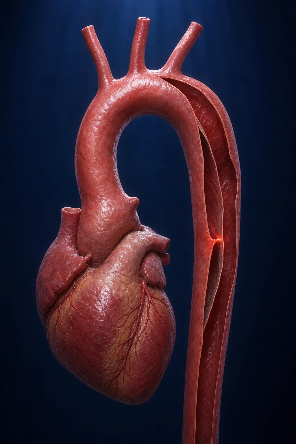
Acute aortic syndrome (AAS) is an umbrella term for three closely related, life-threatening emergencies that share a common final pathway — medial wall stress producing a tear or haemorrhage within the aortic wall — and a near-identical initial resuscitation algorithm: classic aortic dissection, intramural haematoma (IMH), and penetrating aortic ulcer (PAU). [1]
Classic aortic dissection is defined as a tear of the intima (and often the inner media) that allows pulsatile blood to enter and propagate within the media, creating a false lumen separated from the true lumen by an intimal flap. The false lumen may propagate antegrade, retrograde, or both, and may rupture back into the true lumen (re-entry) or out of the aorta entirely into the pericardium, mediastinum, pleura, or abdomen. Acute dissection is defined as onset within 14 days of presentation; subacute is 15 to 90 days; chronic is more than 90 days. IRAD (17-year, n=4428) reports a median time-to-presentation of 4.3 hours and a median time-to-diagnosis of 8.3 hours.[2]
Intramural haematoma (IMH) is a haemorrhage within the media without a demonstrable intimal flap or false lumen on imaging — thought to arise from rupture of the vasa vasorum rather than an intimal tear. IMH accounts for 10 to 30 percent of AAS in modern series and behaves similarly to dissection in the ascending aorta (high-risk, often progresses to overt dissection or rupture) but more favourably in the descending aorta (many resolve with medical therapy). [1]
Penetrating aortic ulcer (PAU) is an ulceration of an aortic atherosclerotic plaque that erodes through the internal elastic lamina into the media, producing a localised haematoma. PAU is typically a disease of the descending aorta in older hypertensive patients with heavy atherosclerosis; rupture risk is significant if the ulcer is deep, the haematoma is large or expanding, or symptoms (especially pain) persist despite medical therapy. [1]
Epidemiologically, AAS is rare but lethal: IRAD reports an overall in-hospital mortality of 27 percent for type A and 9 percent for type B. Type A is roughly twice as common as type B. Onset is typically in the sixth to seventh decade for type B (in the hypertensive, atherosclerotic, smoking patient) and fifth decade for type A (in hypertensive patients, often with connective-tissue disease). Women with dissection are on average older, present later, and have worse outcomes.[2][3]
Classification
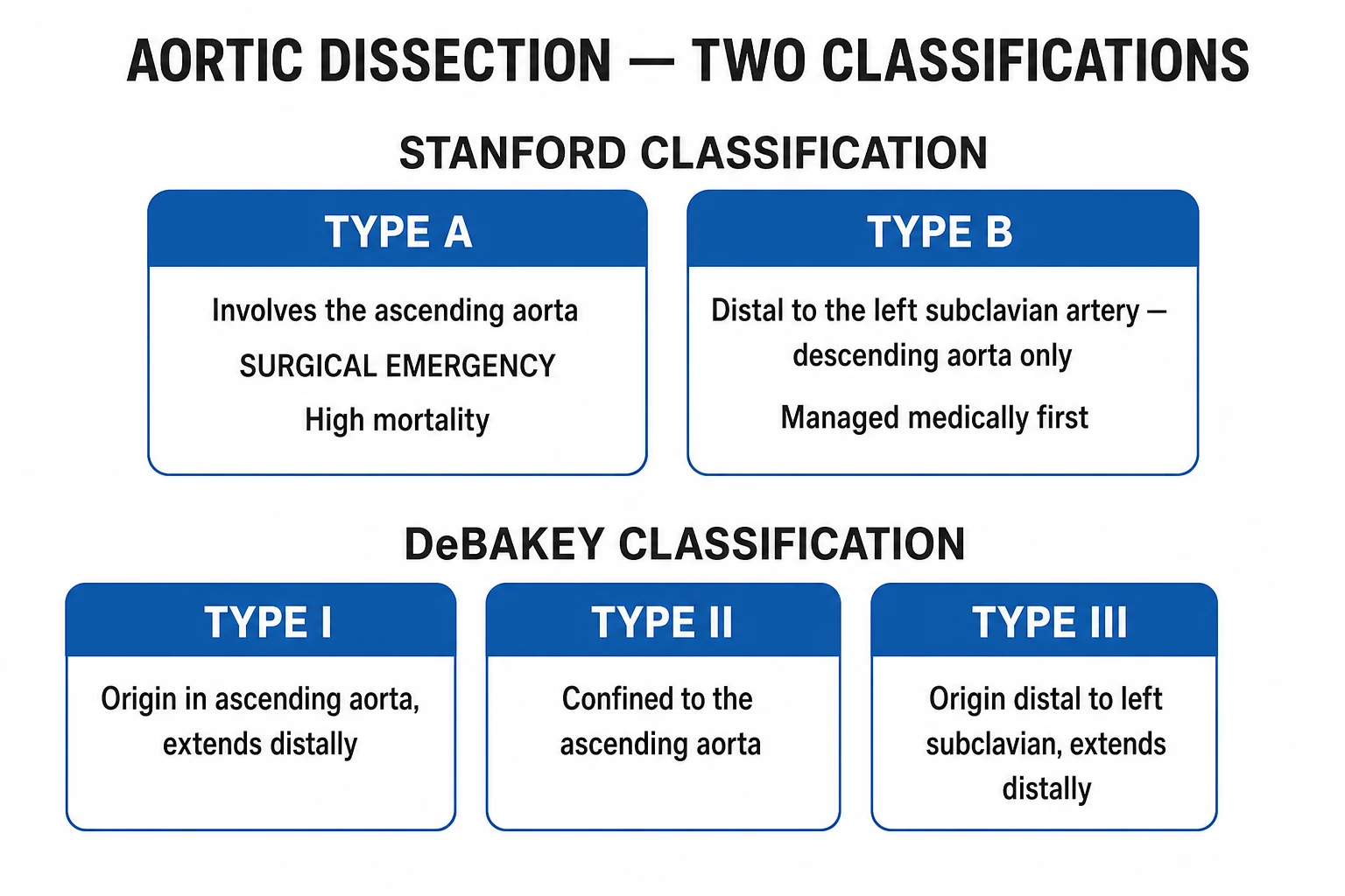
Three classification systems are in clinical use. Each has implications for management. [1]
Stanford — the operative decision: [1]
- Stanford type A — any dissection involving the ascending aorta (with or without descending extension). Surgical emergency.
- Stanford type B — dissection confined to the descending aorta distal to the left subclavian artery, not involving the ascending aorta. Medical management (impulse control); TEVAR for complicated cases. [1]
DeBakey — the anatomic description: [1]
- Type I — originates in the ascending aorta, propagates at least to the arch and often into the descending aorta.
- Type II — confined to the ascending aorta.
- Type III — originates in the descending aorta distal to the left subclavian and propagates distally (IIIa = supradiaphragmatic; IIIb = extends below the diaphragm). [1]
(The DeBakey I / II pattern = Stanford A. DeBakey III = Stanford B.) [1]
Stanford A (≈60%)
- Ascending aorta involvement (with or without arch/descending extension)
- Includes DeBakey I and II
- Mortality ~1 to 2 percent per untreated hour; up to 50 percent die within 48 hours untreated
- Surgery is the default — emergent open repair (supracoronary tube graft, valve-sparing root replacement, or Bentall if aortic valve involved; hemiarch or total arch if arch involved)
- Medical therapy alone is only used as a bridge when surgery is contraindicated (e.g. moribund, severe neurological devastation, patient/family decline)
Stanford B (≈40%)
- Descending aorta only, distal to left subclavian
- Includes DeBakey III
- Initial management is medical (impulse control — HR under 60, SBP 100 to 120 mmHg, analgesia)
- TEVAR indicated for complicated cases — malperfusion, rupture, refractory pain/expansion, uncontrolled hypertension, organ ischaemia
- Uncomplicated B has in-hospital mortality ~10 percent; complicated B treated medically ~20 percent vs TEVAR ~10 percent
For IMH and PAU, classification mirrors anatomy: involving the ascending aorta = treat like type A (surgery in many centres, especially IMH with pericardial effusion or intimal tear seen on TEE); confined to the descending aorta = treat like type B (medical; TEVAR for PAU with persistent pain, expansion or signs of rupture).[1]
Penn classification (for postoperative risk stratification in type A dissection) — adds clinical state at presentation: class Aa (stable, no ischaemia) Ab (localised — branch-vessel malperfusion) Ac (generalised — cardiac tamponade, shock) Ad (combined) — mortality rises from class Aa (~10 percent operative mortality) to Ac (~30 to 50 percent) to Ad (~60 percent).[3]
Aortic dissection: Detecting the High-Risk Patient — the ADD-RS + D-dimer (AORTAs) pathway — a clinical risk-stratification framework used in emergency departments: ADD-RS score (3 points — high-risk conditions, 3 — high-risk pain features, 3 — high-risk exam findings; total 0 to 3 + D-dimer) stratifies patients into low / intermediate / high pretest probability. A score of 0 with a D-dimer under 500 ng/mL within 24 hours of pain onset effectively rules out dissection in low-risk patients (sensitivity 99 to 100 percent in meta-analysis).[4]
Epidemiology & Risk Factors
AAS is uncommon but disproportionately lethal: an estimated 3 to 6 cases per 100,000 person-years — roughly 4 to 10 per 100,000 in modern autopsy series — yet it is a leading cause of sudden cardiovascular death. IRAD (2009 to 2016) shows no decline in incidence but a small improvement in mortality for type A surgery (operative mortality ~22 percent now versus ~25 percent historically).[2]
Risk-factor hierarchy (high-yield for exam): [1]
- Hypertension — the dominant and most prevalent risk factor. Present in 70 to 80 percent of all dissections and about 90 percent of type B in IRAD. Long-standing uncontrolled hypertension, especially nocturnal non-dipping and resistant hypertension, is the principal modifiable risk.
- Age — incidence rises sharply after 50, peaks at 60 to 80. Type A median age ~62 years, type B ~66 years in IRAD.
- Sex — male:female ~2:1 for type A, but women are more likely to present late and have worse mortality. Type B is more common in men.
- Connective-tissue disease — Marfan syndrome (FBN1 mutation): the archetypal cause of type A in young patients — half of Marfan dissections are type A, with lifetime risk up to 50 percent if aortic root more than 50 mm and surgery not performed. Loeys-Dietz syndrome (TGF-β pathway) — more aggressive aortic disease, dissection at smaller diameters (often under 45 mm). Ehlers-Danlos type IV (vascular type) — fragile vessels, surgery treacherous. Familial thoracic aortic aneurysm and dissection (TAAD — ACTA2, MYH11, MYLK, etc.).
- Congenital — bicuspid aortic valve (1 to 2 percent of the population, dissection risk about 5 to 10 times higher; aortic coarctation; Turner syndrome with bicuspid/coatctation).
- Aortic dilatation — ascending aorta diameter above 55 mm (or above 50 mm in Marfan, bicuspid, family history) raises risk substantially; the formula for risk of dissection doubles when the diameter exceeds the aortic size index threshold.
- Atherosclerosis — less a risk for dissection than for PAU, which is the AAS variant most strongly associated with heavy descending aortic plaque.
- Cocaine, methamphetamine and other sympathomimetics — classically associated with dissection (catecholamine surge + acute hypertension + vasospasm; often descending in younger men).
- Pregnancy and the peripartum state — roughly half of dissections under age 40 are pregnancy-associated, concentrated in the third trimester and first 6 weeks postpartum (haemodynamic, hormonal, and aortic-wall remodelling changes).
- Trauma / iatrogenic — motor-vehicle deceleration injuries (deceleration at the aortic isthmus, distal to the left subclavian); catheter-induced (coronary angiography, intra-aortic balloon pump, ECMO cannulation, TAVR, TEVAR misadventure); surgery — cardiopulmonary bypass aortic cannulation, cross-clamp injury.
- Inflammatory aortitis — Takayasu, giant-cell arteritis, Behçet disease — may cause dissection or accelerate IMH.
- Smoking — independent predictor of dissection and post-operative mortality.
- Family history of dissection in a first-degree relative roughly doubles the risk (screening echo for first-degree relatives of dissection probands is recommended). [1]
Dissection is the prototypical condition in which a hypertensive surge interacts with a weakened aortic wall; the resuscitation strategy targets the surge, the elective strategy targets the wall. [1]
Pathophysiology
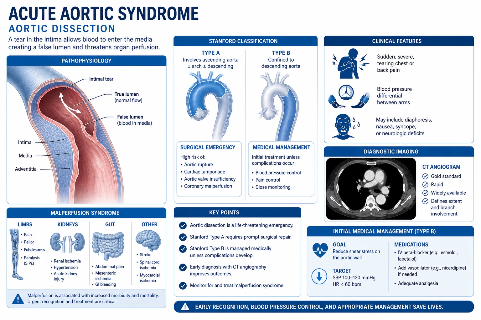
The aorta is a three-layered elastic artery — tunica intima (endothelium on internal elastic lamina), tunica media (smooth muscle and elastic lamellae providing 70 percent of elastic recoil), and tunica adventitia (collagen-rich outer coat, vasa vasorum). Two physical variables determine whether the wall remains intact: the circumferential tensile stress (governed by Laplace's law: stress = pressure × radius / wall thickness) and the rate of rise of pressure (dP/dt) in systole. Hypertension enlarges both — higher pressure increases wall tension; a high dP/dt (e.g. a rapid ventricular ejection in a young hypertensive male with a stiff aorta) transmits abrupt shear to a structurally weak point. [1]
The initiating event is either: [1]
- An intimal tear with pulsatile blood entering the media and propagating longitudinally, splitting the wall — classic dissection. The tear most commonly arises in the right lateral wall of the ascending aorta (highest shear, near the sinotubular junction) or at the aortic isthmus (just distal to the left subclavian artery — the junction of the mobile arch and the fixed descending aorta, the site of deceleration shear in trauma). A dissection with no identifiable entry tear is more consistent with IMH.
- Rupture of a vasa vasorum within the media producing a localised intramural haemorrhage and secondary inflammatory weakening — IMH.
- Ulceration of an atherosclerotic plaque through the internal elastic lamina into the media — PAU. [1]
Wall weakness predisposes via three principal mechanisms: (1) cystic medial degeneration (Erdheim) — loss of smooth muscle, fragmentation of elastic fibres, accumulation of basophilic ground substance — the dominant lesion in Marfan, Loeys-Dietz, and ageing hypertension. (2) Atherosclerotic plaque with medial extension — more typical of PAU. (3) Inflammatory destruction of the media — Takayasu, giant-cell, infectious (syphilitic aortitis — historical, tertiary syphilis). [1]
The malperfusion sequelae are an extension of the same process. The dissection flap may obstruct any branch vessel — coronary ostium (MI, classically RCA → inferior STEMI) — aortic arch vessels (stroke, arm ischaemia) — coeliac/SMA/IMA (mesenteric ischaemia, often painless abdominal pain with lactic acidosis) — renal arteries (AKI, refractory hypertension) — iliac/femoral (limb ischaemia, the classic malperfusion syndrome; 6 P's) — intercostal/lumbar arteries (paraplegia from spinal cord ischaemia). Retrograde extension into the aortic root may distort the valve (acute aortic regurgitation — AR present in up to 50 percent of type A, occasionally presenting as pulmonary oedema with a new diastolic murmur) or rupture into the pericardium (cardiac tamponade — the most common immediate cause of death in type A dissection). [1]
The catastrophic chemistry: as the false lumen becomes a high-pressure space, the intimal flap bows into the true lumen, compromising branch perfusion (dynamic obstruction) or, when the flap extends into the ostium and obstructs it directly (static obstruction), producing organ ischaemia. The pressure differential between true and false lumens determines symptoms: a widely patent false lumen with equalised pressure is often clinically silent; a thrombosed false lumen with persistent true-lumen pressurisation can cause branch ischaemia even when the dissection itself appears minimal. [1]
Clinical Presentation
The classic presentation is unmistakable when complete — but is absent in a third or more of cases, and the exam aim is to recognise the high-risk incomplete patterns. [1]
Pain is the dominant symptom in 85 to 95 percent of cases. It is characteristically: (1) sudden — frequently described by patients as occurring at a precise second ("like a switch was thrown", "the worst moment of my life"); (2) severe at onset — peak intensity within seconds, unlike the crescendo pain of myocardial ischaemia; (3) sharp, tearing, ripping or "shearing" in quality — though IRAD shows sharp is more common than tearing in modern series; (4) radiating — to the back, interscapular region, abdomen, hips or legs, according to dissection propagation; (5) migrating — a pain that moves with propagation is highly suggestive. Pain location tracks the anatomy: anterior chest pain in type A ascending dissection; back, abdominal or flank pain in descending dissection; neck/jaw pain in arch involvement. Painless dissection (about 5 to 15 percent of cases) is more common in elderly, diabetic, syncopal and stroke presentations — a classic trap. [1]
Syncope occurs in 5 to 10 percent, usually from cardiac tamponade (most ominous), from severe hypertension-induced cerebral hypoperfusion, or from arch-vessel compromise. [1]
Hypertension is the most common finding at presentation in type B (about 70 percent); type A often presents with hypertension but also with hypotension or shock in the tamponade subgroup. [1]
Examination findings (the high-yield checklist): [1]
- BP differential — more than 20 mmHg between arms is a classic bedside clue (sensitivity modest, specificity high in a high-risk context). More than 20 mmHg between arm and leg suggests descending involvement affecting the left subclavian.
- Pulse deficit — absent or diminished carotid, brachial, radial or femoral pulse is specific for dissection affecting that branch (and a key exam finding). Document pulses serially.
- Aortic regurgitation murmur — diastolic decrescendo at the left sternal border. Acute severe AR produces a soft murmur because of rapid equalisation of pressures — the absence of a typical wide pulse pressure does not exclude AR.
- Pulmonary oedema in acute AR — sometimes the presenting feature.
- Neurological deficit — stroke (especially right-sided, from carotid malperfusion — common in type A), paraplegia (spinal cord ischaemia from intercostal artery loss in type B), Horner syndrome (sympathetic chain involvement at the isthmus).
- Signs of tamponade — muffled heart sounds, hypotension, raised JVP, pulsus paradoxus; Beck's triad. Note: pericardiocentesis in tamponade from dissection is generally contraindicated — it can precipitate further dissection or rupture.
- Mesenteric ischaemia — pain out of proportion, lactic acidosis, ileus, gut necrosis, often delayed diagnosis because the patient is sedated.
- Limb ischaemia — pulseless cold leg with pallor, paraesthesia, paralysis — the 6 P's.
- Haematuria / oliguria / AKI — renal malperfusion.
- Hoarseness — recurrent laryngeal nerve compression (Ortner syndrome — rare). [1]
Differential Diagnosis
AAS is a diagnostic trap — the initial misdiagnosis rate in IRAD was around 16 to 39 percent (improving with structured protocols and D-dimer use), and missing the diagnosis doubles mortality. Always consider AAS in unexplained acute chest, back, abdominal, or limb pain with any of the high-risk features listed above. The differential in the ED includes: [1]
- Acute coronary syndrome (ACS) / MI — the most common misdiagnosis. STEMI from coronary ostial involvement by a type A dissection is estimated at 1 to 5 percent of type A. Give thrombolytics or anti-platelet/anticoagulant therapy for a presumed MI when the patient actually has a dissection = fatal (haemorrhage into pericardium, mediastinum, or pleura). Always get a CXR (look for widened mediastinum) and bilateral arm BPs before thrombolysis in an atypical MI; faint-mediated thrombolysis bolus is sometimes cited as a cause of death in medicolegal series of missed dissections.
- Pulmonary embolism (PE) — both can present with sudden dyspnoea, chest pain and hypoxaemia. PE may be more pleuritic, with clear evidence of DVT and right-heart strain on echo; dissection more often radiates to the back with BP differential.
- Pneumothorax / tension pneumothorax — unilateral hyper-resonance, tracheal deviation; decompression reverses the picture. A large left pleural effusion in dissection may mimic pneumothorax but is serosanguineous.
- Acute pericarditis / myopericarditis — pleuritic chest pain relieved by leaning forward; pericardial rub; diffuse concave ST elevation and PR depression. Beware the patient with dissection-induced pericardial effusion and tamponade mimicking pericarditis.
- Musculoskeletal (MSK) chest/back pain — reproducible, mechanical, positional; dissection pain is constant and at maximal intensity at onset.
- Aortic aneurysm (AAA) — ruptured or symptomatic — abdominal/back pain with hypotension. Diagnosis may be AAA even if a thoracic dissection is suggested by history — get CT aortogram from neck to pelvis.
- Oesophageal pathology — Boerhaave syndrome (spontaneous oesophageal rupture), oesophageal spasm, GORD — waterbrash, dysphagia; Mallory-Weiss tear — haematemesis after vomiting. Mackler triad for Boerhaave (vomiting, chest pain, subcutaneous emphysema) — CT oesophagogram or contrast swallow.
- Biliary — acute cholecystitis / biliary colic — right upper quadrant pain, Murphy sign; may mimic dissection referred to the abdomen.
- Acute pancreatitis — epigastric pain radiating to back, raised amylase/lipase; dissection can mimic pancreatitis when the false lumen occludes the coeliac axis.
- Renal colic — loin-to-groin pain with haematuria; dissection involving a renal artery can present the same way.
- Spinal pathology — disc herniation, cord compression — gradual onset, neurological pattern matches dermatomes/myotomes; dissection paraplegia is sudden and painless.
- Mesenteric ischaemia — pain out of proportion to examination, lactic acidosis, gut infarction — dissection malperfusion must always be excluded.
- Diabetic ketoacidosis, hyperthyroid storm — both raise BP and may mimic catecholamine crisis; do not miss dissection as the underlying cause. [1]
Clinical & Bedside Assessment
The clinical assessment of a suspected AAS patient is time-critical and proceeds in parallel with resuscitation and investigation. [1]
Immediate bedside actions: [1]
- Airway, breathing, circulation — high-flow oxygen, two large-bore IVs, urinary catheter, continuous ECG, pulse oximetry, invasive arterial line (radial — but check both arm pressures first; the higher side for the line if there is a differential).
- Bilateral arm BP and one leg BP — document the differential explicitly; the higher arm drives titration targets.
- Peripheral pulses — carotid, brachial, radial, femoral, popliteal, dorsalis pedis, posterior tibial — chart the findings serially.
- Focused cardiovascular exam — heart sounds (muffling? new murmur?), JVP (raised in tamponade), palpation of the precordium (wide mediastinum may produce a sternal rock — rare), listen for the diastolic decrescendo of AR.
- Focused neurological exam — conscious level, lateralising signs, spinal cord level if suspected, limb neurology.
- Gut and limb assessment — abdominal tenderness, lactic acidosis, mottling, the 6 P's of limb ischaemia.
- Targeted history — onset (precise second), quality (tearing vs sharp), radiation, migration, associated syncope, neurological or limb symptoms, family history, known hypertension, connective-tissue disease, bicuspid valve, prior aortic surgery, pregnancy, cocaine. [1]
Time-critical investigations in parallel (the diagnostic sequence): [1]
- 12-lead ECG — for MI exclusion (and the dissection-related STEMI trap), LV hypertrophy, ischaemia. Cannot diagnose or exclude dissection.
- CXR — sensitivity modest; signs (widened mediastinum above 8 cm, calcium sign > 5 mm separation of intimal calcium from aortic wall, pleural effusion, left apical pleural cap, tracheal deviation) raise suspicion but absence does not exclude.
- Bedside transthoracic echo (TTE) — views of the ascending aorta (dilation above 45 mm, pericardial effusion, intimal flap if visible, AR severity), rapid screen in unstable patients.
- Bedside TEE — first-line imaging in the haemodynamically unstable patient (sensitivity 97 to 100 percent, specificity 95 to 100 percent for proximal dissection). Can be performed in the operating theatre. Limitations: blind spot in the upper ascending aorta and arch (interference from the air-filled trachea), need for sedation, small risk of oesophageal trauma.
- D-dimer — useful rule-out test for low-probability patients within 24 hours of pain onset (cut-off 500 ng/mL; pooled sensitivity 95 to 99 percent). Specificity is poor (elevated in MI, PE, AAA, sepsis), so a positive D-dimer does not diagnose dissection. ADvISED trial and meta-analysis support its use as an adjunct within structured risk stratification (ADD-RS score plus D-dimer). [1]
Stabilised patient: continue impulse-control and analgesia, proceed to CT aortography (first-line in stable patients; sensitivity and specificity > 95 percent for both classic dissection and IMH; identifies PAU with high accuracy; allows surgical planning — branch-vessel involvement, entry tear location, arch anatomy).[3]
Investigations
The investigation strategy is driven by clinical pretest probability (ADD-RS) and haemodynamic state: [1]
- Stable + low-to-moderate pretest probability — D-dimer as adjunct (if low risk + negative D-dimer within 24 h → can defer CT aortogram if ADD-RS=0). If intermediate risk → CT aortogram.
- Stable + high pretest probability — CT aortogram directly (skip D-dimer).
- Unstable, suspected — bedside TEE (or CT if immediately available in a hybrid suite), proceed to theatre.
- Pregnant, stable, suspected — MRI (without gadolinium) or CT aortogram with low-dose technique if MRI unavailable — D-dimer tends to be elevated in pregnancy and adds little. Discuss with cardiothoracic surgery, radiology, obstetric anaesthesia.
- Severe renal failure or contrast allergy — MRI (without gadolinium); TEE; non-contrast CT (limited utility but detects displaced intimal calcification and pericardial effusion). [1]
CT aortogram
- First-line in stable patients
- ECG-gated, thin-slice, arterial-phase contrast
- Shows intimal flap, true/false lumen, pericardial/pleural effusion, branch-vessel involvement, PAU, IMH, aortic dimensions for surgical planning
- Sensitivity and specificity both above 95 percent for classic dissection
- Radiation (~10 mSv) and iodinated contrast — caveat in pregnancy and CKD
TEE
- First-line in unstable patient, at bedside, in theatre
- Sensitivity 97 to 100 percent and specificity 95 to 100 percent for proximal dissection
- Identifies AR severity, pericardial effusion, coronary ostial involvement
- Limited views of upper ascending aorta and arch (interference from trachea); operator-dependent; small risk of oesophageal trauma
MRI / MRA
- Second-line — preferred in young, pregnant (no gadolinium), CKD (no gadolinium), or those who need serial imaging
- Sensitivity and specificity both above 95 to 98 percent
- Excellent for branch-vessel and aortic-valve anatomy; long acquisition time; limited availability out-of-hours
D-dimer
- Rule-out adjunct in low-risk patients within 24 hours
- Cut-off 500 ng/mL; pooled sensitivity 95 to 99 percent; pooled specificity 40 to 60 percent
- ADvISED + meta-analyses support ADD-RS combined with D-dimer for ED risk stratification
- Positive D-dimer does NOT diagnose dissection — proceed to imaging
- Often elevated in pregnancy; less useful
Additional targeted tests: [1]
- Troponin — frequently mildly elevated in dissection (coronary malperfusion, shock); elevation must not delay imaging when dissection is clinically suspected.
- Renal function — creatinine for branch-vessel malperfusion and for contrast planning.
- Lactate — raised in mesenteric / limb malperfusion; serial monitoring in type B with abdominal/limb pain.
- Blood cultures and inflammatory markers — if inflammatory aortitis considered (Takayasu, syphilis).
- Genetic testing and family screening — for patients under 40 or with syndromic features — Marfan (FBN1), Loeys-Dietz (TGFBR1/2, SMAD3, TGFB2), vascular EDS (COL3A1), familial TAAD (ACTA2, MYH11, MYLK, PRKG1, MAT2A). [1]
Acute Aortic Syndrome — key numbers
Management — Resuscitation
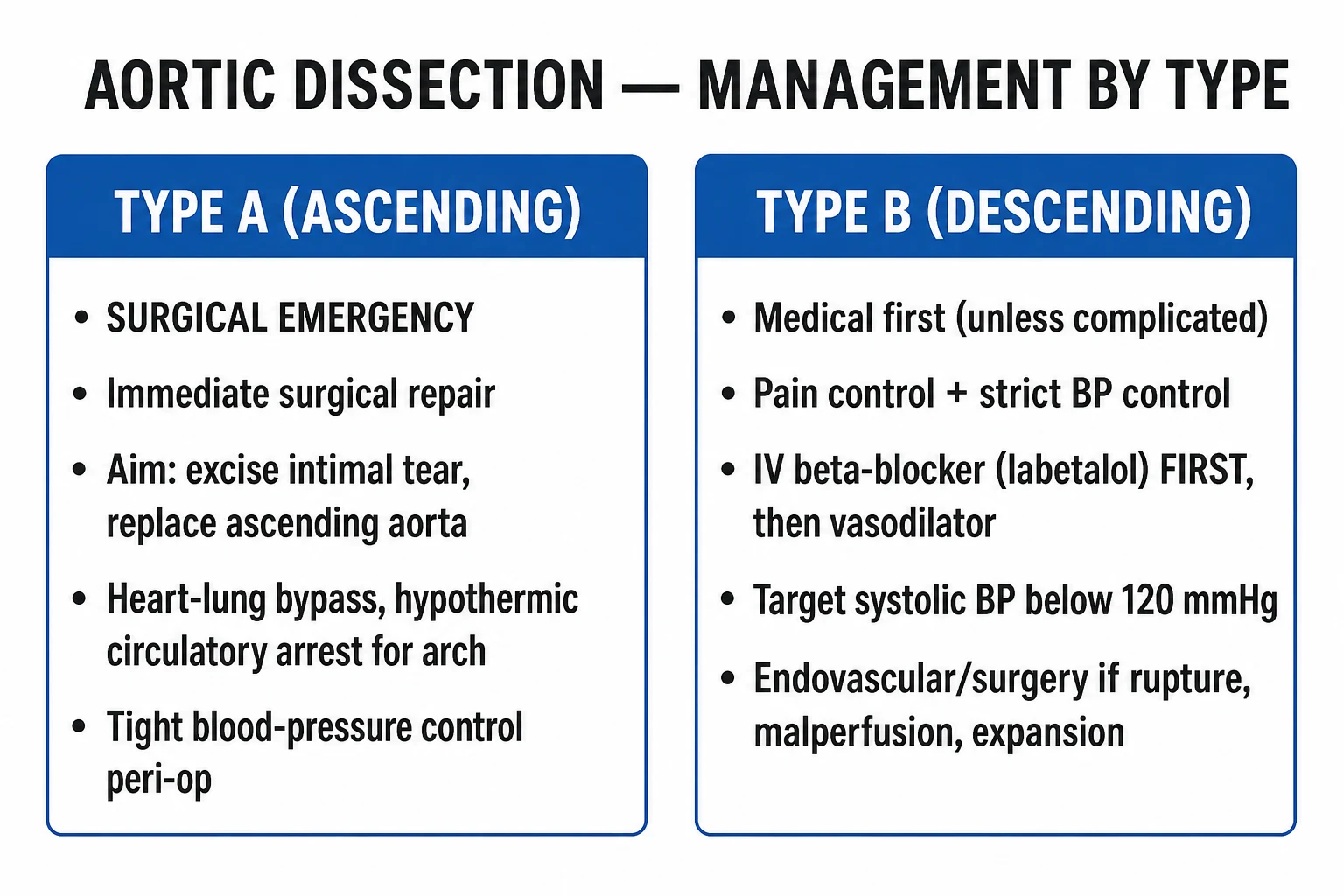
The first principle is everything in parallel: analgesia, rate-control, BP-control, investigation, surgical consultation. Time is myocardium in type A — death without surgery is about 50 percent in 48 hours and 75 percent in 2 weeks; for type B, mortality is unchanged after admission if impulse-control is achieved but rises sharply with malperfusion, expansion or rupture.[1][3]
Resuscitation targets: [1]
- Heart rate: under 60 bpm. Rate-control is the first step — it reduces dP/dt (the rate of rise of aortic pressure), which is the principal determinant of dissection propagation. A heart rate target under 60 bpm is the rationale for first-line IV beta-blockade before any vasodilator.
- Blood pressure: SBP 100 to 120 mmHg (some IRAD/ESC guidance allows to lowest level that maintains end-organ perfusion, often 100 to 120). MAP target 60 to 70.
- Pain control: morphine 5 to 10 mg IV (titrate; reduce dose in elderly; avoid IM injections that obscure imaging windows). Pain drives tachycardia and hypertension; control pain to control BP.
- Avoid hypertension surges — management of pain, suctioning (in intubated patients), bladder distension. [1]
Drug ladder (high-yield dose list): [1]
- Esmolol 500 mcg/kg IV bolus over 1 minute, then 50 to 200 mcg/kg/min infusion (titrate every 5 minutes; max 300 mcg/kg/min) — first-line for rate control because it is ultra-short-acting and titratable. Good in tachycardic, hypertensive patients and when aortic dissection is suspected but not yet confirmed (rapid reversal if diagnosis changes).
- Labetalol 20 to 80 mg IV bolus every 10 minutes (max 300 mg cumulative) OR labetalol infusion 0.5 to 2 mg/min — combined α/β blocker, second-line rate-control option. Use cautiously in acute decompensated heart failure or severe asthma.
- Metoprolol 5 mg IV bolus, repeat every 5 minutes up to 15 mg — pure β1-blocker; option when esmolol/labetalol unavailable.
- Nicardipine 5 mg/hour IV infusion, titrate by 2.5 mg every 5 to 15 minutes up to 15 mg/hour — pure arteriolar vasodilator; add after adequate rate control. Excellent when β-blocker is contraindicated.
- Sodium nitroprusside 0.25 to 10 mcg/kg/min — universal titratable arteriovenous dilator. Add only after rate control (otherwise reflex tachycardia worsens dP/dt); invasive arterial monitoring mandatory; cyanide toxicity with prolonged high-dose infusion (>1.5 mcg/kg/min for > 6 to 8 h, more likely in renal/hepatic failure).
- GTN 5 to 200 mcg/min IV infusion — venodilator with arterial effects at high dose; useful as adjunct (especially if ischaemic chest pain) but tachyphylaxis within hours.
- Morphine 5 to 10 mg IV bolus (or fentanyl 50 to 100 mcg IV titrated) — for analgesia. [1]
Do not give IV fluids to chase a falsely "low" BP: in tamponade with dissection, pericardiocentesis is generally contraindicated (may precipitate re-entry dissection or rupture). Resuscitate as for obstructive shock; proceed to surgery. [1]
Aortic dissection: BLS/ALS note — in cardiac arrest with dissection, chest compressions are likely ineffective and may extend the tear; many patients are unresuscitatable. Pericardiocentesis may temporarily restore output to allow transfer to theatre if the diagnosis is known; in out-of-hospital arrest the diagnosis is rarely known, and survival is anecdotal. [1]
Management — Definitive & Stepwise
Type A — surgery is the default; medical management is a bridge. Emergency open surgery should occur ideally within 6 hours of presentation; outcomes worsen substantially beyond 24 hours. Options include: [1]
- Supracoronary tube graft repair for most type A dissections without valvular involvement.
- Bentall procedure (composite graft + mechanical aortic valve replacement + coronary reimplantation) for aortic root involvement with significant AR or annuloaortic ectasia.
- David procedure (valve-sparing root replacement) for young patients with connective-tissue disease — durable when done in experienced centres.
- Hemiarch replacement when arch involvement is limited; total arch replacement with frozen elephant trunk for extensive arch disease.
- Concomitant procedures — AVR for severe AR, CABG for coronary ostial involvement (usually with the SVG sewn to a separate ostial patch or direct ostial repair), arch debranching. [1]
Type B — medical first; TEVAR for complicated disease. [1]
- Uncomplicated type B — initial management is impulse-control (HR under 60, SBP 100 to 120, analgesia) in HDU/ICU, step down to ward once stable, then chronic oral rate/BP control (bisoprolol 5 to 10 mg daily, amlodipine 5 to 10 mg daily, losartan 50 to 100 mg daily for connective-tissue disease) plus lifelong cardiovascular follow-up (CT aortogram at 1 month, 6 months, 12 months, then annually). About 75 percent stabilise with medical therapy alone; long-term aortic dilatation risk drives imaging follow-up.
- Complicated type B — TEVAR (thoracic endovascular aortic repair) is indicated for: (1) malperfusion — mesenteric, renal, spinal, limb; (2) rapid aortic expansion or impending rupture; (3) refractory pain despite impulse control; (4) uncontrolled hypertension despite adequate medical therapy; (5) rupture — haemothorax, retroperitoneal haematoma. TEVAR closes the entry tear, depressurises the false lumen and restores true-lumen flow. Outcomes: 30-day mortality ~5 to 10 percent, paraplegia risk 2 to 5 percent (lower than open surgery). [1]
Adjuncts in TEVAR: cerebrospinal fluid drainage for high-paraplegia-risk cases (extensive coverage, prior abdominal aortic surgery); permissive hypertension (MAP above 80) post-deployment; staged procedures for malperfusion (proximal TEVAR ± distal open or endovascular fenestration/stenting). [1]
For IMH and PAU — type A IMH behaves like type A dissection (surgical referral in many centres, especially with pericardial effusion, ascending aortic diameter above 50 mm, or intimal tear on TEE); type B IMH is managed medically, with TEVAR for persistent pain, expansion, or rupture. Symptomatic or expanding PAU is often treated with TEVAR in the descending aorta. [1]
Monitoring and follow-up: [1]
- ICU/HDU for at least 24 to 48 hours post-event.
- Serial imaging: CT aortogram (or MRI) at discharge, 1 month, 6 months, 12 months, then annually if stable.
- Aortic size monitoring — a descending aorta above 55 mm, an uncontrolled false lumen, or a rapid growth (>10 mm/year) are criteria for elective intervention in type B.
- Lifelong BP control — target SBP under 130 mmHg in chronic dissection / connective-tissue disease.
- Activity restrictions — heavy lifting avoided; cardiovascular exercise encouraged once stable.
- Cardiac rehabilitation for type A survivors (long ICU stay, sternotomy).
- Family screening in connective-tissue disease (first-degree relatives, screening echo/CT). [1]
Stepwise Management
A pragmatic protocol for a stable patient with a presumptive diagnosis of AAS on arrival (e.g. triage BP 200/110 mmHg, tearing chest pain radiating to back, pulse deficit in left arm): [1]
- Triage and immediate recognition — activate aortic emergency pathway (a multidisciplinary alert across ED, radiology, anaesthesia, cardiothoracic surgery; analogous to the stroke or STEMI pathway). Bilateral BP, pulses, ECG, CXR, IV access.
- Initiate anti-impulse therapy immediately (do not wait for imaging).
- Heart rate target under 60 bpm: esmolol 500 mcg/kg IV bolus then 50 to 200 mcg/kg/min (preferred when aortic dissection suspected and rapid reversal possible) or labetalol 20 mg IV bolus titrated.
- Once rate controlled, BP target SBP 100 to 120 mmHg: add nicardipine 5 mg/hour IV infusion, titrated by 2.5 mg/hour every 5 to 15 minutes to 15 mg/hour; if still high, add nitroprusside 0.25 to 10 mcg/kg/min (ICU-level monitoring, invasive arterial line).
- Analgesia: morphine 5 to 10 mg IV titrated; antiemetic if needed.
- Send investigations in parallel — ECG, troponin, renal function, lactate, ABG, CXR, CT aortogram (or TEE if unstable), FBC, group-and-save, type and crossmatch 4 to 6 units (for surgery).
- Classify on CT: type A vs type B; for type A — call cardiothoracic surgery immediately; for type B complicated — same. For type B uncomplicated — continue medical management.
- Type A pathway — transfer to cardiac surgical centre if not on site; pre-operative optimisation (no anticoagulation if conservative management but crossmatch ready); anti-impulse therapy continues until cardiopulmonary bypass.
- Type B pathway — HDU/ICU monitoring; pain control and rate-control continued for 24 to 48 h; oral conversion when stable (bisoprolol, amlodipine, losartan with connective-tissue disease); CT aortogram at 1 month.
- Complicated type B — TEVAR pathway — prep the angio suite; CSF drain if extended coverage planned; discuss with vascular surgery and anaesthesia.
- Disposition and follow-up — cardiac surgical outpatient clinic 6 weeks post-op (type A); cardiovascular/drug-treatment clinic (type B medical); cardiac rehabilitation; imaging schedule; family screening where indicated; activity and lifestyle counselling. [1]
Specific Subtypes & Scenarios
Marfan and connective-tissue disease — Marfan (FBN1) syndrome is the prototypical cause of type A in the young (under 50). Elective surgery is recommended at aortic root diameter above 50 mm (above 45 mm in some centres with risk modifiers — family history of dissection, rapid growth, severe AR, planned pregnancy). Pregnancy raises risk in the third trimester and first 6 weeks postpartum — pre-pregnancy aortic root assessment, β-blockade throughout pregnancy, consideration of elective surgery pre-conception if root above 45 mm. Loeys-Dietz (TGFBR1/2, SMAD3, TGFB2) — dissection at smaller diameters; surgery at above 42 mm (or earlier with family history of dissection). Ehlers-Danlos type IV (vascular EDS, COL3A1) — avoid arterial instrumentation and surgery where possible; avoid TEE and endovascular access when feasible. [1]
Bicuspid aortic valve — bicuspid patients have a 5 to 10 times higher risk of dissection than tricuspid counterparts; AS/AR management frequently coexists. Surgery threshold generally at aortic diameter above 55 mm (above 50 mm if additional risk factors — family history, rapid growth above 5 mm/year, severe valve dysfunction). [1]
Pregnancy and the peripartum state — third-trimester peripartum dissection is a feared complication; presentation may be confused with PE or normal labour discomfort. Multidisciplinary care (cardiothoracic surgery, obstetric anaesthesia, neonatology, cardiology). For type A in pregnancy — Caesarean under general anaesthesia then aortic surgery for viable fetus; aortic surgery with fetus in utero for non-viable pregnancy. Anti-impulse therapy with labetalol is the first-line rate/BP control; nifedipine as second-line vasodilator. Magnesium is not protective (unlike pre-eclampsia). [1]
Cocaine and methamphetamine dissection — often descending (type B) in young men; classic history of recent use with abrupt hypertension and pain. Benzodiazepines (e.g. midazolam) as first-line for agitation and BP control; β-blocker monotherapy is debated because of unopposed α effect — phentolamine 1 to 5 mg IV for α-blockade or nitroprusside 0.25 to 10 mcg/kg/min after rate control; alternative is labetalol (combined α/β blocker). Avoid pure β-blocker monotherapy. [1]
IMH (intramural haematoma) — type A IMH managed surgically in many centres (especially with pericardial effusion, aortic size above 50 mm, intimal tear on TEE); type B IMH managed medically with close imaging follow-up — many regress; persistent pain, expansion, ulcer-like projections, or peak aortic diameter above 50 mm are indications for TEVAR. [1]
PAU (penetrating aortic ulcer) — typically descending in older atherosclerotic patients. Medical management with risk-factor control for shallow, asymptomatic ulcers; TEVAR for deep/bulging ulcers, persistent pain, expanding haematoma, signs of rupture or aneurysm formation. [1]
Trauma — deceleration aortic injury at the isthmus accounts for ~80 percent; TEVAR is now first-line over open repair in stable patients; medical impulse-control for minimal injuries (grade I — intimal tear only). [1]
Iatrogenic — catheter-induced dissections of the ascending aorta during coronary angiography or TAVR may be sealed with covered stents in the cath lab; arch dissections during circulatory manipulation are managed surgically. [1]
Aortic coarctation — dissection or rupture at the coarctation site; treat the coarctation (surgical or balloon-and-stent) and manage the acute dissection per anatomy. [1]
Recurrent aortic events — survivors of type A repair remain at risk of distal anastomotic complications, chronic descending dissection and aneurysm formation; lifelong imaging and BP control essential. [1]
Complications
- Cardiac tamponade — the most common immediate cause of death in type A dissection (intrapericardial rupture). Beck's triad (hypotension, JVP, muffled heart sounds). Avoid pericardiocentesis if dissection suspected (may precipitate re-entry tear or rupture); proceed to surgery. Subacute tamponade occasionally responds to controlled pericardiocentesis as bridge.
- Acute aortic regurgitation in up to 50 percent of type A — produces acute pulmonary oedema with hypotension; the murmur may be soft. Surgical management includes valve repair/replacement with the root repair.
- Myocardial ischaemia / infarction — coronary ostial malperfusion (commonly RCA → inferior STEMI). The dissection may be discovered during angiographic evaluation of a "STEMI"; thrombolysis or antiplatelet/anticoagulation for presumed MI is dangerous in dissection. Treat with surgery and ostial repair / CABG.
- Stroke — arch-vessel malperfusion (right carotid commonly); type A with stroke has very high operative mortality; surgical decision nuanced (some centres proceed; others consider stroke severity with massive haemorrhagic transformation as relative contraindication).
- Spinal cord ischaemia / paraplegia — intercostal/lumbar artery loss; risk higher in TEVAR with extensive coverage or prior abdominal aortic surgery; mitigate with CSF drain and permissive hypertension.
- Mesenteric ischaemia — coeliac/SMA occlusion, often painless in sedated patients; surveillance by lactate / CT; surgical or endovascular (branch fenestration/stenting) rescue.
- Renal failure — renal malperfusion or contrast-induced; requires renal-dose care, dialysis; affects outcomes.
- Limb ischaemia — iliac/femoral malperfusion, the classic 6 P's; requires endovascular (fenestration, branch stenting) or surgical (femoro-femoral crossover, axillo-femoral) rescue.
- Aortic rupture — into pericardium, mediastinum, pleura (left haemothorax), retroperitoneum or abdomen — usually fatal; emergency surgery or TEVAR.
- Anticoagulation-related haemorrhage — particularly relevant when dissection has been misdiagnosed as MI and anti-coagulation administered.
- Chronic complications of survivors — recurrent dissection, post-surgical graft infection, false-lumen aneurysm, secondary AR, paraplegia, chronic pain. [1]
Prognosis & Disposition
Type A — operative mortality in IRAD has fallen to roughly 22 percent (2022 ACC/AHA cite 15 to 25 percent across centres; high-volume aortic centres report closer to 10 to 15 percent for uncomplicated type A). Survival benefits of surgery are overwhelmingly clear: medical management has a 50 percent mortality at 48 hours and 75 percent at two weeks. Long-term survival of operative survivors is roughly 60 to 70 percent at 10 years. Prognosis worsens with tamponade, shock, stroke, malperfusion (Penn Ac/Ad), advanced age, severe comorbidity.[2][1]
Type B — in-hospital mortality ~10 percent overall. Uncomplicated type B (medically managed) has the best prognosis — long-term survival ~80 percent at 5 years. Complicated type B treated with TEVAR improves outcomes versus medical management alone (INSTEAD-XL and contemporary series: 30-day mortality ~5 to 10 percent; long-term aortic-related survival improved). Mortality drivers: renal failure, malperfusion, age, female sex. Mortality of medical management in malperfusion is ~25 to 30 percent.[1][3]
Disposition: [1]
- Type A — Emergency theatre (cardiothoracic surgery). If the local centre does not have aortic surgery, transfer with the patient on anti-impulse therapy.
- Type B uncomplicated — HDU/ICU; step down when stable.
- Type B complicated — TEVAR suite, hybrid operating theatre, or transfer for the same.
- Rehabilitation for type A post-sternotomy ICU and ward stay; cardiac rehabilitation is standard.
- Follow-up — CT aortogram at discharge, 1 month, 6 months, 12 months, annually; plus echocardiography for valve function post-AVR or root repair; BP monitoring.
- Return to activity — avoid heavy lifting, competitive sport for months; cardiac rehabilitation protocols exist. [1]
Special Populations
- Pregnancy and peripartum — half of dissections under age 40 in women; third-trimester and peripartum are highest-risk windows (acute haemodynamic changes, raised cardiac output, hormonal effects on the aortic wall). Multidisciplinary delivery decision for viable fetus (typically Caesarean before or during the aortic repair) and anti-impulse therapy with labetalol, nifedipine, or methyldopa; ACE/ARB and nitroprusside are contraindicated. Pre-conception counselling for Marfan / Loeys-Dietz / bicuspid with aortic root sizing and elective surgery if above 45 to 50 mm. Pregnancy is generally safe with aortic root under 45 mm.
- Young patients (under 40) — connective-tissue disease more common; family screening (first-degree relatives); genetic referral for Marfan, Loeys-Dietz, vascular EDS and familial TAAD. Lifelong imaging.
- Bicuspid aortic valve patients — surveillance echo every 2 to 5 years by current size, annual above 45 mm; lifestyle (exercise caution with very heavy lifting); pre-conception counselling for women.
- Chronic kidney disease (CKD) patients — avoid iodinated contrast in stable patients where possible (prefer MRI) or use pre-medication; periprocedural renal protection for those undergoing TEVAR or surgery.
- Elderly (above 80) — presentations are often atypical and more frequently painless, syncopal or stroke-like; surgery and TEVAR are still indicated in selected patients; decision must weigh comorbidity, functional status and patient priorities. [1]
Evidence & Guidelines
Key clinical-practice guidelines: [1]
- 2022 ACC/AHA Guideline for the Diagnosis and Management of Aortic Disease (Isselbacher et al.) — the dominant North American reference; updates the 2010 Hiratzka document and incorporates IRAD trends, GenTAC and newer evidence on Marfan/Loeys-Dietz thresholds, TEVAR, and chronic dissection. Class I recommendations include anti-impulse therapy before definitive management, immediate surgery for type A, β-blockade in connective-tissue disease with aortic ectasia, screening first-degree relatives in hereditary disease.[3]
- 2014 ESC Guidelines on the Diagnosis and Treatment of Aortic Diseases (Erbel et al.) — the principal European reference; concept of AAS as a single entity with three subtypes; risk-stratified imaging recommendations; surgery thresholds similar to ACC/AHA but with some differences (type A IMH treated medically in selected cases, ESC).[1]
- 2010 ACCF/AHA/AATS/ACR/ASA/SCA/SCAI/SIR/STS/SVM Guidelines for thoracic aortic disease (Hiratzka et al.) — historically important predecessor and still cited for surgical-threshold tables.[5]
Landmark trials / data: [1]
- IRAD (International Registry of Acute Aortic Dissection) — the largest prospective observational dataset; foundational for epidemiology, mortality, presentation and outcome. 17-year trends (Pape 2015) showed changes in management (more TEVAR for B, more surgical aggressiveness in A) but persistent diagnostic delay.[2]
- Asha meta-analysis (D-dimer) — supports D-dimer below 500 ng/mL + ADD-RS=0 as a high-NPV rule-out in ED low-risk patients within 24 hours of symptom onset.[4]
- ADSORB / INSTEAD-XL trials — TEVAR plus medical therapy improved outcomes vs medical therapy alone in uncomplicated type B at 1 to 5 years, with aortic remodelling and reduced aorta-related mortality when TEVAR was performed acutely/subacutely (ADSORB) and at the subacute stage (INSTEAD-XL).
- GenTAC and other genetic registries — informative for connective-tissue disease and family-screening evidence.
- Griepp/Moon historical works on dP/dt — the foundational haemodynamic rationale for rate-control before vasodilation.
Regional practice nuances: [1]
- UK/Europe — broadly ESC-aligned; NHS aortic-dissection pathways emphasise direct to theatre for type A and rapid TEVAR for complicated type B; ADD-RS + D-dimer for ED risk-stratification.
- North America — ACC/AHA-aligned; aortic emergency pathways in major centres mirror STEMI/stroke pathways; growing role of frozen elephant trunk for arch repair.
- Asia (Japan, Korea) — extensive experience with TEVAR for distal disease and valve-sparing root replacement for type A in connective-tissue disease.
- India — most tertiary centres follow ESC for type A surgery and have adopted TEVAR; awareness challenges remain for early ED diagnosis (high rates of initial misdiagnosis, especially in rural settings). [1]
Exam Pearls
- Acute aortic syndrome = aortic dissection + intramural haematoma + penetrating aortic ulcer; share pathophysiology and initial resuscitation but differ in chronicity and certain management decisions.
- Stanford A = ascending aorta = surgical emergency. Stanford B = descending only = medical impulse-control ± TEVAR. DeBakey I/II ≈ Stanford A; DeBakey III = Stanford B.
- Pain profile: sudden, severe, tearing or sharp, maximal at onset, often radiating to back, interscapular, or migrating along the dissection line.
- Bedside clues: BP differential above 20 mmHg between arms (or arm vs leg); pulse deficit; new AR murmur; neurological deficit; syncope from tamponade. Document higher-arm BP as titration target.
- Type A mortality without surgery: ~1 to 2 percent per hour in the first 48 hours; ~50 percent at 48 h; ~75 percent at 2 weeks. Surgery is the default.
- Anti-impulse triad (apply in EVERY suspected case before imaging): rate first, then BP, then pain. Esmolol 500 mcg/kg IV bolus then 50 to 200 mcg/kg/min; labetalol 20 to 80 mg IV every 10 min (max 300 mg); nicardipine 5 to 15 mg/hour after rate control; nitroprusside 0.25 to 10 mcg/kg/min after rate control; morphine 5 to 10 mg IV.
- Heart rate target: under 60 bpm. BP target: SBP 100 to 120 mmHg (or lowest that maintains perfusion).
- Imaging: CT aortogram in stable patients; TEE in unstable patients; D-dimer + ADD-RS in low-risk ED presentations within 24 h.
- Complicated type B — TEVAR for malperfusion, rupture, refractory pain, expansion, uncontrolled hypertension.
- Never give thrombolysis/anti-coagulant for an "MI" if aortic dissection is in the differential.
- Avoid pericardiocentesis in tamponade from dissection — go to theatre.
- Avoid pure β-blocker monotherapy in cocaine dissection (unopposed α); add phentolamine or use labetalol.
- Pregnancy peripartum: multidisciplinary; labetalol/nifedipine/methyldopa; β-block throughout pregnancy if root ectasia; elective root surgery if above 45 to 50 mm pre-conception.
- Surveillance after type A repair or type B medical/TEVAR: CT aortogram at discharge, 1 m, 6 m, 12 m, then annually. Lifelong BP target SBP under 130 mmHg.
- Family screening: first-degree relatives in Marfan, Loeys-Dietz, vascular EDS, familial TAAD, bicuspid with dissection. [1]
The anti-impulse order — mnemonic
HR-BP-P
Esmolol 500 mcg/kg IV bolus, then 50 to 200 mcg/kg/min OR labetalol 20 to 80 mg IV every 10 min (max 300 mg). Always reduce dP/dt before lowering pressure — the rate of rise of systolic pressure is what propagates dissection.
Add nicardipine 5 to 15 mg/hour IV only after rate is controlled; nitroprusside 0.25 to 10 mcg/kg/min is a second-line add-on for refractory BP — never as monotherapy first.
Use the higher arm (or unaffected leg) for monitoring; intra-arterial line if available. Avoid hypotension — the goal is the lowest level that maintains end-organ perfusion.
Pain drives tachycardia and hypertension; control pain to control BP. Avoid IM injections that obscure imaging windows.
Exam application bank (NEET-PG / INICET)
One-line answer
Acute aortic syndrome (AAS) is a spectrum of life-threatening aortic emergencies — classic aortic dissection (intimal tear with a false lumen), intramural haematoma (IMH), and penetrating aortic ulcer (PAU) — that share a common pathophysiology (medial degeneration and wall shear) and an identical initial resuscitation strategy. Stanford type A (ascending aorta involvement) is a surgical emergency — mortality rises roughly 1 to 2 percent per untreated hour, and emergency open repair (supracoronary tube graft or valve-sparing root replacement, plus aortic arch replacement if required) carries an operative mortality of 15 to 25 percent in IRAD and saves the patient's life in most cases. Stanford type B (descending aorta only, distal to the left subclavian artery) is initially managed medically — impulse-control with IV beta-blockade (esmolol or labetalol) targeting a heart rate under 60 bp
Worked stems (answer without another resource)
Stem 1 — Classic presentation. Map symptoms to mechanism; name the first investigation and first treatment step with dose/route if drug therapy is standard. [1]
Stem 2 — Unstable / complicated. List red flags that force immediate resuscitation, theatre, ICU, antidote, or reperfusion — and what you do in the first 15 minutes. [1]
Stem 3 — Atypical group. Elderly, pregnancy, child, or immunocompromised: how presentation and thresholds change. [1]
Stem 4 — Differential trap. Name the three closest mimics and one discriminator for each. [1]
Stem 5 — Disposition. Who goes home with safety-netting, who is admitted, who needs HDU/ICU/theatre, and what follow-up is mandatory. [1]
Rapid viva checklist
- Definition + classification
- Pathophysiology chain
- Bedside signs / criteria
- Score with exact components (if any)
- Emergency bundle
- Definitive therapy with doses
- Complications of disease and of treatment
- Special populations
- Guideline/trial name if classic
- Three exam traps
Coverage self-check
If you cannot answer any stem above from this page alone, re-read the matching section — the page is intended to be self-sufficient for final-prof and NEET-PG/INICET questions on Acute Aortic Syndrome.
References
- [1]Erbel R, Aboyans V, Boileul C, et al. [2014 ESC Guidelines on the diagnosis and treatment of aortic diseases] Kardiol Pol, 2014.PMID 25524604
- [2]Pape LA, Awais M, Woznicki EM, et al. Presentation, Diagnosis, and Outcomes of Acute Aortic Dissection: 17-Year Trends From the International Registry of Acute Aortic Dissection J Am Coll Cardiol, 2015.PMID 26205591
- [3]Isselbacher EM, Preventza O, Hamilton Black J 3rd, et al. 2022 ACC/AHA Guideline for the Diagnosis and Management of Aortic Disease: A Report of the American Heart Association/American College of Cardiology Joint Committee on Clinical Practice Guidelines Circulation, 2022.PMID 36322642
- [4]Asha SE, Miers JW. A Systematic Review and Meta-analysis of D-dimer as a Rule-out Test for Suspected Acute Aortic Dissection Ann Emerg Med, 2015.PMID 25805111
- [5]Hiratzka LF, Bakris GL, Beckman JA, et al. The (Pro)renin receptor: site-specific and functional linkage to the vacuolar H+-ATPase in the kidney Hypertension, 2009.PMID 19546380
- [6]Shapira O, et al. [Acquired internal intestinal fistulae in children] Khirurgiia (Mosk), 2020.PMID 33301259