Cardiology · Cardiology
Acute Coronary Syndrome
Also known as ACS · Myocardial infarction · MI · Heart attack · STEMI · NSTEMI · Unstable angina
Acute coronary syndrome (ACS) spans STEMI (ST elevation, complete coronary occlusion, emergency reperfusion), NSTEMI (troponin-positive, no ST elevation), and unstable angina (troponin-negative). Diagnosis rests on the 12-lead ECG within 10 minutes and high-sensitivity troponin. Immediate treatment: aspirin 300 mg + a P2Y12 inhibitor + parenteral anticoagulant; STEMI needs primary PCI within 120 minutes (or fibrinolysis if unavailable); NSTE-ACS is risk-stratified with the GRACE score to choose invasive-strategy timing.
On this page & tools
Your progress
Saved locally on this device.
Exam tags
Red flags
Overview & Definition
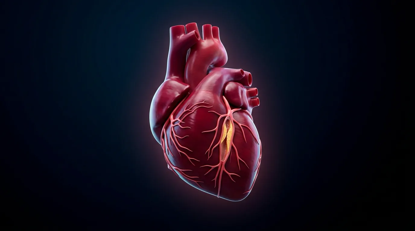
Acute coronary syndrome is not one disease but a spectrum of acute myocardial ischaemia sharing a common origin — an unstable atherosclerotic plaque — and separated by how completely the culprit vessel is occluded and whether myocardium has already died. The umbrella covers three entities: ST-elevation myocardial infarction (STEMI), non-ST-elevation myocardial infarction (NSTEMI), and unstable angina. All three begin with the same trigger — plaque rupture or erosion exposing thrombogenic material to flowing blood — but diverge on the ECG and on cardiac troponin, which is why those two tests, obtained early, do almost all of the diagnostic work.[1]
The Fourth Universal Definition of Myocardial Infarction (2018) is the document examiners quote.[3] It defines acute myocardial injury as a detectable rise and/or fall of cardiac troponin values, with at least one value above the 99th-percentile upper reference limit (URL). That injury becomes an acute myocardial infarction when there is also clinical evidence of acute myocardial ischaemia — at least one of: ischaemic symptoms, new ischaemic ECG changes, development of pathological Q waves, imaging evidence of new loss of viable myocardium or new regional wall-motion abnormality, or identification of a coronary thrombus by angiography or autopsy. The critical teaching point: troponin elevation without an ischaemic clinical context is myocardial injury, not infarction — a distinction examiners probe deliberately, because a troponin rise from sepsis, tachyarrhythmia, pulmonary embolism, stroke, or renal failure is not, by itself, an MI.
This is why type matters as much as diagnosis. The Universal Definition classifies MI into five mechanistic types, and a final-prof candidate is expected to reproduce them:[3]
Type 1
- Spontaneous MI from plaque rupture/erosion/dissection with intraluminal thrombus
- The classic ACS — full reperfusion/DAPT pathway applies
- Example: ruptured LAD plaque with anterior STEMI
Type 2
- MI from oxygen supply-demand mismatch WITHOUT plaque rupture
- Fixed (often non-critical) CAD plus a precipitant
- Causes: tachy/bradyarrhythmia, hypotension/shock, hypoxaemia, severe anaemia, hypertension crisis
- Treat the precipitant — not a default DAPT/reperfusion pathway
Type 3
- MI causing sudden cardiac DEATH before biomarkers could be obtained
- Ischaemic symptoms plus new ischaemic ECG changes or imaging
- Example: cardiac arrest with ST elevation, death before troponin drawn
Type 4 / 5
- Type 4a: MI related to PCI; 4b: stent thrombosis; 4c: restenosis
- Type 5: MI related to CABG
- Procedural MIs — defined by troponin thresholds × URL
In strict usage, "ACS" means Type 1 MI (and unstable angina). Type 2 MI is genuinely common on the wards — a septic, tachycardic patient with known coronary disease and a troponin bump — and over-treating it with loading DAPT and rushing to angiography is a recurring trainee error.[3]
Classification
The working, bedside classification that drives every management decision uses two tests — the ECG and cardiac troponin — and splits ACS into three:[1]
STEMI
- Complete coronary occlusion → transmural ischaemia
- ST elevation in ≥2 contiguous leads (≥1 mm limb / ≥2 mm precordial) OR new LBBB with ischaemic symptoms
- Troponin rises (transmural infarction)
- Needs emergency reperfusion — primary PCI or fibrinolysis
NSTEMI
- Subtotal/partial occlusion → subendocardial ischaemia
- ST depression, T-wave inversion, or a normal ECG
- Troponin rises above 99th-percentile URL with a rise/fall pattern
- Risk-stratified (GRACE/TIMI) for invasive timing
Unstable angina
- Ischaemic symptoms at rest, new-onset, or crescendo pattern
- ECG may show transient changes or be normal
- Troponin stays normal — NO myocyte necrosis
- Increasingly rare since high-sensitivity troponin assays
The mechanistic distinction matters clinically: STEMI is a plumbing emergency — the artery must be opened now, regardless of how the biomarkers eventually turn out — while NSTE-ACS (NSTEMI and unstable angina together) is a risk-stratification problem, where the timing of angiography is chosen from the clinical picture, the ECG, and a validated risk score.[1]
A separate, frequently examined axis is Type 1 versus Type 2 MI (above). Examiners will also ask why unstable angina is disappearing: with high-sensitivity troponin assays, many patients previously labelled "unstable angina" now show a small troponin rise and are reclassified as NSTEMI. Unstable angina persists as a label only for the truly troponin-negative ischaemic presentation, and is still defined by ischaemic symptoms with no troponin rise above the URL.[3]
ECG STEMI thresholds (reproduce exactly — NEET/INICET favourite)
New ST-segment elevation at the J-point in two contiguous leads:
- ≥1 mm (0.1 mV) in all leads other than V2–V3
- In V2–V3: ≥2 mm in men ≥40 years; ≥2.5 mm in men <40 years; ≥1.5 mm in women (regardless of age)
- New LBBB with a compatible history is treated as a STEMI-equivalent (use Sgarbossa / Smith-modified criteria when LBBB is known old)
- Posterior STEMI-equivalent: ST depression V1–V3 + tall R; confirm V7–V9 ≥0.5–1 mm elevation
- LBBB / paced rhythm: look for concordant ST elevation ≥1 mm, concordant ST depression ≥1 mm in V1–V3, or excessively discordant STE (Smith: STE/S ratio ≤ −0.25)
Worked classification stem
A 55-year-old has crushing pain; ECG shows 2 mm ST depression in V1–V3 with tall R waves; troponin will not be back for an hour. Answer: treat as posterior STEMI-equivalent — record V7–V9, activate reperfusion pathway now; do not wait for troponin. Troponin defines infarction size later; the ECG defines the emergency now.
Epidemiology & Risk Factors
Ischaemic heart disease is the leading cause of death worldwide, and in India it has overtaken infectious disease as the number-one killer — a transition the NEET-PG/INICET examiner expects you to know. Globally, ACS accounts for millions of deaths annually, and the burden in South Asia is rising with urbanisation, diabetes prevalence, and tobacco use.[1]
Coronary risk factors divide into non-modifiable and modifiable:[1]
- Non-modifiable — age (risk rises steeply after 55 in men and 65 in women), male sex until menopause narrows the gap, family history of premature coronary disease (a first-degree relative under 55 in men or under 65 in women), and genetic dyslipidaemias (familial hypercholesterolaemia).
- Modifiable — smoking (the single largest population-attributable risk and the one whose cessation most rapidly reduces risk), hypertension, diabetes mellitus (which roughly doubles cardiovascular risk and is now treated as a coronary-risk equivalent), dyslipidaemia (especially elevated LDL-C and low HDL-C), obesity, sedentary lifestyle, chronic kidney disease, and psychosocial stress.[1]
Not all risk factors accelerate atherosclerosis equally — some specifically predispose the plaque to rupture: smoking, hypertension, and hypercholesterolaemia thin the fibrous cap, while diabetes drives inflammation and endothelial dysfunction. Less common, non-atherosclerotic causes of an ACS-type presentation must be named when the patient doesn't fit the typical profile: cocaine-induced coronary spasm, spontaneous coronary artery dissection (SCAD) (especially peripartum women), coronary embolism (endocarditis, atrial fibrillation), coronary arteritis (Kawasaki, Takayasu), and paradoxical embolism through a patent foramen ovale.[1]
Pathophysiology
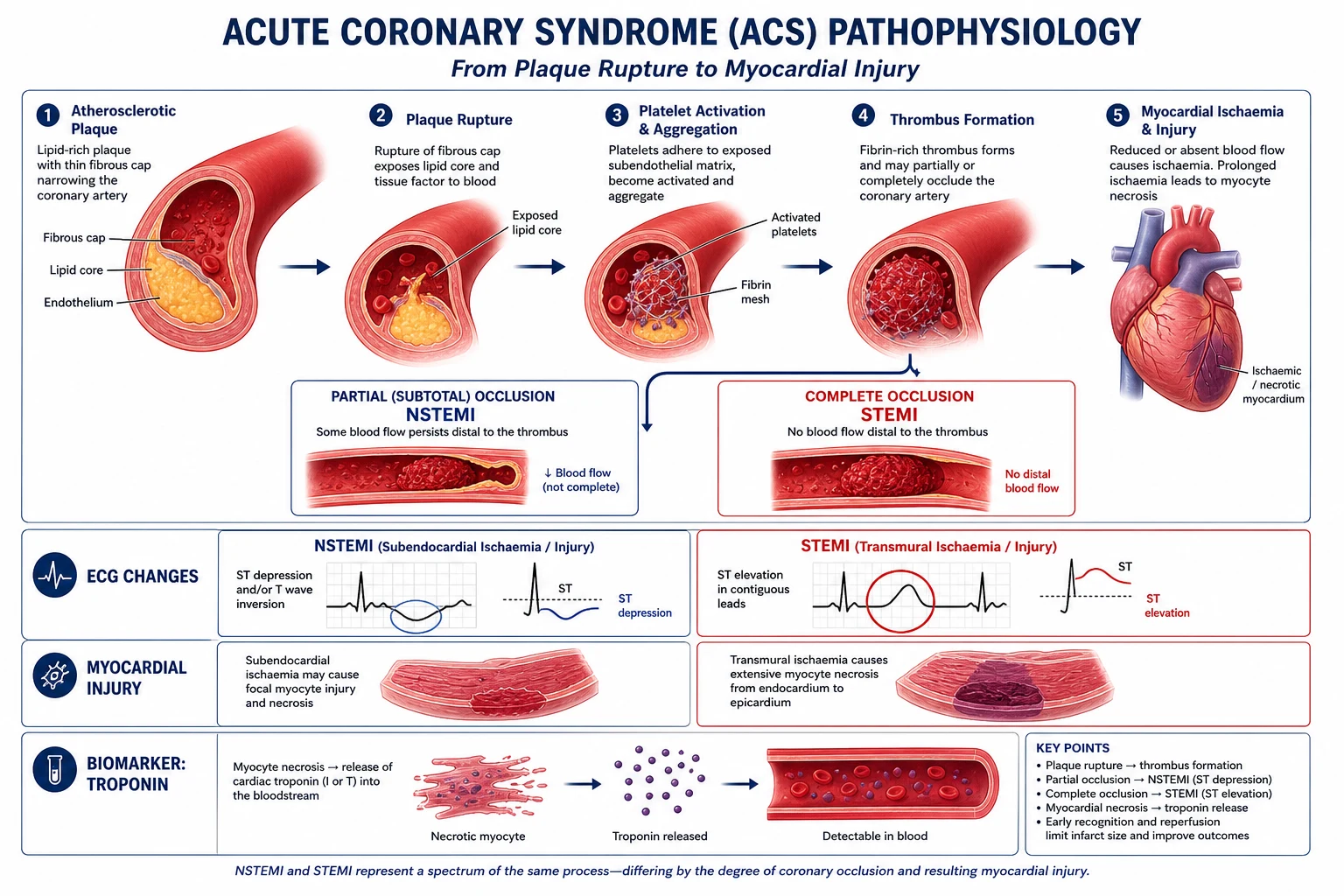
Atherosclerosis builds silently over decades: a fatty streak of lipid-laden macrophages (foam cells) appears in the arterial intima even in adolescence, progressing to a fibrous plaque with a lipid-rich necrotic core covered by a fibrous cap of smooth-muscle cells and collagen. ACS begins when that cap ruptures (the commonest mechanism, in inflamed, lipid-rich, thin-capped plaques) or erodes (more common in women, in plaques without a large lipid core), exposing the thrombogenic lipid core, collagen, and tissue factor to flowing blood.[1]
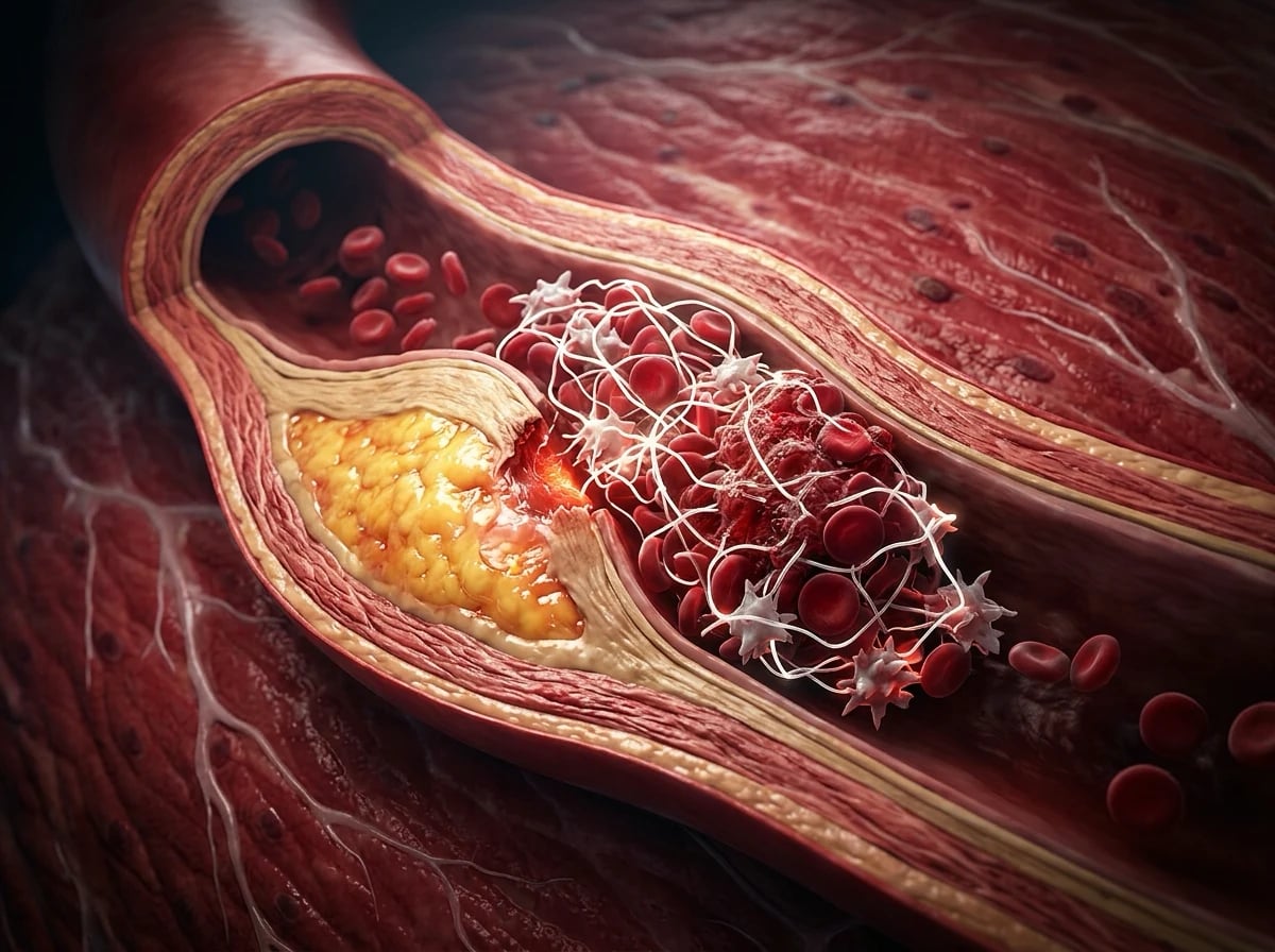
The atherothrombotic cascade then proceeds in parallel along two pathways:[1]
- Platelet activation/aggregation — platelets adhere to exposed subendothelial von Willebrand factor and collagen through glycoprotein Ib and VI, activate, degranulate (releasing ADP, thromboxane A₂, serotonin), and cross-link through glycoprotein IIb/IIIa receptors binding fibrinogen. This is why aspirin (cyclo-oxygenase/thromboxane A₂ blockade) and the P2Y12 inhibitors (ADP-receptor blockade) are the twin pillars of antiplatelet therapy.
- Coagulation cascade — tissue factor activates the extrinsic pathway, generating thrombin, which converts fibrinogen to fibrin and further activates platelets. This is the target of parenteral anticoagulation (heparin, enoxaparin, fondaparinux).[1]
A fibrin-platelet thrombus forms over the ruptured plaque within minutes. What happens next depends entirely on how completely that thrombus occludes the lumen:[1]
- A totally occlusive thrombus cuts off flow to the full thickness of the myocardial wall it supplies — transmural ischaemia — which appears on the ECG as ST elevation and, without rapid reperfusion, progresses to transmural (full-thickness) infarction. This is STEMI.
- A subtotal or non-occlusive (mural) thrombus leaves some flow, producing subendocardial ischaemia (the innermost subendocardium is the most vulnerable layer — it is furthest from the epicardial supply, under the highest wall tension, and last to receive oxygen and first to suffer when supply falls) — seen as ST depression or T-wave inversion, with troponin rise defining NSTEMI if myocyte necrosis occurs, or unstable angina if it does not.[1]
Necrosis begins within roughly 20 minutes of complete occlusion and spreads as a wavefront from subendocardium to subepicardium the longer the artery stays closed — the physiological basis of "time is muscle": by 6 hours about 70% of the myocardium at risk in the supplied territory is irreversibly dead, which is why every minute of reperfusion delay costs viable myocardium. As myocytes die, their membranes rupture and intracellular troponin leaks into the circulation — detectable from 2–4 hours after onset with high-sensitivity assays, peaking at 12–24 hours — the biomarker signal that confirms infarction once the clinical context is established.[1]
Coronary arteries are functional end-arteries with minimal collateral supply in the acute setting, which is why occlusion of a single vessel can be catastrophic and why the ECG territory maps predictably to the culprit artery. The localisation table is core exam material:[1]
ECG localisation of STEMI to culprit coronary artery
Clinical Presentation
Typical ischaemic pain
Classic ACS pain is retrosternal pressure, tightness, or heaviness, often radiating to the left arm, both arms, neck, jaw, or epigastrium, lasting >20 minutes if infarction is evolving, with autonomic features (sweating, nausea, vomiting, dyspnoea, syncope). Effort-related onset that becomes rest pain, crescendo angina, or new limiting angina defines unstable patterns.
Unstable angina patterns (still examinable)
- Rest angina — ischaemic pain at rest, often prolonged
- New-onset severe angina — marked limitation of ordinary activity
- Crescendo angina — previously stable angina becoming more frequent, longer, or lower threshold
Atypical and silent presentations (high-yield traps)
- Women, elderly, diabetics: dyspnoea alone, fatigue, epigastric pain, nausea, confusion, syncope — without classic crushing pain
- Silent MI: discovered on ECG (Q waves) or imaging; common in diabetes/autonomic neuropathy
- Inferior MI: epigastric pain + vagal features (bradycardia, nausea) mimicking acute abdomen/gastritis
- RV infarction: inferior MI + hypotension + clear lungs + raised JVP
- Posterior MI: may have fewer "classic" ECG signs on standard 12-lead
Associated and warning features
- New mitral regurgitation murmur (ischaemic papillary dysfunction)
- Pulmonary oedema (large infarct / ischaemic MR)
- Arrhythmias at presentation (VF/VT, heart block in inferior MI)
- Cardiogenic shock (cold, clammy, oliguria, lactate rise)
Worked stems
Stem 1: 70-year-old diabetic woman with sudden dyspnoea, no chest pain, anterior Q waves and troponin rise → silent NSTEMI/STEMI-equivalent work-up, not "CCF only."
Stem 2: 45-year-old with tearing pain to back and unequal arm BP → dissection until proven otherwise, not MONA first.
Stem 3: Post-prandial epigastric burning in smoker with sweating → ECG before antacid discharge.
Differential Diagnosis
Several life-threatening mimics must be actively excluded before committing to ACS therapy, because antiplatelet and anticoagulant treatment can be catastrophic if the true diagnosis is one of these. The "killers that mimic ACS" are the cornerstone of the chest-pain SAQ:[1]
Life-threatening mimics of ACS — distinguishing features
The single most important differential to exclude clinically before treatment is aortic dissection: tearing, migrating pain radiating to the back, a pulse or blood-pressure differential between limbs (over 20 mmHg), a new aortic regurgitation murmur, and a widened mediastinum on chest X-ray. Anticoagulating a dissection is potentially fatal.[1]
Musculoskeletal chest pain, herpes zoster (shingles — pain may precede the rash), biliary colic/cholecystitis (which can mimic inferior MI and vice versa through shared T7–T9 visceral afferents), and anxiety/panic attacks complete the broader list, but none should be diagnosed without first excluding the killers.[1]
Clinical & Bedside Assessment
Examination in suspected ACS rarely provides a diagnostic sign — its main role is to detect complications and assess haemodynamic status. The focused cardiovascular examination includes:[1]
- Pulse — rate and rhythm (tachycardia suggests hypovolaemia/pain/arrhythmia; bradycardia suggests conduction disease, especially in inferior MI from RCA occlusion of the AV-nodal artery).
- Blood pressure in BOTH arms — a significant differential (over 20 mmHg) raises dissection.
- JVP — raised in right-heart failure or right ventricular infarction.
- Heart sounds — an S3 (third heart sound) signals ventricular dysfunction; a new murmur is a red flag for a mechanical complication (pansystolic → papillary muscle rupture or VSD).
- Lungs — crackles indicate pulmonary oedema.
- Peripheral pulses, perfusion, temperature — cool, shut-down peripheries suggest cardiogenic shock.
- Signs of heart failure — raised JVP, S3, crackles, peripheral oedema.[1]
The Killip classification grades heart-failure severity at presentation and predicts in-hospital mortality directly — it is reproduced verbatim:[1]
Killip classification — heart failure severity at presentation
Higher Killip class escalates the urgency of monitoring and revascularisation and feeds directly into the GRACE risk score. The bedside definitions of cardiogenic shock — hypotension (systolic BP under 90 mmHg), clinical signs of poor perfusion (cold clammy skin, oliguria, altered sensorium), and pulmonary oedema or low cardiac output — mark the patient who needs immediate, not deferred, revascularisation and often mechanical circulatory support.[1]
Investigations
Acute coronary syndrome — the time-critical numbers
The 12-lead ECG, obtained and interpreted within 10 minutes of first medical contact, is the single most important initial test — it separates STEMI (which needs immediate reperfusion regardless of biomarkers) from everything else. STEMI ECG criteria (reproduced exactly):[1]
- ST elevation in at least two contiguous leads: ≥1 mm (0.1 mV) in the limb leads (I, II, III, aVF, aVL) OR ≥2 mm (0.2 mV) in the precordial leads (V1–V6) in men (≥1.5 mm in women), measured at the J-point.
- Contiguous means anatomically adjacent: II/III/aVF (inferior), V1–V4 (anterior/septal), I/aVL/V5–V6 (lateral).
- New left bundle branch block (LBBB) in the context of ischaemic symptoms is treated as a STEMI-equivalent.
- Posterior MI is a STEMI-equivalent: tall R waves with ST depression in V1–V2, confirmed by ST elevation in posterior leads V7–V9.[1]
A normal initial ECG does not exclude ACS — serial ECGs (every 15–30 minutes) and troponin are needed, particularly for circumflex-territory ischaemia, which is notoriously ECG-silent (the LCx supplies the high lateral/posterior wall, poorly represented on the standard 12 leads).[1]
Earlier ECG changes precede the ST elevation that every student recognises. Hyperacute T waves (tall, broad-based, prominent T waves, sometimes with a slight ST take-off) can appear within minutes of occlusion — the earliest electrical sign, easily missed. Other STEMI-equivalents and pitfalls examiners test:[1]
- de Winter T waves — upsloping ST depression in the anterior leads with tall, symmetrical T waves and no STE — an LAD occlusion equivalent.
- Wellens syndrome — deeply inverted or biphasic T waves in V2–V3 in a pain-free patient with critical proximal LAD stenosis; a pending-anterior-MI warning.
- Sgarbossa criteria — three signs diagnosing ischaemia in LBBB/paced rhythm: (1) concordant ST elevation ≥1 mm in any lead; (2) concordant ST depression ≥1 mm in V1–V3; (3) discordant ST elevation ≥5 mm (Smith-modified uses a proportional ratio). Any one criterion is highly specific.
- Right ventricular infarction — ST elevation in V3R–V4R in the setting of inferior STEMI.[1]
High-sensitivity cardiac troponin (hs-cTn) is measured on a rapid algorithm. The ESC 0-hour/1-hour (or 0-hour/3-hour) protocol triages patients into "rule-out", "observe", or "rule-in" bands based on absolute troponin values and the magnitude of change — a rise/fall pattern with at least one value above the 99th-percentile URL, in the correct clinical context, defines myocardial infarction. The 0/1-hour pathway allows safe discharge of low-risk patients within an hour of arrival.[1] Troponin must always be interpreted in context: chronic stable elevations occur in renal failure, and a small rise with a flat (non-rising) pattern suggests chronic injury, not acute infarction.
Two named risk scores are essential for NSTE-ACS and are reproduced exactly as an examiner expects them:[1]
NSTE-ACS risk scores — reproduced verbatim
Adjunct investigations: chest X-ray (pulmonary oedema, a widened mediastinum raising dissection, pneumothorax); bedside echocardiography (regional wall-motion abnormality confirming ischaemia, ejection fraction, mechanical complications, pericardial effusion); full blood count, urea and electrolytes, glucose, and a fasting lipid profile; and coronary angiography, which remains the definitive investigation and the gateway to PCI.[1]
Management — Resuscitation
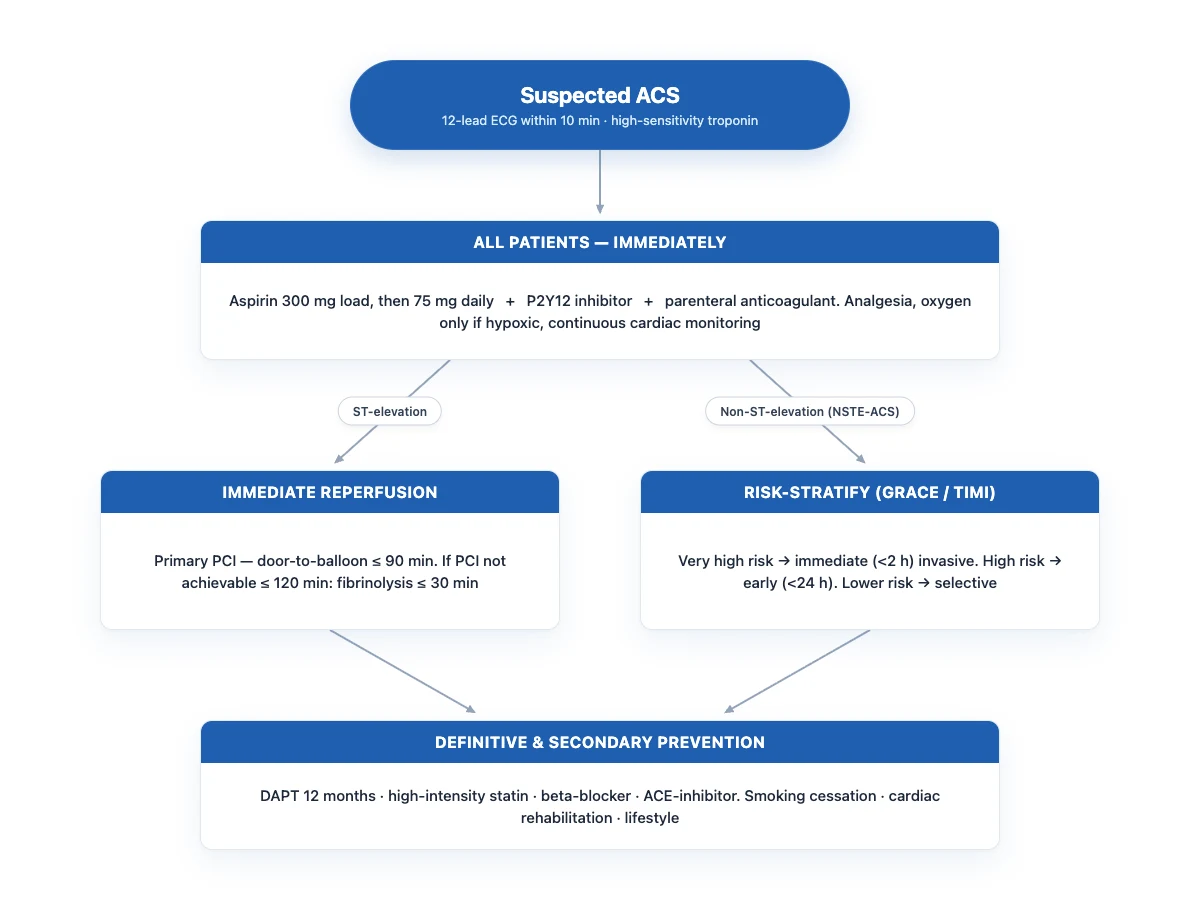
Every patient with suspected ACS receives, at first contact, the MONA-B + anticoagulant bundle adapted to modern evidence:[1]
Immediate first-contact bundle for every suspected ACS
Continuous cardiac monitoring + IV access + defibrillator at bedside
VF is the leading cause of pre-hospital death — the pad/paddles must be ready
12-lead ECG within 10 minutes of first medical contact
The single most time-critical investigation — separates STEMI from NSTE-ACS
Oxygen ONLY if hypoxic (SpO₂ under 90%) — NOT routinely
Routine high-flow O₂ in a normoxic patient is no longer recommended; hyperoxia may worsen outcomes (AIRWAY-2/DETO2X-AMI)
Aspirin 300 mg orally, chewed, then 75–100 mg daily indefinitely
The strongest-evidenced single intervention in acute MI (ISIS-2); survival benefit persists at 10 years
P2Y12 inhibitor loading — ticagrelor 180 mg or clopidogrel 600 mg (or prasugrel 60 mg if PCI-planned)
Given as soon as ACS is suspected and bleeding is excluded — after dissection is considered
Parenteral anticoagulant — fondaparinux 2.5 mg SC / enoxaparin 1 mg/kg SC BD / UFH
Continues until/through PCI; choice by renal function and reperfusion strategy
Analgesia — IV morphine 2.5–5 mg titrated, with an antiemetic
Use sparingly: opioids delay and blunt oral P2Y12 inhibitor absorption
Sublingual GTN for ongoing pain; beta-blocker if pain/rate control needed
AVOID nitrates in RV infarct, hypotension, recent PDE-5-inhibitor use; AVOID beta-blockers in acute HF/cardiogenic shock
Two warnings recur in exams. First, routine high-flow oxygen in a normoxic patient is now contraindicated — the evidence (including the DETO2X-AMI trial) suggests harm from hyperoxia-driven coronary vasoconstriction; oxygen is reserved for SpO₂ under 90% or respiratory distress. Second, morphine is used judiciously because it slows gastric emptying and reduces the absorption (and thus peak levels) of oral ticagrelor and clopidogrel — pain should be treated, but not over-sedated.[4]
Aspirin deserves its own line: the ISIS-2 trial randomised over 17,000 patients with suspected acute MI to intravenous streptokinase, oral aspirin (162 mg/day for one month), both, or neither — aspirin alone reduced 5-week vascular mortality by 23%, as much as streptokinase, and the combination halved mortality. The survival advantage persisted at 10-year follow-up.[2]
Ventricular fibrillation or pulseless VT in the first hours is managed with immediate defibrillation per standard advanced life support — it is the leading cause of pre-hospital death in acute MI and is fully reversible if treated promptly. Continuous monitoring and a defibrillator at the bedside exist precisely for this.[1]
Management — Definitive & Stepwise
After the initial bundle, management forks on the ECG: STEMI to emergency reperfusion, NSTE-ACS to risk-stratified invasive timing.[1]
STEMI — emergency reperfusion (time is muscle)
STEMI reperfusion pathway
PRIMARY PCI if achievable within 120 minutes of first medical contact
Target door-to-balloon under 90 minutes; preferred strategy when available in time
If PPCI NOT achievable within 120 min → FIBRINOLYSIS within 30 min (door-to-needle), if within 12 h of symptom onset
Tenecteplase single IV bolus (weight-based, max 50 mg); alteplase or streptokinase are alternatives
After lysis: RESCUE PCI if failed (persistent pain/ST elevation after 60–90 min) or routine early angiography within 3–24 h if successful
Pharmacoinvasive strategy — bridges the gap in low-resource settings
Antiplatelet + anticoagulant during PCI
Heparin 70–100 U/kg bolus (ACT-guided); ticagrelor preferred post-PCI
Primary PCI (PPCI) is the preferred reperfusion strategy when it can be delivered within 120 minutes of first medical contact (target door-to-balloon under 90 minutes). It achieves higher TIMI-3 flow than lysis, lowers reinfarction and stroke, and is the standard of care in PCI-capable centres. If PPCI cannot be achieved within the 120-minute window and the patient presents within 12 hours of symptom onset (or has ongoing ischaemia), fibrinolysis within 30 minutes of arrival is indicated, followed by a pharmacoinvasive strategy — rescue PCI if lysis fails (persistent pain and ST elevation after 60–90 minutes), or routine early angiography within 3–24 hours if reperfusion is clinically successful. Fibrinolytic options with doses:[1]
Fibrinolytic agents — dosing
Contraindications to fibrinolysis include prior intracranial haemorrhage, known cerebral vascular lesion/malignancy, ischaemic stroke in past 6 months, active bleeding, aortic dissection, and severe uncontrolled hypertension — these patients should be transferred for PCI even if delayed.[1]
NSTE-ACS — risk-stratified invasive timing
NSTE-ACS is not a plumbing emergency; the timing of angiography is chosen from risk features and a validated score. The TIMACS trial established that an early invasive strategy (within 24 hours) reduces the composite of death, MI, or refractory ischaemia in high-risk patients without increasing bleeding.[6]
NSTE-ACS invasive-strategy timing (ESC 2023)
Dual antiplatelet therapy (DAPT) and anticoagulation — the doses
DAPT is the foundation of medical therapy: aspirin indefinitely plus a P2Y12 inhibitor, by default for 12 months after ACS, with the choice of agent guided by efficacy, bleeding risk, cost, and access. The landmark trials are core exam material:[1]
P2Y12 inhibitor comparison and landmark trials
Ticagrelor (PLATO) and prasugrel (TRITON) are more potent than clopidogrel and preferred where affordable and not contraindicated; clopidogrel remains the workhorse in cost-constrained settings and in patients with high bleeding risk or oral anticoagulant needs.[5][4]
Parenteral anticoagulation options include unfractionated heparin (UFH, 70–100 U/kg bolus for PCI), enoxaparin (1 mg/kg subcutaneously twice daily, halved in renal impairment), and fondaparinux (2.5 mg subcutaneously once daily — safest against bleeding, but not used as the sole anticoagulant during PCI, where UFH is added). Anticoagulation is continued until PCI is completed, then typically stopped (or continued for the index hospitalisation in a conservatively managed patient).[1]
Secondary prevention — the five-plus pillars, for every survivor
Every patient leaves hospital on a bundle of disease-modifying therapy, as examinable as the acute ladder:[1]
Secondary-prevention bundle after ACS
Dual antiplatelet therapy for 12 months (aspirin + P2Y12 inhibitor)
Aspirin 75–100 mg lifelong; P2Y12 inhibitor duration individualised by bleeding/ischaemic balance
High-intensity statin
Atorvastatin 80 mg daily — reduce LDL-C to under 1.4 mmol/L (55 mg/dL) and by ≥50%
Beta-blocker
Bisoprolol 5 mg daily (titrate) — especially large/anterior infarct, LVEF under 40%
ACE-inhibitor (or ARB)
Ramipril 2.5 mg daily (titrate to 10 mg) — especially LVEF under 40%, anterior infarct, HF, diabetes, hypertension
Mineralocorticoid receptor antagonist if LVEF under 40% with HF or diabetes
Eplerenone 25–50 mg daily (EPHESUS/REMINDER evidence)
Smoking cessation + cardiac rehabilitation + risk-factor control
Referral to a structured rehab programme; BP, glucose, lipid targets at every review
This bundle, plus lifestyle modification (Mediterranean diet, regular exercise, weight management, alcohol moderation), drives the long-term reduction in recurrent events. Cardiac rehabilitation — a structured programme of exercise, education, and risk-factor modification — reduces mortality and rehospitalisation and should be offered to every eligible patient.[1]
Escalation — cardiogenic shock: managed with inotropic/vasopressor support (noradrenaline may be preferred over dopamine; dobutamine/inotropes for low-output state) and urgent revascularisation. Mechanical circulatory support (intra-aortic balloon pump, percutaneous ventricular assist devices, ECMO) is considered in refractory cases — though the IABP-SHOCK II trial showed no routine mortality benefit from balloon pumping in MI-related cardiogenic shock, so the IABP is now selective rather than default.[7] Suspected mechanical complications (VSD, papillary muscle rupture, free-wall rupture) require urgent echocardiography and cardiothoracic surgical referral.
Specific Subtypes & Scenarios
Right ventricular infarction complicates inferior STEMI (proximal RCA occlusion cutting off the RV marginal branches). The classic triad — hypotension, clear lung fields, and a raised JVP — is confirmed by ST elevation in right precordial leads V3R–V4R. Management is the opposite of left-sided failure: fluid loading to maintain RV preload (the RV is preload-dependent), with nitrates and diuretics avoided because they drop preload-dependent RV output and precipitate profound hypotension. A nitrate-induced drop in BP during treatment of an inferior STEMI should immediately raise this possibility. Right-sided (V3R–V4R) leads should be recorded in every inferior STEMI.[1]
Posterior wall MI is easily missed because the standard 12 leads don't directly visualise the posterior wall — it appears as tall R waves and ST depression in V1–V2 (a mirror image of posterior ST elevation). Confirm with posterior leads V7–V9 showing ST elevation; treat as a STEMI-equivalent and activate reperfusion. A common exam trap is reading V1–V2 ST depression as "ischaemia/NSTEMI" instead of recognising the posterior STEMI-equivalent.[1]
MI in the setting of LBBB — a pre-existing or new LBBB obscures the standard ST-elevation criteria because the QRS is already broad and discordant. The Sgarbossa criteria (and the more sensitive Smith-modified Sgarbossa criteria using a proportional ST/S ratio) identify ischaemic ST changes discordant with the QRS direction to diagnose STEMI in this setting. A new LBBB with ischaemic symptoms is treated as a STEMI-equivalent regardless.[1]
Spontaneous coronary artery dissection (SCAD) — a tear in the coronary artery wall creating a false lumen that compresses the true lumen, causing ACS. Predominantly affects young women without traditional risk factors, often peripartum or with fibromuscular dysplasia and connective-tissue disease. Managed differently from Type 1 ACS: conservative therapy (as the dissection often heals spontaneously) is preferred where the artery is patent; stenting a dissection plane can extend it, and CABG is reserved for extensive/ischaemia-driven dissection.[1]
Coronary vasospasm (Prinzmetal/variant angina) — transient, reversible ST elevation at rest, classically in the early morning, relieved by nitrates and calcium-channel blockers. The underlying plaque may be non-critical; the mechanism is spasm, not rupture. Managed with calcium-channel blockers and nitrates, avoiding beta-blockers (unopposed alpha vasoconstriction).[1]
Initially normal ECG with suspected ACS — repeat the ECG every 15–30 minutes, record right-sided (V3R–V4R) and posterior (V7–V9) leads in inferior/borderline cases, and pursue serial troponin. Circumflex occlusion is the classic ECG-silent STEMI; if clinical suspicion is high and the ECG is normal, bedside echocardiography showing a regional wall-motion abnormality may reveal the territory.[1]
Complications & Pitfalls
Complications separate by timing, and the timing is the most examinable single fact about each:[1]
Post-MI complications — when they strike
The three mechanical complications (VSD, papillary muscle rupture, free-wall rupture) typically strike days 3–5 (after the myocardium has softened from necrosis) and each carries a characteristic murmur — a new pansystolic murmur days after MI is a red flag demanding urgent echocardiography and surgical referral. Papillary muscle rupture (usually the posteromedial papillary muscle, supplied by the RCA alone, versus the anterolateral's dual LAD/LCx supply) produces acute severe mitral regurgitation with sudden pulmonary oedema. Ventricular septal rupture produces a harsh pansystolic murmur with a thrill at the lower left sternal border and biventricular failure. Free-wall rupture produces sudden cardiac tamponade (electromechanical dissociation) and is frequently fatal.[1]
Dressler syndrome — an autoimmune post-MI pericarditis presenting 2–10 weeks after the index event with fever, pleuritic chest pain, a pericardial rub, and a pericardial effusion, with raised inflammatory markers. Managed with NSAIDs and colchicine; steroids are reserved for refractory cases. It is distinct from the acute pericarditis of the first few days, which reflects direct transmural inflammation.[1]
Recurring pitfalls every candidate must name:
- Failing to exclude aortic dissection before anticoagulating.
- Attributing epigastric pain to dyspepsia in a diabetic who is actually infarcting.
- Withholding reperfusion because the ECG is "borderline" — when in doubt, repeat the ECG, record posterior/right-sided leads, and look for STEMI-equivalents.
- Relying on a single negative troponin too early in the rise/fall curve — the 0/1-hour or 0/3-hour algorithm exists precisely to avoid this.
- Missing right ventricular infarction, giving nitrates, and precipitating profound hypotension.
- Treating a Type 2 MI (supply-demand mismatch) with loading DAPT and rushing to angiography instead of treating the precipitant.[1]
Prognosis & Disposition
Mortality after ACS is determined by infarct size, time to reperfusion, baseline risk (age, comorbidity, diabetes, CKD), Killip class at presentation, GRACE score, and mechanical or arrhythmic complications. With modern primary PCI, STEMI in-hospital mortality is roughly 5–6%; without reperfusion it was historically 15–20%. NSTE-ACS in-hospital mortality is often similar or slightly higher because the population is older and more comorbid with multivessel disease.[1]
Killip classification (reproduce exactly)
| Class | Clinical findings | Approximate historical in-hospital mortality |
|---|---|---|
| I | No clinical HF | ~6% |
| II | Mild HF — S3, basal rales, raised JVP | ~17% |
| III | Acute pulmonary oedema | ~30–40% |
| IV | Cardiogenic shock | ~60–80% without urgent revascularisation |
Killip class remains a rapid bedside prognostic tool and disposition cue (CCU/ICU for III–IV).[1]
GRACE score — what it contains and how you use it
GRACE estimates in-hospital and 6-month mortality from: age, heart rate, systolic BP, serum creatinine, Killip class, cardiac arrest at admission, ST-segment deviation, and elevated cardiac biomarkers. Practical thresholds still taught for invasive timing in NSTE-ACS:
- GRACE >140 → high risk → early invasive strategy within 24 h
- GRACE 109–140 → intermediate → invasive strategy generally within 72 h
- GRACE <109 → lower risk → selective/ischaemia-driven approach may be reasonable
TIMI risk score for UA/NSTEMI (0–7) is simpler at the bedside: age ≥65; ≥3 CAD risk factors; known CAD (stenosis ≥50%); aspirin in past 7 days; recent severe angina (≥2 episodes/24 h); ST deviation ≥0.5 mm; positive biomarker. Higher scores predict 14-day death/MI/urgent revascularisation.[1]
Worked disposition stems
Stem A — Killip IV anterior STEMI. A 62-year-old with anterior STEMI, BP 78/50, cool extremities, pulmonary oedema. Disposition: activate cath lab now, CCU/ICU after PCI, consider MCS if refractory shock, avoid routine IABP. Mortality without revascularisation is catastrophic.
Stem B — Low-risk NSTEMI. A 48-year-old with troponin rise, normal ECG after pain resolution, GRACE 80, no ongoing pain. Disposition: monitored ward, dual antiplatelet + anticoagulant, early cardiology review; invasive strategy often during index admission but not emergent if truly low risk and stable. [1]
Stem C — Post-lysis rural transfer. After successful fibrinolysis (ST resolution >50%, pain free), transfer for routine early angiography (3–24 h). If failed lysis (persistent ST elevation/pain at 60–90 min) → rescue PCI. [1]
Secondary prevention and discharge checklist (examinable)
Every survivor should leave with:
- Aspirin lifelong + P2Y12 for 12 months (default; shorten/de-escalate if high bleed risk)
- High-intensity statin (atorvastatin 40–80 mg or rosuvastatin 20–40 mg) — LDL-C goal typically <1.4 mmol/L (<55 mg/dL) and ≥50% reduction in very-high-risk patients per ESC
- Beta-blocker if reduced LVEF, ongoing ischaemia, or arrhythmia indication
- ACE-I/ARB (ARNI later if HFrEF pathway) — especially anterior MI, diabetes, HF, CKD with proteinuria
- MRA if LVEF ≤40% with HF or diabetes post-MI (EPHESUS logic — eplerenone)
- SGLT2 inhibitor if diabetes or if HF with reduced EF evolves
- Cardiac rehabilitation referral, smoking cessation, diet, BP and glycaemic targets
- Driving / return-to-work advice per local rules (often 1 week private driving after successful uncomplicated PCI; longer if LV impairment or bus/lorry) [1]
Long-term prognosis
Recurrent event risk remains elevated indefinitely. Post-MI LV impairment, incomplete revascularisation, ongoing smoking, and non-adherence to DAPT drive early stent thrombosis and late events. LV thrombus after large anterior MI needs anticoagulation (typically 3 months) plus antiplatelet strategy individualised — echo surveillance is mandatory after large anterior infarcts.
[1]Special Populations
Special populations change presentation, reperfusion choice, antithrombotic intensity, and bleeding risk. Examiners use these scenarios heavily on NEET-PG/INICET. [1]
Diabetes mellitus
- Atypical / silent MI is common (autonomic neuropathy blunts pain); present with dyspnoea, nausea, fatigue, or "just unwell."
- Outcomes are worse (larger infarcts, more HF, more restenosis); prefer an early invasive strategy in NSTE-ACS when high-risk.
- Ticagrelor is preferred over clopidogrel when affordable (PLATO diabetic subgroup benefit).
- In-hospital glycaemic control: avoid both marked hyperglycaemia and hypoglycaemia — target roughly 7.8–10 mmol/L (140–180 mg/dL) in critically ill ACS; do not chase tight control with insulin infusions without monitoring.
- High rates of multivessel disease; complete revascularisation strategy discussions are more frequent. [1]
Elderly and frail
- Present with dyspnoea, syncope, delirium, fall, or epigastric pain more than classic crushing pain.
- Higher bleeding risk on DAPT and anticoagulation; use PRECISE-DAPT / ARC-HBR concepts to individualise DAPT duration (often 1–3 months of dual therapy then de-escalation if high bleed risk and low ischaemic risk after uncomplicated PCI).
- Prasugrel is contraindicated ≥75 years (TRITON-TIMI 38 excess ICH/bleeding) and if body weight under 60 kg or prior stroke/TIA.
- Frailty is not an absolute bar to PCI, but goals of care and procedural risk must be explicit.
- Prefer radial access, careful creatinine-adjusted anticoagulants, and shorter triple therapy if on OAC.
Women and pregnancy-associated MI
- Women present later, more atypically, and receive delayed reperfusion more often — a systems failure examiners flag.
- SCAD is a leading cause of MI in pregnancy and the peripartum period and in young women without traditional risk factors; often managed conservatively if stable (dissection heals); PCI can extend the dissection plane.
- Pregnancy ACS work-up: protect the fetus (left lateral tilt, minimise radiation with abdominal shielding, prefer echocardiography and carefully justified angiography).
- Fibrinolysis is relatively contraindicated in pregnancy (placental abruption risk) — PCI is preferred when available.
- MINOCA (MI with non-obstructive coronaries) is more often diagnosed in women — requires CMR, consider spasm, plaque erosion, myocarditis, Takotsubo.
Chronic kidney disease
- Baseline troponin is often chronically elevated — diagnose MI by a rise and/or fall pattern, not a single absolute value.
- Contrast-induced nephropathy risk at angiography: pre-hydrate with isotonic crystalloid when volume status allows, minimise contrast volume, delay non-urgent angiography if eGFR is critically low and no ongoing ischaemia.
- Enoxaparin needs dose adjustment or avoidance at low CrCl; fondaparinux is contraindicated if CrCl under 20 mL/min; unfractionated heparin is often preferred when eGFR is very low because it is fully reverseable and titratable.
- Ticagrelor may need care with uraemic bleeding risk; clopidogrel is commonly used when bleeding risk dominates. [1]
Patients on oral anticoagulation (e.g. AF)
- After PCI for ACS, default modern strategy is short triple therapy (OAC + aspirin + clopidogrel) for up to 1 week to 1 month (shorter if high bleed risk), then dual therapy (OAC + clopidogrel) to 6–12 months, then OAC monotherapy.
- Prefer clopidogrel as the P2Y12 when combined with OAC (less bleeding than ticagrelor/prasugrel).
- Prefer DOAC over warfarin when eligible; continue the lowest effective OAC dose for stroke prevention (do not routinely drop to "ACS half-dose" unless the specific DOAC regimen was trial-tested).
- Always reassess HAS-BLED-style bleeding modifiers and gastroprotection with a PPI.
Cocaine / stimulant-associated ACS
- Mechanism: vasospasm ± plaque rupture + hyperadrenergic state.
- Give benzodiazepines early for agitation/hypertension; nitrates and CCBs for spasm.
- Avoid pure beta-blockers early (theoretical unopposed alpha vasoconstriction) — if beta-blockade needed after vasodilation, labetalol (alpha+beta) is sometimes used carefully.
- Still consider angiography if STEMI criteria met — not all "cocaine chest pain" is spasm alone.
Cardiogenic shock and post-arrest
- Emergency revascularisation remains the cornerstone (SHOCK trial logic).
- Routine IABP is not indicated (IABP-SHOCK II) — reserve MCS (Impella, VA-ECMO) for selected centres and phenotypes.
- Targeted temperature management after ROSC per local protocol; early angiography after OHCA with ST elevation is standard; without ST elevation, individualise.
Evidence, Guidelines & Regional Differences
The 2023 ESC Guidelines for the management of acute coronary syndromes unified the previously separate STEMI and NSTE-ACS European guidelines into a single "one syndrome" document, reflecting the shared pathophysiology and overlapping early management; key recommendations include the 0/1-hour high-sensitivity troponin algorithm, default ticagrelor over clopidogrel, radial-artery access for PCI, complete revascularisation at the index event in STEMI multivessel disease, and de-escalation of antithrombotic therapy in lower-risk patients.[1]
The landmark trials an examiner expects you to cite, and what each changed:[1]
ISIS-2 (1988; 10-yr follow-up 1998)
Population: Over 17,000 patients with suspected acute MI
Key finding
Aspirin alone reduced 5-week vascular mortality by 23% — equal to streptokinase; the combination halved mortality. Survival benefit persisted at 10-year follow-up.
CURE (2001)
Population: Over 12,500 patients with NSTE-ACS
Key finding
20% relative reduction in cardiovascular death, MI, or stroke; increased major bleeding.
PLATO (2009)
Population: Over 18,600 patients with ACS (STEMI and NSTE-ACS)
Key finding
16% relative reduction in cardiovascular death/MI/stroke without an increase in major bleeding (but more non-CABG-related bleeding and dyspnoea).
TIMACS (2009)
Population: Over 3,000 patients with NSTE-ACS
Key finding
No overall mortality difference, but reduced death/MI/refractory ischaemia in high-risk (GRACE above 140) patients.
IABP-SHOCK II (2012; final 12-mo 2013)
Population: 600 patients with acute MI and cardiogenic shock planned for early revascularisation
Key finding
No difference in 30-day or 12-month mortality.
Regional differences — the overall diagnostic and management framework (ECG, troponin, DAPT, timely reperfusion) is globally consistent, but resource-dependent choices differ:[1]
Australian/New Zealand cardiac networks use a hub-and-spoke model with systematic transfer for primary PCI; cardiologists invoke the ESC and ACC/AHA evidence locally.
In India (the NEET-PG/INICET context), timely primary PCI access is uneven; streptokinase remains a widely used, cost-effective fibrinolytic where PPCI cannot be delivered in the 120-minute window, and generic clopidogrel is often favoured over ticagrelor/prasugrel on cost grounds — even though the more potent agents are preferred where affordable.
Controversies worth naming: the routine-oxygen question (now answered — no benefit, possible harm in normoxic patients); the morphine–P2Y12 interaction (measurable pharmacokinetic effect, uncertain clinical impact); complete revascularisation versus culprit-only PCI in STEMI (COMPLETE favoured complete revascularisation); and the optimal DAPT duration and de-escalation strategy (guided by bleeding versus ischaemic risk).[1]
Worked NEET-PG Stems — ACS
- ST elevation V2–V4, 40 min pain, PCI 3 h away → fibrinolysis if within 12 h and no CI; then pharmacoinvasive transfer.
- Inferior STEMI + hypotension + clear lungs → RV infarct; fluids; no nitrates.
- V1–V2 ST depression + tall R → posterior STEMI-equivalent; V7–V9; reperfusion.
- Day 4 post-MI new pansystolic murmur + oedema → mechanical complication; urgent echo/surgery.
- Troponin up in sepsis, no ischaemic ECG → injury/type 2 until proven otherwise; treat cause.
- NSTEMI GRACE 150 → early invasive within 24 h.
- Aspirin forgotten in "possible gastritis" chest pain → never omit without true contraindication after dissection excluded. [1]
Exam Pearls
MONA-B
Plus — do not forget the P2Y12 inhibitor and parenteral anticoagulant, which complete the immediate loading.
TIMI
High-yield one-liners examiners reward:[1]
- STEMI = ST elevation in ≥2 contiguous leads (or new LBBB) + ischaemic symptoms → activate the cath lab; door-to-balloon under 90 minutes is the headline number.
- Troponin-negative + ischaemic symptoms + no diagnostic ECG changes = unstable angina, not NSTEMI — a distinction examiners test precisely.
- ECG localisation: II/III/aVF = inferior = RCA; V1–V4 = anterior = LAD; I/aVL/V5–V6 = lateral = LCx. V3R–V4R for RV; V7–V9 for posterior.
- Posterior MI is the mimic students miss most: tall R with ST depression in V1–V2 is a STEMI-equivalent — check V7–V9.
- Right ventricular infarction: no nitrates, no diuretics — give fluids. The classic "what not to do" trap.
- "Time is muscle": necrosis begins around 20 minutes after complete occlusion; PCI within 120 min, else lysis within 30 min; DAPT 12 months; beta-blocker + ACE-I especially in anterior MI.
- Mechanical complications strike days 3–5 — a new pansystolic murmur then demands urgent echo and surgery.
- Dressler syndrome = 2–10 weeks, autoimmune, NSAIDs + colchicine.
- Aspirin's mortality benefit (ISIS-2) is one of the best-evidenced interventions in all of medicine — never omit it for a "possible" contraindication without checking.
- Killip class predicts in-hospital mortality (I ~6% → IV ~70–80%); GRACE above 140 mandates an early invasive strategy.[2]
Self-test: which fibrinolytic do you give, and when is lysis preferred over PCI?
Primary PCI is preferred if achievable within 120 minutes of first medical contact (door-to-balloon under 90 min). If PPCI cannot be delivered in that window and the patient is within 12 hours of symptom onset, give fibrinolysis within 30 minutes — tenecteplase as a single weight-based IV bolus (max 50 mg) is easiest; alteplase or streptokinase are alternatives. Then pursue a pharmacoinvasive strategy (rescue PCI if lysis fails; routine early angiography within 3–24 h if successful).
Self-test: name the three mechanical complications and their timing/murmur
All strike around days 3–5: (1) papillary muscle rupture → acute severe mitral regurgitation, apical pansystolic murmur, sudden pulmonary oedema; (2) ventricular septal rupture → harsh pansystolic murmur with a thrill at the lower left sternal edge, biventricular failure; (3) free-wall rupture → sudden cardiac tamponade with electromechanical dissociation, often fatal. All need urgent echocardiography and cardiothoracic surgery.
Exam application bank (NEET-PG / INICET)
One-line answer
Acute coronary syndrome (ACS) spans STEMI (ST elevation, complete coronary occlusion, emergency reperfusion), NSTEMI (troponin-positive, no ST elevation), and unstable angina (troponin-negative). Diagnosis rests on the 12-lead ECG within 10 minutes and high-sensitivity troponin. Immediate treatment: aspirin 300 mg + a P2Y12 inhibitor + parenteral anticoagulant; STEMI needs primary PCI within 120 minutes (or fibrinolysis if unavailable); NSTE-ACS is risk-stratified with the GRACE score to choose invasive-strategy timing. [1]
Worked stems (answer without another resource)
Stem 1 — Classic presentation. Map symptoms to mechanism; name the first investigation and first treatment step with dose/route if drug therapy is standard. [1]
Stem 2 — Unstable / complicated. List red flags that force immediate resuscitation, theatre, ICU, antidote, or reperfusion — and what you do in the first 15 minutes. [1]
Stem 3 — Atypical group. Elderly, pregnancy, child, or immunocompromised: how presentation and thresholds change. [1]
Stem 4 — Differential trap. Name the three closest mimics and one discriminator for each. [1]
Stem 5 — Disposition. Who goes home with safety-netting, who is admitted, who needs HDU/ICU/theatre, and what follow-up is mandatory. [1]
Rapid viva checklist
- Definition + classification
- Pathophysiology chain
- Bedside signs / criteria
- Score with exact components (if any)
- Emergency bundle
- Definitive therapy with doses
- Complications of disease and of treatment
- Special populations
- Guideline/trial name if classic
- Three exam traps
Coverage self-check
If you cannot answer any stem above from this page alone, re-read the matching section — the page is intended to be self-sufficient for final-prof and NEET-PG/INICET questions on Acute Coronary Syndrome.
[1]References
- [1]Byrne RA, Rossello X, Coughlan JJ, et al. 2023 ESC Guidelines for the management of acute coronary syndromes Eur Heart J, 2023.PMID 37622654
- [2]Baigent C, Collins R, Appleby P, et al. ISIS-2: 10 year survival among patients with suspected acute myocardial infarction in randomised comparison of intravenous streptokinase, oral aspirin, both, or neither. The ISIS-2 (Second International Study of Infarct Survival) Collaborative Group BMJ, 1998.PMID 9563981
- [3]Thygesen K, Alpert JS, Jaffe AS, et al. Fourth Universal Definition of Myocardial Infarction (2018) J Am Coll Cardiol, 2018.PMID 30153967
- [4]Yusuf S, Zhao F, Mehta SR, et al. Effects of clopidogrel in addition to aspirin in patients with acute coronary syndromes without ST-segment elevation N Engl J Med, 2001.PMID 11519503
- [5]Wallentin L, Becker RC, Budaj A, et al. Ticagrelor versus clopidogrel in patients with acute coronary syndromes N Engl J Med, 2009.PMID 19717846
- [6]Mehta SR, Granger CB, Boden WE, et al. Early versus delayed invasive intervention in acute coronary syndromes N Engl J Med, 2009.PMID 19458363
- [7]Thiele H, Zeymer U, Neumann FJ, et al. Intra-aortic balloon counterpulsation in acute myocardial infarction complicated by cardiogenic shock (IABP-SHOCK II): final 12 month results of a randomised, open-label trial Lancet, 2013.PMID 24011548