Cardiology · Cardiology
Adult Congenital Heart Disease
Also known as Adult congenital heart disease · ACHD · GUCH (grown-up congenital heart) · Eisenmenger syndrome · Repaired Tetralogy of Fallot · Coarctation of aorta
Adult congenital heart disease (ACHD) is the lifetime management of patients with structural heart disease present since birth who survive into adulthood. The commonest lesions encountered in adults are secundum atrial septal defect (ASD), repaired Tetralogy of Fallot (TOF), coarctation of the aorta, bicuspid aortic valve, Ebstein anomaly and the late consequences of atrial-switch (Mustard/Senning) or Fontan surgery. The central diagnostic skill is recognising an unrepaired shunt, the central decision is shunt closure before irreversible pulmonary vascular disease, and the irreversible end-stage of an uncorrected L-to-R shunt is Eisenmenger syndrome — pulmonary arterial hypertension (PAH) at or above systemic level with shunt reversal (R-to-L), central cyanosis, clubbing and secondary erythrocytosis. Pregnancy is contraindicated in Eisenmenger physiology (maternal mortality 30–50%).
On this page & tools
Your progress
Saved locally on this device.
Practise this topic
Exam tags
Red flags
Overview & Definition
Adult congenital heart disease (ACHD), formerly called "grown-up congenital heart" (GUCH) disease, is defined by the 2020 ESC and 2018 AHA/ACC guidelines as the lifetime cardiology care of patients with a structural cardiovascular malformation present since birth who have survived into adulthood — i.e. those over the age of 16 (or sometimes 18) with congenital heart disease (CHD), whether unrepaired, surgically palliated, surgically repaired, or percutaneously treated.[1][2]
ACHD is one of the great public-health success stories of modern cardiology. A century ago, fewer than 20% of CHD patients reached adulthood; today, with surgical and catheter-based care, over 90% of children born with CHD survive to adulthood, and the adult CHD population now outnumbers the paediatric CHD population. Globally there are an estimated 12–15 million ACHD patients, and the prevalence rises roughly 5% per year. The implication for the practising physician: every general cardiology clinic, obstetric service and anaesthetic list will include ACHD patients, and most are poorly served by routine adult cardiology protocols because their anatomy, physiology and pharmacology are unique.[1]
The conceptual core of ACHD is the shunt decision. Most congenital lesions produce an abnormal communication that drives blood from a high-pressure to a low-pressure chamber (a left-to-right shunt). The danger is not the shunt itself but the pulmonary vascular remodelling that the resulting over-circulation produces; once remodelling becomes irreversible, the patient has crossed into Eisenmenger syndrome, and the treatment strategy inverts — the shunt must not be closed. The single most examinable fact in ACHD is the natural history and recognisable bedside phenotype of this transition.[1][3]
The other pillar of ACHD is the repaired patient: surviving into adulthood after neonatal or paediatric surgery is not a cure but a state of altered physiology that requires lifelong surveillance. Repaired Tetralogy of Fallot typically develops pulmonary regurgitation with RV dilatation over two to four decades; coarctation repair leaves a lifelong risk of re-coarctation, aneurysm and hypertension; Mustard/Senning atrial-switch for transposition leaves a systemic right ventricle destined to fail; Fontan circulation palliates single-ventricle physiology but at the cost of chronic venous congestion, protein-losing enteropathy, hepatic congestion and atrial arrhythmia. Recognising these late sequelae is the bulk of adult ACHD practice.[1][2]
Classification
ACHD is classified in two complementary ways: by anatomical complexity (which drives where the patient can be safely followed and by whom) and by haemodynamic pattern (which drives the bedside and imaging reasoning).[1][2]
Anatomical complexity — the ACC/AHA 2018 / ESC 2020 tiers
Simple (mild) complexity
- Native ASD (secundum), isolated small VSD, mild isolated valvular pulmonary stenosis, isolated PDA, bicuspid aortic valve WITHOUT aortopathy or stenosis
- Can often be managed in general adult cardiology with ACHD input
- Small, discrete lesions with no haemodynamic consequence
- Includes repaired ASD, VSD, PDA > 6 months post-procedure with no residual
Moderate complexity
- Repaired Tetralogy of Fallot, repaired coarctation, Ebstein anomaly, partial anomalous pulmonary venous return (PAPVR), ostium primum ASD, sinus venosus ASD, moderate VSD
- Subvalvular / supravalvular AS, coarctation (unrepaired or repaired with residual), pulmonary regurgitation (moderate-severe)
- Requires ACHD specialist follow-up at a regional centre
- Most clinical activity in ACHD clinics
Great (severe) complexity
- Eisenmenger syndrome, cyanotic CHD (all types), Fontan / single-ventricle physiology, transposition (TGA) — atrial-switched (Mustard/Senning) or unrepaired, congenitally-corrected TGA (ccTGA)
- Pulmonary atresia, complex AVSD, systemic right ventricle, double-outlet RV (DORV), interrupted aortic arch
- All moderate lesions with significant residual or sequelae; any CHD with pulmonary vascular disease (PAH)
- MUST be managed at a specialist ACHD centre — general cardiology is unsafe
Haemodynamic classification
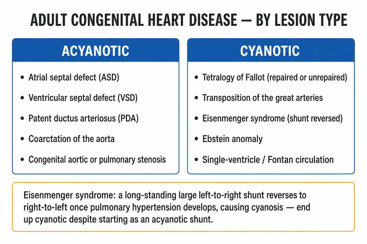
Classifying by what the lesion does to flow pattern is more useful at the bedside: [1]
| Pattern | Mechanism | Examples | Consequence |
|---|---|---|---|
| Acyanotic shunt (L→R) | High-pressure system to low-pressure system | ASD, VSD, PDA, partial AVSD | Volume overload of receiving chamber; pulmonary over-circulation; risk of PAH if untreated |
| Cyanotic shunt (R→L) | Right-sided pressure exceeds left | Eisenmenger, unrepaired TOF, TGA, single ventricle | Hypoxaemia, secondary erythrocytosis, clubbing, paradoxical embolism |
| Obstructive | Fixed outflow obstruction | Coarctation, aortic stenosis, pulmonary stenosis, sub-valvular/supravalvular AS | Pressure overload proximal to obstruction, hypertrophy, collateral formation |
| Regurgitant | Incompetent valve | Bicuspid-related AR, pulmonary regurgitation (post-TOF), Ebstein | Volume overload of receiving chamber, dilatation |
| Complex / mixing | Multiple lesions, single functional ventricle | Fontan, TGA (post-Switch / post-Mustard), heterotaxy | Each lesion has its own physiology and surgical palliation |
Physiological staging (Conway) — used in parallel
The 2018 AHA/ACC guideline also defines a physiological stage (A–D) running in parallel with anatomy: stage A is at risk / no haemodynamic burden; B has mild burden; C has moderate; D has severe with decompensation. Together, anatomy × physiology defines the ACHD AP classification (e.g. "moderate complexity, stage C" = II-C), which drives follow-up intensity.[2]
Epidemiology & Risk Factors
ACHD has become one of the largest cardiology subspecialties because of a single demographic transition. Survival of CHD into adulthood is now over 90%, the adult CHD population now outnumbers the paediatric CHD population, and prevalence is rising ~5% per year in developed nations.[1]
ACHD by the numbers
The commonest unrepaired CHD presenting in adults in the developed world is secundum ASD (an isolated secundum ASD may be silent into the fifth decade). In developing regions — including India — late presentations of rheumatic heart disease mimicking congenital mitral stenosis are still common, but unrepaired large VSD, PDA, and TOF also continue to present in adolescence and adulthood because of cost barriers and limited paediatric surgical access.[1]
[1]Risk factors for late presentation include low socioeconomic status, rural residence, female sex (postponed paediatric surgery in some cultures), co-existing congenital syndromes that delay diagnosis, and migration from a region without CHD services. The single biggest risk factor for an Eisenmenger outcome is non-closure of a large L-to-R shunt in childhood — which is why early surgical or percutaneous closure of significant VSD, PDA and AVSD is one of the most cost-effective interventions in global health.[1][3]
Pathophysiology
The pathophysiology of ACHD divides into shunt physiology (the largest concept), obstructive physiology, regurgitant physiology and systemic right-ventricle / single-ventricle physiology. The unifying concept is wall stress, chamber volume/pressure overload and remodelling.[1]
Shunt physiology and the Qp:Qs ratio
A shunt moves blood between two cardiac chambers or great vessels. Its size is quantified by the pulmonary-to-systemic flow ratio, Qp:Qs:[3]
- Qp:Qs = 1.0 — no shunt (or balanced bidirectional).
- Qp:Qs less than 1.5 — small, haemodynamically insignificant shunt; usually conservative management.
- Qp:Qs 1.5–2.0 — moderate shunt; closure usually indicated if symptomatic or evidence of chamber enlargement.
- Qp:Qs over 2.0 — large shunt; closure indicated to prevent PAH and chamber damage (provided PVR is still acceptable). [1]
A left-to-right shunt delivers extra volume to the pulmonary circulation. The right heart and pulmonary vascular bed accommodate this for years — the patient is asymptomatic — but chronic over-circulation progressively remodels the pulmonary arterioles (medial hypertrophy, intimal proliferation, plexiform lesions — the histological hallmark of irreversible PAH). When PVR approaches systemic vascular resistance (SVR), the shunt decelerates, then reverses (R-to-L). This is the Eisenmenger transition.[1]
The Eisenmenger transition — molecular and haemodynamic
The molecular biology is the same as in idiopathic pulmonary arterial hypertension (IPAH). Endothelial injury from high flow and shear stress shifts the balance toward vasoconstrictors (endothelin-1, thromboxane A2, serotonin) and away from vasodilators (nitric oxide, prostacyclin). Pulmonary artery smooth muscle proliferates, the intima thickens, and characteristic plexiform lesions develop. The vasodilator response is progressively lost — which is why acute vasoreactivity testing is negative in established Eisenmenger. The maladaptive right ventricle hypertrophies, and over years dilates and fails. [1]
Three haemodynamic thresholds define the transition:[1]
- PVR over 3 Wood units without vasoreactivity — elevated, cautious.
- PVR over 5 Wood units or PVR index over 8 WU·m² — high-risk for closure; many centres will not close.
- PVR at or above systemic level (PVR/SVR ratio near 1.0) and/or net right-to-left shunting — Eisenmenger: closure is absolutely contraindicated. [1]
Why the systemic right ventricle fails (TGA after Mustard/Senning; ccTGA)
In d-TGA after atrial switch (Mustard/Senning), and in ccTGA, the morphological right ventricle pumps the systemic circulation. The RV is built for low-pressure, high-volume work (thin walls, sinusoidal fibres) and is poorly adapted to high afterload. Over three to five decades it develops hypertrophy, then dilatation, then systolic dysfunction, often with tricuspid (systemic AV valve) regurgitation and atrial arrhythmia. This is the principal late cause of heart failure and death in this population and is the rationale for primary arterial switch (Jatene) in modern paediatric surgery.[1][3]
Why repaired TOF develops pulmonary regurgitation
Classical TOF repair involves VSD patch closure and right-ventricular outflow tract (RVOT) reconstruction, usually via a transannular patch that enlarges the RVOT but sacrifices pulmonary valve competence. The result is chronic pulmonary regurgitation — well-tolerated for decades but eventually causing RV dilatation, QRS prolongation on ECG, and risk of sustained ventricular tachycardia / sudden cardiac death. The classical late intervention is pulmonary valve replacement (PVR), increasingly percutaneous (Melody, Sapien valves).[2]
Why coarctation produces radio-femoral delay and rib notching
Coarctation is a discrete narrowing (usually juxta-ductal, just distal to the left subclavian). The descending aorta is perfused by collaterals (internal mammary → intercostal → subclavian). The result: upper-limb hypertension, radio-femoral delay, weak/absent femoral pulses, and erosion of the undersides of ribs 4–8 by dilated intercostal arteries (rib notching on chest X-ray). The classic radiological sign is the figure-of-3 sign (the pre-stenotic dilatation of the left subclavian, the coarctation indentation, and the post-stenotic dilatation of the descending aorta).[1][5]
Why ASD produces paradoxical embolism
Even a small ASD or PFO can permit a venous thrombus to cross from right to left atrium during transient RA pressure elevation (cough, Valsalva, pulmonary embolism), causing cryptogenic stroke. This is the rationale for PFO closure in selected young patients with cryptogenic stroke (RESPECT trial), and the rationale for Valsalva precautions and air-bubble avoidance in IV lines in any ACHD patient.[1]
Clinical Presentation
Most unrepaired ACHD lesions present in adulthood in one of four ways: incidental murmur in a young adult; new exertional dyspnoea / atrial arrhythmia in middle age; stroke / TIA from paradoxical embolism; or late decompensation with right-heart failure or Eisenmenger symptoms.[1]
Secundum ASD in the adult — the classic presentation
The prototype of late-presentation ACHD is the secundum ASD. The L-to-R shunt at atrial level volume-loads the right atrium and right ventricle for decades. Most patients are asymptomatic until the fourth to sixth decade, when:[1][3]
- Exertional dyspnoea (the commonest first symptom).
- Atrial fibrillation or flutter (the right atrium dilates and triggers re-entry).
- Right-heart failure (peripheral oedema, hepatic congestion, ascites).
- Paradoxical embolism (cryptogenic stroke).
- Rarely, recurrent respiratory infections from chronic pulmonary over-circulation. [1]
The bedside signs (see Bedside Assessment) form a triad that examiners love: wide, fixed split S2; pulmonary flow murmur; RV precordial heave. Palpable RV impulse at the lower left sternal edge; prominent pulmonary artery pulsation in the second left ICS. [1]
Coarctation of aorta in an adult
Presents with upper-body hypertension (often in young men), headaches, epistaxis, leg claudication or leg fatigue (cold feet on exercise). Physical findings are the diagnostic gift: radio-femoral delay, weak or absent femoral pulses, blood pressure differential between arms and legs (arm-leg systolic gradient over 20 mmHg), and an interscapular bruit from collaterals. Associated bicuspid aortic valve (50–85%) produces an ejection click and/or aortic murmur. Turner syndrome should be considered in any female with coarctation.[1][5]
Eisenmenger syndrome — the cyanotic phenotype
The classic Eisenmenger presentation combines central cyanosis, digital clubbing, and a long history of a "hole in the heart" never closed:[1][3]
- Central cyanosis — bluish discolouration of lips, tongue, nailbeds (best seen under natural light, warming the hands).
- Digital clubbing — loss of the nail-bed angle (Schamroth's sign positive: when distal phalanges of identical fingers from opposite hands are placed nail-to-nail, the normal diamond window is lost).
- Secondary erythrocytosis — haemoglobin often over 18 g/dL, haematocrit over 55% (a compensatory response to chronic hypoxaemia via renal erythropoietin; distinct from polycythaemia vera — white cells and platelets are normal).
- Hyperviscosity symptoms — headaches, visual disturbance, tinnitus, fatigue, paresthaesia (related to haematocrit; but iron deficiency from routine phlebotomy worsens symptoms and must be avoided).
- Haemoptysis — from rupture of dilated bronchial collaterals or in situ pulmonary artery thrombosis; the most-feared complication.
- Right-heart failure late — elevated JVP, hepatomegaly, peripheral oedema.
- Syncope / sudden death — from arrhythmia or massive pulmonary embolism/haemorrhage.
- Hyperuricaemia and gout (from increased erythrocyte turnover).
- Cerebral abscess / venous sinus thrombosis (because the lungs no longer filter venous bacteria/clot — bypassing the pulmonary filter via R-to-L shunt). [1]
The type of shunt predicts the distribution of cyanosis (exam gold):[3]
- VSD / AVSD Eisenmenger — uniform central cyanosis.
- PDA Eisenmenger — differential cyanosis: hands (pre-ductal, perfused by left ventricle) pink; feet (post-ductal, perfused by pulmonary artery through the PDA) cyanosed.
- ASD Eisenmenger — late, milder cyanosis; ASD has the lowest pressure gradient and usually the slowest PAH progression. [1]
Late complications of repaired Tetralogy of Fallot
Patients with repaired TOF typically present in adulthood with:[2]
- Pulmonary regurgitation (the dominant lesion) — RV heave, Eisenmenger-like diastolic murmur (early diastolic, low-pitched, left parasternal — Graham-Steell-like), RV dilatation on imaging.
- Residual RVOT obstruction or branch pulmonary artery stenosis.
- Atrial arrhythmia (typical atrial flutter — incisional macro-re-entry around the atriotomy/scar — is the hallmark).
- Ventricular arrhythmia and sudden cardiac death — risk rises sharply when QRS duration exceeds 180 ms on ECG.
- Aortic root dilatation and aortic regurgitation (over-riding aorta, bicuspid aortic valve).
- Residual VSD. [1]
Ebstein anomaly
Congenital downward displacement of the septal and posterior tricuspid leaflets into the RV produces severe tricuspid regurgitation, "atrialisation" of the RV inlet (a portion of the RV becomes functionally part of the RA), a small functional RV, and commonly an ASD or PFO (with R-to-L shunting in severe cases — cyanotic Ebstein). Adults present with arrhythmia (especially accessory-pathway-mediated SVT — WPW), right-heart failure, dyspnoea, or incidental murmur. The classic ECG is giant P waves (right atrial enlargement), RBBB and a long PR interval. WPW with right-sided pathway produces a left-bundle-branch-block-pattern delta wave.[1][5]
Atypical presentations
- Pregnancy unmasking a previously silent shunt — decreased SVR and increased cardiac output reverse a borderline L-to-R ASD into R-to-L, producing exertional dyspnoea or paradoxical embolism.
- Elderly with ASD presenting with atrial fibrillation and right-heart failure mimicking left-heart disease.
- Repaired coarctation presenting with refractory hypertension or aortic dissection in a young adult.
- Fontan adult with new-onset protein-losing enteropathy (PLE) — peripheral oedema, ascites, chronic diarrhoea, hypoalbuminaemia; a marker of failing Fontan circulation. [1]
Differential Diagnosis
The examiner wants differentials framed by the dominant bedside finding, with the discriminating feature for each.[1]
Wide fixed split S2 (ASD differential)
- ASD secundum (commonest cause; pulmonary flow murmur, RV heave)
- Ostium primum ASD (with cleft mitral valve → MR)
- Sinus venosus ASD (often with PAPVR; high placement)
- Partial AVSD (primum ASD + cleft MV)
- Large VSD (rare to have truly fixed split)
- RBBB and RV pacing can mimic — but no pulmonary flow murmur, no RV volume overload on echo
Cyanosis + clubbing in an adult
- Eisenmenger syndrome (CHD history, secondary erythrocytosis, large PA on CXR)
- Idiopathic pulmonary arterial hypertension (no shunt; clear lung fields; no clubbing until very late)
- Chronic lung disease (COPD, pulmonary fibrosis — lung signs, less erythrocytosis)
- Pulmonary AV malformation (HHT/Osler-Weber-Rendu; telangiectasia, epistaxis, family history)
- Methaemoglobinaemia (chocolate-brown blood, no response to oxygen)
Differential cyanosis (hands pink, feet blue)
- PDA with Eisenmenger (the classic — pre-ductal SpO2 normal, post-ductal low)
- Interrupted aortic arch (neonatal; rare in adults)
- Aorto-pulmonary window with Eisenmenger (rare)
- Subclavian steal (one arm only — not feet)
- Coarctation with PDA (mixed picture)
Radio-femoral delay / weak femoral pulses
- Coarctation of aorta (the classic; upper-limb HTN, rib notching)
- Aortic dissection involving subclavian/femoral origin (acute presentation)
- Leriche syndrome / aortoiliac occlusive disease (older, vascular risk factors, no upper-body HTN)
- Subclavian artery stenosis (one-sided; subclavian steal)
- Takayasu arteritis (pulseless disease, bruits, raised inflammatory markers)
Pulmonary flow murmur in an adult
- ASD (with fixed split S2 — the discriminator)
- Pulmonary valve stenosis (ejection click, no fixed split S2)
- Innocent Still's murmur (children; vibratory; disappears on sitting forward)
- Anaemia / hyperthyroidism / pregnancy (high-flow, both murmurs soft)
Late diastolic murmur + RV heave
- Pulmonary regurgitation (repaired TOF; idiopathic dilatation of PA)
- Graham-Steell murmur of pulmonary hypertension (high-pitched early diastolic, LSB)
- Aortic regurgitation (different site — right 2nd ICS, apex; bounding pulse)
Three clinical discriminators are pure viva currency: [1]
- ASD vs pulmonary stenosis: both produce a pulmonary area ejection murmur, but only ASD has the wide, fixed split S2 (PS has a wide but variably split S2 that varies with respiration).
- PDA-Eisenmenger vs coarctation: differential cyanosis is shared, but coarctation has upper-body hypertension and rib notching without central cyanosis; PDA-Eisenmenger has cyanosis in the feet only.
- Eisenmenger clubbing vs chronic lung disease clubbing: Eisenmenger has the shunt history, secondary erythrocytosis with normal platelets/WCC, prominent pulmonary artery on CXR, and clear lung fields (the remodeled pulmonary vasculature is the problem, not parenchyma).[1]
Clinical & Bedside Assessment
The bedside examination in ACHD yields more specific diagnostic information than in almost any other cardiology subspecialty. The examiner wants the specific manoeuvres and their physiology.[1][5]
Secundum ASD — the bedside phenotype
- Pulse: usually regular, small-volume if shunt is large.
- JVP: may show prominent a-wave if pulmonary hypertension is developing.
- Precordium: RV heave at the lower left sternal edge (volume-overloaded RV); palpable pulmonary artery pulsation in the 2nd left ICS.
- Auscultation:
- S1 normal; S2 widely and fixed split (the fixed split is the discriminator — delayed P2 from prolonged RV ejection that does not vary with respiration because the RV is already volume-loaded).
- Pulmonary flow murmur: ejection systolic, 2nd/3rd left ICS, from increased flow across a normal pulmonary valve.
- Tricuspid flow murmur: diastolic rumble at lower left sternal edge if Qp:Qs is very large.
- A systolic thrill is uncommon.
- With advancing age: atrial fibrillation, signs of right-heart failure. [1]
Coarctation — the bedside phenotype
- All four limbs palpated simultaneously to compare radio-radial, radio-femoral, and femoro-femoral pulses.
- Radio-femoral delay: simultaneous palpation of radial and femoral — the femoral upstroke is delayed and weak.
- Blood pressure: arms hypertensive; legs hypotensive. Arm-to-leg systolic gradient over 20 mmHg is significant. Measure BP in both arms (left subclavian may be involved) and one leg.
- Precordium: LV heave from hypertension; ejection click and/or aortic murmur if bicuspid valve co-exists.
- Auscultation over the scapula: interscapular bruit from collateral flow through intercostal and scapular arteries.
- Look for Turner phenotype (short stature, webbed neck, widely spaced nipples) in females. [1]
Eisenmenger — the bedside phenotype
- Hands: central cyanosis, clubbing (Schamroth's sign positive).
- Face: central cyanosis (lips, tongue, conjunctival suffusion).
- Neck: raised JVP with prominent a-wave (pulmonary hypertension, non-compliant RV).
- Precordium: RV heave; palpable P2 (severe PAH).
- Auscultation:
- Loud P2 (pulmonary hypertension).
- Right-sided S4.
- Pulmonary ejection click.
- Graham-Steell murmur — high-pitched, early-diastolic, blowing, left parasternal, from functional pulmonary regurgitation due to dilated pulmonary artery.
- The original shunt murmur disappears (the gradient has been abolished).
- Differential cyanosis check: measure SpO2 at the right hand (pre-ductal) and a foot (post-ductal); a drop of over 3% (typically much greater) post-ductally indicates PDA with Eisenmenger. [1]
Repaired Tetralogy of Fallot — the bedside phenotype
- Precordium: RV heave from chronic PR.
- Auscultation: absent or single S2 (P2 absent because the pulmonary valve is regurgitant or replaced by a patch), low-pitched early-diastolic murmur at the upper left sternal edge (pulmonary regurgitation), and a residual ejection murmur if RVOT obstruction persists.
- ECG: RBBB (from the RVOT incision/patch), QRS duration over 180 ms is a marker of high risk of sustained VT/SCD.[2]
Reproduced bedside tests
- Schamroth's window test — place the distal phalanges of the same fingers of opposite hands nail-to-nail; loss of the normal diamond-shaped window = clubbing.
- Pre- and post-ductal SpO2 — diagnostic of differential cyanosis.
- Müller's manoeuvre (inspiration against a closed glottis) increases the venous return and accentuates right-sided murmurs; Valsalva decreases RV filling and softens right-sided murmurs (the opposite effect on HOCM).
- Squat-to-stand: in pulmonary stenosis the murmur falls on standing (reduced venous return); in ASD it is little changed. [1]
Investigations
Investigation is layered: ECG and CXR for pattern recognition; transthoracic echocardiography (TTE) for anatomy, shunt size, and chamber size; cardiac MRI for quantification; cardiac CT for anatomy where MRI is contraindicated; cardiopulmonary exercise testing (CPET) for prognosis; cardiac catheterisation for haemodynamics and PVR.[1]
ECG — high-yield patterns
| Lesion | Classic ECG finding |
|---|---|
| ASD secundum | RSR' in V1 (incomplete RBBB), right-axis deviation, RV volume overload |
| Ostium primum ASD | Left-axis deviation + RBBB (the LAD distinguishes from secundum) |
| Coarctation | LVH from hypertension |
| Ebstein | Giant P waves (right atrial enlargement), long PR, RBBB, pre-excitation / WPW (right-sided pathway, LBBB-pattern delta) |
| Eisenmenger | RVH with strain (right-axis deviation, dominant R in V1, right precordial T inversion), P pulmonale |
| Repaired TOF | RBBB, QRS over 180 ms high-risk; Q waves in inferior and right precordial leads from VSD patch and ventriculotomy |
| Mustard/Senning (TGA) | Sinus node dysfunction, atrial arrhythmia, RVH (systemic RV) |
| ccTGA | AV dissociation (conduction system malformation), Q waves in septal leads |
| Fontan | Sinus node dysfunction, atrial arrhythmia, low-voltage QRS |
Chest X-ray
- ASD — cardiomegaly, prominent pulmonary artery segment, plethoric lung fields (pulmonary plethora from over-circulation); normal-size aortic knuckle.
- Coarctation — rib notching (undersides of ribs 4–8 from dilated intercostal collaterals), figure-of-3 sign (pre-stenotic dilatation of the left subclavian, coarctation notch, post-stenotic dilatation of descending aorta), LVH.
- Eisenmenger — large central pulmonary arteries with pruned, oligaemic peripheral lung fields (the radiological signature of PAH); RV enlargement.
- Ebstein — massive cardiomegaly ("box-shaped heart") from severe right atrial enlargement, small pulmonary trunk.
- TOF (untreated) — boot-shaped heart (coeur en sabot: upturned apex from RVH + concave pulmonary segment).
- Pulmonary AR / repaired TOF — dilated pulmonary artery. [1]
Echocardiography — the workhorse
TTE defines lesion, size, haemodynamic effect, and ventricular function. Specific uses:[1][2]
- ASD: type (secundum vs primum vs sinus venosus vs coronary sinus), size, shunt colour Doppler direction, estimate Qp:Qs, rim measurements for device closure (need adequate aortic, atrial, SVC, IVC and posterior rims; deficient retro-aortic rim is the commonest reason for surgical rather than percutaneous closure), RV size and pulmonary pressure.
- VSD: location (membranous, muscular, inlet, outlet), size, jet velocity (a high-velocity jet across a small VSD reflects a large LV-RV pressure gradient — protective against PAH).
- PDA: size, ductal flow pattern, pulmonary pressures from the jet velocity.
- Coarctation: site, gradient (peak and mean), associated BAV, aortic dimensions.
- Ebstein: tricuspid displacement distance, severity of TR, ASD/PFO presence.
- Repaired TOF: PR severity (jet deceleration time, regurgitant fraction), RV size (RVEDV), RV function, residual RVOT obstruction, aortic root.
- Pulmonary pressures: estimate from tricuspid regurgitant jet velocity (RVSP = 4 × V² + RA pressure). [1]
Transoesophageal echocardiography (TOE) is essential for inferior rim visualisation of ASD, intracardiac shunts not well seen on TTE, and intra-procedural guidance for device closure. [1]
Cardiac MRI — the gold standard for RV and great vessels
CMR is the gold standard for RV volumes, mass and function (which TTE struggles with given the unusual geometry) and for quantifying shunts (Qp:Qs by flow mapping) and great vessel anatomy. Indications: any moderate-or-greater ACHD lesion, repaired TOF surveillance (annual/biennial), systemic RV, coarctation (3D anatomy and gradient), suspected PAPVR or sinus venosus ASD.[1]
Cardiac CT — for coronary anatomy and metallic artefact
CT is used for coronary artery assessment pre-operatively (instead of invasive angiography in younger patients), for pulmonary venous anatomy, and where MRI is contraindicated (implantable devices, claustrophobia). High spatial resolution makes it the test of choice for stent surveillance after coarctation stenting. [1]
Cardiac catheterisation — haemodynamics and PVR
Catheterisation is mandatory before shunt closure if there is any concern about PVR. Key measurements:[1][3]
- Pulmonary artery pressure (systolic, diastolic, mean).
- Pulmonary vascular resistance (PVR) in Wood units.
- PVR/SVR ratio.
- Qp:Qs.
- Pulmonary vasoreactivity (response to inhaled nitric oxide 10–20 ppm or 100% oxygen) — if vasoreactive, the patient may respond to targeted PAH therapy and closure may be reconsidered. [1]
Closure is contraindicated if PVR is over 5 Wood units (or PVR index over 8 WU·m²) and does not fall below 5 WU with vasodilator challenge. Below these thresholds, closure is generally safe and indicated. [1]
Cardiopulmonary exercise testing (CPET)
CPET (peak VO2, VE/VCO2 slope) is the strongest predictor of mortality in ACHD and is used to time interventions and transplantation referral. A peak VO2 below 15 mL/kg/min (or under 50% predicted) and VE/VCO2 slope over 35 indicate advanced disease. [1]
Blood tests
- Full blood count — secondary erythrocytosis in cyanotic disease (haemoglobin over 18 g/dL, haematocrit over 55%); iron studies are essential (iron deficiency is common from inappropriate phlebotomy or menorrhagia and worsens symptoms).
- Renal function, liver function — hepatorenal congestion in advanced disease.
- BNP / NT-proBNP — rising trends predict ventricular dysfunction.
- Urate — often high in cyanotic disease.
- Coagulation — deranged in Fontan (hepatic congestion) and Eisenmenger (frequent bleeding AND thrombosis).
- Pregnancy test in any woman of reproductive age before imaging. [1]
Exercise testing
- Symptom-limited CPET (above) is the most useful.
- Routine exercise ECG is not useful in cyanotic disease — desaturation limits interpretation. [1]
Special: modified WHO classification for pregnancy (reproduced verbatim)
The modified WHO risk classification (mWHO) is the ESC's preferred framework for counselling pregnant women with cardiovascular disease and is reproduced verbatim:[4]
[1]Two named scoring systems also exist (CARPREG II and ZAHARA) — these assign weighted points to findings (prior cardiac events, baseline NYHA, systemic ventricular dysfunction, left-heart obstruction, history of arrhythmia) and stratify risk of maternal cardiac complications. mWHO is preferred by the ESC.[4]
[1]Management — Resuscitation
Most ACHD patients are not in resuscitation — but three scenarios are time-critical and frequently mishandled by general physicians: Eisenmenger haemoptysis, atrial arrhythmia in cyanotic disease, and right-heart failure in the systemic RV.[1]
Eisenmenger haemoptysis
The single most-feared complication of Eisenmenger syndrome and a leading cause of death. Mechanism: rupture of dilated bronchial collaterals or in situ pulmonary artery thrombosis with distal infarction and haemorrhage.[1]
Immediate management:
- Sit the patient upright, leaning toward the bleeding side (if lateralised) to protect the contralateral lung.
- High-flow oxygen — to keep SpO2 in the patient's usual range (do NOT chase normoxia; their baseline may be 75–85%, and sudden over-oxygenation can worsen pulmonary vasoconstriction mismatch).
- Avoid over-resuscitation — small-volume crystalloid; avoid fluid overload that worsens RV failure.
- Cautious correction of coagulopathy — Eisenmenger patients have a mixed bleeding/thrombotic state; fresh frozen plasma and platelets only if bleeding severe; avoid antifibrinolytics if thrombotic risk high.
- Bronchial artery embolisation (interventional radiology) is the definitive acute treatment for localised bleeding.
- Contact ACHD specialist team immediately and consider transfer. [1]
AVOID:
- Anticoagulation unless a clear concurrent indication — Eisenmenger carries both bleeding and thrombosis risk.
- Vasopressors that raise PVR (e.g. high-dose noradrenaline) — prefer agents that do not raise pulmonary pressures.
- Sudden vasodilatation (which can worsen R-to-L shunt). [1]
Atrial arrhythmia in cyanotic ACHD
Atrial flutter / fibrillation in Eisenmenger or repaired TOF is haemodynamically dangerous because the RV is preload-dependent.[1]
- AVN-blocking drugs (beta-blockers, calcium-channel blockers, digoxin) are DANGEROUS — by slowing AV nodal conduction they can promote 1:1 atrial flutter conduction (a rate of 250–300/min) and cardiovascular collapse; digoxin also loses efficacy in hypoxaemia.
- Preferred management: synchronised DC cardioversion if unstable; intravenous amiodarone 300 mg over 1 hour then 900 mg over 24 hours if stable; anticoagulate (LMWH then warfarin or DOAC carefully — see Special Populations).
- Seek expert EP input — catheter ablation (often cavotricuspid isthmus ablation for atrial flutter) is highly effective and preferred. [1]
Right-heart failure / systemic RV decompensation
- Diuretics — furosemide 40–80 mg IV with potassium-sparing adjunct; monitor renal function.
- Spironolactone 25–50 mg OD for aldosterone antagonism and diuretic synergy.
- ACE-inhibitors and beta-blockers have NOT shown the same mortality benefit in systemic-RV or RV-failure as in LV failure — use cautiously, low dose, slow titration.
- Avoid pure afterload reducers that drop SVR and worsen R-to-L shunt in Eisenmenger.
- In Eisenmenger / PAH specifically: targeted PAH therapy (see below). [1]
Endocarditis prophylaxis — what the patient actually needs
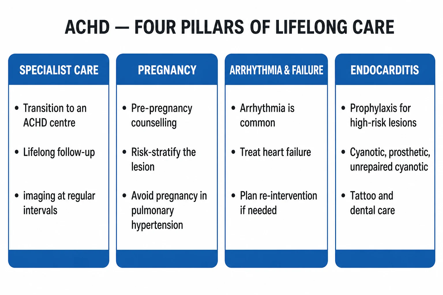
Prophylaxis is restricted (NICE/ESC/AHA all converge) to the highest-risk cardiac lesions undergoing high-risk dental procedures (gum and periapical manipulation, oral mucosal incision).[1][2]
[1]Management — Definitive & Stepwise
Definitive management in ACHD is closure of correctable shunts and obstruction in those with reversible physiology, targeted PAH therapy in Eisenmenger, valve and conduit interventions in repaired lesions, and heart or heart-lung transplantation for end-stage disease.[1][2]
Step 1 — Secundum ASD closure
Closure is indicated for any of:[1][2]
- Symptomatic secundum ASD (dyspnoea, atrial arrhythmia, paradoxical embolism), regardless of shunt size.
- Asymptomatic ASD with significant shunt (Qp:Qs over 1.5) and right-heart enlargement on imaging, with acceptable PVR (PVR under 5 WU, PVR/SVR under 0.33).
- Right-heart enlargement on imaging, even if asymptomatic. [1]
Percutaneous device closure (Amplatzer Septal Occluder) is preferred for secundum ASD if all of:
- Diameter under 38 mm (stretched balloon).
- Adequate rims (over 5 mm) on all sides — particularly the retro-aortic rim, posterior-inferior rim (IVC) and superior rim (SVC).
- No other cardiac surgery required. [1]
Surgical closure (direct suture or pericardial/PTFE patch) is preferred for:
- Primum, sinus venosus, and coronary sinus defects (these cannot be closed percutaneously).
- Secundum ASD with deficient rims, very large size, or associated anomalies needing surgery (PAPVR, cleft mitral valve, severe TR).
- Eisenmenger physiology — contraindicated. [1]
Anticoagulation: aspirin 75 mg OD for 6 months and endocarditis prophylaxis for 6 months post-device closure. Periprocedural heparin during the procedure. [1]
Complications of device closure: device embolisation (rare), atrial arrhythmia (transient), pericardial effusion/tamponade (rare cardiac erosion — emergency), headache (stop antiplatelet if migraine-like). [1]
Step 2 — Coarctation intervention
- Peak-to-peak gradient over 20 mmHg on catheterisation, OR
- Hypertension uncontrolled on three drugs with anatomical coarctation on imaging (any gradient), OR
- Aneurysm formation at the repair site, OR
- Re-coarctation after previous surgery with significant gradient. [1]
Stenting (percutaneous, covered stent preferred) is first-line for native or recurrent adult coarctation with suitable anatomy — lower complication rate than surgery, shorter hospital stay, equally effective gradient reduction. [1]
Surgical repair (resection with end-to-end anastomosis, or interposition graft, or extra-anatomic bypass) is reserved for long-segment coarctation, aneurysm not amenable to stent, or co-existing arch disease needing surgery. [1]
Post-intervention: lifelong surveillance for re-coarctation, aneurysm at repair site, and refractory hypertension (which may persist despite anatomical cure — the baroreceptors and renovascular system have adapted). MRI or CT annually for 5 years then biennially. Beta-blockade as first-line antihypertensive (renin-angiotensin activation blunted), ACE-inhibitor if residual hypertension. [1]
Step 3 — Pulmonary valve replacement in repaired TOF
Indications for PVR (2020 ESC; thresholds similar in 2018 AHA/ACC):[1][2]
- RV end-diastolic volume index (RVEDVi) over 150 mL/m², OR
- RV end-systolic volume index (RVESVi) over 80–90 mL/m², OR
- RV EF under 47%, OR
- QRS duration over 180 ms, OR
- Symptoms (arrhythmia, heart failure, exertional intolerance), OR
- Severe PR with RV dilatation. [1]
Percutaneous PVR (Melody valve, Sapien XT/Apollo) is preferred where the RVOT and pulmonary artery anatomy is suitable (right ventricle-to-pulmonary artery conduit or bioprosthetic valve in place) — preserves surgical option and lower operative risk. [1]
Surgical PVR is preferred for: native RVOT unsuitable for percutaneous valve, concomitant lesions needing surgery (residual VSD, severe TR, aortic root dilatation), or very large RVOT. [1]
Adjunctive: at surgery, tricuspid valve repair, atrial flutter ablation (right atrial maze), VSD patch revision and PFO/ASD closure are considered. [1]
Step 4 — Eisenmenger medical therapy
Closure of the defect is CONTRAINDICATED. Medical management extends life and improves symptoms.[1][3][6]
Targeted pulmonary arterial hypertension (PAH) therapy (the cornerstone): [1]
- Bosentan (dual endothelin-receptor antagonist) — first-line; the BREATHE-5 trial (Galie 2006) showed improved pulmonary vascular resistance and 6-minute-walk distance in Eisenmenger. Start at 62.5 mg BD for 4 weeks then 125 mg BD; monthly LFTs (hepatotoxicity — discontinue if AST/ALT over 3× ULN); contraindicated in pregnancy (teratogenic).
- Sildenafil (PDE-5 inhibitor) — 20 mg TDS (titrate to 40–80 mg TDS); cheaper, well-tolerated; headache and hypotension the main side effects; add-on or alternative.
- Tadalafil — 40 mg OD; alternative PDE-5 inhibitor with once-daily dosing.
- Macitentan — 10 mg OD (dual ERA; MAESTRO study supported benefit).
- Prostacyclin analogues — inhaled iloprost or IV epoprostenol for advanced disease. [1]
Supportive measures: [1]
- Supplemental oxygen — controversial; symptom relief, no survival benefit. Use if symptomatic improvement.
- Anticoagulation — selectively, in those with atrial arrhythmia, prior thromboembolism, or mechanical valve; INR target 2.0–2.5 (lower than for non-Eisenmenger because of bleeding risk). DOACs are not routinely recommended in Eisenmenger (limited data).
- Avoid iron deficiency — supplement oral iron if ferritin or transferrin saturation low; avoid routine phlebotomy (worsens iron deficiency and outcomes).
- Phlebotomy ONLY for symptomatic hyperviscosity (headaches, visual disturbance) with haematocrit over 65% AND volume-depleted — remove 250–500 mL with concomitant volume replacement. Not for "high haemoglobin" alone.
- Vaccinations — annual influenza, pneumococcal, COVID-19.
- Avoid: pregnancy (contraception counselling mandatory), high-altitude exposure, dehydration, isometric heavy lifting, smoking.
- Dehydration prevention — adequate fluid intake, especially during illness. [1]
Heart-lung or bilateral lung transplantation is the only definitive therapy for end-stage Eisenmenger, considered when 6MWD falls below 350 m, peak VO2 below 10 mL/kg/min, syncope, or refractory heart failure; 5-year survival after transplant is 50–70%. [1]
Step 5 — Specific lesion management
| Lesion | Definitive management |
|---|---|
| VSD | Surgical or percutaneous closure if Qp:Qs over 2, symptomatic, or with aortic regurgitation from prolapsing right coronary cusp. Contraindicated in Eisenmenger. |
| PDA | Percutaneous device closure if PVR acceptable; small PDA can be observed. |
| Pulmonary stenosis | Percutaneous balloon valvuloplasty (first-line) if peak gradient over 50 mmHg; surgical for dysplastic valve. |
| Ebstein | Surgical Cone repair (or tricuspid valve replacement + ASD closure) if symptomatic, cyanotic, severe TR, or arrhythmia. |
| Bicuspid valve | AVR for severe AS/AR (see aortic-stenosis topic); aortic surveillance (intervention at 5.5 cm; 5.0 cm with risk factors). |
| ccTGA | Tricuspid (systemic AV) valve repair/replacement for severe regurgitation; pacemaker for AV block; CRT if dyssynchrony. |
| Mustard/Senning (TGA) | Conversion to arterial switch (selected centres); heart failure therapy; arrhythmia ablation; consider transplant. |
| Fontan (failing) | Fenestration, protein-losing enteropathy management (heparin, octreotide, budesonide, spironolactone), conversion to extracardiac conduit, cardiac transplantation. |
Specific Subtypes & Scenarios
ASD types — and why the type matters
The type of ASD determines whether percutaneous closure is possible:[1][5]
- Secundum ASD (75%) — defect in the fossa ovalis; percutaneous device closure is the default if rims are adequate.
- Primum ASD (15–20%) — part of partial AVSD; with a cleft anterior mitral leaflet producing mitral regurgitation. Surgical closure with mitral repair is mandatory; never device closure.
- Sinus venosus ASD (5–10%) — at the SVC or IVC orifice; commonly associated with partial anomalous pulmonary venous return (PAPVR) (right upper pulmonary vein draining to SVC). Surgical (often with Warden procedure — redirecting the anomalous vein).
- Coronary sinus ASD (rare) — unroofed coronary sinus; often with persistent left SVC. Surgical closure. [1]
Repaired Tetralogy of Fallot — long-term follow-up
Every repaired TOF patient needs lifelong ACHD specialist follow-up.[1][2]
- Annual review with ECG (QRS duration trend), echo (RV size and function, PR, residual lesions), clinical status.
- Cardiac MRI every 1–3 years (RV volumes, PR fraction) — drives the timing of PVR.
- Holter monitoring for sustained VT in those with QRS over 180 ms, syncope, or symptoms.
- CPET for prognostic stratification.
- Aortic surveillance — ascending aorta dilatation in 15% (over-riding aorta, bicuspid valve).
- Endocarditis prophylaxis only in the high-risk categories above. [1]
Pregnancy in repaired TOF is generally well-tolerated if: oxygen saturation normal, no significant residual lesions, RV function acceptable, no arrhythmia. Severe PR with RV dilatation raises risk — consider PVR before pregnancy. [1]
Coarctation — surgical types and complications
Surgical techniques historically used: resection with end-to-end anastomosis (most common, lowest recurrence), subclavian flap angioplasty, interposition graft, patch aortoplasty (highest aneurysm risk — now avoided). Late complications:[1][5]
- Re-coarctation — 7–15%, more common with subclavian flap and patch.
- Aneurysm at repair site — particularly after patch aortoplasty; surveillance CT/MRI.
- Persistent or recurrent hypertension — even after anatomical cure.
- Paraplegia (rare, from spinal cord ischaemia) — risk at original surgery and re-operation.
- Aortic dissection or rupture — risk lifelong. [1]
Ebstein anomaly
The Cone reconstruction (Da Silva) is the modern surgical procedure of choice — it mobilises the functional tricuspid leaflets into a competent cone at the true annulus. Indications for surgery: symptoms, cyanosis, severe TR, arrhythmia unresponsive to ablation, paradoxical embolism. WPW pathways are typically ablated before or at surgery. Pregnancy is generally well-tolerated in milder forms.[1]
Fontan circulation — the failing Fontan
The Fontan palliation (for single-ventricle physiology: tricuspid atresia, hypoplastic left heart, double-inlet LV) connects the systemic venous return directly to the pulmonary arteries, bypassing the right heart. Late complications in adults:[1]
- Atrial arrhythmia (very common — Fontan atrium is large and scarred).
- Thrombosis (Fontan pathway, pulmonary embolism) — most centres use lifelong aspirin 75 mg OD or warfarin.
- Protein-losing enteropathy (PLE) — loss of protein into the gut from chronic elevated venous pressure; presents with oedema, ascites, chronic diarrhoea, hypoalbuminaemia, hypocalcaemia. Treatment: spironolactone, budesonide, octreotide, heparin, high-protein high-MCT diet, and ultimately transplantation.
- Plastic bronchitis — rare but characteristic cast-forming airway obstruction.
- Fontan-associated liver disease (FALD) — chronic venous congestion → fibrosis → cirrhosis and hepatocellular carcinoma; annual liver ultrasound + AFP from adolescence.
- Renal dysfunction from chronic venous congestion.
- Cyanosis from Fontan fenestration or veno-venous collaterals. [1]
Transposition of the great arteries
d-TGA (aorta from RV, pulmonary artery from LV — parallel circulations, incompatible with life without shunting) is treated by:[1]
- Arterial switch (Jatene, modern) — the current standard; coronary arteries are re-implanted. Late complications: supravalvular pulmonary stenosis, coronary ostial stenosis, neo-aortic root dilatation.
- Mustard / Senning (atrial switch, historical) — baffles redirect venous return at atrial level. Late complications: systemic RV failure, atrial arrhythmia, baffle obstruction/leak, sinus node dysfunction. Pregnancy is mWHO III.
- ccTGA (congenitally-corrected: AV and ventriculo-arterial discordance, "double switch") — natural circulation is corrected but the morphological RV pumps the systemic circulation. Late complications as for systemic RV; complete heart block is common (1–2% per year). [1]
Bicuspid aortic valve and aortopathy
BAV affects 1–2% of the population (commonest congenital cardiac lesion); associated aortopathy affects the ascending aorta and root. Risk of dissection is 8× that of the general population. Surveillance of the ascending aorta annually if 4.0–4.5 cm, with surgical replacement at 5.0 cm (or 4.5 cm if AVR is planned, with risk factors, or rapid growth over 0.5 cm/yr). Coarctation coexists in 5–10% of BAV patients — every BAV needs an aortic screen and every coarctation a BAV screen.[1][2]
Complications & Pitfalls
Complications of untreated / late-presenting ACHD
- Pulmonary vascular disease / Eisenmenger — the dominant complication; irreversible.
- Heart failure — systemic RV failure, RV failure from chronic volume overload (ASD, repaired TOF).
- Arrhythmia — atrial fibrillation and flutter (RA dilatation, atrial scars); ventricular tachycardia and sudden cardiac death (repaired TOF, large QRS).
- Paradoxical embolism — stroke, TIA, visceral abscess (bypasses pulmonary filter).
- Endocarditis — particularly restrictive lesions (bicuspid, VSD, PDA).
- Haemoptysis — Eisenmenger.
- Aortic dissection / rupture — coarctation, BAV aortopathy, Turner.
- Protein-losing enteropathy, plastic bronchitis, FALD — Fontan.
- Secondary erythrocytosis complications — hyperviscosity, gout, iron deficiency (iatrogenic from phlebotomy), bleeding tendency.
- Renal and hepatic dysfunction from chronic venous congestion. [1]
Pitfalls in management
- Closing a shunt in established Eisenmenger — removes the "pop-off", acutely raises RV afterload, precipitates fatal RV failure. The diagnosis of irreversibility must precede any decision to close.
- Treating Eisenmenger atrial arrhythmia with AVN-blocking drugs — collapses the patient.
- Routine phlebotomy for high haematocrit — causes iron deficiency, which paradoxically worsens hyperviscosity (microcytic, less-deformable RBCs).
- Dehydration in cyanotic ACHD — precipitates thrombosis; keep well-hydrated.
- Giving DOACs to a Fontan patient — limited data, higher bleeding/thrombosis risk; warfarin or aspirin is preferred.
- Pregnancy in mWHO IV — maternal mortality 30–50% in Eisenmenger.
- Flying at altitude unpressurised or climbing to altitude — worsens hypoxaemia in cyanotic ACHD.
- Isometric heavy weight-lifting — raises PVR and SVR unpredictably.
- Assuming a "normal" blood pressure is reassuring in coarctation — arm BP can be normal even with significant re-coarctation because collaterals compensate; measure four-limb BP.
- Missing 22q11 deletion in conotruncal disease — affects offspring recurrence risk and the patient's own care (calcium, immune, psychiatric). [1]
Prognosis & Disposition
Prognosis in ACHD spans from near-normal life expectancy (small unrepaired ASD, mild PS, well-repaired isolated lesions) to markedly reduced survival (Eisenmenger, failing Fontan, systemic RV failure).[1]
Eisenmenger syndrome
Median survival from diagnosis is approximately 30–40 years younger than the general population. The contemporary cohort on targeted PAH therapy has a 3-year survival of around 80% and 10-year survival around 55–75%. Leading causes of death: heart failure (30%), sudden cardiac death (25%), haemoptysis (15%), peri-operative (10%), pregnancy-related, thromboembolism, sepsis.[1]
ASD closure
Outcomes after secundum ASD closure (device or surgery) are excellent in properly selected patients — RV dimensions regress, symptoms improve, exercise capacity rises. The AMAZE trial addressed whether routine ASD closure in older adults with minimal symptoms (over 50 years) improved outcomes vs medical therapy alone; migraine improved but the trial did not show a significant reduction in the primary composite (death, stroke, TIA, HF) at ~3.5 years, suggesting watchful waiting is reasonable in selected elderly patients with small shunts and no RV enlargement.[1]
Coarctation repair
Survival after contemporary repair is excellent (over 90% at 20 years), but lifelong surveillance is mandatory for re-coarctation, aneurysm, persistent hypertension, and associated BAV aortopathy. Coronary artery disease is the leading cause of late death in coarctation patients. [1]
Repaired TOF
Survival is 90–95% at 25 years post-repair. Risk factors for late death include QRS over 180 ms, severe PR with RV dilatation, ventriculotomy, older age at repair, residual VSD, and aortic regurgitation. Timely PVR reduces arrhythmic risk. [1]
Fontan
5- and 10-year survival are 95% and 90% respectively, but 20-year survival falls to 70–80% with attrition from PLE, FALD, arrhythmia, and thrombosis. Transplantation referral is timed by CPET, PLE, and recurrent arrhythmia. [1]
Disposition
Lifelong ACHD specialist follow-up is the rule for all but the simplest lesions. The intensity (annual, biennial, every 5 years) is set by the ACHD AP classification. Transition clinics from paediatric to adult services are critical — loss to follow-up at transition is a well-documented cause of preventable late decompensation, particularly in repaired TOF and Fontan. [1]
Special Populations
Pregnancy — the highest-stakes ACHD consultation
Pre-conception counselling is non-negotiable for any woman with ACHD. The 2018 ESC pregnancy guideline uses the mWHO classification (reproduced above) as its core decision tool.[4]
Pregnancy contraindicated in ACHD — mWHO IV
ACHE
severe AS, symptomatic severe MS, severe coarctation
EF under 40% or NYHA III-IV; previous peripartum cardiomyopathy with residual impairment
any PAH, including Eisenmenger — maternal mortality 30-50%
Marfan with aorta over 45 mm, vascular EDS, severe systemic RV failure
Specific situations: [1]
- mWHO I-II (small ASD, repaired coarctation, mild PS, repaired ASD/VSD/PDA without residual, repaired TOF with no significant residual): pregnancy usually well-tolerated. Antenatal care with shared obstetric-cardiology; vaginal delivery preferred.
- mWHO II–III (systemic RV, Fontan, repaired cyanotic without residual cyanosis, mechanical valve): pregnancy possible with intensive specialist monitoring; deliver at level 3 maternal medicine centre; consider low-dose aspirin in Fontan for placental flow; LMWH in mechanical valve carefully managed.
- mWHO III (severe regurgitant lesions, moderate stenotic lesions, systemic RV EF 40–50%): high risk; counsel against pregnancy or pre-pregnancy intervention (BAV for AS, valvuloplasty, PVR for repaired TOF).
- mWHO IV — pregnancy contraindicated; termination offered. [1]
Mechanical valve and pregnancy — anticoagulation
Mechanical valves need lifelong anticoagulation, which makes pregnancy high-risk.[4]
- Warfarin is teratogenic (weeks 6–12) and causes fetal haemorrhage (third trimester).
- LMWH does not cross the placenta but is less reliable in mechanical valves (valve thrombosis risk).
- Regimen (2018 ESC): switch warfarin to dose-adjusted LMWH twice daily (target anti-Xa 0.8–1.2 U/mL 4–6 h post-dose) from week 6 to week 12; resume warfarin (INR 2.5–3.5) weeks 12–36; switch back to LMWH/IV UFH at 36 weeks; stop heparin 4–6 h before planned delivery. Continue warfarin postpartum (safe in breastfeeding).
- DOACs are contraindicated in pregnancy and mechanical valves. [1]
Contraception in ACHD
Safe and effective contraception is part of ACHD care — unplanned pregnancy in mWHO IV can be fatal. [1]
- Highly effective options: copper or levonorgestrel IUD (Mirena) — first-line for almost all ACHD patients, including cyanotic, Fontan, pulmonary hypertension.
- Progestogen-only pill (POP) — safe in all.
- Depot medroxyprogesterone — generally safe but theoretical bone-density concern with long use.
- Combined oral contraceptive (oestrogen-containing) is CONTRAINDICATED in: cyanotic disease, Fontan, pulmonary hypertension (PVR over 4 WU), atrial arrhythmia, mechanical valve, prior thromboembolism, systemic RV failure — because of thrombosis risk.
- Barrier methods alone are inadequate (high failure rate).
- Sterilisation (tubal ligation) carries anaesthetic risk in complex ACHD; partner vasectomy is simpler. [1]
ACHD and non-cardiac surgery
Risk stratification by lesion and physiology. High-risk features: cyanotic disease, pulmonary hypertension, heart failure, systemic RV, Fontan, severe obstructive lesions. Principles:[1]
- Avoid dehydration (maintains preload, reduces viscosity crisis in cyanotic).
- Strict air-bubble avoidance in all IV lines (paradoxical embolism).
- Avoid sudden vasodilatation (epidural, vasodilators) in cyanotic/Eisenmenger (worsens shunt).
- Maintain SVR in left-sided obstruction (severe AS, severe coarctation).
- Anticoagulation bridging for mechanical valves.
- VTE prophylaxis balanced against bleeding risk. [1]
ACHD in the elderly
Late-presenting unrepaired ASD in the over-65s (incidental murmur or atrial fibrillation); repaired patients developing late sequelae at older age (PVR decades post-TOF, aneurysm post-coarctation); and the cumulative comorbidity of CABG, hypertension, and diabetes compounding congenital lesions. Multidisciplinary input is essential. [1]
Evidence, Guidelines & Regional Differences
The landmark trials and guidelines
Guideline comparison
2020 ESC ACHD (Europe)
- Anatomy (simple/moderate/great) + physiology A-D combined into AP class
- Shunt closure if PVR under 5 WU and PVR/SVR under 0.33
- PVR in repaired TOF at RVEDVi over 150 mL/m² or RVESVi over 80 mL/m² or QRS over 180 ms
- Bosentan first-line in Eisenmenger (Class I)
- modified WHO classification for pregnancy (Class I)
2018 AHA/ACC ACHD (US)
- ACHD AP classification (anatomy + physiology A-D)
- Same PVR thresholds; emphasis on 'ACHD heart team' and centre designation
- Endocarditis prophylaxis for: unrepaired cyanotic, prosthetic material within 6 months of complete repair, residual defect adjacent to prosthetic, prior IE, prosthetic valve
- Recommends MRI as primary modality for RV in repaired TOF
- Coarctation stenting as first-line in suitable adult anatomy
NICE (UK)
- Aligns with ESC; recommends specialist ACHD centre for all moderate/great complexity
- mWHO framework for pregnancy; level 3 maternal medicine centre for mWHO III-IV
- Endocarditis prophylaxis only for prosthetic, prior IE, unrepaired cyanotic, repaired-with-residual — no longer for acquired valvular disease
- Bridging anticoagulation in mechanical valve pregnancy follows ESC framework
Regional deltas
[1] [1]Exam Pearls
- The "ASD triad" at the bedside: wide fixed split S2 + pulmonary flow murmur + RV heave.
- Eisenmenger definition (verbatim): an uncorrected L-to-R shunt that has produced irreversible PAH (PVR at or above systemic level) with shunt reversal (R-to-L), central cyanosis, clubbing, secondary erythrocytosis. The original shunt murmur disappears.
- Differential cyanosis = PDA Eisenmenger (pre-ductal pink, post-ductal cyanotic). The reverse (hands blue, feet pink) is essentially impossible — except for D-TGA + PDA + PPHN.
- PVR threshold for closure: under 5 Wood units (PVR/SVR under 0.33). Above this, closure is contraindicated.
- PVR thresholds for repaired TOF: RVEDVi over 150, RVESVi over 80, RV EF under 47, QRS over 180 ms — any one is an indication.
- Pregnancy in Eisenmenger: maternal mortality 30–50%; mWHO IV, pregnancy contraindicated.
- AVN-blocking drugs (beta-blockers, CCBs, digoxin) are dangerous in Eisenmenger atrial arrhythmia — use amiodarone, anticoagulate.
- Routine phlebotomy is wrong in Eisenmenger — only for symptomatic hyperviscosity with haematocrit over 65% AND volume-depleted; replete iron.
- Endocarditis prophylaxis: only for unrepaired cyanotic, prosthetic material within 6 months, residual defect adjacent to prosthetic, prior IE, prosthetic valve.
- Bosentan (BREATHE-5) is first-line PAH therapy in Eisenmenger; monitor LFTs monthly.
- Coarctation triad: upper-limb hypertension, radio-femoral delay, rib notching on CXR with figure-of-3 sign.
- Every coarctation patient needs a BAV screen, and vice versa.
- Turner syndrome → coarctation; Williams → supravalvular AS; Noonan → pulmonary stenosis; Down → AVSD; 22q11 → TOF / interrupted aortic arch / truncus.
- Repaired TOF — late problem is pulmonary regurgitation; QRS over 180 ms predicts sudden death.
- Ebstein — giant P waves, RBBB, WPW with right-sided pathway (LBBB-pattern delta wave).
- Repaired coarctation — lifelong surveillance for re-coarctation, aneurysm, persistent hypertension, and coronary artery disease (leading late cause of death).
- Systemic RV (TGA after Mustard/Senning, ccTGA) — destined to fail; ACE-i/beta-blocker less proven than in LV failure; consider early heart-failure referral.
- Fontan — risk: atrial arrhythmia, thrombosis, PLE, FALD; aspirin or warfarin lifelong; avoid oestrogen-containing contraception.
- Schamroth's window test for clubbing.
- Transition of care at age 16 from paediatric to adult ACHD is the single most important system intervention — loss to follow-up causes preventable death.
- Cryptogenic stroke in a young adult → think PFO/ASD (paradoxical embolism). [1]
Exam application bank (NEET-PG / INICET)
One-line answer
Adult congenital heart disease (ACHD) is the lifetime management of patients with structural heart disease present since birth who survive into adulthood. The commonest lesions encountered in adults are secundum atrial septal defect (ASD), repaired Tetralogy of Fallot (TOF), coarctation of the aorta, bicuspid aortic valve, Ebstein anomaly and the late consequences of atrial-switch (Mustard/Senning) or Fontan surgery. The central diagnostic skill is recognising an unrepaired shunt, the central decision is shunt closure before irreversible pulmonary vascular disease, and the irreversible end-stage of an uncorrected L-to-R shunt is Eisenmenger syndrome — pulmonary arterial hypertension (PAH) at or above systemic level with shunt reversal (R-to-L), central cyanosis, clubbing and secondary erythrocytosis. Pregnancy is contraindicated in Eisenmenger physiology (maternal mortality 30–50%).
Worked stems (answer without another resource)
Stem 1 — Classic presentation. Map symptoms to mechanism; name the first investigation and first treatment step with dose/route if drug therapy is standard. [1]
Stem 2 — Unstable / complicated. List red flags that force immediate resuscitation, theatre, ICU, antidote, or reperfusion — and what you do in the first 15 minutes. [1]
Stem 3 — Atypical group. Elderly, pregnancy, child, or immunocompromised: how presentation and thresholds change. [1]
Stem 4 — Differential trap. Name the three closest mimics and one discriminator for each. [1]
Stem 5 — Disposition. Who goes home with safety-netting, who is admitted, who needs HDU/ICU/theatre, and what follow-up is mandatory. [1]
Rapid viva checklist
- Definition + classification
- Pathophysiology chain
- Bedside signs / criteria
- Score with exact components (if any)
- Emergency bundle
- Definitive therapy with doses
- Complications of disease and of treatment
- Special populations
- Guideline/trial name if classic
- Three exam traps
Coverage self-check
If you cannot answer any stem above from this page alone, re-read the matching section — the page is intended to be self-sufficient for final-prof and NEET-PG/INICET questions on Adult Congenital Heart Disease.
[1] [1]References
- [1]Baumgartner H, De Backer J, Babu-Narayan SV, et al. 2020 ESC Guidelines for the management of adult congenital heart disease Eur Heart J, 2021.PMID 32860028
- [2]Stout KK, Daniels CJ, Aboulhosn JA, et al. 2018 AHA/ACC Guideline for the Management of Adults With Congenital Heart Disease: A Report of the American College of Cardiology/American Heart Association Task Force on Clinical Practice Guidelines Circulation, 2019.PMID 30586767
- [3]Baumgartner H, Bonhoeffer P, De Groot NMS, et al. ESC Guidelines for the management of grown-up congenital heart disease (new version 2010) Eur Heart J, 2010.PMID 20801927
- [4]Regitz-Zagrosek V, Roos-Hesselink JW, Bauersachs J, et al. 2018 ESC Guidelines for the management of cardiovascular diseases during pregnancy Eur Heart J, 2018.PMID 30165544
- [5]Warnes CA, Williams RG, Bashore TM, et al. ACC/AHA 2008 Guidelines for the Management of Adults with Congenital Heart Disease: Executive Summary: a report of the American College of Cardiology/American Heart Association Task Force on Practice Guidelines (writing committee to develop guidelines for the management of adults with congenital heart disease) Circulation, 2008.PMID 18997168
- [6]Galie N, Beghetti M, Gatzoulis MA, et al. (BREATHE-5). Bosentan therapy in patients with Eisenmenger syndrome: a multicenter, double-blind, randomized, placebo-controlled study Circulation, 2006.PMID 16801459