Cardiology · Cardiology
Aortic Stenosis
Also known as Aortic stenosis · AS · Calcific aortic stenosis · Bicuspid aortic valve stenosis · Degenerative aortic stenosis
Aortic stenosis (AS) is obstruction of left ventricular outflow at the valve level, caused most often by calcific degeneration (elderly) or a bicuspid valve (younger); rheumatic disease predominates in young Indian patients. Severe AS produces the classic symptom triad of angina, syncope and heart failure, a slow-rising small-volume pulse (pulsus parvus et tardus) and a crescendo-decrescendo ejection systolic murmur radiating to the carotids. Diagnosis rests on echocardiography with the continuity equation. Aortic valve replacement (AVR) is the only survival-modifying therapy; choice of surgical AVR (SAVR) versus transcatheter AVR (TAVI/TAVR) is driven by age, surgical risk and anatomy.
On this page & tools
Your progress
Saved locally on this device.
Exam tags
Red flags
Overview & Definition
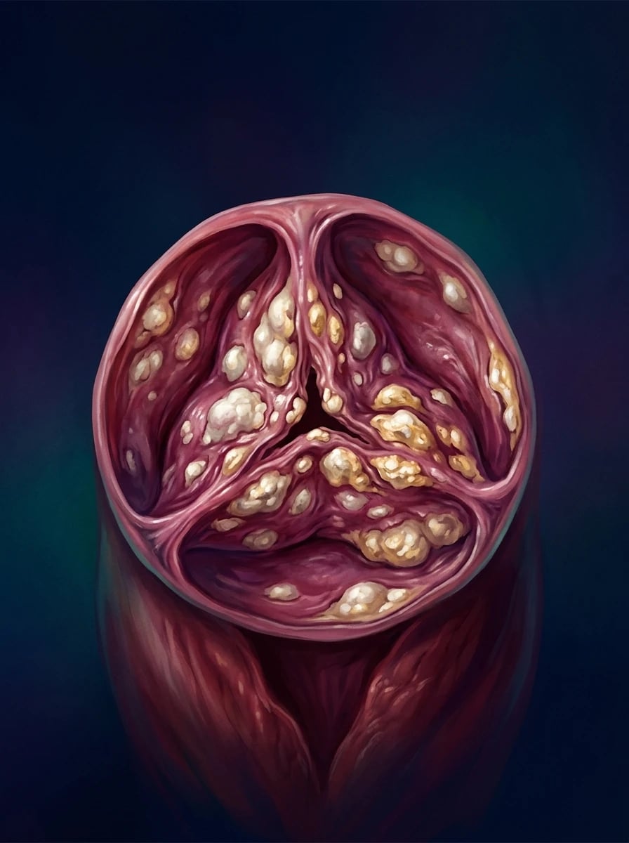
Aortic stenosis (AS) is fixed obstruction to left ventricular outflow at the level of the aortic valve, produced by thickening, fibrosis and calcification of the valve cusps with reduction of the effective aortic valve area (AVA).[1]
Two qualifications matter at the outset. First, although "aortic stenosis" usually means valvular obstruction, the LV outflow tract can be obstructed at three anatomical levels — valvular (the valve itself), subvalvular (discrete membranous ridge, tunnel, or hypertrophic cardiomyopathy) and supravalvular (above the sinuses, e.g. Williams–Beuren syndrome). Only valvular AS is discussed in detail here; the other two are considered in the differential. Second, AS is no longer regarded as passive "wear and tear" — calcific AS is an active, regulated, osteogenic inflammatory process with a biology remarkably like atherosclerosis, which is why the statin hypothesis was reasonable (and why its failure matters — see Evidence below).[8][9]
The clinical skill in AS is not naming the murmur but recognising severe disease from the bedside, stratifying symptom risk, using echocardiography to define severity (especially the low-flow low-gradient variants), and referring at the correct threshold — because AVR is the only intervention that changes survival, and the window closes once the LV decompensates.[1][2]
Classification
AS is classified by aetiology and by haemodynamic severity (the latter drives every management decision).[1]
Aetiological classification
Calcific (degenerative) AS
- Commonest cause in the developed world; over 70 years
- Tri-leaflet valve; calcification begins at base of cusp, spreads toward centre
- Active osteogenic process — valve interstitial cells → osteoblast phenotype (RUNX2, BMP-2)
- Risk factors mirror atherosclerosis: age, male sex, HTN, dyslipidaemia, smoking, diabetes, CKD
Bicuspid aortic valve (BAV)
- Prevalence 1–2% of population — commonest congenital cardiac lesion
- Presents 1–2 decades earlier (40–60 yrs)
- Associated aortopathy: ascending aortic dilatation, coarctation, dissection risk
- Ejection click often present; valve remains flexible until late
Rheumatic AS
- Predominant cause in young Indian / developing-world patients
- Commissural fusion (MCC) — medial cusps scar and fuse; mitral valve almost always also involved
- Tri-leaflet valve with thickened, tethered cusps; calcification commissural
- Declining incidence where rheumatic fever prevention is effective
Rare causes
- Radiation-induced (mantle radiotherapy, 10–20 yr latency)
- End-stage renal disease (accelerated calcification)
- Drug-induced valvulopathy (fenfluramine, ergotamines) — usually regurgitant
- Homocystinuria, Paget's disease, Fabry disease

Haemodynamic severity (the echocardiographic grades)
Severity is graded by peak aortic jet velocity (Vmax), mean transvalvular gradient and aortic valve area (AVA, calculated by the continuity equation). For patients with small body size, the indexed AVA (AVA/BSA) is preferred.[1][2]
| Grade | Peak velocity (Vmax) | Mean gradient | AVA | Indexed AVA |
|---|---|---|---|---|
| Normal | under 2.0 m/s | under 5 mmHg | 3.0–4.0 cm2 | — |
| Mild | 2.0–2.9 m/s | under 20 mmHg | over 1.5 cm2 | over 0.85 |
| Moderate | 3.0–3.9 m/s | 20–39 mmHg | 1.0–1.5 cm2 | 0.60–0.85 |
| Severe | at least 4.0 m/s | at least 40 mmHg | under 1.0 cm2 | under 0.6 |
| Very severe | at least 5.0 m/s | at least 60 mmHg | under 1.0 cm2 | under 0.6 |
The indexed AVA matters because a small patient may have an absolute AVA of 0.9 cm2 that is not truly severe once indexed to body surface area; conversely, a large patient may be severe with an AVA of 1.1 cm2 once indexed. Very severe AS (Vmax over 5 m/s) carries a high event rate and is now an interventional trigger (Class I in ESC 2021 if Vmax at least 5.5 m/s; Class 2a in ACC/AHA 2020 at Vmax at least 5 m/s).[1][2]
A separate classification — low-flow low-gradient AS — is essential because the standard thresholds misclassify it. Two subtypes are defined:
- Classical low-flow low-gradient: LVEF under 40%, AVA under 1.0 cm2, mean gradient under 40 mmHg. Confirmed as true-severe by dobutamine stress echo (DSE) or CT aortic calcium score.
- Paradoxical low-flow low-gradient: LVEF at least 50% but stroke volume index under 35 mL/m2 (restrictive, small, hypertrophied LV), AVA under 1.0 cm2, mean gradient under 40 mmHg — typically elderly women with hypertension.[2]
Epidemiology & Risk Factors
AS is the commonest native valve lesion requiring intervention in high-income countries.[2] Calcific AS has a prevalence of about 0.4% in adults overall, rising sharply with age: roughly 2–3% in those aged 75 and over and over 5% after age 85. As life expectancy rises, the burden of AS — and of TAVI — continues to grow.
[1]AS by the numbers
Risk factors for calcific AS overlap closely with atherosclerotic risk factors — age, male sex, hypertension, hyperlipidaemia (raised Lp(a) is particularly linked), smoking, diabetes, metabolic syndrome and chronic kidney disease. Patients with clinically significant AS also have higher rates of systemic atherosclerosis, and the two processes share pathobiology.[8][9]
Pathophysiology
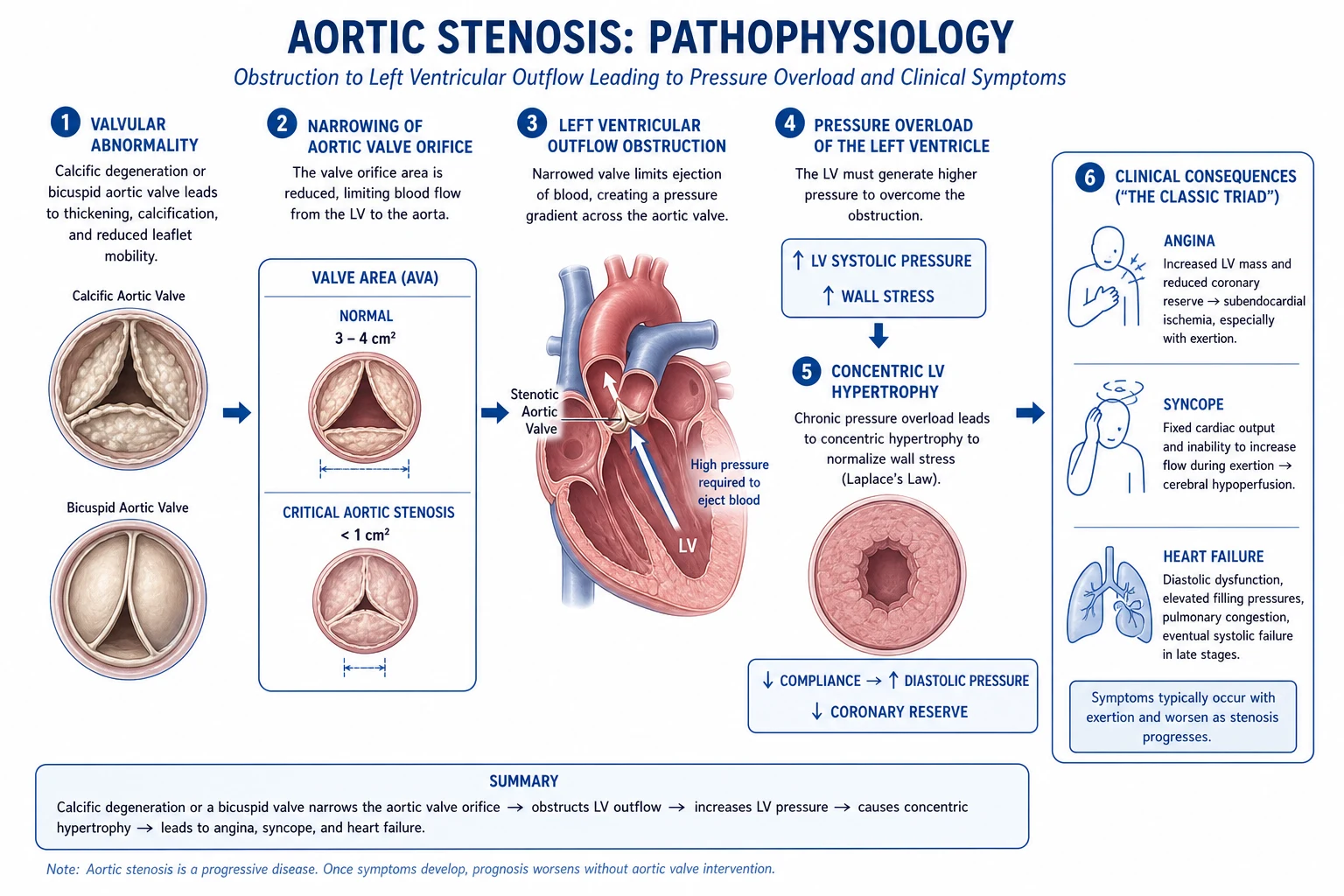
AS is a disease of mechanical obstruction superimposed on an active biological process, with haemodynamic and cellular consequences that explain every symptom. [1]
Mechanical obstruction and the LV response
Narrowing the aortic orifice raises the pressure gradient between LV and aorta. To generate a normal stroke volume across a stenotic valve, the LV must generate higher systolic pressure — i.e. chronic pressure overload. The LV compensates by undergoing concentric hypertrophy (new sarcomeres added in parallel), thickening the wall to normalise wall stress per Laplace's law (wall stress = P × r / 2h, where P is cavity pressure, r the radius, h the wall thickness). This adaptation preserves ejection fraction for years — the patient is asymptomatic even with severe AVA reduction.[1]
Why angina with normal coronaries
About half of AS patients with angina have no obstructive coronary disease. Three mechanisms converge:
- Increased demand — the hypertrophied LV has more myocardium to perfuse and generates higher systolic pressures.
- Decreased supply — the thick wall compresses intramural vessels during systole, and the low diastolic perfusion gradient (elevated LVEDP, low post-stenotic aortic diastolic pressure) reduces sub-endocardial flow.
- Reduced coronary flow reserve — even anatomically normal coronaries cannot vasodilate further on demand (reserve falls below the normal three- to four-fold). The patient develops demand ischaemia and subendocardial ischaemia, the substrate for angina, exertional dyspnoea and ventricular arrhythmia. [1]
Why syncope
Exertion causes peripheral vasodilatation; in AS the LV cannot raise cardiac output across the fixed obstruction (preload- and afterload-limited), so systemic arterial pressure and cerebral perfusion fall → exertional pre-syncope or syncope. Syncope may also be arrhythmic — ventricular tachycardia/fibrillation from ischaemic, hypertrophied myocardium, or conduction disease from calcium extension into the septum and His–Purkinje tissue. [1]
Progression to heart failure
Three stages follow one another as AVA narrows:[1]
- Compensated phase — concentric hypertrophy, normal EF, often asymptomatic.
- Diastolic dysfunction — the hypertrophied LV is stiff, LVEDP rises, atrial contribution (the S4) becomes critical; exertional dyspnoea, orthopnoea and pulmonary congestion appear.
- Systolic dysfunction — once afterload mismatch and interstitial fibrosis overwhelm the hypertrophic response, EF falls and the heart dilates; the murmur may soften as forward output drops. This is the late, decompensated phase with the worst prognosis.
Active osteogenic biology
Calcific AS is not passive wear: valve interstitial cells undergo osteoblastic differentiation under the control of RUNX2, BMP-2 and osteopontin; lipoprotein deposition, inflammation (T-lymphocytes, macrophages), angiotensin-converting enzyme and matrix vesicles/apatite produce ectopic bone-like nodules. Warfarin accelerates calcification (by inhibiting matrix Gla-protein carboxylation), explaining why some bioprosthetic valves calcify faster in anticoagulated patients.[8]
Clinical Presentation
Most patients with mild-to-moderate AS are asymptomatic, picked up on examination (murmur) or echo. Symptoms indicate severe disease and a sharply reduced survival until AVR. [1]
The classic symptom triad
The severe-AS triad and untreated survival
ASH
Median survival ~5 years untreated — often demand ischaemia with normal coronaries
Median survival ~3 years untreated — exertional, fixed cardiac output / arrhythmia
Median survival ~2 years untreated — worst of the triad; urgent AVR
Other common symptoms include exertional dyspnoea (the commonest early symptom), orthopnoea/PND (decompensating LV), fatigue and effort intolerance (low forward output), and — less commonly — palpitations from atrial fibrillation. Sudden cardiac death occurs in 1–2% of asymptomatic severe AS per year and is the rationale for surveillance and timely AVR.[1]
Bedside signs
- Pulse: slow-rising and small volume = pulsus parvus et tardus — best felt at the carotid; in critical AS the pulse is threshold (nearly imperceptible). A carotid thrill (shudder) may accompany it.
- Apex: sustained, heaving, non-displaced (concentric LVH); displaced only late when systolic dysfunction supervenes.
- Auscultation (right 2nd ICS, aortic area):
- Ejection systolic murmur — crescendo–decrescendo, harsh, radiating to the carotids (and to the apex in older patients — the Gallavardin phenomenon, a high-pitched musical component). Intensity roughly tracks severity only early — a soft murmur in late disease is ominous.
- Ejection click — high-pitched, immediately after S1, indicates a mobile (often bicuspid) valve; disappears once the valve calcifies.
- A2 soft or absent — the calcified, immobile valve can no longer snap shut; S2 becomes single or shows paradoxical (reversed) splitting (delayed A2 falls behind P2 because of prolonged LV ejection).
- S4 gallop — stiff, hypertrophied LV with a forceful atrial kick (requires sinus rhythm).
- Other: a basal ejection thrill at the right 2nd ICS indicates critical stenosis. Signs of right heart failure (raised JVP, hepatomegaly, peripheral oedema) appear late. [1]
Atypical presentations (always examined for)
- Elderly / frail: may present in pulmonary oedema or cardiogenic shock with only a soft or inaudible murmur (low-output state) — the "low-gradient" or critical AS presentation.
- Low-output state: low gradient despite critical stenosis; murmur under-graded.
- Atrial fibrillation: precipitates decompensation (loss of atrial kick).
- Heyde syndrome: AS with anaemia from gastrointestinal bleeding due to acquired von Willebrand disease type 2A — high-shear destruction of large vWF multimers causing angiodysplasia of the right colon; valve replacement typically resolves it.
- Endocarditis: fever, emboli, new murmur (bicuspid valves at higher risk). [1]
Differential Diagnosis
The bedside question is "is this ejection systolic murmur AS, or something else?". The pulse, the radiation, and the response to dynamic manoeuvres settle most cases. [1]
Aortic stenosis
- Right 2nd ICS, radiates to carotids
- Pulsus parvus et tardus (slow-rising)
- Ejection click (if mobile/bicuspid)
- Soft A2, paradoxical S2, S4
- Valsalva/standing → SOFTER; squatting → LOUDER
HOCM (hypertrophic cardiomyopathy)
- Left sternal edge, NO carotid radiation
- Jerky/bisferiens pulse
- Valsalva/standing → LOUDER; squatting → SOFTER (opposite of AS!)
- Brisk rising pulse (not slow)
- Murmur ↓ with handgrip (↑ afterload reduces obstruction)
Pulmonary stenosis
- Left 2nd ICS, radiates to left shoulder/back (NOT carotids)
- Wide, fixed split S2
- Systolic ejection click louder on expiration
- Right ventricular heave; prominent a-wave in JVP
- Normal carotid pulse
Mitral regurgitation
- Apex, pansystolic (not ejection), radiates to axilla (NOT carotids)
- Soft S1, S3 common
- Louder with handgrip (↑ afterload)
- Displaced, hyperdynamic apex
Aortic sclerosis
- Ejection systolic murmur — but Vmax under 3.0 m/s
- Preserved A2, normal pulse, no haemodynamic significance
- No LVH; no symptoms
- Distinguished only by echocardiography
Subvalvular / supravalvular AS
- Subvalvular: discrete membrane — no ejection click, murmur LSB
- Supravalvular: Williams syndrome, elfin facies, hypercalcaemia, peripheral pulmonary stenosis
- Right brachial pulse stronger than left in supravalvular AS
- Echo/localisation imaging defines the level
VSD
- Pansystolic at lower-left sternal edge
- Harsh, often with thrill
- Biventricular heave if large
- Distinguished by echo
Differentiating AS from HOCM at the bedside is a favourite viva question — they are opposite on Valsalva and squatting (the HOCM murmur gets louder where the AS murmur gets softer), the HOCM pulse is jerky/bifid rather than slow-rising, and the HOCM murmur does not radiate to the carotids.[1]
Clinical & Bedside Assessment
The examiner wants the dynamic auscultation reproduced and explained — not just stated. [1]
Dynamic manoeuvres and their physiology
| Manoeuvre | Effect on AS murmur | Why | Effect on HOCM |
|---|---|---|---|
| Valsalva (strain phase) | Softer | ↓ preload → ↓ stroke volume → ↓ turbulent jet | Louder (less LV volume → more obstruction) |
| Standing up | Softer | ↓ venous return (preload) | Louder |
| Squatting | Louder | ↑ venous return (preload) AND ↑ afterload → ↑ stroke volume | Softer (more LV volume reduces dynamic obstruction) |
| Handgrip | No change or softer | ↑ afterload; no effect on fixed AS but ↓ gradient in some | Softer (↑ afterload ↑ LV volume, less obstruction); makes MR/AR louder |
Key exam point: squatting and Valsalva move AS and HOCM in opposite directions. The pulse in AS is slow-rising (parvus et tardus); in HOCM it is jerky/bisferiens. [1]
Carotid assessment
Palpate the carotid with the pads of the fingers (not the thumb — your own pulse). In severe AS the upstroke is delayed, weak and sustained — "parvus et tardus". A palpable carotid thrill (shudder) supports critical stenosis. Compare with the brachial/radial: in supravalvular AS the right arm pulse is stronger than the left (the Coanda effect directs the high-velocity jet preferentially to the innominate). [1]
Ejection click and S2
- Ejection click: high-pitched, just after S1, at the apex/left sternal edge. Indicates a mobile, often bicuspid valve. It disappears as the valve calcifies, so its absence in calcific AS is expected; its presence should prompt a search for bicuspid morphology.
- S2: in severe AS, A2 is soft or absent (immobile valve). If LV ejection is markedly prolonged, A2 is delayed and falls after P2, producing paradoxical (reversed) splitting of S2 — best heard at the left 2nd/3rd ICS, with the two components moving further apart in expiration.
- S4: low-pitched presystolic sound at the apex — the stiff, hypertrophied LV receiving its atrial kick. Implies a stiff ventricle; requires sinus rhythm. [1]
Investigations
Echocardiography — the definitive test
Transthoracic echocardiography (TTE) is first-line and usually sufficient. It defines:[1][2]
- Valve morphology — bicuspid vs tri-leaflet vs rheumatic; degree and distribution of calcification.
- Peak aortic jet velocity (Vmax) — by continuous-wave Doppler, aligned to the jet.
- Mean and peak transvalvular gradients.
- Aortic valve area (AVA) by the continuity equation: AVA = (CSA_LVOT × VTI_LVOT) / VTI_aortic, where CSA is cross-sectional area and VTI is velocity–time integral. The continuity equation is examined in nearly every AS viva.
- LV function — EF, wall thickness, mass, diastolic function (E/A, E/e'); stroke volume and stroke-volume index (essential for low-flow low-gradient AS).
- Associated lesions — aortic regurgitation, mitral disease; aortic root/ascending aorta dimensions (especially in BAV).
Dobutamine stress echocardiography — for low-flow low-gradient AS
When AVA is small but the gradient is low (under 40 mmHg) and EF is reduced, the question is whether the stenosis is true-severe (the valve really is critically narrow) or pseudo-severe (low flow makes a moderate valve look severe). Dobutamine stress echo increases contractility and flow:[2]
- True-severe AS: gradient rises with flow to at least 40 mmHg, AVA stays under 1.0 cm2.
- Pseudo-severe AS: AVA enlarges with flow to over 1.0 cm2; the valve was not truly critical.
- Absence of contractile reserve (stroke volume rise under 20%) carries a poor prognosis and high peri-procedural risk.
CT aortic valve calcium scoring (sex-specific thresholds: over 3000 AU in men, over 1600 AU in women) is an alternative confirmation, especially in paradoxical low-flow low-gradient AS with normal EF. [1]
ECG
- LV hypertrophy (Sokolow–Lyon: SV1 + RV5/V6 at least 35 mm; or Cornell criteria) with strain pattern (lateral ST depression and T-wave inversion).
- Left atrial abnormality (bifid P in V1).
- Conduction abnormalities — first-degree AV block, LBBB, AF.
- In advanced disease, secondary repolarisation changes. [1]
Chest X-ray
Often normal early. Look for cardiomegaly, signs of pulmonary venous congestion/oedema, calcification of the aortic valve (best on a lateral or heavily penetrated film), post-stenotic dilatation of the ascending aorta, and (in BAV) an enlarged aortic knuckle. [1]
CT coronary angiography / invasive coronary angiography
Pre-operative coronary angiography is mandatory before AVR in patients over 40, those with risk factors, or any suspicion of ischaemia — because concomitant CABG at the time of SAVR improves outcomes in significant CAD. ECG-gated cardiac CT is essential before TAVI — for annular sizing (which determines prosthesis size and risk of paravalvular leak or annular injury), iliofemoral access assessment, and calcium burden/annular calcium distribution (predicts conduction injury and paravalvular leak).[2]
Exercise testing
Exercise testing is CONTRAINDICATED in symptomatic severe AS (risk of syncope, ventricular arrhythmia, death). It has a limited, specialist role in asymptomatic severe AS — to unmask symptoms, an abnormal blood-pressure response (drop, or failure to rise over 20 mmHg), or ST changes that would re-classify the patient as symptomatic (and therefore AVR-indicated).[1]
Biomarkers
BNP / NT-proBNP is increasingly used in apparently asymptomatic AS: a markedly raised level (more than three-fold the upper limit of normal) predicts symptom development and is an ESC Class 2a trigger for AVR, on the basis that "asymptomatic" patients with high BNP are probably exercising less and hiding symptoms. [1]
[1]Management — Resuscitation
A patient with severe AS who presents in acute pulmonary oedema or cardiogenic shock is a high-risk emergency — the fixed outflow obstruction means the LV is exquisitely preload- and afterload-sensitive.[1]
Immediate measures (ABCDE):
- Sit upright, high-flow oxygen to target SpO2 94–98% (88–92% if chronic CO2 retainer); CPAP/NIV reduces work of breathing and preload.
- Cautious IV loop diuretic — furosemide 20–40 mg IV (lower than usual; over-diuresis collapses the underfilled, hypertrophied LV and drops output catastrophically).
- IV access, monitoring, bloods (including troponin, BNP, lactate, cultures if sepsis suspected).
- Treat precipitants: AF (rate-control cautiously — digoxin preferred over negative-inotrope beta-blockers; cardioversion if unstable), ischaemia, anaemia, infection, arrhythmia. [1]
AVOID (these are classic traps):
- Vasodilators — nitrates, ACE-inhibitors, hydralazine: drop afterload, collapse the LV output, profound hypotension.
- Aggressive diuresis — empty the stiff LV, drop preload, drop output.
- Negative inotropes — beta-blockers and non-dihydropyridine calcium-channel-blockers (verapamil/diltiazem) depress the compensation that is keeping the LV ejecting. [1]
Refractory / cardiogenic shock:
- Cautious inotrope — dobutamine 2.5–5 µg/kg/min IV (β1 agonist) to augment contractility and forward output (and may raise gradient).
- Mechanical support — intra-aortic balloon pump (IABP) if necessary (with caution — raises afterload that the LV must pump against); VA-ECMO in extremis as a bridge.
- Balloon aortic valvuloplasty (BAV) — percutaneous balloon inflation splits fused commissures and acutely lowers the gradient; used as a bridge to definitive AVR/TAVI in the critically ill, or as palliation in those not fit for AVR. It is NOT definitive therapy — restenosis occurs within ~6 months. [1]
Definitive AVR should be planned during the index admission once stabilised — survival without it is poor.[1]
Management — Definitive & Stepwise
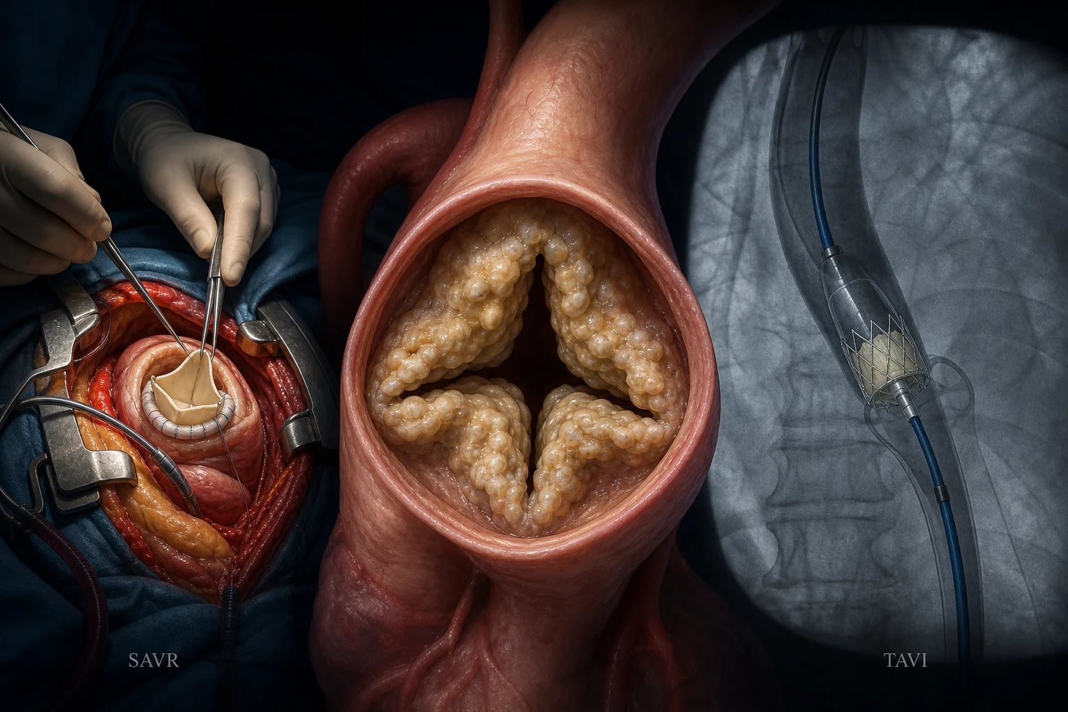
Aortic valve replacement (AVR) is the ONLY therapy proven to improve survival in severe AS. No drug halts or reverses the valve process. Two routes — surgical AVR (SAVR) and transcatheter AVR (TAVI/TAVR) — are now available across the entire risk spectrum.[1][2]
Class I indications for AVR (2020 ACC/AHA)
AVR is indicated in any of:[1]
- Symptomatic severe AS (any of the triad — angina, syncope, heart failure, or symptomatic exertional BP drop).
- Asymptomatic severe AS with LVEF under 50%.
- Severe AS when other cardiac surgery is planned (CABG, mitral surgery, ascending aorta repair).
- Asymptomatic severe AS undergoing TAVR/SAVR (when low surgical risk) — Class 2a in 2020 ACC/AHA; Class I in 2021 ESC if Vmax at least 5.5 m/s.
Class 2a indications in asymptomatic severe AS
- Vmax 4.0–4.9 m/s with rapid progression (increase over 0.3 m/s/yr).
- Very severe AS (Vmax at least 5.0 m/s) — 2020 ACC/AHA 2a; ESC 2021 Class I if at least 5.5 m/s.
- Abnormal exercise test — symptoms or BP drop/failure to rise over 20 mmHg on exercise.
- Markedly raised BNP (more than three-fold ULN), confirmed on repeat, with no other cause.
- Severe AS with rapid mean-gradient progression at least 5 mmHg/yr. [1]
Choice of SAVR vs TAVI (heart-team decision)
The 2020 ACC/AHA guideline uses age cut-points:[1]
| Age / profile | Preferred approach |
|---|---|
| Under 65 yrs (life expectancy over 20 yrs) | SAVR preferred |
| 65–80 yrs | Either SAVR or TAVI — heart-team decision based on anatomy, comorbidity, life expectancy |
| Over 80 yrs (life expectancy under 10 yrs) | TAVI preferred |
The 2021 ESC/EACTS guideline uses a single cut-point — under 75 yrs → SAVR; over 75 yrs or high surgical risk/inoperable → TAVI — with the heart team arbitrating the 70–80 band.[2]
SAVR is favoured when: young patient; bicuspid anatomy with unfavourable TAVI landing zone; need for concomitant cardiac surgery (CABG, mitral repair, ascending aorta replacement); unfavourile vascular access (tortuous, calcified iliofemoral); annular or LVOT calcium that raises TAVI risk of rupture or paravalvular leak; infective endocarditis. [1]
TAVI is favoured when: elderly/frail; high or prohibitive surgical risk (STS/ EuroSCORE); hostile chest (previous thoracotomy, radiation); porcelain aorta; favourable iliofemoral access and annular anatomy; poor surgical candidate for extracorporeal circulation. [1]
Prosthetic choice (mechanical vs bioprosthetic for SAVR): mechanical valves in patients under 50 (or those needing anticoagulation anyway); bioprosthetic over 50, in women planning pregnancy, or where anticoagulation is impractical. The ACURATE / SURTAVI / PARTNER trials confirmed TAVI non-inferiority across risk strata. [1]
Medical therapy (supportive only)
- No drug halts calcific AS. The SEAS trial (ezetimibe + simvastatin, NEJM 2008) and ASTRONOMER trial (rosuvastatin, Circulation 2010) both failed to show any reduction in progression or clinical events, despite the atherosclerosis-like biology.[8][9]
- Treat comorbidities carefully: hypertension (ACE-i cautiously, in low doses, monitoring for hypotension); heart failure (diuretics; avoid over-diuresis); atrial fibrillation (rate-control with digoxin rather than beta-blockers if critical AS).
- Endocarditis prophylaxis is no longer routinely advised for unrepaired native valves, but is reasonable in high-risk subsets (previous endocarditis, prosthetic valve) before dental procedures.
- Surveillance — see below.
Balloon aortic valvuloplasty (BAV)
Not definitive. Restenosis in ~6 months; used as a bridge to AVR in the critically ill, in pregnancy, or as palliation in patients not fit for AVR. In young patients with non-calcified bicuspid AS and no calcification it may have a role, but AVR remains definitive.[1]
Specific Subtypes & Scenarios
Low-flow low-gradient AS
Two subtypes — both frequently missed by standard thresholds:[2]
| Feature | Classical LF-LG | Paradoxical LF-LG |
|---|---|---|
| LVEF | Under 40% | At least 50% |
| Stroke volume index | Low (under 35 mL/m2) | Low (under 35 mL/m2) |
| AVA | Under 1.0 cm2 | Under 1.0 cm2 |
| Mean gradient | Under 40 mmHg | Under 40 mmHg |
| LV pattern | Dilated, thin-walled (systolic dysfunction) | Small, thick, restrictive (diastolic dysfunction) |
| Typical patient | Male, prior MI, ischaemic cardiomyopathy | Elderly woman, hypertension |
| Confirmation | Dobutamine stress echo (contractile reserve) | CT calcium score, DSE, echo of diastology |
| Prognosis | Poor without AVR; high procedural risk | Often good with TAVI; high if untreated |
Confirming true severity is the point: a small AVA with low gradient could be pseudo-severe (a moderate valve made to look tight by low flow) or truly severe. DSE and CT calcium score distinguish these and decide who is referred for AVR. [1]
Bicuspid aortic valve disease
BAV affects 1–2% of the population and is the commonest congenital cardiac lesion. The valve has two cusps (with a raphe marking the fused commissure), predisposes to early AS and/or AR, and is associated with an aortopathy affecting the ascending aorta and root (risk of dilatation, dissection, rupture). Coarctation of the aorta coexists in 5–10% (and vice versa — every coarctation patient needs a BAV screen). Surveillance of the ascending aorta is mandatory: annual echocardiography if over 4.0 cm, with CT/MRI when not well seen on echo; surgical replacement at 5.5 cm (5.0 cm with risk factors or planned SAVR), and concomitant aortic repair at SAVR if over 4.5 cm. [1]
Asymptomatic severe AS
Most are safe in the short term (event rate about 1% per year), but risk rises with Vmax (event rate ~30% by 2 years if Vmax over 5 m/s), very severe AS, BNP elevation, LVEF decline, and rapid progression. Surveillance TTE every 6–12 months for severe AS (every 3–5 years for moderate, every 3–5 years for mild). Patient education is critical: report any new symptom (especially exertional dyspnoea, chest pain, pre-syncope) immediately — symptoms re-classify the patient as AVR-indicated. [1]
Rheumatic AS
Defined by commissural fusion (especially the medial commissures producing a fish-mouth orifice), almost always with co-existing mitral disease (mitral stenosis or regurgitation). Predominant in young patients in India and other rheumatic-fever-endemic regions. The ejection click is usually absent (calcified), and the murmur may be softer. Management is identical (AVR when severe and symptomatic), often combined with mitral surgery and BMV if needed. [1]
Complications & Pitfalls
Complications of untreated severe AS
- Heart failure — diastolic then systolic; the leading cause of death.
- Sudden cardiac death — typically once symptomatic; rare (under 1%/yr) when truly asymptomatic.
- Atrial fibrillation — precipitates decompensation by loss of atrial kick; raises stroke risk.
- Infective endocarditis — particularly bicuspid valves.
- Conduction disease — calcium extends into the septum and AV node (heart block).
- GI bleeding (Heyde syndrome) — acquired vWF deficiency from high shear. [1]
Procedural complications
TAVI:
- Conduction disturbance — new left bundle branch block in up to 30–50%; permanent pacemaker in roughly 5–10% with balloon-expandable (SAPIEN) valves and 15–20% with self-expanding (CoreValve/Evolut) valves — the latter sits deeper in the LVOT, closer to the conduction tissue.[6]
- Paravalvular leak — more common than after SAVR; even mild–moderate leak worsens survival, hence the importance of CT annular sizing.
- Stroke — peri-procedural (embolic protection devices are now used).
- Vascular access complications — iliofemoral dissection, bleeding.
- Coronary obstruction — rare but catastrophic (especially when the native leaflet occludes the ostium, more likely with a low coronary ostium and a sinus of Valsalva).
- Prosthetic valve deterioration — over many years; bioprosthetic valves (TAVI and SAVR) calcify, especially in younger patients.
SAVR:
- Standard operative risks — stroke (~1–2%), bleeding, infection, AF (~30%), renal injury.
- Paravalvular leak, prosthetic endocarditis, structural valve deterioration (bioprosthetic, 10–15 years).
- Anticoagulation (mechanical prostheses) — lifelong warfarin with INR target 2.5 (range 2.0–3.0; higher with mitral-position or older-generation valves). [1]
Pitfalls
- Softening of the murmur is NOT improvement — it is a late, dangerous sign of worsening LV function (reduced gradient because the failing LV can no longer generate the gradient across the stenotic valve). Same for a "less impressive" gradient on echo — check EF and stroke volume.
- Treating AS as heart failure with vasodilators/ACE-i in the emergency department — can precipitate collapse.
- Missed bicuspid aortopathy — the valve may be addressed and the ascending aorta forgotten, leading to later dissection.
- Failure to confirm true severity in low-flow low-gradient AS — pseudo-severe AS does not need AVR.
- Delaying AVR for asymptomatic patients who "feel well" once symptoms develop — survival then drops sharply. [1]
Prognosis & Disposition
The central prognostic fact in AS is the symptom threshold.[1]
- Asymptomatic severe AS: event rate (death or AVR) about 1% per year, normal life expectancy in the short term — but risk rises with Vmax (very severe AS event rates reach 30% by 2 years), LVEF decline, BNP elevation, and rapid progression.
- Symptomatic severe AS: median survival after the classic symptoms, untreated — angina ~5 years, syncope ~3 years, heart failure ~2 years; overall 2-year mortality from symptom onset roughly 50%.
- After successful AVR: symptoms improve dramatically; survival approaches that of the age-matched population (about 85–90% 5-year survival post-TAVI/SAVR), provided EF recovers and there is no significant paravalvular leak.
- Discharge and follow-up: post-AVR patients need lifelong surveillance — echo at discharge, 6 months, then annually; prosthetic-specific considerations (anticoagulation for mechanical, endocarditis prophylaxis). Asymptomatic severe AS in the community needs 6-monthly echo with symptom review. [1]
The take-home rule: once severe AS is symptomatic, AVR should not be delayed — there is no plateau and no medical alternative. [1]
Special Populations
Pregnancy
Pregnancy imposes a 30–50% rise in blood volume and cardiac output and a fall in afterload — poorly tolerated by a fixed LV outflow obstruction. Asymptomatic mild-to-moderate AS usually tolerates pregnancy with careful monitoring. Symptomatic severe AS carries a significant risk of pulmonary oedema, syncope and maternal death. Management:[1][2]
- Pre-conception counselling is ideal: AVR before pregnancy if severe AS, or BAV as a bridge in symptomatic severe AS to allow pregnancy.
- During pregnancy: avoid volume depletion (epidural preferred over general anaesthesia for delivery; avoid supine hypotension; left lateral position), beta-blockade cautiously for rate, monitor in a joint obstetric–cardiology clinic, deliver in a cardiac centre.
- Severe symptomatic AS in pregnancy: BAV in the second trimester as a bridge; TAVI/SAVR reserved for refractory cases (high foetal radiation risk with TAVI).
Elderly / frail
Frailty is now a formal part of the heart-team assessment (e.g. Rockwood Clinical Frailty Scale, gait speed, grip strength). TAVI is the preferred route in the over-80s, the frail, and those with hostile chests or prohibitive surgical risk, on the basis of the PARTNER and Evolut low-risk and intermediate-risk data.[3][4][5][6][7]
End-stage renal disease (ESD/CKD)
ESD accelerates valve calcification (calcific AS is markedly more prevalent and progresses faster in dialysis patients), raises surgical risk (high mortality, peri-procedural complications), and biases choice toward TAVI in suitable anatomy. Outcomes remain worse than in non-renal patients. [1]
Bicuspid valve
See Specific Subtypes — the ascending aorta needs lifelong surveillance (annual echo/CT/MRI from 4.0 cm), and concomitant aortic repair or replacement is performed at SAVR when the aorta exceeds 4.5 cm. [1]
Evidence, Guidelines & Regional Differences
The landmark TAVI trials — across the risk spectrum
The evidence base for TAVI is one of the most rapidly assembled in cardiology. The major trials established non-inferiority (and often superiority) of TAVI to SAVR across every risk stratum: [1]
Statin failure — SEAS and ASTRONOMER
Despite the atherosclerosis-like biology, lipid-lowering does not halt AS:
- SEAS (Rossebø, NEJM 2008) — simvastatin + ezetimibe in mild-to-moderate AS — no effect on AS progression, AVR need, or cardiovascular events (and a small cancer signal that was not reproduced).[8]
- ASTRONOMER (Chan, Circulation 2010) — rosuvastatin in asymptomatic mild-to-moderate AS — no slowing of haemodynamic progression.[9]
The lesson: the biology resembles but is not identical to atherosclerosis — once calcification is established, the osteogenic process is driven by mechanisms (RUNX2, BMP-2, valvular calcification pathways) that statins do not modify. Investigation now targets these specific pathways (e.g. denosumab, warfarin alternatives). [1]
Regional guideline differences
2020 ACC/AHA (US)
- Under 65 → SAVR; over 80 → TAVI; 65–80 → either (heart team)
- Class I AVR for symptomatic severe AS; EF under 50%
- Very severe AS (Vmax at least 5 m/s): Class 2a
- STS-PROM for surgical risk
2021 ESC/EACTS (Europe)
- Under 75 → SAVR; over 75 or high-risk → TAVI
- Class I AVR if Vmax at least 5.5 m/s in asymptomatic
- Class 2a for BNP over 3x ULN, rapid progression
- EuroSCORE II for surgical risk
Exam Pearls
- Severe AS one-line trio: Vmax at least 4.0 m/s OR mean gradient at least 40 mmHg OR AVA under 1.0 cm2 (indexed under 0.6 cm2/m2).
- Single survival-modifying therapy: AVR — no drug halts progression.
- Symptom triad and survival: angina 5, syncope 3, heart failure 2 years untreated.
- Valsalva/standing makes AS SOFTER but HOCM LOUDER (opposite!) — the favourite bedside discriminator. Squatting reverses both.
- Pulsus parvus et tardus (slow-rising small pulse) + ejection systolic murmur radiating to the carotids = AS at the bedside.
- Ejection click = bicuspid / mobile valve; disappears when calcified.
- S4 = stiff hypertrophied LV (needs sinus rhythm).
- Angina with normal coronaries in AS = demand ischaemia + reduced coronary flow reserve.
- Heyde syndrome: AS + GI bleed from angiodysplasia (acquired vWF type 2A from high shear) — resolves after AVR.
- Paradoxical (reversed) S2 splitting = delayed A2 from prolonged LV ejection.
- Low-flow low-gradient AS: classical (EF under 40) vs paradoxical (EF at least 50, SVI under 35 mL/m2) — confirm with dobutamine stress echo or CT calcium score.
- Post-TAVI pacemaker: balloon-expandable (SAPIEN) ~5–10%; self-expanding (CoreValve/Evolut) ~15–20% — new LBBB or high-grade AV block are the commonest reasons.
- Softening murmur = worsening AS (failing LV can no longer generate the gradient) — never interpret as improvement.
- Bicuspid valve = screen the ascending aorta and check for coarctation (and vice versa).
- Exercise testing is contraindicated in symptomatic severe AS. [1]
Exam application bank (NEET-PG / INICET)
One-line answer
Aortic stenosis (AS) is obstruction of left ventricular outflow at the valve level, caused most often by calcific degeneration (elderly) or a bicuspid valve (younger); rheumatic disease predominates in young Indian patients. Severe AS produces the classic symptom triad of angina, syncope and heart failure, a slow-rising small-volume pulse (pulsus parvus et tardus) and a crescendo-decrescendo ejection systolic murmur radiating to the carotids. Diagnosis rests on echocardiography with the continuity equation. Aortic valve replacement (AVR) is the only survival-modifying therapy; choice of surgical AVR (SAVR) versus transcatheter AVR (TAVI/TAVR) is driven by age, surgical risk and anatomy.
Worked stems (answer without another resource)
Stem 1 — Classic presentation. Map symptoms to mechanism; name the first investigation and first treatment step with dose/route if drug therapy is standard. [1]
Stem 2 — Unstable / complicated. List red flags that force immediate resuscitation, theatre, ICU, antidote, or reperfusion — and what you do in the first 15 minutes. [1]
Stem 3 — Atypical group. Elderly, pregnancy, child, or immunocompromised: how presentation and thresholds change. [1]
Stem 4 — Differential trap. Name the three closest mimics and one discriminator for each. [1]
Stem 5 — Disposition. Who goes home with safety-netting, who is admitted, who needs HDU/ICU/theatre, and what follow-up is mandatory. [1]
Rapid viva checklist
- Definition + classification
- Pathophysiology chain
- Bedside signs / criteria
- Score with exact components (if any)
- Emergency bundle
- Definitive therapy with doses
- Complications of disease and of treatment
- Special populations
- Guideline/trial name if classic
- Three exam traps
Coverage self-check
If you cannot answer any stem above from this page alone, re-read the matching section — the page is intended to be self-sufficient for final-prof and NEET-PG/INICET questions on Aortic Stenosis.
[1] [1]References
- [1]Otto CM, Nishimura RA, Bonow RO, et al. 2020 ACC/AHA Guideline for the Management of Patients With Valvular Heart Disease: A Report of the American College of Cardiology/American Heart Association Joint Committee on Clinical Practice Guidelines Circulation, 2021.PMID 33332150
- [2]Vahanian A, Beyersdorf F, Praz F, et al. 2021 ESC/EACTS Guidelines for the management of valvular heart disease Eur Heart J, 2022.PMID 34453165
- [3]Smith CR, Leon MB, Mack MJ, et al. Transcatheter versus surgical aortic-valve replacement in high-risk patients N Engl J Med, 2011.PMID 21639811
- [4]Leon MB, Smith CR, Mack MJ, et al. Transcatheter aortic valve replacement versus surgical valve replacement in intermediate-risk patients: a propensity score analysis Lancet, 2016.PMID 27053442
- [5]Reardon MJ, van Mieghem NM, Popma JJ, et al. Surgical or Transcatheter Aortic-Valve Replacement in Intermediate-Risk Patients N Engl J Med, 2017.PMID 28304219
- [6]Popma JJ, Deeb GM, Yakubov SJ, et al. Transcatheter Aortic-Valve Replacement with a Self-Expanding Valve in Low-Risk Patients N Engl J Med, 2019.PMID 30883053
- [7]Mack MJ, Leon MB, Thourani VH, et al. Five-Year Outcomes of Transcatheter or Surgical Aortic-Valve Replacement N Engl J Med, 2020.PMID 31995682
- [8]Rossebo AB, Pedersen TR, Boman K, et al. Intensive lipid lowering with simvastatin and ezetimibe in aortic stenosis N Engl J Med, 2008.PMID 18765433
- [9]Chan KL, Teo K, Dumesnil JG, et al. Effect of Lipid lowering with rosuvastatin on progression of aortic stenosis: results of the aortic stenosis progression observation: measuring effects of rosuvastatin (ASTRONOMER) trial Circulation, 2010.PMID 20048204