cardiology · cardiology
Arrhythmogenic Cardiomyopathy
Also known as Arrhythmogenic right ventricular cardiomyopathy · ARVC · Arrhythmogenic right ventricular dysplasia · ARVD · Arrhythmogenic cardiomyopathy · ACM
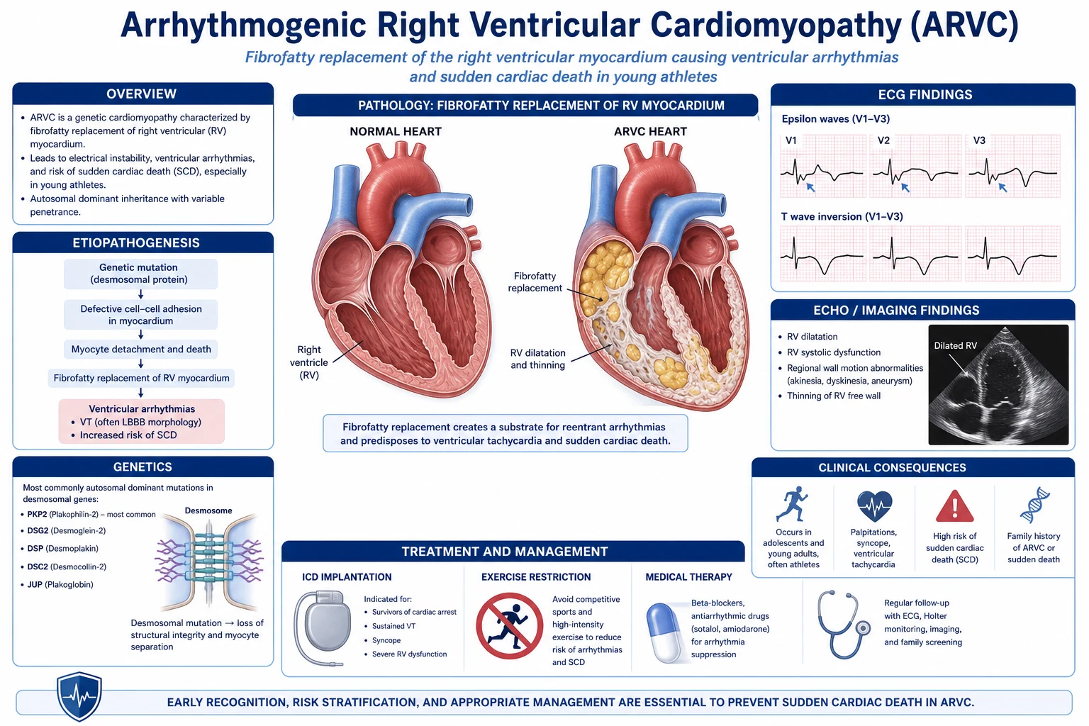
Arrhythmogenic cardiomyopathy (ACM) is an inherited heart-muscle disease in which progressive fibro-fatty replacement of ventricular myocardium (classically the right ventricle) produces ventricular arrhythmias, heart failure and sudden cardiac death (SCD) in apparently healthy young people — most notably competitive athletes. Inheritance is usually autosomal dominant and the genes are predominantly desmosomal (PKP2 plakophilin-2, DSP desmoplakin, DSG2 desmoglein-2, DSC2 desmocollin-2, JUP plakoglobin); non-desmosomal genes include TMEM43, LMNA, PLN, DES, CDH2, SCN5A, CTNNA3. The ECG hallmark is the epsilon wave (a low-amplitude deflection at the end of the QRS in V1-V3) with T-wave inversion V1-V3 (without RBBB); cardiac MRI shows RV dilatation, regional wall-motion abnormalities and late gadolinium enhancement. Diagnosis uses the 2010 Modified Task Force Criteria (six categories — imaging, biopsy, repolarisation, depolarisation, arrhythmia, family history) refined by the 2020 Padua criteria for CMR quantification. Management combines lifestyle restriction (no competitive or endurance sport), beta-blockade (sotalol, metoprolol, bisoprolol), ICD for the high-risk (sustained VT, aborted SCD, severe RV/LV dysfunction, unexplained syncope), catheter ablation of drug-refractory VT, and heart failure therapy / transplantation for the burnt-out phase.
On this page & tools
Your progress
Saved locally on this device.
Exam tags
Red flags
Overview & Definition
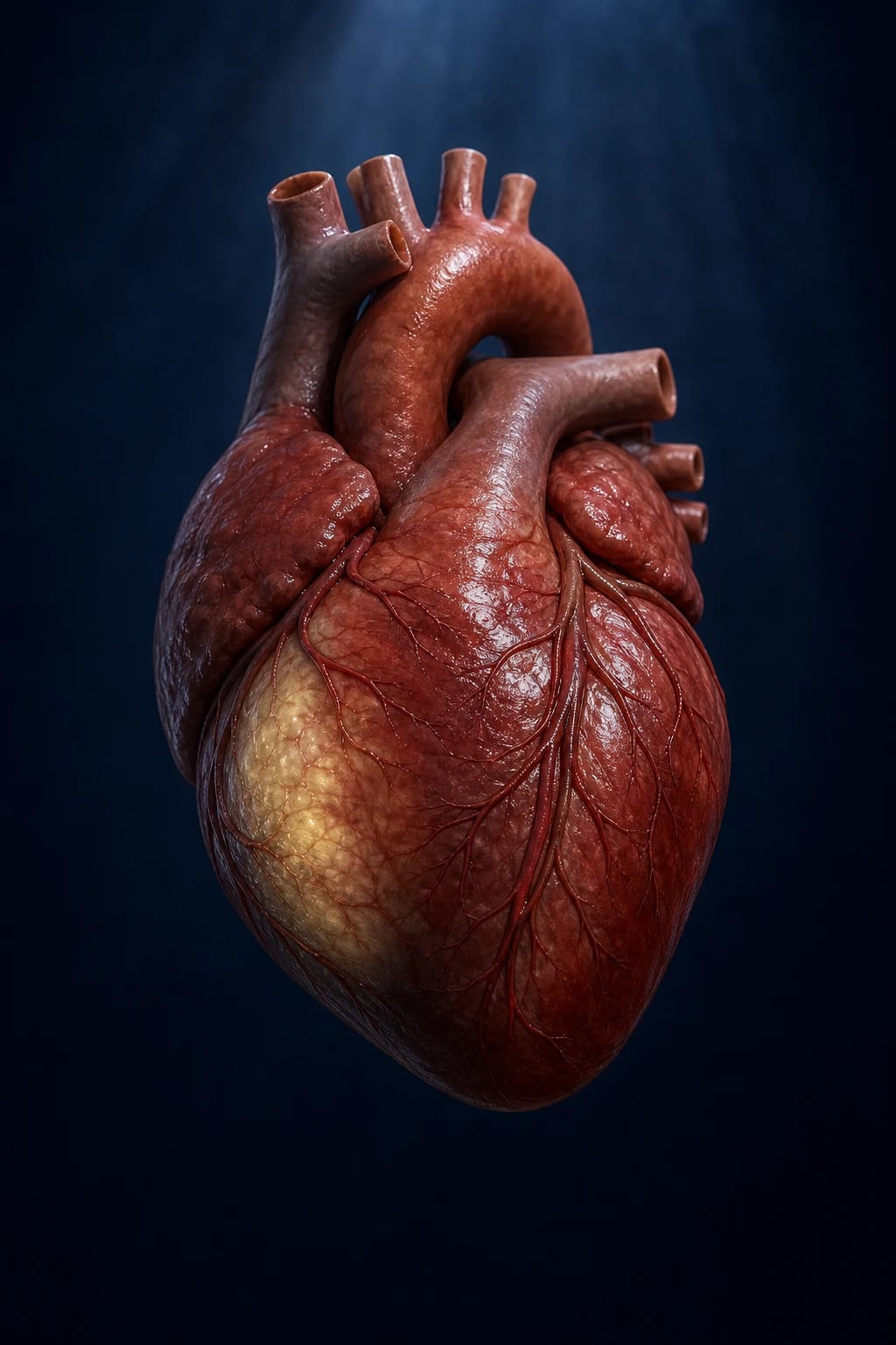
Arrhythmogenic cardiomyopathy (ACM) — historically called arrhythmogenic right ventricular cardiomyopathy/dysplasia (ARVC/ARVD) — is an inherited heart-muscle disease characterised by progressive replacement of ventricular myocardium by fibro-fatty and fibrous tissue, predominantly affecting the right ventricle but increasingly recognised in left-dominant and biventricular forms.[1][5]
The disease is defined by four pillars: (i) a genetic substrate (predominantly desmosomal mutations, autosomal dominant); (ii) a structural phenotype of fibro-fatty substitution beginning at the RV outflow tract, inflow and apex (the "triangle of dysplasia"); (iii) an electrical phenotype of slow conduction producing epsilon waves, late potentials, and re-entrant ventricular arrhythmias; and (iv) a clinical phenotype of palpitations, syncope, and sudden cardiac death that is exertion-related in the young.[1][8]
In 1994, the original European Society of Cardiology / International Society and Federation of Cardiology Task Force (McKenna, Thiene) defined diagnostic criteria; the criteria were quantitatively revised in 2010 (Marcus) and further refined for cardiac MRI in 2020 (the Padua criteria, Corrado/Bauce). The current umbrella term "arrhythmogenic cardiomyopathy" (replacing ARVC) reflects that the left ventricle is involved in over half of cases.[3][5]
[1]Classification
ACM is classified along three axes — by ventricular phenotype, by genotype, and by clinical phase: [1]
Classic RV-dominant (ARVC)
- Fibro-fatty replacement of RV myocardium; 'triangle of dysplasia' (RV inflow, outflow, apex)
- ECG: epsilon wave, TWI V1-V3 (no RBBB); VT with LBBB + superior axis
- Most common phenotype; highest yield on Padua CMR criteria; PKP2 in 40-50%
Left-dominant ACM
- Fibro-fatty / scar replacement of LV myocardium (subepicardial/mid-wall lateral wall)
- ECG: TWI in lateral leads (V4-V6, I, aVL) or inferolateral; absent/minimal epsilon wave
- Under-recognised (Sen-Chowdhry 2008); commonly DSP, LMNA, PLN, DSG2 mutations
Biventricular ACM
- Both RV and LV scarred; overlap with dilated cardiomyopathy phenotype in burnt-out phase
- Right and left heart failure; high arrhythmic burden
- Often late-stage classic ARVC, or PLN / TMEM43 founder variants
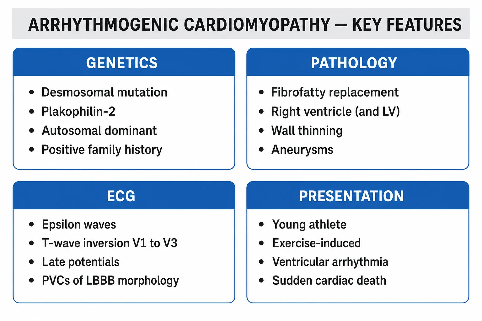
By genotype (the exam-relevant gene list): [1]
- Desmosomal genes (~50-60% of index cases):
- PKP2 (plakophilin-2) — the single commonest (~40% of familial ARVC; truncating variants).
- DSP (desmoplakin) — left-dominant and biventricular forms, carvajal syndrome (skin + woolly hair + ACM), familial exercise-triggered myocarditis.
- DSG2 (desmoglein-2) and DSC2 (desmocollin-2) — ~5-10% each.
- JUP (junction plakoglobin / gamma-catenin) — Naxos disease (recessive; woolly hair, palmoplantar keratoderma, ARVC).[8]
- Non-desmosomal genes: TMEM43 (Newfoundland founder, lethal without ICD), LMNA (overlap with DCM and conduction disease — high SCD risk), PLN (p.Arg14del; Netherlands/Spain founder; biventricular with mid-wall LV LGE), DES (desmin), CDH2 (N-cadherin), CTNNA3 (alpha-T-catenin), SCN5A (overlap with Brugada), TTN, RBM20.[9][10]
By clinical phase (the four-stage natural history of Marcus/Basso): [1]
- Concealed (asymptomatic) phase — silent structural change; the patient may still present with SCD as the first symptom.
- Overt electrical (arrhythmic) phase — palpitations, syncope, NSVT/sustained VT, often exertion-triggered.
- Right-ventricular dysfunction phase — RV failure with raised JVP, peripheral oedema, hepatomegaly.
- Biventricular ("burnt-out") phase — overlaps with dilated cardiomyopathy; both RV and LV failure, refractory arrhythmias. [1]
ACM — high-yield numbers
Epidemiology & Risk Factors
ACM has an estimated population prevalence of 1 in 2,000-5,000 and accounts for ~5% of all SCD in the under-35 age group in the United States, rising to over 20% of athletic SCD in the Veneto region of Italy (a founder-effect / high-penetrance region).[4][6]
Demographic and clinical risk factors (high-yield): [1]
- Young age — clinical presentation typically 20-40 yr; SCD peak in adolescents and young adults.
- Male sex — 3:1 male predominance in overt disease (partly biological, partly because males participate more often in competitive endurance sport). Note: women are equally genetically affected but have lower penetrance and later presentation.
- Competitive athletic status — endurance sport (marathon, cycling, rowing, swimming) multiplies arrhythmic risk 5-fold by loading the RV and triggering recurrent myocyte detachment.[6]
- Family history of SCD under 50 yr, of cardiomyopathy, or of a known desmosomal mutation.
- Mediterranean / founder ancestry — Italy (Veneto), Greece (Naxos island), the Netherlands (PLN p.Arg14del), Newfoundland (TMEM43), South Africa.[4]
- Homozygous / compound-heterozygous / digenic desmosomal mutations — earlier onset, more severe phenotype, higher SCD risk.[9]
Genotype-specific penetrance and SCD risk (high-yield): [1]
- PKP2 truncating variants — penetrance ~ 60-80%, classical RV phenotype, ICD benefit clear for sustained VT.
- DSP — left-dominant, fibrosis > fat, exercise-triggered myocarditis-like presentation, high SCD risk even with preserved EF.[10]
- LMNA — overlap with DCM and conduction disease; high SCD risk at LVEF above 35%; ICD indicated earlier than EF-based thresholds would suggest.
- PLN p.Arg14del — biventricular, mid-wall basal-inferolateral LV LGE; high arrhythmia burden.
- TMEM43 p.S358L — Newfoundland founder, near-100% penetrance in males, almost universally fatal in untreated males by age 50 — prophylactic ICD recommended.[2]
Pathophysiology
The unifying model is the "wounded healer" / desmosomal-to-nuclear signalling hypothesis.[8]
Step 1 — Defective desmosome. A desmosomal gene mutation (PKP2, DSP, DSG2, DSC2 or JUP) produces a mechanically weakened intercalated disc. Cardiac desmosomes anchor intermediate filaments (desmin) to cadherins (desmoglein, desmocollin) via plakoglobin and plakophilin, and embed them into the cytoskeleton through desmoplakin. Defects in any component produce a stretched, friable junction. [1]
Step 2 — Mechanical stress precipitates myocyte detachment. The right ventricle is preferentially affected because it has the thinnest wall and highest wall stress under exercise. Repetitive RV stretch during endurance exercise shears cardiomyocytes apart — accounting for the triangle of dysplasia (RV inflow, outflow, apex, where wall stress is highest) and for the strongly exercise-related arrhythmic phenotype. [1]
Step 3 — Myocyte death and inflammatory repair. Detached myocytes die by apoptosis and necrosis. A macrophage-driven inflammatory infiltrate (often misdiagnosed as myocarditis) clears debris, and the gap is repaired by fibroblast activation and adipocyte replacement (the characteristic fibro-fatty scar). [1]
Step 4 — Adipogenesis through Hippo-YAP and Wnt/beta-catenin. Desmosomal dysfunction releases plakoglobin (gamma-catenin) from the desmosome into the cytoplasm, where it competes with beta-catenin for nuclear translocation. This suppresses canonical Wnt signalling and activates the Hippo pathway effector YAP/TAZ, diverting cardiac progenitor cells toward adipogenic differentiation — explaining the histological hallmark of fat interspersed with surviving myocytes.[8]
Step 5 — Slow conduction and re-entry. The fibro-fatty scar electrically insulates surviving myocardial strands, producing slow, fractionated conduction (the surface epsilon wave and late potentials on signal-averaged ECG). This slow conduction + unidirectional block provides the substrate for re-entrant ventricular tachycardia, with a macro-re-entrant circuit around RV scar producing the classical LBBB-morphology VT with superior axis (exit near the RV apex / inflow). [1]
Why the disease progresses from RV to LV: with advancing scar and RV dilatation, wall stress and shared intercalated disc loading propagate disease across the septum into the LV, producing left-dominant and biventricular phenotypes — explaining why ACM and dilated cardiomyopathy converge in the burnt-out phase. [1]
Clinical Presentation
The presentation reflects the four phases and is highly variable, even within families.[4][5]
Symptoms (frequency high-yield): [1]
- Palpitations — the commonest symptom (~ 30-50%); may be isolated PVCs, NSVT, or sustained VT.
- Syncope / pre-syncope — exertion-related; presages SCD and mandates urgent evaluation.
- Sudden cardiac arrest — the first presentation in ~ 5-10%, often during or shortly after exercise.
- Symptoms of right-heart failure in the burnt-out phase — peripheral oedema, raised JVP, abdominal distension (hepatomegaly), effort intolerance.
- Atypical chest pain ( RV dilatation or coexistent pericarditis-like inflammation). [1]
Symptom-trigger context — the exam favourite: [1]
- Symptoms are typically exertion-related (during or immediately after endurance activity). An athlete with palpitations or syncope during sport must be assumed to have ACM, hypertrophic cardiomyopathy, long QT, anomalous coronary artery, or myocarditis until proven otherwise.[6]
Examination (often normal between events): [1]
- Unremarkable in early disease — a normal clinical examination does NOT exclude ACM.
- Auscultation — S3 (RV or LV) in advanced disease; wide, fixed split S2 if RV dilatation delays pulmonary valve closure; murmur of tricuspid regurgitation (holosystolic, louder with inspiration) in RV failure.
- Signs of right-heart failure — raised JVP with prominent a-wave, peripheral oedema, hepatomegaly, ascites.
- Dermatological phenotype (genotype-specific): Naxos disease = woolly hair + palmoplantar keratoderma + ARVC (recessive JUP); Carvajal syndrome = woolly hair + striate palmoplantar keratoderma + left-dominant ACM (recessive DSP). [1]
Atypical presentations — high-yield subgroups: [1]
- The athlete at pre-participation screening — asymptomatic, detected by ECG (TWI V1-V3, epsilon waves) and frequent PVCs with LBBB morphology on routine screening.
- The patient with "myocarditis" — exercise-triggered troponin-positive chest pain with patchy subepicardial LGE; DSP variants may masquerade as recurrent myocarditis — a family history of SCD or DCM is the clue.[10]
- The older patient with "dilated cardiomyopathy" — burnt-out biventricular ACM mislabelled as idiopathic DCM; the prior history of palpitations or syncope, RV predominance, and late potential / epsilon wave suggest the diagnosis.
- The "Brugada-like" ECG — SCN5A / PKP2 variants can produce a type-2 Brugada pattern that resolves with exercise.
- The pregnant carrier — penetrance may rise postpartum; symptomatic deterioration in the peripartum period is well described. Surveillance echo + Holter in each trimester.
Differential Diagnosis
The clinical problem of "young patient with VT, syncope, abnormal ECG or RV abnormality" has a defined differential; each diagnosis carries a different management implication.[2]
Idiopathic RV outflow-tract (RVOT) VT / PVCs
- Benign, structurally normal heart; focal mechanism (cAMP-mediated triggered activity)
- ECG: LBBB with INFERIOR axis (tall R in II/III/aVF), smooth onset; responds to adenosine/verapamil
- No epsilon wave, no TWI V1-V3, no desmosomal mutation; RF ablation usually curative
Hypertrophic cardiomyopathy (HCM)
- Asymmetric septal hypertrophy (>15 mm); dynamic LVOT obstruction
- ECG: deep TWI V2-V6, large Q waves; murmur louder with Valsalva
- SCD risk by HCM-SCD score; genetics sarcomeric (MYH7, MYBPC3)
Dilated cardiomyopathy (DCM)
- LV dilatation with LVEF below 45%; ARVC may converge to this phenotype in burnt-out phase
- ECG: LBBB, poor R-wave progression, atrial fibrillation
- Distinguish: prior VT with LBBB-superior axis, RV predominance, epsilon wave, desmosomal mutation favour ACM
Cardiac sarcoidosis
- Granulomatous infiltrative cardiomyopathy; RV and LV scar; commonly causes high-degree AV block in young adults
- CMR: septal/subepicardial LGE (non-coronary distribution); PET-CT active inflammation; bilateral hilar lymphadenopathy
- Tissue biopsy non-caseating granuloma; corticosteroid/immunosuppressant-responsive
Myocarditis (viral or exercise-triggered, DSP-positive)
- Acute troponin-positive chest pain with regional wall-motion abnormality and patchy LGE
- Biopsy: lymphocytic or giant-cell infiltrate
- Recurrent exertion-triggered myocarditis suggests DSP — cascade to ACM
Brugada syndrome
- coved ST elevation V1-V3 in baseline ECG that may normalise; sodium-channel (SCN5A) mutation
- No structural change on CMR; arrhythmia polymorphic VT/VF, not sustained monomorphic VT
- Provocation with fever or sodium-channel blocker; ICD for syncope/SCD survivors
Congenital anomalous coronary artery (e.g. ALCAPA, inter-arterial RCA)
- Exertional syncope/SCD in the young; ischaemic substrate
- Coronary CT angiography diagnostic; no epsilon wave
- Surgical repair
Distinguishing features always worth stating in a viva: [1]
- Axis of the LBBB VT — inferior axis = benign RVOT VT, superior axis (negative in inferior leads) = ARVC until proven otherwise.
- The QRS during VT — ARVC VT is typically slurred with notching (scar-related conduction delay); RVOT VT is smooth and rapid.
- ECG response — epsilon wave, terminal activation delay (≥55 ms in V1-V3), and TWI V1-V3 without RBBB are diagnostic of ACM and absent in idiopathic RVOT VT. [1]
Clinical & Bedside Assessment
Bedside examination is often normal in early disease — the diagnosis is driven by ECG and imaging, not auscultation.[1][4]
ECG — the cornerstone. Reproduce the high-yield findings: [1]
- Epsilon wave — a small-amplitude deflection (low-amplitude 'blip') at the end of the QRS complex, best seen in V1-V3. Pathognomonic but only present in ~30% of overt cases; represents late activation of surviving RV myocyte strands within scar.
- T-wave inversion V1-V3 (without complete RBBB) — the single most common repolarisation abnormality (~ 50-85% of overt disease); in adults over 14 yr TWI in V1-V3 (in the absence of RBBB) is a major Task Force criterion.[1]
- Prolonged S-wave upstroke (≥ 55 ms) in V1-V3 — a depolarisation abnormality, even without a frank epsilon wave.
- Localised QRS prolongation (> 110 ms) in V1-V3 with relatively normal QRS duration in V6 (the "parietal block" of ARVC).
- Terminal activation delay (TAD) ≥ 55 ms from nadir of S wave to end of QRS / onset of T wave in V1-V3.
- Incomplete or complete RBBB — relatively common but does NOT exclude the diagnosis; TWI extending to V4-V6 is highly suggestive even with RBBB.
- Low QRS voltages in limb leads (especially in advanced disease).
- Ventricular ectopy — frequent PVCs of LBBB morphology with superior axis; NSVT or sustained VT with the same morphology.
- Signal-averaged ECG (SAECG) — late potentials (filtered QRS > 114 ms, RMS40 below 20 microV, LAS above 38 ms) — a minor Task Force criterion.
ECG of ARVC — remember 'EPSILON'
EPSILON
small deflection at end of QRS in V1-V3
>= 55 ms in V1-V3
RV inflow/apical exit — negative in inferior leads
TWI V1-V3 without RBBB (major criterion)
on signal-averaged ECG
superior axis + notched QRS separates ARVC from benign RVOT VT
RV inflow, outflow and apex scarred
the discriminating repolarisation rule
Holter (24-hour, extended if high suspicion) — quantifies PVC burden (a PVC count over 1,000 per 24 h is a minor Task Force criterion; over 10,000 per 24 h is high-risk), captures NSVT, and characterises VT morphology.[1]
Exercise ECG — used cautiously because ACM arrhythmias are exertion-triggered; may unmask TWI, PVCs, or VT during recovery. Do not perform in overt disease without monitoring. [1]
Signal-averaged ECG — detects late potentials; one of the four SAECG criteria (filtered QRS duration, RMS40, low-amplitude signal duration) gives a minor Task Force criterion. [1]
Investigations
The diagnostic strategy is built around the 2010 Modified Task Force Criteria (six categories) — two major, one major + two minor, or four minor from different categories establishes a definite diagnosis.[1]
1. Imaging — global or regional dysfunction (echo, CMR, angiography). [1]
- Transthoracic echocardiogram — first-line:
- RV regional wall-motion abnormalities (akinesia, dyskinesia, aneurysm) of the triangle of dysplasia.
- Major criterion: RVOT parasternal long-axis PLAX ≥ 32 mm or parasternal short-axis PSAX ≥ 36 mm, or fractional area change (FAC) ≤ 33%.
- Minor criterion: RVOT PLAX 29-32 mm / PSAX 32-36 mm, or FAC above 33% to ≤ 40%.[1]
- Left-dominant ACM: LV regional akinesia/dyskinesia with LVEF 35-53% (major) or preserved LVEF with scar.
- Cardiac MRI (CMR) — gold-standard imaging for tissue characterisation:
- Qualitative: RV akinesia/dyskinesia/diastolic bulging.
- Quantitative (Padua criteria, 2020): RVEF ≤ 40% with RV end-diastolic volume ≥ 110 mL/m² (male) or ≥ 100 mL/m² (female) = major; RVEF above 40% to ≤ 45% with elevated RVEDVi = minor. LVEF ≤ 45% with LV end-diastolic volume ≥ 95 mL/m² (male) or ≥ 80 mL/m² (female) = major.[3]
- Late gadolinium enhancement (LGE) — subepicardial / mid-wall in the RV (inflow, outflow, apex) and the LV inferolateral wall (not subendocardial, which would imply ischaemia). Note: isolated LGE is no longer in the 2010 TFC but is included in the Padua criteria and is a strong predictor of arrhythmia.[3]
- Fat-water separation techniques may show intramyocardial fat (sensitive but not specific; normal epicardial fat must be excluded).
- RV angiography (historical gold standard; rarely performed today) — akinesia/dyskinesia/aneurysm of the RV triangle of dysplasia.
2. Tissue characterisation — endomyocardial biopsy. [1]
- Residual myocytes below 60% by morphometric analysis (with fibrous replacement of the RV free wall) = major criterion; 60-75% = minor.
- Biopsy the RV septum (free-wall biopsy risks perforation); sensitivity is moderate because the septum is often spared.
- Histology: fibro-fatty replacement with surviving strands of myocardium interspersed — pathognomonic. [1]
3. Repolarisation abnormalities. [1]
- Major: TWI V1-V3 (or beyond) in the absence of complete RBBB in an individual over 14 yr.
- Minor: TWI V1-V2 (without RBBB) or TWI V4-V6 (left-dominant form); TWI V1-V4 with complete RBBB. [1]
4. Depolarisation / conduction abnormalities. [1]
- Major: epsilon wave (reproducible low-amplitude signal at end of QRS in V1-V3).
- Minor: late potentials on SAECG; terminal activation duration QRS ≥ 55 ms in V1-V3 (without complete RBBB); QRS duration in V1-V3 / V6 ratio ≥ 1.2. [1]
5. Arrhythmias. [1]
- Major: non-sustained or sustained VT of LBBB morphology with superior axis (negative in II, III, aVF; very specific for RV apex/inflow exit).
- Minor: sustained or non-sustained VT of LBBB morphology with inferior axis (RVOT); frequent PVCs of LBBB morphology (over 500/24 h on Holter); atrial fibrillation / flutter in advanced disease. [1]
6. Family history. [1]
- Major: ACM in a first-degree relative (definite diagnosis on Task Force or pathogenic mutation); pathological autopsy in a first-degree relative.
- Minor: history of premature SCD (under 35 yr) in a first-degree relative; definite ACM in a second-degree relative. [1]
Definite diagnosis
- 2 major, OR 1 major + 2 minor, OR 4 minor from different categories (2010 TFC)
Borderline
- 1 major + 1 minor, OR 1 major, OR 2-3 minor from different categories
Possible
- 1 minor criterion only (proceed to CMR + genetic testing + family cascade)
Additional investigations: [1]
- Genetic testing — cascade screening for PKP2, DSG2, DSC2, DSP, JUP, TMEM43, PLN, LMNA, DES, CDH2, SCN5A, CTNNA3. A pathogenic mutation is a major Task Force criterion and triggers family cascade screening.
- High-sensitivity troponin and NT-proBNP — biomarkers of myocardial injury and strain; correlate with disease extent.
- 12-lead during symptoms / Holter / loop recorder — to capture and characterise arrhythmia.
- Electrophysiological study (EPS) — historically used for risk stratification; the ARVC risk model (Cadrin-Tourigny) does not include EPS and routine EPS is no longer recommended.[7]
Management — Resuscitation
The time-critical scenario is monomorphic or polymorphic VT with haemodynamic compromise in a young patient.[2]
- ABCDE. High-flow oxygen only if hypoxic.
- Pulseless VT/VF → immediate defibrillation (200 J biphasic) + CPR per ALS algorithm; amiodarone 300 mg IV after the third shock, then 900 mg per 24 h; adrenaline 1 mg IV every 3-5 min once defibrillation has failed.
- Stable monomorphic VT — amiodarone 150-300 mg IV over 10-20 min (or procainamide if available), synchronised DC cardioversion if unstable.
- Unstable VT (hypotension, syncope, heart failure, ischaemia) — synchronised DC cardioversion under sedation.
- Correct electrolytes — potassium above 4.0 mmol/L, magnesium above 2.0 mg/dL.
- Treat triggers — ischaemia, fever, electrolyte disturbance, pro-arrhythmic drugs, sympathomimetics including recreational stimulants.
- Once stabilised, plan ICD (see Definitive Management) — these patients are at very high risk of recurrence. [1]
Management — Definitive & Stepwise
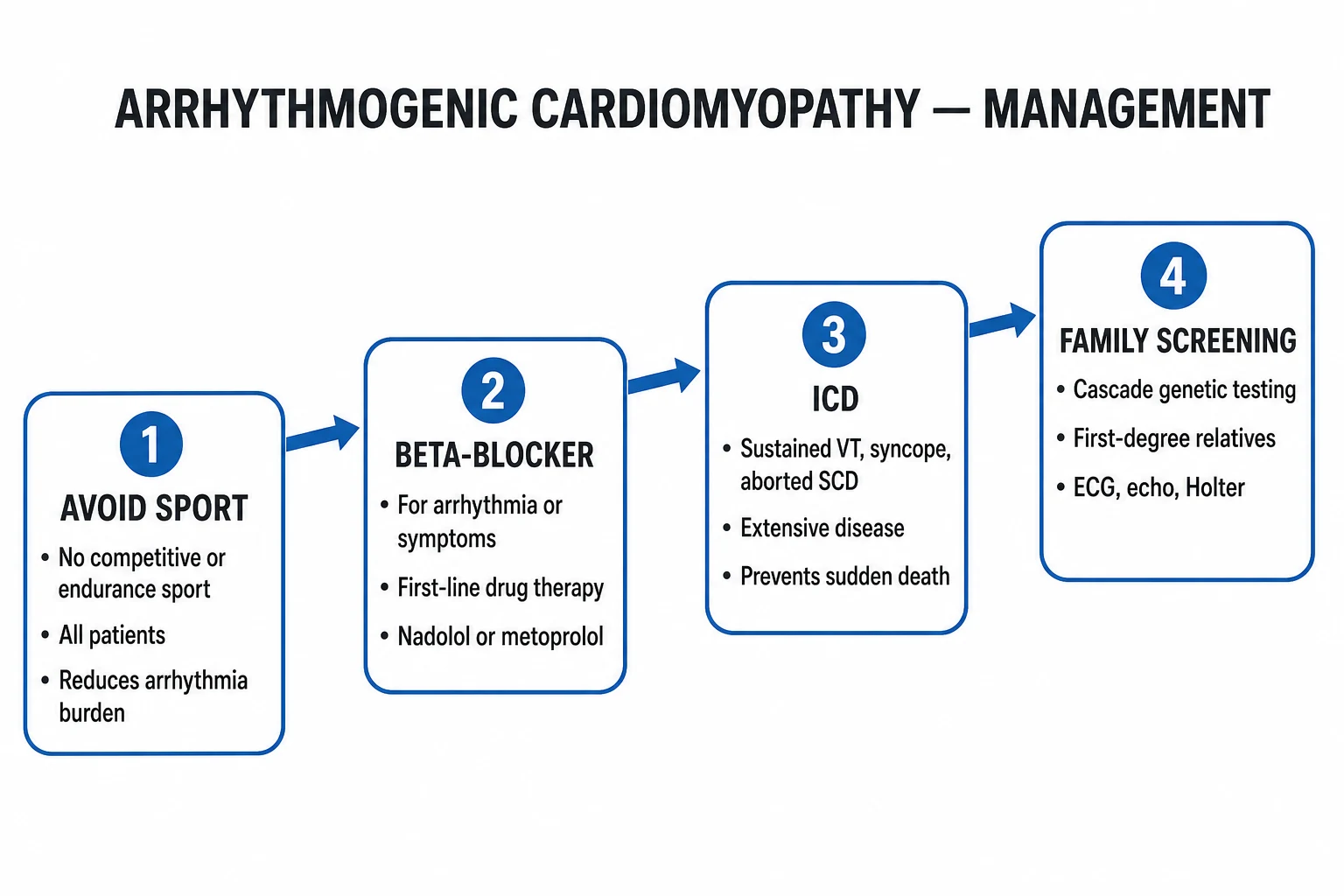
Management has four parallel streams: lifestyle, pharmacological, device therapy, and catheter / surgical.[2]
1. Lifestyle (the most under-prescribed intervention): [1]
- No competitive sport — all competitive and endurance sport is prohibited in definite ACM; the 2015 Bethesda #36 / 2020 IOC / ESC 2021 recommendation is absolute. The risk of SCD during sport is multiplied 5-fold and the progression of disease is also accelerated.
- Recreational sport — low-intensity, non-competitive activity (e.g. gentle walking, golf) is permitted in mutation carriers without phenotypic expression; no isometric weightlifting at high intensity.
- Occupational restrictions — commercial pilot, commercial driver, diver, heavy goods vehicle licence — all affected. [1]
2. Pharmacological therapy (anti-arrhythmic and heart-failure): [1]
- Beta-blockers (first-line anti-arrhythmic):
- Sotalol — historically most effective in ARVC; start 80-160 mg orally twice daily (renal dosing; QTc monitoring; avoid with severe LV dysfunction).
- Metoprolol succinate 23.75-190 mg orally daily, or bisoprolol 1.25-10 mg orally daily — first-line when sotalol not tolerated.
- Carvedilol 3.125-25 mg orally twice daily — preferred if LV dysfunction coexists.
- Rationale: suppress adrenergically triggered arrhythmia; reduce RV wall stress.
- Amiodarone — 100-200 mg orally daily (after loading 800-1600 mg/day for 1-3 weeks). Reserved for sotalol failure / intolerance; monitor TFTs, LFTs, lung function, QTc. Combined with beta-blocker for refractory arrhythmia.
- Mexiletine or flecainide — uncommonly used; mexiletine 200 mg orally three times daily may help sodium-channel-positive genotypes; flecainide carries pro-arrhythmic risk.
- Heart-failure therapy in the burnt-out phase — identical to guideline-directed DCM therapy: ACE inhibitor / ARB / ARNI, beta-blocker, MRA (spironolactone / eplerenone), SGLT2 inhibitor (dapagliflozin / empagliflozin); diuretics for congestion; digoxin and anticoagulation for atrial fibrillation (CHA2DS2-VASc-based).
- Avoid QT-prolonging drugs in sotalol/amiodarone-treated patients (macrolides, fluoroquinolones, ondansetron, methadone). [1]
3. Implantable cardioverter-defibrillator (ICD) — primary and secondary prevention. [1]
ICD is the only therapy proven to reduce SCD in ACM. Indications (2022 ESC / International Task Force):[2]
- Class I (mandatory):
- Survivors of SCD due to VT/VF (secondary prevention).
- Sustained VT (haemodynamically stable or unstable) with structural disease.
- Class IIa (should be considered):
- Unexplained syncope with structural disease ( presumed arrhythmic).
- NSVT on Holter with inducible VT on EPS in patients with structural disease (the role of EPS is declining — many centres now offer ICD on extensive scar / multiple risk factors alone).
- Class IIb (consider):
- Severe RV or LV systolic dysfunction (LVEF ≤ 35%, RVEF severely reduced) — EF-guided primary prevention.
- LMNA / DSP / PLN / TMEM43 carriers with high-risk features (male sex, NSVT, marked LGE) — ICD at LVEF above 35%, because SCD risk is high even with preserved EF.
- Class III (not indicated): asymptomatic gene carriers without phenotype — ICD not routinely offered; lifestyle, Holter, and imaging surveillance. [1]
ICD considerations specific to ACM: transvenous leads may fail in advanced RV disease (poor sensing/thresholds, perforation of thinned RV free wall) — subcutaneous ICD (S-ICD) is increasingly preferred where VT is monomorphic and painless anti-tachycardia pacing is not required; epicardial leads for those who fail transvenous. [1]
4. Catheter ablation — endocardial + epicardial. [1]
- Indicated for drug-refractory, recurrent monomorphic VT, ICD storms, or unacceptable shock burden.
- ARVC VT is macro-re-entrant with critical isthmus on the epicardial RV surface; epicardial ablation is often required and superior to endocardial-only.
- Recurrence is common (50-70% within 3 yr) because disease progresses — ablation reduces ICD shocks but does not prevent SCD; an ICD remains essential.
- Periprocedural coronary angiography prior to epicardial ablation to avoid arterial injury. [1]
5. Surgical and advanced heart failure therapy: [1]
- Cardiac sympathetic denervation — for refractory VT storm (bilateral or left thoracoscopic).
- Heart transplantation — for refractory heart failure or intractable VT storm in the burnt-out phase; survival excellent; arrhythmia-free post-transplant.
- LVAD / mechanical circulatory support — bridge to transplant in end-stage disease. [1]
Specific Subtypes & Scenarios
- Classic RV-dominant ACM (ARVC) — the textbook phenotype. Diagnosis by 2010 TFC; PKP2 in ~ 40%; epsilon wave + TWI V1-V3 + LBBB-superior VT. Treat with beta-blocker + ICD if high-risk + no competitive sport.[1]
- Left-dominant ACM — LV subepicardial / mid-wall scar, lateral TWI, often DSP, LMNA, PLN, DSG2. No epsilon wave (LV scar does not slow RV activation). Lower threshold for ICD in LMNA / DSP even with preserved LVEF.[5]
- Biventricular ACM — late-stage or PLN/TMEM43 founder phenotypes; both RV and LV failure; biventricular ICD considerations; high arrhythmia burden.
- Naxos disease (recessive JUP) — woolly hair, non-epidermolytic palmoplantar keratoderma, 100% penetrance of ARVC by adolescence; ICD is standard.[8]
- Carvajal syndrome (recessive DSP) — striate palmoplantar keratoderma + woolly hair + left-dominant ACM.
- Familial exercise-triggered myocarditis (DSP) — recurrent troponin-positive chest pain with patchy LGE; genetic cascade is the key to diagnosis and to avoiding inappropriate "viral myocarditis" labels.[10]
- ACM in the athlete — pre-participation ECG screening detects TWI V1-V3 and LBBB PVCs; disqualification from competitive sport is mandatory once definite. Corrado 2003 showed athletic SCD from ARVC fell in Veneto after Italy introduced mandatory ECG screening of athletes.[6]
- LMNA-associated ACM — high SCD risk at preserved LVEF; ESC 2022 recommends ICD even at LVEF above 35% when there is NSVT, male sex, NSVT, or non-missense variant.[2]
- TMEM43 p.S358L Newfoundland — near-universal male penetrance; prophylactic ICD from age 18 yr recommended.[2]
- ACM in the asymptomatic gene carrier — imaging and Holter surveillance annually through adolescence and at least biennially thereafter; restrict competitive sport.
Complications & Pitfalls
Cardiac complications: [1]
- Sudden cardiac death — the dominant mode of death in young patients; exertion-related.
- Recurrent monomorphic VT / VT storm / appropriate ICD shocks — most common sustained arrhythmia; ablation reduces shock burden.
- Inappropriate ICD shocks — due to atrial fibrillation / SVT with aberrancy or T-wave oversensing; programme discriminators and beta-blockade help.
- Right-heart failure — RV dilatation, tricuspid regurgitation, hepatomegaly, ascites, cachexia.
- Biventricular heart failure — burnt-out phase; treat as DCM.
- Atrial fibrillation / flutter — in advanced disease; anticoagulate by CHA2DS2-VASc.
- Device-related complications — ICD lead perforation of thinned RV free wall, infection, pneumothorax; S-ICD preferred in thinned RV.
- Endocardial / epicardial thrombus and pulmonary embolism — scarred RV aneurysms are thrombogenic; consider anticoagulation in RV akinesia with low flow. [1]
Classic pitfalls (examiner favourites): [1]
- Mislabelling ARVC VT as benign RVOT VT — always check axis (superior vs inferior) and QRS morphology (notched vs smooth) before declaring an RVOT VT benign.
- Missing TWI V1-V3 in a young athlete — dismissed as "normal early repolarisation" or "juvenile pattern" past age 14; TWI V1-V3 in any athlete over 14 yr without RBBB requires evaluation.
- Failing to screen family — first-degree relatives need ECG, Holter, echo and CMR; ~ 30% of "sporadic" cases turn out to have an affected relative on cascade screening.
- Permitting the patient to return to competitive sport — the single most preventable cause of SCD.
- Misdiagnosing DSP myocarditis as viral — recurrent episodes, exertional trigger, or family history of SCD/DCM demand genetic testing.[10]
- Placing a transvenous ICD lead in a paper-thin RV free wall — high perforation risk; consider S-ICD.
- Treating the arrhythmia but not the heart failure — burnt-out disease needs full GDMT.
- Failing to anticoagulate atrial fibrillation in burnt-out disease — stroke risk is high.
- Accepting a single normal ECG/MRI — penetrance evolves over years; re-test gene carriers every 1-2 yr.
Prognosis & Disposition
Overall annual SCD rate in definite ACM is approximately 2.5-3% per year without therapy; ICD therapy reduces SCD substantially. Five-year mortality from heart failure in the burnt-out phase approaches that of DCM.[4][7]
Predictors of adverse outcome (high-yield): [1]
- History of sustained VT, aborted SCD, or unexplained syncope.
- Young age at presentation, male sex.
- Severe RV or LV systolic dysfunction (RVEF < 40%, LVEF ≤ 35%).
- Extensive late gadolinium enhancement (LV or RV) on CMR — even with normal EF.
- High non-sustained VT burden / frequent PVCs on Holter.
- Proband status (more severe than relatives detected on cascade).
- High-risk genotype — LMNA, DSP, PLN, TMEM43; compound/digenic heterozygosity.[9]
- Inappropriate sinus tachycardia, fibrosis, and RV aneurysms.
Cadrin-Tourigny / Padua risk-prediction models integrate these factors to estimate annual arrhythmic event probability; an annual risk above 5-10% generally supports primary prevention ICD.[7]
Disposition: [1]
- Outpatient with annual review for low-risk gene carriers — ECG, Holter, echo; CMR every 2-3 yr.
- Specialist inherited cardiac conditions clinic for all definite ACM.
- Cardiology + clinical genetics MDT for cascade testing and counselling.
- Psychological support and driving / occupational advice — group 2 driving licence (HGV / PCV) forfeited; private driving restricted after ICD / syncope / VT.
- Pre-conception counselling for women with ACM — pregnancy is generally well tolerated in stable disease; avoid ACE inhibitor / ARB / MRA / SGLT2i in pregnancy. [1]
Special Populations
- Athletes — the highest-stakes population. Pre-participation ECG + family history is the screening test; definite ACM = lifetime disqualification from competitive sport (Bethesda #36 / ESC 2021). Light recreational activity permitted.
- Asymptomatic gene carriers — imaging and Holter surveillance annually through adolescence, at least biennially in adults; no competitive sport even without phenotype.
- Children and adolescents (under 14 yr) — TWI V1-V3 may be a normal juvenile pattern; do not over-diagnose ACM before age 14 yr unless other criteria are present.[1]
- Pregnancy — generally well tolerated in stable, compensated ACM; manage in joint obstetric-cardiology clinic; switch teratogenic drugs (ACE inhibitor/ARB → labetalol/nifedipine; MRA → stop; amiodarone → use only if essential; sotalol acceptable but monitor); regional anaesthesia preferred; ICD available for high-risk deliveries. Penetrance may worsen postpartum — re-image at 6 months.
- The elderly — burnt-out biventricular disease often labelled DCM; reconsider the diagnosis when there is prior VT, RV predominance, or a family history of SCD.
- LMNA / TMEM43 carriers — lower ICD threshold; the SCD risk at LVEF above 35% is high.
- Patients with an existing ICD — regular device interrogation, driving restrictions, MRI-conditional device preferred for future imaging.
Evidence, Guidelines & Regional Differences
The key documents and trials: [1]
- Marcus 2010 (Modified Task Force Criteria, Circulation) — quantitative redefinition of the 1994 criteria; six diagnostic categories; remains the international diagnostic standard.[1]
- Corrado/Bauce 2020 (Padua criteria, International Journal of Cardiology) — quantitative CMR thresholds for RV and LV volumes / function; increased sensitivity over 2010 TFC; integrated into ESC 2022.[3]
- ESC 2022 Ventricular Arrhythmia and SCD Guideline — current European standard for risk stratification and ICD thresholds, including genotype-specific advice (LMNA, DSP, PLN, TMEM43).[2]
- Sen-Chowdhry 2008 (JACC) — defined left-dominant ACM as a distinct entity; shifted the umbrella term from ARVC to ACM.[5]
- Marcus 2010 / Xu 2010 (JACC) — compound and digenic heterozygosity contributes to ARVC, explaining variable penetrance and severity within families.[9]
- Cadrin-Tourigny 2019 (Eur Heart J) — derived a predictive risk model for ventricular arrhythmias in mutation carriers; integrates clinical, Holter and CMR features.[7]
- Poller 2020 (J Am Heart Assoc) — DSP truncating variants cause familial recurrent exercise-triggered myocarditis masquerading as viral.[10]
Regional differences: [1]
[1]- United States (AHA/ACC/HRS 2017) — pre-participation screening emphasises history and physical, with ECG added selectively; Bethesda #36 (2015) sports recommendations; S-ICD widely used.
- Italy (COCIS, Corrado) — mandatory 12-lead ECG pre-participation screening for all competitive athletes since 1982; sports disqualification is regulated by COCIS.
- Europe generally (ESC 2022) — genotype-stratified ICD thresholds; S-ICD preferred for thinned RV.
- India (ICMR / NMC) — no national pre-participation screening; diagnosis is often delayed to presentation with SCD or sustained VT; cascade testing underused; access to CMR and genetic testing is regional. ICD and ablation facilities are concentrated in tertiary centres.
- International consensus — 2024 — broad agreement on no competitive sport, beta-blockade first, ICD for sustained VT / aborted SCD / high-risk genotypes, genetic cascade screening of first-degree relatives. [1]
Controversies: [1]
- EPS-guided ICD — declining role; modern risk models rely on NSVT, genotype, and CMR rather than EPS.
- The role of CMR-only LGE — included in Padua but excluded from 2010 TFC; trials ongoing to refine risk.
- S-ICD vs transvenous ICD — S-ICD preferred in thinned RV, young patients (longer lifetime lead burden), and high infection risk, but lacks ATP.
- Return to recreational sport — permitted at low intensity; some centres permit moderate intensity in low-risk gene carriers.
- Gene therapy — preclinical; not clinical reality. [1]
Exam Pearls
- ARVC is the leading cause of SCD in young Italian athletes (Veneto data); HCM is the leading cause in US athletes.[6]
- Epsilon wave = small terminal deflection in V1-V3, seen in ~30% of overt cases — most specific ECG finding.
- TWI V1-V3 without RBBB (in adults over 14 yr) = the single most common repolarisation criterion.
- LBBB-superior-axis VT (negative in inferior leads) = classical ARVC VT from RV inflow/apex; inferior axis suggests benign RVOT VT.
- The "triangle of dysplasia" = RV inflow + RV outflow + RV apex — the three preferentially scarred regions.
- PKP2 (plakophilin-2) is the single most common gene (~40%).
- Naxos disease = JUP (recessive) = woolly hair + palmoplantar keratoderma + ARVC.
- Carvajal syndrome = DSP (recessive) = striate keratoderma + left-dominant ACM.
- LMNA, DSP, PLN, TMEM43 = high-risk genotypes warranting ICD at LVEF above 35%.
- Padua 2020 criteria: RVEF ≤ 40% + RVEDVi ≥ 110 mL/m² (male) or ≥ 100 mL/m² (female) = major.
- Modified Task Force 2010: 2 major OR 1 major + 2 minor OR 4 minor from different categories = definite.
- SAECG late potentials = minor depolarisation criterion.
- PVC burden over 1,000 per 24 h = minor arrhythmia criterion; over 10,000 = high-risk.
- Sotalol 80-160 mg BD is the classical first-line anti-arrhythmic; amiodarone reserved for failure / intolerance.
- ICD is the only therapy proven to reduce SCD; ablation reduces shocks but does not prevent SCD.
- No competitive or endurance sport — the single most under-prescribed intervention; risk multiplier of ~5-fold.
- Subepicardial / mid-wall LGE (NOT subendocardial) — distinguishes non-ischaemic scar from ischaemic.
- First-degree relatives need ECG, Holter, echo, CMR and genetic cascade testing — ~ 30% of "sporadic" cases have an affected relative.
- TWI in V1-V3 in a child under 14 yr may be normal juvenile pattern — do not over-diagnose ACM.
- Idiopathic RVOT VT vs ARVC VT — inferior axis + smooth QRS + adenosine-sensitive = benign; superior axis + notched QRS + scar on CMR = ARVC.
Exam application bank (NEET-PG / INICET)
One-line answer
Arrhythmogenic cardiomyopathy (ACM) is an inherited heart-muscle disease in which progressive fibro-fatty replacement of ventricular myocardium (classically the right ventricle) produces ventricular arrhythmias, heart failure and sudden cardiac death (SCD) in apparently healthy young people — most notably competitive athletes. Inheritance is usually autosomal dominant and the genes are predominantly desmosomal (PKP2 plakophilin-2, DSP desmoplakin, DSG2 desmoglein-2, DSC2 desmocollin-2, JUP plakoglobin); non-desmosomal genes include TMEM43, LMNA, PLN, DES, CDH2, SCN5A, CTNNA3. The ECG hallmark is the epsilon wave (a low-amplitude deflection at the end of the QRS in V1-V3) with T-wave inversion V1-V3 (without RBBB); cardiac MRI shows RV dilatation, regional wall-motion abnormalities and late gadolinium enhancement. Diagnosis uses the 2010 Modified Task Force Criteria (six categories — imag
Worked stems (answer without another resource)
Stem 1 — Classic presentation. Map symptoms to mechanism; name the first investigation and first treatment step with dose/route if drug therapy is standard. [1]
Stem 2 — Unstable / complicated. List red flags that force immediate resuscitation, theatre, ICU, antidote, or reperfusion — and what you do in the first 15 minutes. [1]
Stem 3 — Atypical group. Elderly, pregnancy, child, or immunocompromised: how presentation and thresholds change. [1]
Stem 4 — Differential trap. Name the three closest mimics and one discriminator for each. [1]
Stem 5 — Disposition. Who goes home with safety-netting, who is admitted, who needs HDU/ICU/theatre, and what follow-up is mandatory. [1]
Rapid viva checklist
- Definition + classification
- Pathophysiology chain
- Bedside signs / criteria
- Score with exact components (if any)
- Emergency bundle
- Definitive therapy with doses
- Complications of disease and of treatment
- Special populations
- Guideline/trial name if classic
- Three exam traps
Coverage self-check
If you cannot answer any stem above from this page alone, re-read the matching section — the page is intended to be self-sufficient for final-prof and NEET-PG/INICET questions on Arrhythmogenic Cardiomyopathy.
References
- [1]Marcus FI, McKenna WJ, Sherrill D, et al. Diagnosis of arrhythmogenic right ventricular cardiomyopathy/dysplasia: proposed modification of the task force criteria Circulation, 2010.PMID 20172911
- [2]Zeppenfeld K, Tfelt-Hansen J, de Riva M, et al. 2022 ESC Guidelines for the management of patients with ventricular arrhythmias and the prevention of sudden cardiac death Eur Heart J, 2022.PMID 36017572
- [3]Corrado D, Perazzolo Marra M, Zorzi A, et al. (Padua group, Bauce B senior). Diagnosis of arrhythmogenic cardiomyopathy: The Padua criteria Int J Cardiol, 2020.PMID 32561223
- [4]Marcus FI, Zareba W, Calkins H, et al. (North American Multidisciplinary Study). Arrhythmogenic right ventricular cardiomyopathy/dysplasia clinical presentation and diagnostic evaluation: results from the North American Multidisciplinary Study Heart Rhythm, 2009.PMID 19560088
- [5]Sen-Chowdhry S, Syrris P, Ward D, Asimaki A, Sevdalis E, McKenna WJ. Left-dominant arrhythmogenic cardiomyopathy: an under-recognized clinical entity J Am Coll Cardiol, 2008.PMID 19095136
- [6]Corrado D, Basso C, Rizzoli G, Schiavon M, Thiene G. Does sports activity enhance the risk of sudden death in adolescents and young adults? J Am Coll Cardiol, 2003.PMID 14662259
- [7]Cadrin-Tourigny J, Bosman LP, Nozza A, et al. (Bhonsale A contributing). A new prediction model for ventricular arrhythmias in arrhythmogenic right ventricular cardiomyopathy Eur Heart J, 2019.PMID 30915475
- [8]Marcus F, Towbin JA, Zareba W, et al. The mystery of arrhythmogenic right ventricular dysplasia/cardiomyopathy: from observation to mechanistic explanation Circulation, 2006.PMID 17060394
- [9]Xu T, Yang Z, Vatta M, et al. (Multidisciplinary Study of Right Ventricular Dysplasia). Compound and digenic heterozygosity contributes to arrhythmogenic right ventricular cardiomyopathy J Am Coll Cardiol, 2010.PMID 20152563
- [10]Poller W, Gattenlohner S, Haas J, et al. Familial Recurrent Myocarditis Triggered by Exercise in Patients With a Truncating Variant of the Desmoplakin Gene J Am Heart Assoc, 2020.PMID 32410525