Cardiology · Cardiology
Atrial Fibrillation
Also known as AF · Afib · Auricular fibrillation
Atrial fibrillation (AF) is the commonest sustained cardiac arrhythmia: irregularly irregular rhythm, absent P waves, fibrillatory f-waves. Care follows the ABC pathway: Avoid stroke (anticoagulate if CHA2DS2-VASc ≥2 in men, ≥3 in women, with a DOAC preferred), Better symptom control (rate vs rhythm control), Cardiovascular risk-factor optimisation. Unstable AF with rapid ventricular response needs emergency synchronised DC cardioversion.
On this page & tools
Your progress
Saved locally on this device.
Practise this topic
Exam tags
Red flags
Overview & Definition
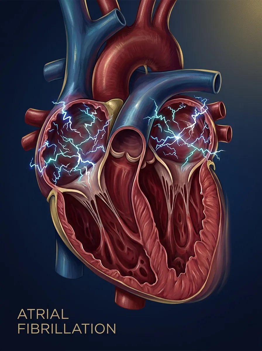
Atrial fibrillation (AF) is a supraventricular tachyarrhythmia defined by uncoordinated atrial electrical activation with consequent loss of effective atrial mechanical contraction. In place of the single organised sinus impulse that normally propagates across both atria each cycle, AF is a storm of chaotic depolarisation waves — the atria quiver rather than contract, and the ventricles respond to a rapid, unpredictable barrage of impulses conducted through the atrioventricular (AV) node. [1]
The diagnosis rests on three ECG hallmarks that every candidate must be able to recite verbatim:[1]
- An irregularly irregular ventricular (RR) rhythm — no pattern to the spacing between beats.
- Absent P waves — no discrete atrial depolarisation is visible.
- Fibrillatory f-waves — fine, rapid, low-amplitude undulations of the baseline (best seen in lead V1) replacing organised atrial activity, at a rate of 350 to 600 per minute. [1]
When the f-waves are coarse (amplitude greater than 0.5 mm in V1) the rhythm can superficially resemble atrial flutter; the decisive discriminator is the absolute irregularity of the RR intervals in AF, contrasted with the regular (or regularly irregular) ventricular response of flutter. [1]
AF is documented objectively on a 12-lead ECG, or — at minimum — a single-lead rhythm strip of at least 30 seconds demonstrating all three hallmarks. A single irregular pulse on examination is not diagnostic; the rhythm must be captured electrically. The ESC, ACC/AHA/ACCP/HRS, and NICE guidelines are unified on this standard.[1][2]
The term "lone AF" — formerly used for AF in younger patients with no identifiable cause — is now discouraged: with modern imaging and risk-factor assessment, virtually all patients have an identifiable driver (most often hypertension, sleep-disordered breathing, or subclinical cardiomyopathy).[1]
Classification
AF is classified by its temporal pattern, not by ventricular rate or symptom burden. The thresholds are examiner favourites because they govern decisions about rhythm-versus-rate strategy. [1]
First diagnosed
- Any first-ever episode, regardless of duration or symptoms
- Every patient is a 'first-diagnosed' patient once
- Triggers a work-up for a reversible cause and a CHA2DS2-VASc score
Paroxysmal
- Self-terminating, usually within 48 hours and nearly always within 7 days
- Episodes are recurrent but sinus rhythm restores spontaneously
- 'Pill-in-the-pocket' flecainide is an option in selected patients with a structurally normal heart
Persistent
- Continuous and lasting longer than 7 days, OR terminating only with cardioversion (electrical or pharmacological)
- Rhythm control is a reasonable and often preferred goal, especially early
- Anticoagulation decision is governed by CHA2DS2-VASc, not by pattern
Long-standing persistent
- Continuous for longer than 12 months when a rhythm-control strategy is still being pursued
- Ablation is often considered as the rhythm-control modality of choice
- Distinguished from permanent by the *intent* to restore sinus rhythm
Permanent
- AF accepted by patient and clinician; no further attempt at rhythm control
- Rate control and comorbidity management become the focus
- Re-classification back to long-standing persistent is allowed if the patient or clinician changes their mind
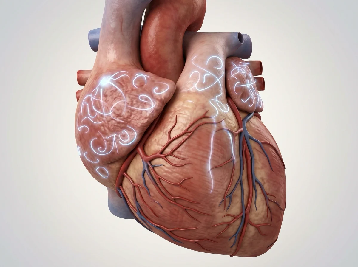
Valvular versus non-valvular AF — the single most important classification for the anticoagulation question, and the most heavily tested. AF complicating moderate-to-severe mitral stenosis or a mechanical prosthetic heart valve is "valvular AF" and mandates warfarin (target INR 2.0–3.0). DOACs are contraindicated here: the RE-ALIGN trial showed that dabigatran in mechanical-valve patients was associated with more thromboembolic and bleeding events than warfarin. All other AF is "non-valvular", where DOACs are preferred over warfarin. The 2023 ACC/AHA/ACCP/HRS guideline explicitly retains this dichotomy.[1][2]
Note that mild mitral regurgitation of degenerative origin, bioprosthetic valves without other risk, and mitral valve repair do not by themselves qualify as "valvular AF" in the contemporary sense — these patients are managed with DOACs. The defining lesion is rheumatic mitral stenosis or a mechanical valve. [1]
Epidemiology & Risk Factors
AF is the commonest sustained cardiac arrhythmia and one of the few cardiovascular epidemics still growing in absolute numbers, driven by ageing populations and the global rise in obesity, hypertension, and diabetes. Key figures a candidate should hold: [1]
AF by the numbers
Hypertension is the commonest single driver of AF, present in over 70% of patients. The full list of modifiable and cardiac risk factors is recalled by the high-yield mnemonic HIPAT-H: [1]
High-yield drivers of AF
HIPAT-H
Commonest driver; also drives heart failure and stroke risk
Coronary disease and prior MI; atrial scarring provides the substrate
COPD, OSA, PE, pneumonia — hypoxia and right-heart strain
Binge ('holiday heart syndrome') and chronic excess; a direct atrial toxin
Check TSH in every new AF — treatable and reversible driver
Bidirectional: HF causes AF and AF worsens HF (tachycardiomyopathy)
Beyond HIPAT-H, recognise obesity (dose-response with AF incidence and recurrence after ablation), diabetes, valvular and rheumatic heart disease (dominant cause in the Indian subcontinent), pericarditis and myocarditis, post-cardiac-surgery state (30–40% of coronary bypass and valve patients), endurance-athlete remodelling, and genetic predisition (family history doubles risk).[1]
The complication burden is what makes AF dangerous — the arrhythmia itself is rarely the immediate cause of death. The four cardinal complications are: ischaemic stroke and systemic thromboembolism (typically a large, severe cardioembolic stroke with high disability and mortality), heart failure (both a cause and a consequence — uncontrolled ventricular rate produces a potentially reversible tachycardia-induced cardiomyopathy), cognitive decline and dementia (linked to silent cerebral micro-infarcts), and roughly doubled all-cause mortality.[1]
Pathophysiology
AF is sustained by two overlapping and synergistic mechanisms. Understanding both is what makes the rationale for ablation intuitive. [1]
1. Focal triggers. In many patients — particularly those with paroxysmal AF — the arrhythmia is initiated by rapidly firing ectopic foci, most of which arise from the muscular sleeves of myocardial tissue extending 1–3 cm into the pulmonary veins where they join the left atrium. These sleeves possess intrinsic automaticity and can generate the rapid bursts that trigger AF. The therapeutic implication is direct: electrically isolating the pulmonary veins from the left atrium (pulmonary vein isolation, PVI) removes the trigger and is the cornerstone of catheter ablation.[1]
2. Multiple re-entrant wavelets. Once initiated, AF is sustained by multiple chaotic re-entrant wavelets circulating through atrial tissue. The substrate for re-entry is heterogeneous conduction and shortened, dispersed refractory periods — a function of atrial dilation, fibrosis, and the myopathy of long-standing AF. The older multiple-wavelet hypothesis (Moe, refined by Allessie) explains why AF begets AF: each episode shortens atrial refractoriness and promotes fibrosis, lowering the threshold for the next episode and making termination harder. [1]
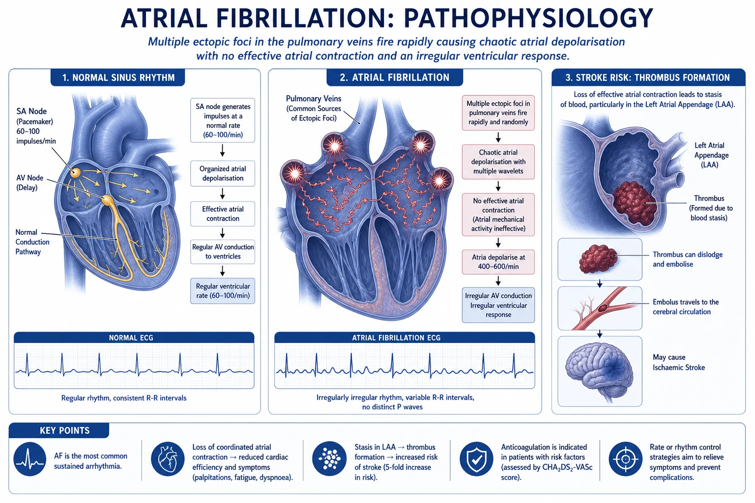
Electrical and structural remodelling is the molecular substrate for the "AF begets AF" adage. Within days of sustained AF, atrial myocytes down-regulate the L-type calcium current and up-regulate inward-rectifier potassium currents, shortening the action potential duration and effective refractory period — exactly the change that favours more re-entry. Over months to years, fibrosis, fatty infiltration, and atrial dilation further disrupt conduction, which is why early rhythm control (EAST-AFNET 4) and aggressive risk-factor modification (weight loss, blood-pressure control, sleep-apnoea treatment) are increasingly emphasised.[3][1]
The irregularly irregular ventricular response is the bedside signature of AF and a direct mechanical consequence of the chaotic atrial rhythm. The AV node is bombarded by 350–600 atrial depolarisations per minute but conducts only a variable, unpredictable subset to the ventricles — governed by concealed conduction, concealed discharge, and refractoriness within the node itself. The result is the diagnostic irregular irregularity of the RR intervals. The ventricular rate in untreated AF is typically 100–160 bpm, but it can be much faster in sympathetic-driven states (sepsis, hyperthyroidism, exercise, post-operative) or deceptively slow when the AV node is diseased or blunted by drugs (beta-blocker, digoxin). [1]
Why AF causes stroke — the mechanism that underpins the entire anticoagulation enterprise. In sinus rhythm, the left atrial appendage (LAA) contracts and empties with each beat. In AF, the atria do not contract effectively; blood stagnates in the LAA, a long, blind-ended, trabeculated pouch where flow is naturally sluggish. Stasis plus endocardial dysfunction and hypercoagulability (the Virchow triad, applied to AF) produces thrombus, which embolises — typically to the brain, producing a large-territory, often devastating, cardioembolic stroke. Over 90% of AF-related thrombus originates in the LAA, which is why anticoagulation and, in selected patients, LAA occlusion (e.g. Watchman device) are the two stroke-prevention strategies.[1]
Clinical Presentation
The presentation of AF spans an enormous spectrum — from the patient who is entirely asymptomatic and discovered at a routine blood-pressure check, to the patient who arrives in cardiogenic shock. [1]
Typical symptoms are driven by the loss of the atrial contribution to filling (the "atrial kick", worth 15–30% of stroke volume), the rapid ventricular rate, and the irregularity: [1]
- Palpitations — classically described as fast and irregular, often coming on abruptly and ceasing suddenly in paroxysmal AF.
- Dyspnoea and reduced exercise tolerance — from loss of atrial kick and tachycardia.
- Fatigue — often the dominant and most under-recognised symptom, especially in the elderly.
- Chest pain — from rapid rate and underlying ischaemia, or from the arrhythmia itself.
- Presyncope or syncope — from rate-related hypotension or, rarely, a sinus pause on termination of paroxysmal AF ("brady-tachy syndrome").
- Polyuria — atrial natriuretic peptide release from the stretched atria (an under-taught but classic clue). [1]
Asymptomatic ("silent") AF is strikingly common — up to one-third of all AF — and is often detected only on an opportunistic pulse check, a routine ECG, a smartwatch, or when a stroke is the first manifestation. A substantial fraction of "cryptogenic" stroke is attributable to undiagnosed paroxysmal AF, which is why prolonged rhythm monitoring (insertable cardiac monitor) is part of the cryptogenic-stroke work-up. [1]
Atypical presentation in the elderly is a deliberate examiner test. Rather than complaining of palpitations, the older AF patient may present with falls, confusion or delirium, new or worsening heart failure, an incidental finding on admission for another problem, or simply worsening exercise capacity. A pulse check on every elderly patient — and an ECG when irregular — is the high-yield habit. All major guidelines stress opportunistic screening for an irregular pulse in those over 65.[1][2]
Bedside signs that reward the careful examiner: [1]
- Irregularly irregular pulse — the cardinal sign. Confirm at the radial and the apex.
- Pulse deficit — the apical (heart) rate exceeds the radial (wrist) rate, because some ventricular beats are too weakly contractile (short filling time) to transmit a palpable peripheral pulse. A deficit of more than 10 bpm is clinically significant.
- Variable intensity of the first heart sound (S1) — because the AV filling interval changes beat to beat, altering mitral-valve closure force.
- Absent "a" waves in the jugular venous pulse (JVP) — there is no coordinated atrial contraction to generate the venous "a" wave.
- Signs of an underlying cause: a thyroid goitre or eye signs of thyrotoxicosis, the murmurs of mitral valve disease, crackles and a third heart sound of heart failure, or fever and a septic focus. [1]
Differential Diagnosis
The differential of an irregularly irregular pulse is short, high-yield, and distinguished almost entirely on the ECG: [1]
Atrial fibrillation
- No discrete P waves; fine fibrillatory f-waves
- Absolutely irregular RR intervals
- Ventricular rate usually 100–160 bpm untreated
Atrial flutter with variable block
- Sawtooth flutter (F) waves, best in II/III/V1
- Atrial rate ~300 bpm; ventricular response variable (2:1, 3:1, 4:1)
- Often coexists with or degenerates into AF
Multifocal atrial tachycardia (MAT)
- Three or more distinct P-wave morphologies
- Classically in severe COPD with hypoxia
- Irregularly irregular but P waves are present
Frequent ectopics
- Atrial or ventricular premature beats interrupt a normal baseline
- Normal sinus P waves and a regular underlying rhythm between ectopics
- Carotid sinus massage may abolish the irregularity
Sinus arrhythmia
- 'Regularly irregular' — varies with respiration
- Normal P-wave morphology; common in the young and athletes
- Benign; requires no treatment
Pre-excited AF (Wolff-Parkinson-White) is the must-not-miss mimic and the most dangerous trap in AF management. The ECG shows a broad, irregularly irregular tachycardia with delta waves and QRS morphology that varies beat to beat. It can easily be confused with ventricular tachycardia — but VT is essentially always regular, whereas pre-excited AF is irregular. The management is life-critical and distinct: never give AV-nodal blockers (adenosine, verapamil, diltiazem, beta-blocker, or digoxin) — these block the AV node, accelerating conduction down the accessory pathway and risking degeneration to ventricular fibrillation. The correct drugs are intravenous procainamide, ibutilide, or amiodarone, or synchronised DC cardioversion if unstable. [1]
Clinical & Bedside Assessment
The focused assessment of a patient with suspected AF serves three goals: confirm the rhythm, assess haemodynamic stability, and search for an underlying cause. [1]
Confirm the rhythm. Manual pulse palpation reveals an irregularly irregular pulse, and a pulse deficit (apical rate greater than radial) supports the diagnosis. But the pulse is not enough — diagnosis requires objective ECG documentation: a 12-lead ECG, or at minimum a single-lead rhythm strip of at least 30 seconds demonstrating the three hallmarks (absent P waves, f-waves, irregularly irregular RR).[1]
Assess stability. The single most important bedside judgement in AF is whether the patient is haemodynamically unstable — this determines whether the next step is drugs (stable) or electricity (unstable). The four instability criteria are: hypotension (systolic BP under 90 mmHg or signs of shock), acute heart failure (pulmonary oedema, hypoxia), ongoing ischaemic chest pain, or reduced conscious level. Any one mandates emergency synchronised DC cardioversion, not a rate-control drug.[1][2]
Search for a cause. Examine for: thyroid signs (goitre, tremor, eye signs) of thyrotoxicosis; murmurs of valvular disease (especially the rumbling mid-diastolic of mitral stenosis); signs of heart failure (raised JVP, basal crackles, S3, peripheral oedema); signs of sepsis or pneumonia; and stigmata of alcohol excess. Every new AF patient gets thyroid function tests as part of first-line bloods. [1]
Investigations
The first-line work-up for new AF has three aims: confirm the rhythm, find a cause, and assess stroke and bleeding risk to plan anticoagulation. [1]
12-lead ECG (or 30-s rhythm strip)
- Diagnostic: absent P waves, f-waves, irregularly irregular RR
- Look for LVH, prior MI, bundle branch block, pre-excitation, QT
- Repeat if the first ECG is normal and paroxysmal AF is suspected
Transthoracic echocardiogram
- Chamber size (especially left atrial enlargement), LV systolic and diastolic function
- Valve disease — defines 'valvular' vs 'non-valvular' for anticoagulation
- Identifies cardiomyopathy and pulmonary hypertension
Bloods
- TFTs (TSH, free T4) in every patient — thyrotoxicosis is reversible
- FBC, U&E, LFTs, coagulation; HbA1c and lipids for cardiovascular risk
- Troponin if ischaemia suspected; BNP/NT-proBNP if heart failure suspected
Ambulatory ECG monitoring is reserved for detecting paroxysmal AF when the resting ECG is normal but suspicion is high (e.g. cryptogenic stroke, unexplained palpitations). Options range from a 24–72 hour Holter monitor, through patient-activated external loop recorders (1–4 weeks), to implantable cardiac monitors / insertable loop recorders (up to 3 years) for the elusive paroxysm — the latter is the most sensitive tool for detecting silent AF after cryptogenic stroke.[1]
Transoesophageal echocardiography (TOE/TEE) has two roles. First, to exclude left atrial appendage thrombus before cardioversion or ablation when the clinician wants to shorten the mandatory 3-week pre-cardioversion anticoagulation window. Second, to assess the severity of valvular lesions suspected on transthoracic echo. A thrombus seen on TOE is an absolute contraindication to cardioversion until it has been treated with anticoagulation and resolved. [1]
CHA2DS2-VASc — stroke-risk score (reproduced verbatim)
CHA2DS2-VASc components
CHA2DS2-VASc, reproduced exactly as the examiner expects:[1]
- C — Congestive heart failure / LV dysfunction — 1 point
- H — Hypertension (treated or untreated) — 1 point
- A2 — Age 75 years or older — 2 points
- D — Diabetes mellitus — 1 point
- S2 — Prior stroke, TIA, or systemic thromboembolism — 2 points
- V — Vascular disease (prior MI, peripheral arterial disease, aortic plaque) — 1 point
- A — Age 65–74 years — 1 point
- Sc — Sex category, female — 1 point [1]
Maximum = 9 (men) or 10 (women, with the sex point). Anticoagulate at a score of 2 or more in men or 3 or more in women; consider at 1 in men. Female sex as the sole point (a score of 1 in a woman under 65 with no other risk) does not by itself mandate anticoagulation. Worked example: a 78-year-old woman with hypertension and type 2 diabetes scores 2 (age) + 1 (HTN) + 1 (DM) + 1 (sex) = 5 — anticoagulate. [1]
HAS-BLED — bleeding-risk score (reproduced verbatim)
HAS-BLED is used alongside CHA2DS2-VASc not to decide whether to anticoagulate but to flag correctable bleeding-risk factors:[1]
- H — Hypertension (uncontrolled, systolic over 160 mmHg) — 1 point
- A — Abnormal renal or liver function — 1 point each (max 2)
- S — Prior stroke — 1 point
- B — Prior major bleeding or bleeding predisposition — 1 point
- L — Labile INR (time in therapeutic range under 60%, on warfarin) — 1 point
- E — Elderly (age over 65) — 1 point
- D — Drugs (concurrent antiplatelet or NSAID) or excess alcohol — 1 point each (max 2) [1]
Maximum = 9. A score of 3 or more signals high bleeding risk and prompts closer monitoring, attention to modifiable factors (control the blood pressure, stop the NSAID, reduce alcohol), and preferential use of agents with lower bleeding profiles (apixaban). A high HAS-BLED score is explicitly not a reason to withhold anticoagulation — the stroke risk in AF almost always exceeds the bleeding risk, and the modifiable factors flagged by HAS-BLED should be corrected, not used to deny therapy.[1]
Management — Resuscitation
The immediate, time-critical decision in any patient presenting with AF is whether they are haemodynamically unstable. This single judgement bifurcates the entire pathway and is heavily examined. [1]
Acute AF — first 30 minutes
ABCDE assessment
Airway, high-flow oxygen if hypoxic or in shock, two large-bore IV cannulae, continuous cardiac monitoring, pulse oximetry and non-invasive BP. Identify and treat reversible precipitants (sepsis, hypovolaemia, hypoxia, thyrotoxicosis).
Decide: stable or unstable?
Unstable = hypotension (SBP under 90), acute heart failure/pulmonary oedema, ongoing ischaemic chest pain, or reduced GCS. Any one = cardiovert.
If unstable — synchronised DC cardioversion
Sedation/general anaesthesia with airway support. Synchronised biphasic shock starting at 120–200 J, escalating 200→300→360 J as needed. Correct electrolytes (K greater than 4.0, Mg greater than 2.0) and consider amiodarone 300 mg IV if first shock fails.
If stable — rate or rhythm control
Rate control with IV metoprolol, bisoprolol, verapamil, or digoxin (avoid CCB/digoxin in HFrEF). Rhythm control with IV amiodarone, ibutilide, or vernakalant in selected patients (e.g. first episode, onset within 48 h, young, symptomatic).
Pre-excited AF — special case
Broad irregular tachycardia with delta waves: NEVER AV-nodal blockers. Use IV procainamide, ibutilide, amiodarone, or synchronised cardioversion.
Peri-cardioversion anticoagulation
Same rule applies whether cardioversion was elective or emergency: heparin/DOAC anticoagulation for at least 4 weeks afterwards (and 3 weeks before, if elective or TOE-guided).
Management — Definitive & Stepwise
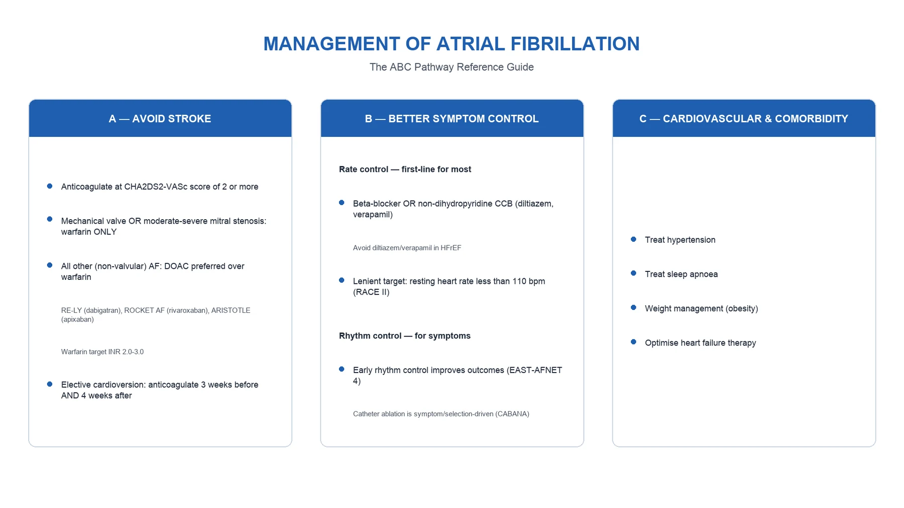
Modern AF care is organised around the ABC pathway — a framework adopted by the 2020 ESC guideline and echoed by the 2023 ACC/AHA/ACCP/HRS update and NICE NG196.[1][2]
A — Avoid stroke (anticoagulation)
Anticoagulation is the single most evidence-supported intervention in AF and the largest contributor to survival. Anticoagulate when CHA2DS2-VASc is 2 or more in men, or 3 or more in women; consider at intermediate scores; generally withhold at 0 (men) or 1 (women, sex point alone).[1][2]
For non-valvular AF, a DOAC is preferred over warfarin — DOACs have a more predictable dose response, no routine INR monitoring, fewer food and drug interactions, and equivalent or lower stroke rates with less intracranial bleeding. The four approved DOACs and their pivotal trials: [1]
Apixaban (factor Xa inhibitor — ARISTOTLE)
First-line DOAC for non-valvular AF; superior to warfarin with less bleeding and lower mortality
Dose
5 mg orally twice daily
Rivaroxaban (factor Xa inhibitor — ROCKET AF)
Non-inferior to warfarin; once-daily dosing aids adherence
Dose
20 mg orally once daily with food
Dabigatran (direct thrombin inhibitor — RE-LY)
150 mg BID superior to warfarin for stroke prevention; non-inferior for bleeding
Dose
150 mg orally twice daily
Edoxaban (factor Xa inhibitor — ENGAGE AF-TIMI 48)
Once-daily factor Xa inhibitor; non-inferior to warfarin with less bleeding
Dose
60 mg orally once daily
RE-LY
N Engl J Med (Connolly et al.)
18,113 patients with non-valvular AF randomised to dabigatran 110 or 150 mg BID vs warfarin
Key finding
Dabigatran 150 mg BID reduced stroke/systemic embolism vs warfarin with similar major bleeding; less intracranial and life-threatening bleeding
Practice change
Established dabigatran as the first DOAC superior to warfarin — ushered in the DOAC era
ROCKET AF
N Engl J Med (Patel et al.)
14,264 high-risk non-valvular AF patients randomised to rivaroxaban 20 mg OD vs warfarin
Key finding
Rivaroxaban non-inferior for stroke/systemic embolism; significantly less intracranial and fatal bleeding
Practice change
Provided the once-daily DOAC option
ARISTOTLE
N Engl J Med (Granger et al.)
18,201 non-valvular AF patients randomised to apixaban 5 mg BID vs warfarin
Key finding
Apixaban superior to warfarin for stroke prevention with less major bleeding and lower all-cause mortality
Practice change
Made apixaban the preferred DOAC for many clinicians, especially in the elderly and CKD
Warfarin (target INR 2.0–3.0) is mandatory for valvular AF (mechanical valve or moderate-to-severe mitral stenosis) and remains an option for non-valvular AF where DOACs are unavailable, unaffordable, or contraindicated (e.g. severe renal failure, antiphospholipid syndrome).[1][2]
Left atrial appendage occlusion (e.g. Watchman device) is an option for stroke prevention when long-term anticoagulation is contraindicated (e.g. prior life-threatening bleed) and the patient is at high thromboembolic risk — typically followed by a period of anticoagulation and then dual antiplatelet therapy.[2]
B — Better symptom control (rate or rhythm)
The choice between rate control (leave the patient in AF, control the ventricular response) and rhythm control (try to restore and maintain sinus rhythm) is symptom-driven, not mortality-driven. AFFIRM showed no survival advantage for a routine rhythm-control strategy over rate control.[4]
Rate control is first-line for most patients, especially older patients with mild symptoms and permanent AF. [1]
Bisoprolol ( cardioselective beta-blocker — first-line rate control)
First-line rate control in AF; preferred in coexisting IHD or HF
Dose
5 mg orally once daily (range 2.5–10 mg OD)
Diltiazem (non-DHP calcium-channel blocker — rate control)
First-line rate control when beta-blocker contraindicated; NOT in HFrEF (negative inotrope)
Dose
180–240 mg orally once daily (modified release); 60 mg modified release BD
Digoxin (cardiac glycoside — adjunctive rate control)
Adjunct in sedentary or HFrEF patients; slow onset; less effective in active/sympathetic states
Dose
0.125–0.25 mg orally once daily
The rate-control target is lenient — a resting ventricular rate under 110 bpm — per the RACE II trial, which showed lenient control non-inferior to strict (under 80 bpm resting, under 110 bpm on mild exercise) and easier to achieve.[5] The lenient target applies to stable, permanent AF; tighter control is reasonable in symptomatic patients or those with HFrEF where tachycardiomyopathy is a concern.
Rhythm control is indicated for: (a) symptomatic patients in whom rate control fails to control symptoms, (b) first-episode AF, (c) AF with a treated reversible trigger (e.g. thyrotoxicosis controlled), (d) AF complicating heart failure with reduced ejection fraction (HFrEF) where restoring sinus rhythm may improve cardiac output, and (e) younger patients in whom early rhythm control is favoured by EAST-AFNET 4.[3]
Flecainide (class IC antiarrhythmic — rhythm control)
Pharmacological cardioversion and maintenance; 'pill-in-the-pocket' in selected patients
Dose
50–100 mg orally twice daily; 200 mg single dose (pill-in-the-pocket)
Amiodarone (class III antiarrhythmic — rhythm control)
Effective in HFrEF and structural heart disease; pharmacological cardioversion
Dose
200 mg TDS for 1 week, then 200 mg BD for 1 week, then 200 mg OD maintenance
Sotalol and dronedarone (class III antiarrhythmics — rhythm control)
Maintenance of sinus rhythm in selected patients
Dose
Sotalol 80–160 mg BD; dronedarone 400 mg BD
Electrical cardioversion (synchronised DC shock) restores sinus rhythm in 70–90% when AF onset is recent. The two anticoagulation strategies for elective cardioversion: [1]
- Conventional (3-and-4 rule): anticoagulate for at least 3 weeks before and at least 4 weeks after cardioversion, regardless of CHA2DS2-VASc score, because the atria remain mechanically "stunned" for weeks after electrical restoration to sinus rhythm.
- TOE-guided: anticoagulate from presentation, perform TOE to exclude LAA thrombus, cardiovert if clear, then anticoagulate for at least 4 weeks afterwards.[1]
Catheter ablation (pulmonary vein isolation) is the most effective rhythm-control strategy, achieving sinus rhythm in 60–80% of paroxysmal AF and 40–60% of persistent AF after one or more procedures. CABANA showed no overall mortality benefit of ablation over drug therapy in the intention-to-treat analysis, so ablation is symptom- and selection-driven — favoured for symptomatic paroxysmal AF refractory to antiarrhythmics, and increasingly for AF complicating HFrEF (where subgroup analyses suggest benefit).[9][2]
C — Cardiovascular and comorbidity optimisation
Every AF review should address the modifiable drivers that independently predict AF recurrence, progression, and complication burden: [1]
- Treat hypertension to target (under 130/80 mmHg in most).
- Weight loss — each unit of BMI reduction is associated with AF regression (LEGACY and CARDIO-FIT trials).
- Screen and treat obstructive sleep apnoea with CPAP.
- Reduce alcohol to safe limits or abstain.
- Manage diabetes, ischaemic heart disease, and heart failure to guideline targets.
- Address sedentary lifestyle — moderate exercise is protective.[1][2]
Specific Subtypes & Scenarios
Paroxysmal AF — a "pill-in-the-pocket" strategy (single oral dose of flecainide 200 mg or propafenone 600 mg taken at the onset of symptoms, after medical supervision and confirmation of a structurally normal heart) is appropriate for patients with infrequent, well-tolerated episodes. For frequent or bothersome recurrence, catheter ablation (PVI) is superior to antiarrhythmic drugs for maintaining sinus rhythm and is the rhythm-control modality of choice.[2]
AF with rapid ventricular response and haemodynamic compromise — emergency synchronised DC cardioversion. There is no role for rate-control drugs as the first intervention in the unstable patient. [1]
Pre-excited AF (WPW) — broad, irregular tachycardia with delta waves. Never AV-nodal blockers (adenosine, verapamil, diltiazem, beta-blocker, digoxin). Use IV procainamide, ibutilide, or amiodarone, or synchronised cardioversion if unstable. After restoration of sinus rhythm, refer for accessory-pathway ablation.[2]
Post-cardiac-surgery AF — occurs in 30–40% of patients after coronary bypass or valve surgery, typically on day 2–3, and is often self-limiting. Rate control (beta-blocker, with amiodarone for refractory cases or pre-existing LV dysfunction) is first-line. Anticoagulation follows the usual CHA2DS2-VASc-based decision, accounting for post-operative bleeding risk.[1]
AF in HFrEF — beta-blocker for rate control (avoid diltiazem/verapamil as negative inotropes); digoxin or amiodarone for additional rate control. Catheter ablation is particularly favoured — subgroup analyses of CASTLE-AF and the CABANA HFrEF subgroup suggest benefit in survival and LV function. Dronedarone is contraindicated in unstable HF (PALLAS).[9]
Valvular AF (mitral stenosis or mechanical valve) — warfarin only (target INR 2.0–3.0 for mitral stenosis; mechanical-valve target depends on valve position and type, typically 2.5–3.5). DOACs are contraindicated.[1]
AF in hyperthyroidism — treat the underlying thyrotoxicosis (antithyroid drugs, radioiodine, surgery); beta-blocker (propranolol) for rate control; anticoagulate by CHA2DS2-VASc while thyrotoxic, often discontinued once euthyroid and in sinus rhythm. Cardioversion is unlikely to succeed while the patient is thyrotoxic. [1]
Complications & Pitfalls
Stroke / systemic embolism
- 4–5× increased risk; AF causes 15–20% of all ischaemic strokes
- Typically a large-territory, severe cardioembolic stroke with high disability
- Risk modified by CHA2DS2-VASc; reduced 64% by anticoagulation
Heart failure
- Bidirectional: AF worsens HF; HF predisposes to AF
- Tachycardia-induced cardiomyopathy from prolonged uncontrolled rate — POTENTIALLY REVERSIBLE with rate/rhythm control
- Loss of atrial kick reduces cardiac output by 15–30%
Cognitive decline / dementia
- Linked to silent cerebral micro-infarcts and chronic hypoperfusion
- Even 'silent' AF carries cognitive risk
- Anticoagulation may attenuate decline
Bleeding from anticoagulation
- Direct consequence of the cornerstone therapy
- Minimise by managing HAS-BLED factors: control BP, stop NSAIDs, reduce alcohol
- Reversal agents: idarucizumab (dabigatran), andexanet alfa (rivaroxaban/apixaban/edoxaban)
Death
- AF roughly doubles all-cause mortality
- Driven by stroke, HF, and underlying cardiovascular disease
- Mortality reduced by anticoagulation, rate/rhythm control, and comorbidity optimisation
The classic examiner-loved pitfalls, each of which can maim or kill a patient: [1]
- Giving AV-nodal blockers in pre-excited AF (WPW) — accelerates conduction down the accessory pathway and precipitates VF.
- Using a DOAC in valvular AF (mechanical valve or moderate-severe mitral stenosis) — RE-ALIGN and registries show harm or inferior efficacy; warfarin is mandatory.
- Withholding anticoagulation because the HAS-BLED score is high — HAS-BLED flags correctable risk factors; it does not contraindicate treatment.
- Cardioverting without adequate peri-procedural anticoagulation — the atria are mechanically stunned and the thromboembolic risk is high immediately post-cardioversion.
- Treating the rhythm without addressing the trigger — thyrotoxicosis, sepsis, alcohol, pulmonary embolism, uncontrolled hypertension all need treatment or AF will recur.
- Missing the diagnosis of paroxysmal AF in a cryptogenic stroke — prolonged monitoring (implantable loop recorder) is part of the work-up.
- Assuming an irregular pulse is AF without an ECG — flutter with variable block, MAT, and frequent ectopics can all mimic AF. [1]
Prognosis & Disposition
Prognosis is determined by the same three pillars as management — stroke prevention, symptom control, and comorbidity management. Untreated AF roughly doubles all-cause mortality, mostly via stroke and heart failure; this excess mortality is largely abolished by appropriate anticoagulation, rate/rhythm control, and aggressive risk-factor modification.[1]
The landmark prognosis trials a candidate should hold: [1]
EAST-AFNET 4
N Engl J Med (Kirchhof et al.)
2,789 patients with recently diagnosed AF (within 1 year) and cardiovascular conditions randomised to early rhythm control vs usual care
Key finding
Early rhythm control reduced the composite of death from CV causes, stroke, hospitalisation for HF or acute coronary syndrome (HR 0.79; 96% CI 0.66–0.94)
Practice change
Shifted guidelines toward EARLY (within 1 year) rhythm control in appropriate patients, rather than deferring it
AFFIRM
N Engl J Med (Wyse et al.)
4,060 AF patients at high stroke/death risk randomised to rhythm control vs rate control
Key finding
No survival advantage for routine rhythm control over rate control (5-year mortality 23.8% vs 21.3%, HR 1.15, P=0.08); more hospitalisation and adverse drug effects with rhythm control
Practice change
Established rate control as a legitimate first-line strategy; rhythm control became symptom- and selection-driven
RACE II
N Engl J Med (Van Gelder et al.)
614 patients with permanent AF randomised to lenient (resting HR under 110) vs strict (under 80 resting, under 110 on mild exercise) rate control
Key finding
Lenient control non-inferior for CV morbidity/mortality (3-year composite 12.9% vs 14.9%); easier to achieve (97.7% vs 67.0% met target)
Practice change
Made lenient rate control (resting HR under 110 bpm) the default target in stable permanent AF
Disposition. Stable, rate-controlled AF is managed as an outpatient — the patient is discharged on oral rate-control medication, anticoagulation if indicated, with primary-care or cardiology follow-up. Admission is warranted for: haemodynamic instability, rapid AF requiring IV rate or rhythm control, new embolic stroke, suspected serious underlying cause (acute MI, decompensated HF, thyrotoxic storm, sepsis), or initiation of complex therapy (e.g. IV amiodarone with monitoring).[1]
Follow-up and safety-net — at each review: confirm anticoagulation adherence (DOAC or INR if warfarin) and re-check CHA2DS2-VASc and HAS-BLED; assess symptom control and recheck heart rate; review renal and hepatic function for dose adjustments; reinforce modifiable risk-factor targets (weight, blood pressure, alcohol, sleep apnoea); and arrange ECG monitoring for paroxysmal AF recurrence. [1]
Special Populations
Heart failure with reduced ejection fraction (HFrEF). The AF–HFrEF relationship is bidirectional and dangerous. Rate control with a beta-blocker is preferred (avoid diltiazem/verapamil as negative inotropes). Digoxin or amiodarone provide additional rate control. Catheter ablation is favoured over antiarrhythmic drugs in HFrEF — CASTLE-AF showed ablation reduced a composite of death and HF hospitalisation in selected HFrEF patients with AF, and is now a class I indication in the 2023 ACC/AHA/ACCP/HRS update for selected HFrEF patients. Dronedarone is contraindicated in unstable HF.[2][9]
The elderly. CHA2DS2-VASc is heavily age-weighted (age 65–74 = 1; age 75+ = 2), so almost all elderly AF patients warrant anticoagulation — and HAS-BLED is also high. Apixaban is often the preferred agent: its dose-reduction criteria (2.5 mg BD if two or more of age 80+, weight 60 kg or under, creatinine 1.5 mg/dL or more) make it safer in the elderly, and ARISTOTLE showed lower major bleeding than warfarin in older subgroups. Falls and frailty are not absolute contraindications — the stroke risk usually exceeds the bleeding risk even when falls are accounted for.[8]
Chronic kidney disease (CKD). DOAC doses must be adjusted by creatinine clearance: [1]
- Apixaban — usable down to low eGFR; standard 5 mg BD, reduced to 2.5 mg BD by the two-of-three rule above.
- Rivaroxaban — 20 mg OD down to CrCl 50; 15 mg OD at CrCl 15–50; avoid under 15.
- Dabigatran — 150 mg BD at CrCl 30–50 (75 mg BID in some regions/elderly); 75 mg BID at CrCl 15–30; contraindicated under 15. Dabigatran is the most renally cleared (80%).
- Edoxaban — 60 mg OD down to CrCl 50; 30 mg OD at CrCl 15–50; avoid if CrCl over 95 (ineffective).
- Warfarin is the fallback in advanced CKD (CrCl under 15) where DOAC data are limited.[1][2]
Valvular AF (mitral stenosis or mechanical valve). Warfarin only — DOACs contraindicated (RE-ALIGN for mechanical valves; subgroup data for mitral stenosis).[1]
Pregnancy. Anticoagulation in pregnant AF patients is by low-molecular-weight heparin (e.g. enoxaparin), which does not cross the placenta. Warfarin is teratogenic (especially weeks 6–12) and is avoided until the second trimester in mechanical-valve patients who require it; DOACs are avoided (limited safety data). Rate control with beta-blocker (metoprolol preferred; avoid atenolol due to fetal growth restriction) or digoxin. AF is uncommon in pregnancy and should prompt a search for a precipitant (thyrotoxicosis, anaemia). [1]
Post-cardiac-surgery AF. Common (30–40%), usually transient, peaks day 2–3. Rate control (beta-blocker) first-line; short-course amiodarone for refractory cases or pre-existing LV dysfunction. Anticoagulation follows CHA2DS2-VASc, weighing bleeding risk.[1]
Evidence, Guidelines & Regional Differences
The two governing guidelines, both deeply evidence-based: [1]
- 2020 ESC Guidelines (Hindricks et al., 2021) — introduced the ABC pathway, DOAC-preference framework for non-valvular AF, early rhythm control (echoing EAST-AFNET 4), and aggressive risk-factor modification.[1]
- 2023 ACC/AHA/ACCP/HRS Guideline (Joglar et al., 2024) — broadly concordant with ESC; explicitly endorses DOAC preference, LAA occlusion for selected patients, catheter ablation in HFrEF, and risk-factor modification.[2]
NICE NG196 (UK) is similarly aligned: DOAC preferred for non-valvular AF, ABC pathway, lenient rate control, catheter ablation for drug-refractory symptomatic paroxysmal AF. [1]
CABANA
JAMA (Packer et al.)
2,204 symptomatic AF patients (age 65+ or with stroke risk) randomised to catheter ablation vs drug therapy
Key finding
No significant reduction in the primary composite (death, disabling stroke, serious bleeding, cardiac arrest) with ablation (8.0% vs 9.2%, HR 0.86, P=0.30); significant crossover (27.5% of drug group crossed to ablation)
Practice change
Did NOT make ablation a default mortality play; ablation remains symptom-driven, with benefit in HFrEF and symptomatic paroxysmal AF
Regional deltas that examiners test: [1]
- US (ACC/AHA, 2023 update) — DOAC-preferred; catheter ablation class I for drug-refractory symptomatic paroxysmal AF and selected HFrEF; LAA occlusion for contraindication to anticoagulation.
- Europe (ESC, 2020) — ABC pathway; same DOAC preference; emphasis on early rhythm control and risk-factor modification.
- UK (NICE NG196) — concordant with above; DOAC first-line; same rate/rhythm framework.
- India (NEET-PG / INICET context) — rheumatic and valvular AF are far more common than in high-income settings (rheumatic heart disease remains endemic), so warfarin retains a larger practical role. INR-monitoring access, DOAC cost, and adherence are real-world constraints on the "DOAC-preferred" default. The ABC framework and DOAC preference still apply where available and affordable.
The current controversy is the shift from routine rate control toward early rhythm control (EAST-AFNET 4) and aggressive risk-factor modification (weight, OSA, BP, alcohol) — a paradigm that moves the locus of care upstream, before atrial remodelling is established.[3]
Exam Pearls
- ECG hallmark: irregularly irregular rhythm, absent P waves, f-waves — the commonest sustained arrhythmia.
- CHA2DS2-VASc: age 75+ and prior stroke score 2 each; everything else (CHF, HTN, DM, vascular, age 65–74, female sex) scores 1; anticoagulate at 2 or more in men, 3 or more in women (female sex alone is not enough).
- HAS-BLED 3 or more = high bleeding risk — flags correctable factors, does NOT deny anticoagulation.
- Mechanical valve or moderate-severe mitral stenosis = warfarin only (DOAC contraindicated).
- Pre-excited AF (WPW): broad irregular tachycardia with delta waves; NEVER AV-nodal blockers — procainamide/amiodarone or cardiovert.
- Cardioversion anticoagulation: 3 weeks before AND 4 weeks after (or TOE-guided), regardless of CHA2DS2-VASc.
- Apixaban dose reduction: 2.5 mg BD if two or more of age 80+, weight 60 kg or under, creatinine 1.5 mg/dL or more.
- DOAC reversal: idarucizumab (dabigatran), andexanet alfa (rivaroxaban, apixaban, edoxaban).
- "AF begets AF" — atrial remodelling shortens refractoriness and promotes fibrosis; pulmonary veins are the usual focal trigger (basis for ablation).
- Rate vs rhythm: AFFIRM — no survival advantage for routine rhythm control; EAST-AFNET 4 — early rhythm control improves composite CV outcome; RACE II — lenient rate control (resting HR under 110 bpm) non-inferior to strict; CABANA — ablation is not a default mortality play (symptom-driven; benefit in HFrEF).
- Lenient rate target: resting ventricular rate under 110 bpm in stable permanent AF.
- Pill-in-the-pocket: single-dose flecainide 200 mg (or propafenone 600 mg) for infrequent, well-tolerated paroxysmal AF in a structurally normal heart — always with an AV-nodal blocker on board.
- Tachycardia-induced cardiomyopathy is potentially reversible with rate or rhythm control.
- Polyuria in new AF — atrial natriuretic peptide release (classic exam clue).
- Absent "a" waves in the JVP and variable-intensity S1 are the bedside signs of atrial chaos. [1]
Self-test: A 76-year-old woman with hypertension and diabetes presents with new AF. CHA2DS2-VASc? Anticoagulation?
CHA2DS2-VASc = 2 (age 75+) + 1 (HTN) + 1 (DM) + 1 (age 65–74 not applicable — she is over 75) ... recalculate: age 75+ = 2, HTN = 1, DM = 1, female sex = 1 = 5. Anticoagulate — a DOAC (apixaban 5 mg BD, dose-reduced only if she meets two-of-three criteria) is preferred. HAS-BLED to flag correctable bleeding risk, but a high score does not deny anticoagulation.
Exam application bank (NEET-PG / INICET)
One-line answer
Atrial fibrillation (AF) is the commonest sustained cardiac arrhythmia: irregularly irregular rhythm, absent P waves, fibrillatory f-waves. Care follows the ABC pathway: Avoid stroke (anticoagulate if CHA2DS2-VASc ≥2 in men, ≥3 in women, with a DOAC preferred), Better symptom control (rate vs rhythm control), Cardiovascular risk-factor optimisation. Unstable AF with rapid ventricular response needs emergency synchronised DC cardioversion.
Worked stems (answer without another resource)
Stem 1 — Classic presentation. Map symptoms to mechanism; name the first investigation and first treatment step with dose/route if drug therapy is standard. [1]
Stem 2 — Unstable / complicated. List red flags that force immediate resuscitation, theatre, ICU, antidote, or reperfusion — and what you do in the first 15 minutes. [1]
Stem 3 — Atypical group. Elderly, pregnancy, child, or immunocompromised: how presentation and thresholds change. [1]
Stem 4 — Differential trap. Name the three closest mimics and one discriminator for each. [1]
Stem 5 — Disposition. Who goes home with safety-netting, who is admitted, who needs HDU/ICU/theatre, and what follow-up is mandatory. [1]
Rapid viva checklist
- Definition + classification
- Pathophysiology chain
- Bedside signs / criteria
- Score with exact components (if any)
- Emergency bundle
- Definitive therapy with doses
- Complications of disease and of treatment
- Special populations
- Guideline/trial name if classic
- Three exam traps
Coverage self-check
If you cannot answer any stem above from this page alone, re-read the matching section — the page is intended to be self-sufficient for final-prof and NEET-PG/INICET questions on Atrial Fibrillation.
References
- [1]Hindricks G, Potpara T, Dagres N, et al. 2020 ESC Guidelines for the diagnosis and management of atrial fibrillation developed in collaboration with the European Association for Cardio-Thoracic Surgery (EACTS): The Task Force for the diagnosis and management of atrial fibrillation of the European Society of Cardiology (ESC) Developed with the special contribution of the European Heart Rhythm Association (EHRA) of the ESC Eur Heart J, 2021.PMID 32860505
- [2]Joglar JA, Chung MK, Armbruster AL, et al. 2023 ACC/AHA/ACCP/HRS Guideline for the Diagnosis and Management of Atrial Fibrillation: A Report of the American College of Cardiology/American Heart Association Joint Committee on Clinical Practice Guidelines Circulation, 2024.PMID 38033089
- [3]Kirchhof P, Camm AJ, Goette A, et al. Early Rhythm-Control Therapy in Patients with Atrial Fibrillation N Engl J Med, 2020.PMID 32865375
- [4]Wyse DG, Waldo AL, DiMarco JP, et al. A comparison of rate control and rhythm control in patients with atrial fibrillation N Engl J Med, 2002.PMID 12466506
- [5]Van Gelder IC, Groenveld HF, Crijns HJ, et al. Lenient versus strict rate control in patients with atrial fibrillation N Engl J Med, 2010.PMID 20231232
- [6]Connolly SJ, Ezekowitz MD, Yusuf S, et al. Dabigatran versus warfarin in patients with atrial fibrillation N Engl J Med, 2009.PMID 19717844
- [7]Patel MR, Mahaffey KW, Garg J, et al. Rivaroxaban versus warfarin in nonvalvular atrial fibrillation N Engl J Med, 2011.PMID 21830957
- [8]Granger CB, Alexander JH, McMurray JJ, et al. Apixaban versus warfarin in patients with atrial fibrillation N Engl J Med, 2011.PMID 21870978
- [9]Packer DL, Mark DB, Robb RA, et al. Effect of Catheter Ablation vs Antiarrhythmic Drug Therapy on Mortality, Stroke, Bleeding, and Cardiac Arrest Among Patients With Atrial Fibrillation: The CABANA Randomized Clinical Trial JAMA, 2019.PMID 30874766