Cardiology · Cardiology
Bradycardia and AV Block
Also known as Sinus bradycardia · Sick sinus syndrome · Atrioventricular block · Heart block · Complete heart block · Stokes-Adams attacks
Bradyarrhythmia in adults means a resting heart rate below 60 bpm (the clinical threshold drops to below 50 bpm when truly symptomatic, and athletic resting bradycardia of 30 to 40 bpm is physiological). The two master categories are sinus node dysfunction (sinus bradycardia, sinus arrest, sinoatrial block, sick sinus syndrome) and atrioventricular (AV) block — 1st degree (PR over 200 ms, benign), 2nd degree Mobitz I (Wenckebach, usually benign, AV nodal, atropine-responsive), 2nd degree Mobitz II (infranodal, high risk of progression to complete block, Class I indication for pacing even when asymptomatic), and 3rd degree (complete heart block, AV dissociation, Class I pacing). Common aetiologies include age-related idiopathic fibrosis and calcification (Lev disease and Lenegre disease), inferior MI with right coronary artery occlusion supplying the AV node, iatrogenic drug toxicity (beta-blocker, calcium-channel blocker, digoxin), electrolyte disturbance (hyperkalaemia, hypermagnesaemia), post-cardiac surgery (aortic valve replacement, septal myectomy, TAVI, congenital repair), Lyme carditis, Chagas disease, sarcoidosis, amyloidosis, myocarditis, and high vagal tone (athletes, sleep apnoea, cough, micturition). Pathophysiology centres on failure of impulse formation (SA node) or conduction (AV node, His–Purkinje), often with ischaemia (the SA and AV nodes are supplied by the RCA in 80–90 percent of patients, making inferior MI the classic cause). Clinical presentation ranges from asymptomatic through fatigue, exercise intolerance, dizziness, breathlessness, to Stokes-Adams syncope, falls and sudden cardiac death when long pauses occur. Diagnosis rests on the 12-lead ECG (PR interval, Mobitz pattern, dissociation) extended by ambulatory ECG/Holter (24 h), patch/event monitors (7–14 days), implantable loop recorder (up to 3 years), exercise testing and electrophysiology study with AV Wenckebach cycle length. Management is the Resuscitation Council UK / ACC/AHA/HRS 2018 bradycardia algorithm: atropine 500 mcg IV bolus (max 3 mg), second-line isoprenaline 5 mcg/min IV infusion titrated, adrenaline 1 mg IV for arrest, transcutaneous or transvenous pacing, and the definitive single-chamber (VVI) or dual-chamber (DDD) permanent pacemaker for irreversible block. Mobitz II, high-grade AV block, complete heart block and symptomatic sinus node dysfunction are Class I indications for pacing per 2018 ACC/AHA/HRS and 2013 ESC pacing guidelines. Temporary pacing is required when the cause may reverse (drug toxicity, inferior MI, post-cardiac surgery, Lyme, myocarditis) — a bridge to recovery or to definitive permanent device implantation.
On this page & tools
Your progress
Saved locally on this device.
Exam tags
Red flags
Overview & Definition
Bradyarrhythmia is any rhythm in which the heart rate is inappropriately slow for the clinical state, conventionally a resting sinus rate under 60 bpm in an adult, with the symptomatic threshold around 40 to 50 bpm and physiological extremes down to 30 bpm in highly trained endurance athletes during sleep. The condition is not a single disease but a family of disorders, each with a different anatomical lesion, prognosis and treatment.[1][3]
Sinus bradycardia is a sinus-node–driven rhythm under 60 bpm with every P wave conducting 1:1 to the QRS. It may be physiological (sleep, athletic conditioning), drug-induced (beta-blocker, calcium-channel blocker, digoxin, clonidine, ivabradine), vagally mediated (cough, micturition, trigeminal stimulation, sleep apnoea), reflect raised intracranial pressure (Cushing reflex), hypothermia, severe hypothyroidism, or be a feature of inferior MI with right coronary artery (RCA) ischaemia of the SA node. Sinus arrest is sudden cessation of P-wave generation producing a pause (no P wave on the ECG); if the escape pacemaker does not fire, asystole follows and Stokes-Adams syncope occurs. Sinoatrial (SA) exit block has the same surface ECG appearance as sinus arrest but is a conduction block within the atrium (atrial tissue unable to transmit the impulse); type I resembles Wenckebach in the SA node; type II looks like a missed beat with the pause an exact multiple of the underlying PP interval. Sick sinus syndrome (SSS) is the umbrella term covering all of these plus tachy-brady syndrome (alternating AF with sinus pauses), and is the commonest indication for permanent pacing in the elderly.[1][2]
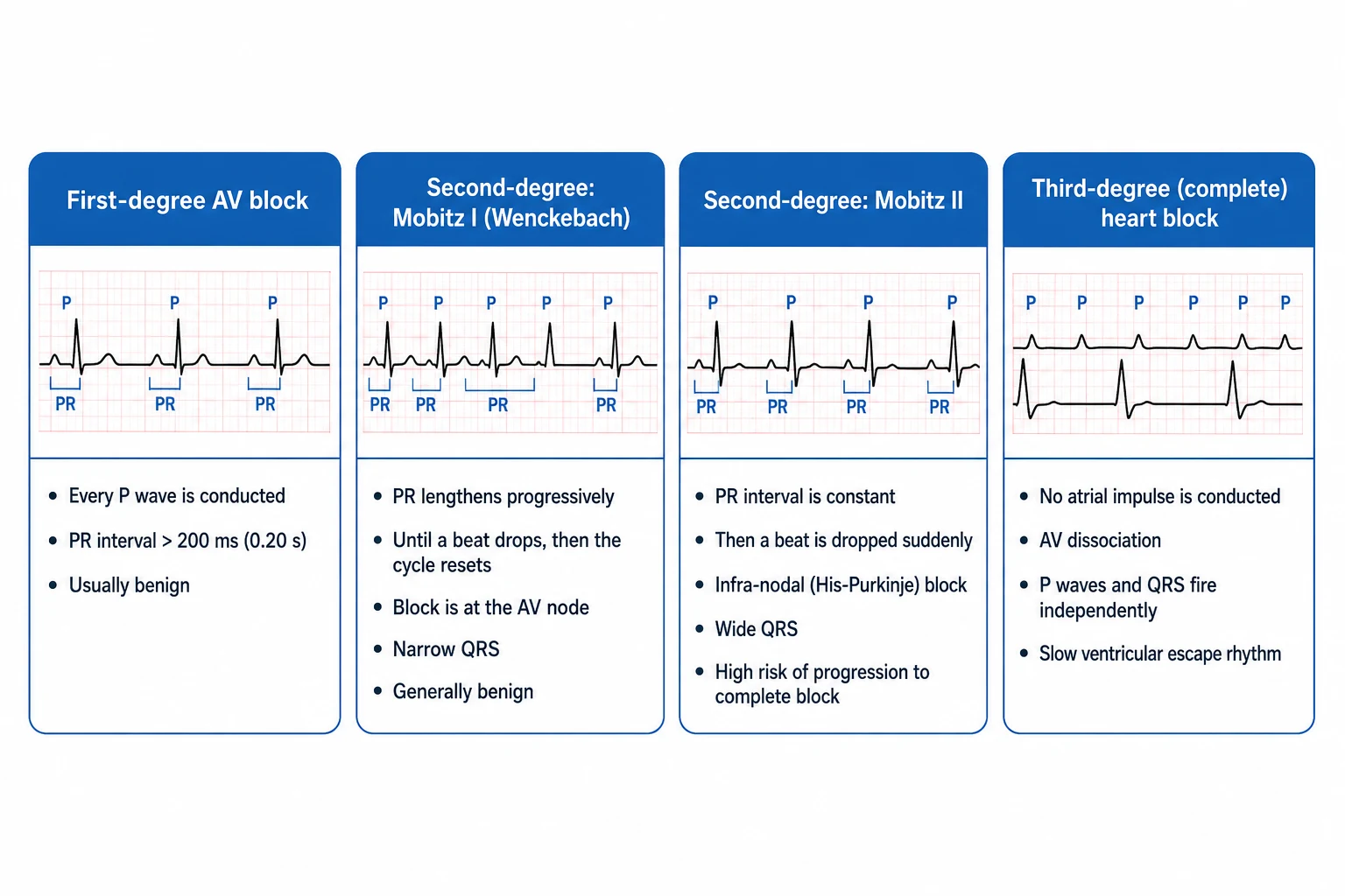
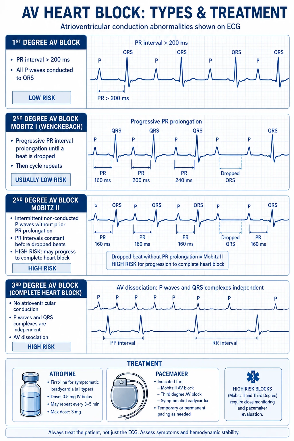
AV block is failure of atrial impulse conduction to the ventricles, graded by the relationship between P waves and QRS complexes. 1st-degree AV block — every P conducted, PR interval over 200 ms (over 300 ms on intracardiac recordings; one large-toe-to-three-large-toe width on the ECG at 25 mm/s), almost always at the level of the AV node, benign. 2nd-degree AV block — some P waves not conducted. Mobitz type I (Wenckebach) — progressive PR prolongation until a P is dropped, the pause less than twice the preceding PP, almost always AV nodal (proximal to the His bundle) and benign; atropine-responsive, vagal, often nocturnal in young adults. Mobitz type II — fixed (normal or slightly prolonged) PR with sudden non-conducted P waves; the lesion is infranodal (His–Purkinje), with high risk of progression to complete block and syncope, and is a Class I indication for permanent pacing even when asymptomatic. High-grade (advanced) AV block — two or more consecutive non-conducted P waves with sinus rhythm, requires AV nodal disease or infranodal disease. 3rd-degree (complete) AV block (CHB) — complete AV dissociation, atrial and ventricular rhythms independent; the ventricular escape is junctional (narrow QRS, 40–60 bpm) — relatively stable, or ventricular (wide QRS, 20–40 bpm) — unstable, syncope, sudden cardiac death. The anatomical correlate of congenital CHB is failure of AV node/His bundle development; of acquired Mobitz II and infranodal CHB, Lev disease (calcification of the aortic and mitral annuli extending into the conduction system) and Lenegre disease (idiopathic sclerotic degeneration of the conduction system).[1][3][4]
The clinical importance of bradyarrhythmia is its causation of syncope, falls, injury and sudden cardiac death when pauses are long enough to cause cerebral hypoperfusion (typically over 6 seconds), and its drain on cardiac output in patients with stiff, hypertrophied or failing ventricles (the same 35 bpm that is asymptomatic in a healthy 25-year-old produces florid heart failure in a 75-year-old with LV dysfunction). The exam skill is (1) pattern recognition on the 12-lead ECG (sinus brady vs SA block vs Wenckebach vs Mobitz II vs CHB), (2) anatomical localisation (nodal — narrow QRS, vagal, atropine-responsive — vs infranodal — wide QRS, structural, pacing-required), (3) search for reversible causes (drugs, ischaemia, electrolytes, Lyme, hypothyroidism), (4) prompt resuscitation for unstable bradycardia, and (5) definitive pacing for irreversible disease per 2018 ACC/AHA/HRS and 2013 ESC pacing.[1][3]
Classification
Bradyarrhythmias are classified by anatomical level of the block (which determines prognosis and treatment), by ECG pattern (the surface morphology), and by aetiology (which determines reversibility). [1]
Sinus bradycardia
- Regular P before every QRS, rate under 60 bpm
- Almost always benign when asymptomatic and a known athlete or during sleep
- Pathological when drug-induced, hypothyroid, post-inferior MI, raised ICP, severe hypoxia, sick sinus syndrome
Sinus arrest / SA exit block
- Sudden absence of P waves producing a pause; underlying PP not visible
- Pause not a multiple of PP — sinus arrest; pause equal to multiple of PP — SA exit block type II
- Long pause (over 6 seconds) risks Stokes-Adams syncope and indicates pacing
- Seen in sick sinus syndrome and vagal syncope
Sick sinus syndrome (SSS)
- Bradycardia plus sinus pauses plus bradycardia-tachycardia (AF with long sinus pauses on termination)
- Common indication for permanent pacing in the elderly (50 to 60 percent of implants over 70)
- Tachy-brady variant carries the highest stroke risk — anticoagulate per AF guidelines
1st-degree AV block
- PR over 200 ms, every P conducted
- Almost always AV nodal (proximal to His), benign
- Marked prolongation (over 300 ms) can produce symptoms by causing AV dyssynchrony (pacemaker syndrome equivalent)
- Rarely nodal disease progresses to higher-grade block
2nd-degree Mobitz I (Wenckebach)
- Progressive PR prolongation then a dropped QRS
- Usually AV nodal — narrow QRS, vagal, nocturnal in healthy young adults, inferior MI, drug effect
- Generally benign — observe; rarely needs pacing; **atropine improves** (nodal block speeds up)
- If associated with bifascicular block and symptoms, consider electrophysiology study
2nd-degree Mobitz II
- Fixed PR with sudden non-conducted P wave (no progressive PR prolongation)
- Infranodal lesion (His–Purkinje) — wide QRS usual, syncope risk high
- **Class I pacing even when asymptomatic** — risk of progression to CHB is roughly 50 percent within 1 year
- If associated with bifascicular block or syncope, the recommendation intensifies
3rd-degree (complete) AV block (CHB)
- P and QRS dissociated — atrial and ventricular rates independent
- Narrow QRS escape (junctional, 40 to 60 bpm, more stable) vs wide QRS escape (ventricular, 20 to 40 bpm, syncope common)
- Congenital — autoimmune maternal anti-Ro/SSA antibodies causing neonatal lupus; pacing if symptomatic or resting rate under 50
- Acquired — Class I pacing for permanent or persistent block; if reversible (drug, MI, post-op, Lyme), temporary pacing suffices
Bradycardia and AV block — key numbers
Epidemiology & Risk Factors
The epidemiology of bradyarrhythmia follows the two disease populations: (1) young, healthy patients with vagal or athletic bradycardia (and rare congenital CHB) and (2) older patients with degenerative conduction disease and ischaemic, drug, metabolic and surgical aetiologies.[1][4]
Sinus bradycardia and vagal bradycardia — resting sinus bradycardia is common: roughly 1 in 4 healthy adults have a daytime resting HR below 60 bpm, and up to 50 to 60 percent of trained endurance athletes have resting rates in the 30s at night on Holter. Vagal tone dominates in the young; vasovagal syncope is the commonest cause of syncope in young adults (and the second commonest overall, after cardiac syncope in older patients). [1]
AV block — 2nd- and 3rd-degree AV block are uncommon in the general population (prevalence under 1 percent) but rise with age: complete heart block is rare under 60 (under 5 per 100 000) and rises to over 200 per 100 000 over 80. Lev disease (calcific extension from aortic and mitral annuli into the conduction system, associated with hypertension, diabetes, and aortic sclerosis) and Lenegre disease (idiopathic sclerotic fibrosis of conduction tissue, often familial) account for the majority of acquired AV block in the elderly.[1][3]
Ischaemia and infarction — the SA node is supplied by the SA nodal artery (a proximal branch of the RCA in 60 to 70 percent) and by the left circumflex in 30 to 40 percent; the AV node is supplied by the AV nodal artery, a distal branch of the RCA in 80 to 90 percent. Hence inferior MI produces sinus bradycardia, sinus arrest and AV nodal block (typically Mobitz I / Wenckebach), which is usually self-limiting within 5 to 7 days but may require temporary pacing; anterior MI with proximal left anterior descending occlusion is rarer but produces infranodal block (Mobitz II / CHB) because of extensive septal necrosis — this is much more dangerous and the lesion is permanent.[3][4]
Drug toxicity is the commonest reversible cause in clinical practice — beta-blocker overdose, calcium-channel blocker overdose (especially verapamil and diltiazem), digoxin toxicity, amiodarone-induced sinus bradycardia and AV block (via calcium-channel blocking effect — common in the elderly), ivabradine (Iƒ-channel blocker), clonidine, and lithium. Electrolyte disturbance — hyperkalaemia (peaked T → PR prolongation → loss of P → sinusoidal → asystole — typically above 6.5 mmol/L), hypermagnesaemia, severe hypocalcaemia. Endocrine — hypothyroidism, Addisonian crisis, hypopituitarism.[1][4]
Inflammatory and infiltrative — Lyme disease (Borrelia burgdorferi) is the classic cause of reversible high-grade AV block in endemic areas (Northeast US, Northern Europe, parts of UK), occurring in 1 to 10 percent of untreated erythema migrans; the block is nodal, often responds to IV ceftriaxone, and may require temporary pacing. Cardiac sarcoidosis — infiltrative granulomatous disease of the septum with conduction block and ventricular arrhythmia, often requiring MRI and biopsy. Chagas disease (Trypanoma cruzi) — chronic apical aneurysm and conduction fibrosis, common in Latin America. Amyloidosis (AL and ATTR) — infiltrative cardiomyopathy with conduction block. Myocarditis — viral or autoimmune, may produce transient high-grade block.[3][4]
Post-cardiac surgery and structural — aortic valve replacement (mechanical valve impinging on the conduction system, especially right coronary cusp), transcatheter aortic valve implantation (TAVI) (membranous septum compression, commonest cause of new LBBB and CHB after TAVI — conduction disturbance rate 10 to 30 percent), ventricular septal defect (VSD) repair, tetralogy of Fallot repair, atrial septal defect closure, septal myectomy (HOCM), and alcohol septal ablation all carry risk of new high-grade AV block. [1]
Sleep apnoea is an increasingly recognised cause of nocturnal bradycardia and pauses (the diving reflex / vagal surge at apnoea termination), and is the commonest cause of bradycardia in middle-aged obese patients otherwise unexplained. Vagal reflex — cough syncope (during severe cough), micturition syncope (typically elderly men at night — vagally mediated bradycardia, often with vasodepression), swallow syncope, defaecation syncope.[3][4]
Pathophysiology
The pathophysiology of bradyarrhythmia is best understood by tracing the anatomical site of impulse failure — impulse formation (SA node), impulse conduction from atrium to AV node, AV node to His bundle, and His–Purkinje to ventricle — and the blood supply, autonomic input, metabolic environment and intrinsic disease at each site.[1][3]
1. The SA node is a crescent of specialised atrial tissue at the junction of the superior vena cava and right atrium, richly innervated by parasympathetic (vagal) and sympathetic fibres, and containing automatic cells whose intrinsic rate is reduced by vagal acetylcholine acting on M2 muscarinic receptors (slowing the funny current, Iƒ, and reducing the slope of phase 4 depolarisation) and increased by sympathetic noradrenaline acting on β1-adrenoceptors (increasing Iƒ and the calcium current). The result in health is a heart rate that varies with respiration (RSA, respiratory sinus arrhythmia), position, exertion, emotion and sleep. Pathological sinus bradycardia arises when (1) excessive vagal tone (athletes, cough, micturition, carotid sinus massage, raised ICP, inferior MI — all reflex vagal bradycardia), (2) intrinsic SA nodal disease (sick sinus syndrome — idiopathic fibrosis, amyloid, sarcoid, ischaemia), (3) extrinsic drug effect (beta-blocker, calcium-channel blocker, digoxin, amiodarone, ivabradine), or (4) metabolic or endocrine failure (hypothyroidism, hyperkalaemia). Sinus arrest is the sudden failure of impulse generation; the pause length is a function of the latency of the next available escape pacemaker (junctional = 1 to 2 seconds; ventricular = up to 6+ seconds).[1]
2. The AV node is the only electrical connection between atria and ventricles under normal conditions — the annulus fibrosus insulates the rest — and is the most vagally-influenced segment of the conduction system. At rest, vagal dominance keeps the AV nodal Wenckebach cycle length around 500 ms (rate 120 bpm) — a physiological upper rate limit above which AV nodal Wenckebach appears as an atrial-driven response (loss of 1:1 conduction with atrial pacing above 120). The same vagal dominance explains why Mobitz I is so commonly benign — vagal surges transiently depress AV nodal conduction and the PR lengthens. The AV node is also the commonest site of iatrogenic drug block (beta-blocker, digoxin, verapamil/diltiazem, amiodarone) and of ischaemic block in inferior MI because the AV nodal artery is a distal branch of the RCA in 80 to 90 percent of patients. Lyme carditis characteristically attacks the AV node, producing a reversible nodal block.[3][4]
3. The His–Purkinje system (the infranodal conduction) consists of the bundle of His (penetrating the central fibrous body and dividing into left and right bundles), fascicles (left anterior fascicle, left posterior fascicle), and the Purkinje network. This segment is largely not vagally-supplied, is dependent on fast sodium channels (phase 0 upstroke) rather than the slow calcium current, and has low automaticity — the escape rate from the His bundle is roughly 40 to 60 bpm, from a fascicle 30 to 40 bpm, and from the ventricle 20 to 40 bpm, the latter unreliable and unstable. The infranodal conduction is susceptible to (1) idiopathic fibrosis (Lenegre disease, accelerating in middle age), (2) calcific infiltration from aortic and mitral valve disease (Lev disease), (3) anterior MI with proximal LAD occlusion and septal necrosis, (4) infiltrative disease (sarcoid, amyloid), and (5) drugs that depress sodium channels (Class IA antiarrhythmics — procainamide, disopyramide; Class IC — flecainide, propafenone — these drugs are contraindicated in patients with structural heart disease and the result can be the unmasking of bundle branch block or the precipitation of asystole when used in patients with unrecognised conduction disease). Mobitz II and CHB with wide QRS are the surface ECG signs of infranodal block.[1][3]
4. The electrophysiological mechanisms of the patterns are worth understanding for the exam. Wenckebach is the gradual decrement in AV nodal conduction with each successive atrial impulse, until one impulse fails to conduct — the pause is less than twice the preceding RR (because during the dropped beat, the AV node recovers and the next PR after the dropped beat is the shortest of the cycle). Mobitz II is the sudden failure of an all-or-none His–Purkinje fibre — fixed PR with a sudden dropped QRS. CHB is the failure of all conduction — the atria and ventricles beat independently, the ventricular escape rhythm determining symptoms (junctional escape is relatively well tolerated, ventricular escape is syncopic and dangerous). Sinus arrest is the failure of impulse formation; the pause has no underlying sinus P waves, and an escape pacemaker must fire or asystole ensues.[3]
5. The role of autonomic tone is the mechanistic link between vagal syndromes and bradyarrhythmia — the vasovagal reflex classically has a cardioinhibitory component (sudden vagally-mediated bradycardia or asystole — Class II indication for pacing under 2018 ESC syncope guidelines) and a vasodepressor component (sympathetic withdrawal and venous pooling, pacing does not help); the carotid sinus reflex produces the same picture in older patients with carotid hypersensitivity (a 3-second pause or 50 mmHg systolic drop on carotid sinus massage in patients over 40 is diagnostic); and the cough, micturition, swallow and defaecation reflexes are all vagally-mediated. [1]
6. The relationship to myocardial ischaemia deserves explicit emphasis: bradycardia (especially sinus bradycardia) is common in inferior MI because the RCA supplies both the SA and AV nodes, vagal reflexes (Bezold–Jarisch reflex — a vagal surge from ischaemic myocardium) compound the picture, and the resulting block is usually transient (resolves as the infarct stabilises and vagal tone normalises) — temporary pacing suffices. Anterior MI with proximal LAD occlusion and septal necrosis destroys the His–Purkinje system and produces infranodal block that is permanent and requires permanent pacing.[1][3]
Clinical Presentation
The clinical presentation of bradyarrhythmia spans four gradients: (1) asymptomatic (incidental on ECG or device interrogation), (2) fatigue / exercise intolerance / breathlessness, (3) presyncope (dizziness, lightheadedness, nausea, sweating) and (4) syncope (transient loss of consciousness) or sudden cardiac death when long pauses occur. The exam skill is recognising which clinical picture is bradyarrhythmia.[1][4]
Symptoms (the patient may be unable to give a long history after syncope): [1]
- Cardiac symptoms of the underlying cause — chest pain of acute MI (crushing central, radiation, autonomic features), palpitation of preceding tachyarrhythmia (in brady-tachy SSS, the patient may describe the AF that terminated in a pause), breathlessness of pulmonary oedema if the slow rate cannot match cardiac output demand.
- Fatigue, lethargy, exercise intolerance — the commonest symptom of mild bradycardia; the patient describes inability to climb stairs, exertional breathlessness, "I'm slowing down" — often misattributed to ageing.
- Presyncope / dizziness / lightheadedness — the cardinal "I had to sit down" symptom of transient cerebral hypoperfusion from bradyarrhythmia; pauses may be hours apart, days apart, or weeks apart.
- Stokes-Adams syncope — sudden loss of consciousness without warning, with rapid recovery (no post-ictal confusion, no Todd paresis) caused by a sudden fall in cardiac output from an asystolic pause (CHB, sinus arrest, vasovagal cardioinhibitory). The patient may recall darkness before the eyes, falling, and rapid recovery (seconds to a minute) — a key differentiator from seizure (which has tonic-clonic activity, tongue biting, urinary incontinence and post-ictal confusion) and from orthostatic syncope (preceded by lightheadedness on standing).
- Falls and injury — bradyarrhythmic syncope in the elderly may present as a fall with hip fracture or head injury, masquerading as a mechanical fall — a 12-lead ECG should be performed in every unexplained fall.
- Sudden cardiac death — bradyarrhythmia (especially CHB) is a recognised, though less common, cause of out-of-hospital cardiac arrest; Mobitz II and acquired CHB are the most lethal patterns.[1][4]
Vital signs and bedside observation: [1]
- Resting HR below 50 bpm — bradycardia by definition. Below 40 bpm strongly suggests pathological block; below 30 bpm in a non-athlete is bradyarrhythmia until proven otherwise.
- Irregular irregular rate — AF with bradycardia, AF with pauses, sinus arrhythmia, Wenckebach pattern, SA block / sinus arrest.
- Pulse character — the slow-rising pulse of low stroke volume, the regular slow rhythm of CHB ("cannon waves" in the JVP because of atrial contraction against a closed tricuspid valve — see below).
- Blood pressure — orthostatic, supine-to-standing drop of over 20 mmHg systolic (orthostatic hypotension is a common cause of apparent brady-symptom overlap, especially in the elderly with autonomic dysfunction). [1]
Examination (organ by organ, looking for the cause): [1]
- JVP — slow-rate raises the chance of cannon a waves in CHB, where the atria contract against a closed tricuspid valve; the slow-rate wave at the JVP has no relationship to the rate of the arterial pulse. Distended neck veins are uncommon unless decompensation has set in.
- Heart sounds — varying intensity of S1 in CHB (the variable AV relationship alters the timing of mitral leaflet closure); soft S1 if PR is long; loud S1 if P and QRS are close together.
- Carotid sinus massage — should be performed with continuous ECG monitoring after auscultation of the carotids and exclusion of carotid bruit; a pause over 3 seconds or a systolic BP drop over 50 mmHg with symptom reproduction confirms carotid sinus hypersensitivity (the cardioinhibitory form is the indication for pacing under 2018 ESC syncope guidelines).[4]
- Thyroid — goitre, slow-relaxing ankle reflex for hypothyroidism (a treatable cause of sinus bradycardia).
- Skin and complexion — pallor, jaundice (Addisonian crisis), erythema migrans (Lyme).
- Neurological — pupil asymmetry and extensor posturing suggest raised ICP rather than bradyarrhythmia.
Atypical presentations (high-yield): [1]
- Elderly faller — Stokes-Adams presenting as hip fracture.
- Young athlete — asymptomatic bradycardia on pre-participation screening ECG; reassurance in the absence of symptoms or pauses.
- Pregnancy — raised HR expected; bradycardia with dizziness or presyncope suggests the rare peripartum cardiomyopathy with conduction block or beta-blocker over-treatment for hypertension.
- Post-cardiac surgery (AVR, TAVI, septal repair) — new CHB or Mobitz II requires pacing; observation for 5 to 7 days post-TAVI before proceeding to permanent device because conduction recovery is common.[1][3]
Differential Diagnosis
The first task is to separate true bradyarrhythmia from mimics, particularly because the management of a syncopal patient with apparent bradycardia on a single ECG can be misleading. The differential covers reflex syndromes, orthostatic causes, other cardiac causes of syncope, and non-cardiac mimics.[3][4]
Vasovagal (reflex) syncope
- Prodrome of warmth, nausea, sweating, blurred vision ('I could feel it coming') — followed by syncope
- Bradycardia often paired with vasodepression — pacing alone does not help (VPS-II trial)
- Trigger identifiable — pain, emotional upset, prolonged standing, hot bath, venesection
- Cardioinhibitory vasovagal (asystolic, on tilt) — Class IIb pacing consideration in highly-selected patients over 40 with recurrent syncope
Carotid sinus hypersensitivity
- Triggered by head turning, tight collar, shaving over neck, carotid sinus massage
- Cardioinhibitory — over 3-second pause on carotid sinus massage with symptom reproduction
- Vasodepressor — over 50 mmHg systolic BP drop on massage
- Mixed form — both
- Class I pacing for symptomatic cardioinhibitory CSS over 40
Orthostatic hypotension
- Supine to standing drop in systolic over 20 mmHg (or diastolic over 10 mmHg)
- Symptoms within 3 minutes of standing (initial) or after prolonged standing (delayed)
- Autonomic failure (Parkinson disease, dementia, diabetes), volume depletion (diuretics, haemorrhage, vomiting), drug-induced (alpha-blocker, vasodilator)
- Reassure, hydration, discontinue culprit drug, midodrine, fludrocortisone
Neurocardiogenic syncope
- Older umbrella term — used to describe mixed vasovagal and reflex syndromes
- Diagnosed by **head-up tilt testing** — positive if syncope reproduced with bradycardia and hypotension
- Treatment is mostly non-pharmacological — hydration, salt, counter-manoeuvres (leg crossing, hand grip)
- Drug-refractory recurrent cardioinhibitory vasovagal — may be considered for pacing per ISSUE-3 (pacing reduces syncope burden but not mortality)
Cardiac non-bradyarrhythmic syncope
- **VT in structural heart disease** (ischaemic cardiomyopathy, HOCM, ARVD) — sudden collapse, ICD candidate, NOT a pacing indication
- Aortic stenosis, HOCM, PE, tamponade — flow obstruction syncope, NOT bradyarrhythmic
- Supraventricular tachycardia (AVNRT, AVRT) with rate-related hypotension — bradycardia after termination, not the cause
- Pulmonary embolism with right-heart strain — sinus tachycardia, not bradycardia; misdiagnosed bradyarrhythmia is a delay to CTPA
Non-cardiac syncope mimics
- **Seizure** — tonic-clonic activity, tongue biting, urinary incontinence, post-ictal confusion, **no rapid recovery**
- **Psychogenic pseudosyncope** — prolonged apparent loss of consciousness with normal haemodynamics and no EEG correlate
- Vertebrobasilar TIA — focal neurological signs, vertigo, diplopia, dysarthria
- Subarachnoid haemorrhage — sudden severe headache, meningism, focal signs
- Hypoglycaemia — confusion, sweating, known diabetes, low capillary glucose
The single best discriminator at the bedside is the ECG combined with continuous rhythm monitoring during symptoms and a detailed history of the trigger, prodrome and recovery. Prolonged unexplained syncope with normal ECG and Holter requires implantable loop recorder (ILR) — the SPAIN, ISSUE-3 and other trials demonstrated diagnostic yield above 30 percent in unexplained recurrent syncope with ILR, and pacing in detected bradyarrhythmia reduced syncope burden in the ISSUE-3 / ISSUE-3-derived populations.[1][3][4]
Clinical & Bedside Assessment
The bedside assessment of bradyarrhythmia is ABCDE first, ECG in every suspected case, continuous monitoring, and a focused search for the cause.[1][4]
A — Airway and Breathing: [1]
- Secure airway if patient has syncope or impaired consciousness.
- Supplemental oxygen to SpO2 94 to 98 percent if hypoxic (88 to 92 percent in COPD / CO2 retainers); excess oxygen in inferior MI may cause vasoconstriction and worsen output — keep SpO2 under 99 percent if no hypoxaemia. [1]
B — Breathing pattern observation: Slow, deep breaths suggest excess vagal tone (vasovagal); Cheyne–Stokes respiration suggests heart failure, raised ICP, or post-cardiac-arrest; Biot respiration suggests brainstem lesion. [1]
C — Circulation: [1]
- Heart rate and rhythm — palpate the radial pulse for 60 seconds; count a full minute because bradyarrhythmia often fluctuates.
- Blood pressure — supine and standing; postural drop suggests volume, autonomic or drug cause.
- ECG monitoring — continuous ECG telemetry with rate and rhythm alarms; print a rhythm strip at every event.
- 12-lead ECG — performed immediately. Look for:
- P-wave morphology — sinus vs low atrial vs absent (sinus arrest / AF with pauses)
- PR interval — 1st-degree block, Wenckebach periodicity, fixed PR with dropped beat
- QRS width — narrow = nodal escape, wide = ventricular escape (worse prognosis)
- R–R relationship — regular slow = sinus brady or CHB; irregular slow = AF with bradycardia or Wenckebach with variable conduction; PP equal with missed P = SA exit block type II; PP unequal with missed P = sinus arrest
- Bundle branch patterns — bifascicular block plus PR prolongation is a precursor to CHB (Class IIa pacing if symptomatic or syncopal, Class I pacing if EP study HV over 70 ms).
- Carotid sinus massage (with continuous ECG and BP) — in patients over 40 with unexplained syncope, performed in supine position, gentle unilateral massage over 5 seconds; positive if pause over 3 seconds or systolic drop over 50 mmHg with symptom reproduction. No longer contraindicated in the post-stroke window per 2018 ESC syncope guidelines (the previous 3-month exclusion was removed).[4]
- JVP — slow rate with cannon a waves is diagnostic of AV dissociation (CHB); otherwise inspect for raised JVP of heart failure or pulmonary hypertension.[1][3]
D — Disability: [1]
- Blood glucose — to exclude hypoglycaemia as a mimic, particularly in patients on insulin or sulphonylureas.
- Neurological — pupil asymmetry, conscious level, focal signs for stroke / raised ICP.
- Differential — focal neurology with bradycardia suggests raised ICP (Cushing reflex — bradycardia + hypertension + irregular breathing). [1]
E — Exposure: [1]
- Skin — jaundice (Addison, jaundice), erythema migrans (Lyme), cyanosis (hypoxia), pallor (anaemia), surgical scars (post-cardiac surgery).
- Neck circumference — sleep apnoea phenotype.
- Cardiovascular examination — heart sounds, murmurs (aortic sclerosis/Lev), prosthetic valves (mechanical AVR, mitral repair), pulmonary crepitations (heart failure), peripheral oedema. [1]
The "bradycardia bundle" at the bedside (Resuscitation Council UK / ACLS bradycardia algorithm): [1]
- Confirm the rhythm on ECG.
- Assess for adverse features — syncope, hypotension (SBP under 90), confusion, signs of shock, ischaemic chest pain, acute heart failure. If any adverse feature is present, treat aggressively.
- Identify and treat the reversible cause — drugs, ischaemia, electrolyte, hypoxia.
- If symptomatic bradycardia — atropine 500 mcg IV bolus, repeat every 3 to 5 minutes to a maximum of 3 mg total (3 mg is the fully vagolytic dose).
- If no response — second-line — isoprenaline (isoproterenol) 5 mcg/min IV infusion, titrated up to 20 mcg/min OR adrenaline (epinephrine) 2 to 10 mcg/min infusion OR transcutaneous pacing OR transvenous pacing.
- If stable but irreversible cause — same-day permanent pacing (Mobitz II, CHB).[1][3]
Investigations
First-line (in parallel with resuscitation): [1]
- 12-lead ECG — the single most useful investigation for diagnosis. Look for sinus vs junctional vs ventricular rate, PR interval and relationship, QRS width, bundle branch pattern, evidence of ischaemia (ST elevation / depression in MI), and QT interval (drug effect). Repeat ECG during symptoms to capture transient rhythm.
- Continuous ECG telemetry — for the in-patient; triggers capture of all bradycardic episodes with rate, duration and rhythm correlation to symptoms. Alarm settings must include asystolic pauses over 3 seconds, rate under 40 bpm sustained 30 seconds, and Mobitz II / CHB.
- Ambulatory ECG (Holter) — 24-hour to 48-hour continuous recording in patients with frequent symptoms (at least once per 24 hours); captures roughly 50 percent of culprit rhythms in syncopal patients. Preferred at presentation before more invasive testing.
- Patch / event monitor — 7 to 14 days — for patients with less frequent symptoms. The Zio patch (continuous up to 14 days) has a diagnostic yield of over 50 percent in selected patients.[4]
- Implantable loop recorder (ILR) — up to 3 years — for recurrent unexplained syncope with normal ECG and Holter; Class I indication in this group per 2018 ESC syncope guidelines. ILR-guided therapy (pacing for bradyarrhythmia, ablation for tachyarrhythmia, ICD for VT) reduces syncope burden in ISSUE-3-derived populations.[4]
Second-line: [1]
- Transthoracic echocardiogram (TTE) — mandatory in suspected bradyarrhythmia: structural heart disease, valvular disease (aortic sclerosis, calcification, rheumatic, congenital), LV and RV function, infiltrative disease appearances, pericardial effusion. The TTE guides whether a new conduction block is the consequence of an underlying cardiomyopathy (sarcoid, amyloid, ARVD) which will change management.[1][3]
- Exercise stress testing — for exertional bradycardia or suspected chronotropic incompetence; failure to reach 80 percent of maximum predicted HR with symptoms is diagnostic. Also exposes ischaemia as a cause. Use caution — Mobitz II and CHB may deteriorate with exercise (infranodal disease worsens with catecholamine surge as the atrial rate increases and the diseased His–Purkinje cannot conduct).[1][3]
- Electrophysiology study (EPS) — invasive — for bifascicular block plus syncope (syncope unexplained despite ECG and ILR, in a patient with bifascicular block — measure HV interval; HV over 70 ms predicts progression to CHB); suspected SSS with pauses (sinus node recovery time over 1.5 to 2 seconds is positive). Less commonly used since ILR became available, but central to decision-making where ILR is unavailable or pacing is being considered.[1][3]
Blood tests: [1]
- Electrolytes — sodium, potassium, magnesium (hyperkalaemia, hypermagnesaemia commonest), calcium (hypocalcaemia prolongs QT and predisposes to arrhythmia).
- Renal function — uraemia, hyperkalaemia.
- Thyroid function — TSH for hypothyroidism (a common treatable cause); over-replacement in levothyroxine also causes tachyarrhythmia.
- Cardiac enzymes (troponin) — for suspected MI; inferior STEMI is a common cause of sinus and AV nodal block.
- Digoxin level — if patient on digoxin (toxicity is in the differential of any bradyarrhythmia on digoxin).
- Lyme serology — in endemic areas with high-grade AV block, especially summer months, history of tick exposure or erythema migrans.
- Autoimmune — anti-Ro/SSA antibodies in suspected neonatal lupus / congenital CHB in offspring.
- Arterial blood gas — for hypoxia, hypercapnia and acid–base state.[3][4]
Advanced: [1]
- Cardiac MRI — for cardiac sarcoidosis (T2-weighted oedema, late gadolinium enhancement in septum and LV free wall), myocarditis, amyloidosis, ARVD. Useful in unexplained AV block in a young patient.
- CT coronary angiography / coronary angiography — when ischaemia is the cause (inferior MI producing AV block, anterior MI producing infranodal block) — guides primary PCI.
- Right ventricular biopsy — endomyocardial, for giant-cell myocarditis, cardiac sarcoidosis, transplant rejection; aetiology-specific high-yield but rarely used.[3]
Named scores / criteria — reproduced verbatim
2018 ACC/AHA/HRS Guideline summary (Kusumoto et al. 2019)[1][2][7]
| Pattern | Class I pacing (must do) | Class IIa (reasonable to do) |
|---|---|---|
| Symptomatic sinus bradycardia / SSS | Persistently symptomatic, irreversible; pause over 3 s; chronotropic incompetence | Symptomatic SSS without pauses if reversible cause not identified |
| Acquired Mobitz II | Yes (even when asymptomatic) | — |
| Acquired CHB | Yes (regardless of symptoms) | — |
| Post-MI AV block | Persistent second- or third-degree block after MI | Transient block with bifascicular block and HV over 70 ms at EP study |
| Post-cardiac surgery, post-TAVI | Persistent after 5 to 7 days observation | New LBBB with HV over 70 ms |
| Asymptomatic bifascicular block + syncope | If HV over 70 ms | Reasonable if HV 55 to 70 ms |
| Lyme, drug-induced | — | If symptomatic and required, temporary; pacing is rare |
| Reflex syncope (vasovagal, CSS) | Cardioinhibitory CSS over 40 with recurrent syncope | Recurrent asystolic vasovagal syncope over 40 refractory to non-pharmacological treatment |
2013 ESC Pacing Guidelines (Brignole et al.)[3]
- Class I — symptomatic SSS (pauses / bradycardia), Mobitz II, CHB, post-MI persistent 2nd/3rd-degree block, post-cardiac-surgery persistent block, bifascicular block with syncope and HV over 70 ms.
- Class IIa — symptomatic SSS without documented bradycardia but clear causal relationship, asymptomatic bifascicular block with HV over 70 ms.
- Class IIb — asymptomatic SSS in over 70 with pauses over 6 seconds, cardioinhibitory reflex syncope with recurrent syncope and tilt-positive asystolic pattern.
- Class III (no benefit) — asymptomatic sinus bradycardia, asymptomatic vagal brady, reversible drug-induced block, end-stage terminal illness, severe cognitive impairment. [1]
2018 ESC Syncope Guidelines (Brignole et al.)[4]
| Reflex subtype | Diagnostic criterion | Treatment |
|---|---|---|
| Cardioinhibitory carotid sinus syndrome | CSM-induced pause over 3 s with symptom reproduction | Class I pacing (dual-chamber) over age 40 |
| Vasodepressor CSS | CSM-induced SBP drop over 50 mmHg with symptom reproduction | No proven drug; midodrine, fludrocortisone, supportive measures |
| Vasovagal syncope | Tilt-induced, situational trigger, prodrome | Education, hydration, counter-pressure manoeuvres; pacing if cardioinhibitory refractory (over 40, asystolic on tilt) |
| Orthostatic hypotension | Standing SBP drop over 20 mmHg or diastolic over 10 mmHg | Volume, drug review, midodrine, fludrocortisone |
High-yield numbers for examination
Management — Resuscitation
The acute management of bradyarrhythmia is staged, dopamine-tinged with atropine as the first-line, escalating to pacing as the second-line, as codified in the Resuscitation Council UK / European Resuscitation Council 2021 bradycardia algorithm and the 2018 ACC/AHA/HRS bradycardia guideline.[1][3]
ABCDE first, in parallel with diagnostic work-up: [1]
- Oxygen to SpO2 94 to 98 percent (88 to 92 percent in COPD).
- Two large-bore cannulae; continuous ECG and BP monitoring; 12-lead ECG (do not delay).
- Capillary blood glucose.
- Bedside echo if cause is unclear.
- Send bloods — FBC, U&E (potassium, magnesium, calcium), troponin, thyroid, digoxin level, Lyme serology, ABG. [1]
If the patient has adverse features — syncope, hypotension, confusion, ischaemic chest pain, acute heart failure — treat aggressively: [1]
- Atropine 500 mcg IV bolus (this is the Resuscitation Council UK dose; ACLS uses 1 mg) — increases SA node rate and AV nodal conduction by blocking vagal acetylcholine. Repeat every 3 to 5 minutes to a maximum of 3 mg total (3 mg fully vagolytic — additional doses have no further vagal effect). Atropine works for vagal bradycardia, sinus bradycardia, Mobitz I, SA exit block — and does NOT work for Mobitz II, infranodal CHB (the lesion is below the vagal-influenced AV node). Cardiac transplant recipients have denervated hearts and will not respond to atropine — use isoprenaline or pacing.[1][3]
- Second-line — if atropine fails or is contraindicated (Mobitz II, infranodal block, transplant recipient) — isoprenaline (isoproterenol) IV infusion 5 mcg/min, titrated up to 20 mcg/min to achieve HR over 50 bpm or restore symptoms. Adrenaline (epinephrine) 2 to 10 mcg/min (often 5 to 10 mcg/min) is an alternative; dopamine 5 to 20 mcg/kg/min for inotropic support if bradycardia is accompanied by hypotension in the context of MI or sepsis. Aminophylline — 100 mg IV bolus over 1 minute, then infusion — is an alternative in atropine-resistant bradycardia post-cardiac surgery or MI (adenosine receptor antagonism).[1][3]
- Transcutaneous pacing (TCP) — demand mode, rate 60 to 80 bpm, current set to capture (start 30 mA, increase until QRS capture confirmed on monitor, then dial back to lowest capturing current). Effective in minutes, no specialised equipment beyond a defibrillator with pacing function, uncomfortable (the patient will feel chest wall muscle contraction) — always give sedation and analgesia (midazolam 1 to 2 mg IV for anxiety, morphine 2 to 5 mg IV for analgesia; or ketamine 10 to 30 mg IV). TCP is a bridge to definitive transvenous or permanent pacing.[1][4]
- Transvenous pacing — placement of a pacing wire via the internal jugular or femoral vein into the right ventricle under fluoroscopy. Indicated when TCP fails to capture, when pacing is required for over 30 minutes, or when pacemaker implantation is being deferred. Risks — pneumothorax, arterial puncture, RV perforation, infection; requires operator experience and fluoroscopy or blind placement.
Cardiac arrest with bradyarrhythmia: if the patient is in asystolic or severe bradycardic arrest, follow the standard ALS algorithm — adrenaline (epinephrine) 1 mg IV every 3 to 5 minutes, atropine 3 mg IV (the full vagolytic dose) once only, identify and treat the reversible cause (the 4 H's: hypoxia, hypo/hyperkalaemia, hypovolaemia, hypothermia, plus the 4 T's: thrombosis, tamponade, tension pneumothorax, toxins). Precordial thump may be considered at the start of a witnessed monitored arrest with asystole or VF (rare in bradyarrhythmia; more relevant for VT).[1][3][4]
Adjunctive therapy in MI-related bradyarrhythmia: [1]
- Inferior MI with AV block — reperfusion is the definitive therapy; sinus and nodal blocks usually recover within 7 days. Temporary pacing for symptomatic bradycardia (atropine as first-line, transvenous pacing for atropine non-response).
- Anterior MI with infranodal block — urgent reperfusion (primary PCI); block is usually permanent because the septal necrosis destroys the His–Purkinje system — permanent pacing required.[1]
Management — Definitive & Stepwise
The definitive management of bradyarrhythmia is cause-specific and staged: identify and treat the cause, support the patient through the acute phase, and implant a permanent pacemaker when the cause is irreversible and the bradyarrhythmia is clinically significant.[1][3]
Step 1 — Identify and treat the reversible cause (within minutes to hours)
The first priority is to search for a reversible cause because successful treatment obviates the need for permanent pacing. [1]
- Drug toxicity — withhold the offending drug (beta-blocker, calcium-channel blocker, digoxin, amiodarone, ivabradine). For digoxin toxicity use Digoxin Immune Fab (DigiFab) in symptomatic bradycardia; supportive care with atropine and pacing as required. For beta-blocker overdose, glucagon 5 to 10 mg IV bolus then infusion plus high-dose insulin / dextrose euglycaemic therapy and IV lipid emulsion in severe cases. For calcium-channel blocker overdose, calcium gluconate 30 mL of 10 percent IV plus high-dose insulin / dextrose and vasopressors. Hyperkalaemia — calcium gluconate 30 mL of 10 percent IV over 5 min, insulin / dextrose 10 units in 50 mL 50 percent dextrose, salbutamol nebulisation, sodium bicarbonate if acidotic, and consideration of renal replacement therapy. Hypermagnesaemia — calcium gluconate (the membrane-stabilising action).[1][3][4]
- Ischaemia — emergency reperfusion (primary PCI within 90 minutes for STEMI, fibrinolysis if PCI unavailable within 120 minutes). Bradyarrhythmia in inferior MI is usually self-limiting (resolves as the RCA territory becomes reperfused or the vagal surge subsides); temporary pacing as required.
- Lyme carditis — IV ceftriaxone 2 g once daily for 14 to 21 days for high-grade AV block in hospitalised adults; oral doxycycline 100 mg twice daily for lower-severity disease. Temporary pacing may be required, but the block is typically reversible within 1 to 2 weeks and permanent pacing is rare if the antibiotic course is completed.[4]
- Hypothyroidism — levothyroxine replacement, starting at 25 to 50 mcg daily in the elderly, titrated every 4 to 6 weeks; bradycardia resolves over weeks.
- Obstructive sleep apnoea — CPAP reduces nocturnal bradycardia and improves oxygenation.
- Vagal syndromes — vasovagal: non-pharmacological (education, salt, hydration, counter-pressure manoeuvres, head-up tilt sleeping); carotid sinus hypersensitivity: dual-chamber pacing for the cardioinhibitory form over age 40.
Step 2 — Pacemaker implantation (the definitive pacing therapy) — if the cause is irreversible
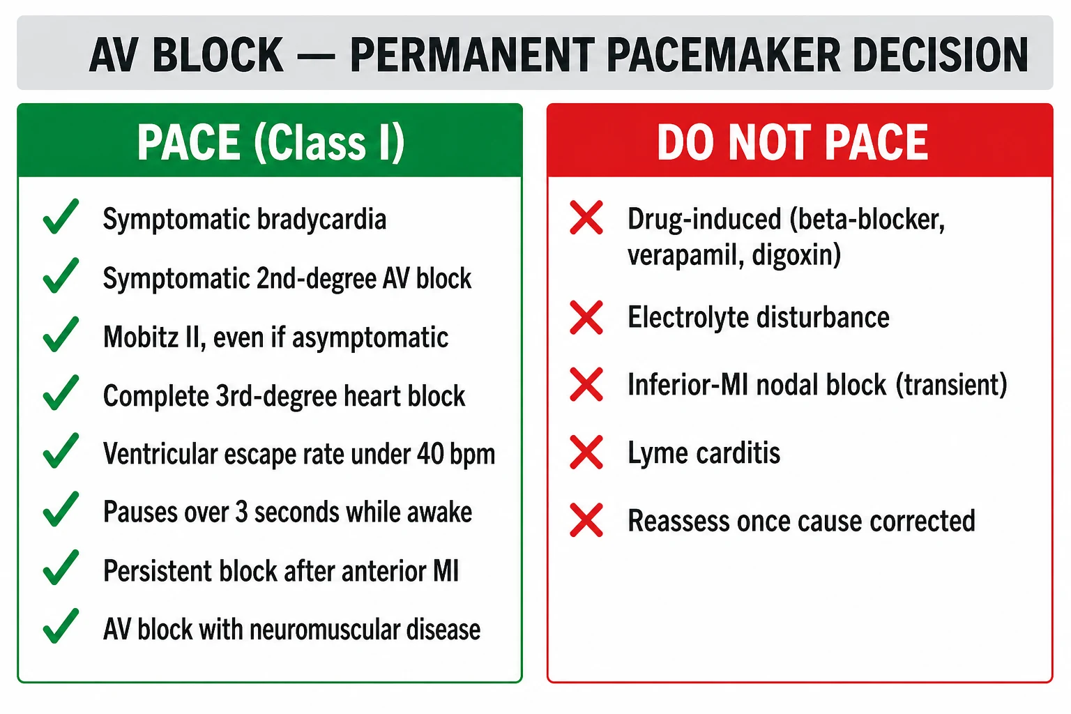
Class I permanent pacing indications (per 2018 ACC/AHA/HRS and 2013 ESC guidelines):[1][3]
- Symptomatic sinus node dysfunction including the brady-tachy form of SSS.
- Acquired Mobitz II AV block — even when asymptomatic.
- Acquired complete heart block — even when asymptomatic.
- Post-MI persistent second- or third-degree AV block.
- Post-cardiac-surgery or post-TAVI persistent block.
- Asymptomatic bifascicular block with syncope and HV over 70 ms on EP study.[1][3]
Pacemaker codes (NBG; NASPE/BPEG Generic): [1]
- VVI — ventricular pacing, ventricular sensing, inhibited mode (single-chamber ventricular). Used when atrial sensing is unnecessary (chronic AF with bradycardia).
- AAI — atrial pacing, atrial sensing, inhibited mode. Used in isolated sinus node dysfunction with intact AV conduction.
- DDD — dual-chamber pacing and sensing, with atrial tracking (P-synchronous ventricular pacing) and rate-responsive modes. The standard for sinus node dysfunction and AV block in sinus rhythm (preserves AV synchrony, reduces pacemaker-syndrome, improves exercise capacity).[1][3]
Choosing the device (the practical algorithm):[3]
Sinus node dysfunction (intact AV conduction)
- **AAI** if atrial lead reliable and AV node Wenckebach above 130 bpm
- **DDD** if any AV nodal disease present
- Rate-responsive (DDDR / AAIR) for chronotropic incompetence — adjusts pacing rate to activity
AV block in sinus rhythm
- **DDD** is standard — preserves AV synchrony, improves haemodynamics, reduces AF over VVI
- Minimise right ventricular apical pacing — promote intrinsic conduction (programmed to long AV interval); His bundle or left bundle branch area pacing increasingly used to avoid pacing-induced cardiomyopathy
- MRI-conditional leads if MRI anticipated
Chronic AF with bradycardia
- **VVI** single-chamber ventricular pacemaker
- Avoid tracking mode (DDD) — no P wave to track and atrial lead is unnecessary; rate response (VVIR) for activity
- Consider AV-node ablation + pacing ('ablate-and-pacemaker') for AF with rapid ventricular response not controlled by drugs
Reflex syncope (over 40)
- **DDDR** with **closed-loop stimulation / rate-drop hysteresis** — pacing responds to a sudden rate drop rather than to absolute rate
- ISSUE-3 / ISSUE-3-derived — pacing reduces syncope burden in cardioinhibitory vasovagal syncope but does NOT eliminate it (because vasodepression is the dominant mechanism in many)
- Carotid sinus syndrome — dual-chamber pacing reduces syncope recurrence from 60 percent to 10 percent
Biventricular pacing (CRT)
- Bundle branch block (LBBB over 150 ms) with LVEF under 35 percent and NYHA II-IV — **Class I** per 2022 AHA/ACC/HFSA HF guideline and 2021 ESC pacing/CRT guideline
- AV block requiring frequent ventricular pacing with LV dysfunction — **CRT upgrade**
- Not for isolated bradyarrhythmia with normal LV function
His bundle / conduction system pacing
- Alternative to RV apical pacing and biventricular pacing — direct capture of the His bundle or left bundle branch area
- Preserves normal ventricular activation, avoids pacing-induced cardiomyopathy
- Increasingly used in AV block, particularly in younger patients and those at risk of pacing-induced LV dysfunction
Step 3 — Adjunctive therapies and chronic management
- Anticoagulation in brady-tachy SSS / paroxysmal AF — per CHA2DS2-VASc score, no different from other AF populations.
- Driving and occupational restrictions — symptomatic bradyarrhythmia mandates driving cessation in many jurisdictions; permanent pacemaker restores licence eligibility after a defined period of freedom from syncope.
- Sport and exercise — paced patients with stable restoration of normal heart rate and no evidence of underlying structural disease can usually return to recreational sport; competitive collision sport is restricted in some devices because of the lead vulnerability.
- Follow-up — device clinic review at 1 week, 1 month, every 6 to 12 months, with remote monitoring for the majority of devices. Battery end-of-life (EOL) and lead failure are anticipated complications. [1]
Stepwise Management Protocol
The Resuscitation Council UK / European Resuscitation Council 2021 bradycardia algorithm is the bedside decision-tree that brings the resuscitation, cause identification and definitive therapy together.[1][3][4]
| Step | Action | Threshold / dose |
|---|---|---|
| 1 | Assess rhythm and adverse features | Syncope, SBP under 90, confusion, chest pain, acute heart failure |
| 2 | Identify and treat reversible cause | Drugs, ischaemia, electrolyte, hypoxia, raised ICP, hypothermia |
| 3 | If adverse features — Atropine 500 mcg IV bolus | Repeat every 3 to 5 min to max 3 mg total |
| 4 | If atropine fails or Mobitz II / infranodal — second-line | Isoprenaline 5 mcg/min IV OR adrenaline 2 to 10 mcg/min OR TCP OR transvenous pacing |
| 5 | If in arrest — ALS algorithm | Adrenaline 1 mg IV every 3 to 5 min; atropine 3 mg IV once |
| 6 | If stable but cause irreversible — permanent pacing | Single-chamber (VVI/AAI) or dual-chamber (DDD) within 48 to 72 hours |
| 7 | If reversible cause — observe / temporary pacing | Repeat ECG and Holter after recovery; reassess need for permanent |
| 8 | Disposition | CCU for symptomatic bradycardia / Mobitz II / CHB; ward monitor for stable sinus brady; outpatient if asymptomatic and reversible |
Subtypes & Scenarios
- Inferior MI with AV block — RCA supplies SA node (60 to 70) and AV node (80 to 90). Sinus bradycardia, sinus arrest and nodal block (Mobitz I) common. Usually self-limiting within 5 to 7 days. Temporary pacing for symptomatic or hypotension patients. Atropine-responsive.[3][4]
- Anterior MI with new AV block — proximal LAD occlusion produces septal necrosis and infranodal block (Mobitz II, CHB). Mortality high because of the size of MI. Permanent pacing required.[1][3]
- Lyme carditis — Borrelia burgdorferi produces reversible nodal block in endemic areas; Class IIa admission for high-grade block with IV ceftriaxone 2 g once daily; temporary pacing suffices in most.[4]
- Post-cardiac surgery — particularly aortic valve replacement (mechanical valve impact on conduction tissue), TAVI (10 to 30 percent new LBBB; 2 to 8 percent new CHB), ventricular septal defect repair, tetralogy of Fallot repair, septal myectomy / alcohol septal ablation. Observe for 5 to 7 days before permanent device because conduction recovery is common.[3]
- Sleep apnoea — central or obstructive sleep apnoea produces nocturnal bradycardia and pauses at apnoea termination (diving reflex). CPAP reduces nocturnal bradycardia and improves symptoms.[4]
- Athletic bradycardia — trained endurance athletes have resting HR 30 to 60 bpm with sinus rhythm, no pauses, no symptoms — physiological and not an indication for pacing. Distinguish by absence of symptoms, normal Holter (apart from low HR), and absence of pauses.[1][3]
- Reflex syncope over 40 — vasovagal syncope with cardioinhibitory component (asystole on tilt); pacing (DDDR with rate-drop feature) reduces syncope burden by about 50 percent but does not abolish it (because vasodepression is the co-driver) (ISSUE-3 / VPS-II trial).[8]
- Drug-induced — beta-blocker, calcium-channel blocker, digoxin, amiodarone, ivabradine, clonidine; withhold drug, supportive care; Fab for digoxin; glucagon for beta-blocker; calcium for CCB; pacing as bridge if needed.[1][3]
- Carotid sinus hypersensitivity — cardioinhibitory, vasodepressor or mixed. Class I dual-chamber pacing in cardioinhibitory form over age 40 with recurrent syncope.[4]
- Infiltrative disease — cardiac sarcoidosis (septal LGE on MRI, granulomatous inflammation, ventricular arrhythmia; can mimic idiopathic AV block in a young patient — consider MRI with gadolinium and FDG-PET), amyloidosis (AL or ATTR with low voltage ECG despite increased wall thickness), haemochromatosis (rare, often with diabetes and skin pigmentation).[3][4]
- Hyperkalaemia — produces progressive ECG changes (peaked T, PR prolongation, P-wave loss, sinusoidal, asystole). Treatment — calcium gluconate 30 mL of 10 percent IV, insulin / dextrose, salbutamol nebulisation, sodium bicarbonate, renal replacement therapy.[1][3]
- Post-cardiac transplant — denervated heart, resting HR rises to 90 to 110 bpm typically; atropine ineffective (no vagal innervation). Isoprenaline, pacing required if bradycardic.[1]
- Congenital CHB — autoimmune maternal anti-Ro/SSA antibodies (neonatal lupus); pacing if symptomatic, resting rate under 50, exercise intolerance, pauses, ventricular dysfunction. Up to two-thirds require pacing in childhood or early adulthood.[3]
Complications & Pitfalls
Cardiac complications: [1]
- Stokes-Adams syncope — sudden asystolic pause producing cerebral hypoperfusion; injury from falls; rare sudden cardiac death if prolonged.
- Heart failure — chronic bradycardia produces LV dilatation from low output; pacing restores output (and reverse remodels).
- Progression to higher-grade block — Mobitz I progresses to Mobitz II or CHB in 20 to 30 percent over 5 years; Mobitz II progresses to CHB in 50 percent within 1 year.
- Tachy-brady syndrome — paroxysmal AF on a background of SSS, with long sinus pauses on AF termination; high stroke risk; anticoagulation required.
- Pacemaker complications — pneumothorax, lead dislodgement, infection (early and late — pocket erosion, endocarditis), lead fracture, venous thrombosis, pacemaker syndrome (AV dyssynchrony in VVI pacing, manifests as fatigue, hypotension, pulsations in the neck — converted to DDD or reduced ventricular pacing). [1]
Pitfalls (errors that kill): [1]
- Mistaking Mobitz II for Mobitz I — narrow QRS with Wenckebach is nodal; wide QRS Mobitz II is infranodal — only the latter is a Class I pacing indication. Always look for the QRS width and the presence or absence of progressive PR prolongation.[1][3]
- Atropine for Mobitz II / infranodal block — atropine is NOT effective and may paradoxically worsen the block by accelerating the sinus rate against an infranodal lesion (atropine-induced atrial tachycardia can produce 2:1 or higher infranodal block). Reach for pacing first line.[1]
- Giving excess atropine — after 3 mg total, the vagolytic effect is complete; further doses have no benefit and delay reaching the second-line (transcutaneous pacing).[1][3]
- Misdiagnosing vasovagal syncope as AV block — the prodrome (warmth, nausea, sweating, blurred vision) and trigger are absent in AV block; carotid sinus massage and tilt testing can distinguish but pacing is not first-line for vasovagal.[4]
- Misdiagnosing seizure as Stokes-Adams syncope — Stokes-Adams has rapid recovery without post-ictal state; seizure has tonic-clonic activity, tongue biting, urinary incontinence, post-ictal confusion; an ECG and a witness description usually establish the diagnosis. Always do an ECG in unexplained loss of consciousness.[1][4]
- Forgetting Lyme in summer bradycardia — endemic areas, history of tick exposure / erythema migrans; IV ceftriaxone and temporary pacing; permanent pacing rarely needed.[4]
- Treating hyperkalaemia with atropine alone — atropine does not reverse the membrane effect of hyperkalaemia; give calcium gluconate 30 mL of 10 percent IV and insulin / dextrose.
- Pacing the elderly patient with cognitive impairment without careful discussion — the benefit of pacing in advanced dementia is limited; involve the patient and family in the decision.[3][4]
Prognosis & Disposition
Prognosis depends on the underlying pattern and cause. Asymptomatic sinus bradycardia in a young athlete is benign; Mobitz II and CHB in a patient with anterior MI carries a 50 percent one-year mortality without pacing (mostly from the underlying LV dysfunction, not the block per se — pacing improves the block but does not magically restore dead myocardium). [1]
Predictors of adverse outcome: age over 75, LVeF under 35, bundle branch block with syncope, HV over 70 ms on EP study, persistent Mobitz II, CHB with wide-complex escape, non-reversible cause, delay to pacing, recurrent syncope, falls and injury, underlying structural heart disease (LV dysfunction, valvular, infiltrative). [1]
Disposition: [1]
- Critical care / coronary care unit — symptomatic bradycardia with adverse features, Mobitz II, CHB (especially with wide-complex escape or haemodynamic instability), post-MI bradyarrhythmia, post-cardiac-surgery, post-TAVI new block in the first 24 to 48 hours.
- Telemetry ward — stable sinus bradycardia, Mobitz I without symptoms, post-pacemaker insertion.
- Outpatient — asymptomatic sinus bradycardia, asymptomatic 1st-degree AV block, asymptomatic Mobitz I, bradycardia in a known athlete.
- Specialist pacing clinic follow-up — every 6 to 12 months with remote monitoring; battery end-of-life identified 6 to 12 months in advance to allow elective generator replacement before emergency.[1][3]
Special Populations
- Athletes — resting bradycardia to 30 bpm and pauses to 2 seconds at night are physiological; investigation is triggered by symptoms, pauses over 3 seconds, Mobitz II or higher-grade block, or syncope. International criteria allow sinus bradycardia and 1st-degree AV block without further work-up; the bradycardia reverses with deconditioning.[1]
- Elderly (over 75) — high prevalence of SSS and AV block (Lev / Lenegre); falls the commonest presentation. Driving licence cessation / restoration rules vary by jurisdiction. Careful shared-decision making for pacing in cognitive impairment and end-stage disease.[3]
- Pregnancy — heart rate rises by 10 to 15 bpm in pregnancy; persistent bradycardia is unusual and warrants investigation. Lidocaine, sotalol, amiodarone are problematic. Pacing is safe (radiation-shielded implantation) when required.[3]
- Paediatric — congenital CHB (maternal anti-Ro/SSA antibodies), post-cardiac-surgery for congenital heart disease (VSD, ToF, TGA repair), long QT with bradycardia-dependent torsades. Pacing when symptomatic or resting HR below 50 bpm; epicardial leads in small children because of size constraints.[3]
- Cardiac transplant — denervated heart, resting HR 90 to 110 bpm normally; bradycardia or pauses uncommon but is treated with pacing (atropine ineffective).[1]
- Bifascicular block with syncope — Class IIa pacing if EP study HV over 70 ms; ILR if EP study inconclusive; many patients have a bradyarrhythmic mechanism identified by ILR within 2 years.[1][3]
Evidence, Guidelines & Regional Differences
Key guidelines: [1]
- 2018 ACC/AHA/HRS Guideline on the Evaluation and Management of Patients With Bradycardia and Cardiac Conduction Delay (Kusumoto et al., JACC 2019 — full text PMID 30412709, executive summary PMID 30412710, Heart Rhythm PMID 30412778). The most modern North American guideline on bradyarrhythmia.[1][2][7]
- 2013 ESC Guidelines on Cardiac Pacing and Cardiac Resynchronization Therapy (Brignole et al., Europace PMID 23801827) — the European reference for pacing indications; updated by the 2021 ESC guideline (PMID 24030452).[3]
- 2018 ESC Guidelines for the Diagnosis and Management of Syncope (Brignole et al., EHJ PMID 29562304) — covers the reflex syncope (vasovagal, CSS), orthostatic hypotension and the indications for pacing in reflex syncope (carotid sinus massage criteria, ILR use, ISS).[4]
- Resuscitation Council UK / European Resuscitation Council 2021 Adult Bradycardia Algorithm — the bedside ACLS / ERC tool for acute bradycardia management (atropine, second-line isoprenaline / adrenaline / pacing).
Landmark trials and statements every exam candidate should know: [1]
- VPS-II trial (Menozzi et al., EHJ PMID 23882233) — pacing does NOT reduce syncope recurrence in vasovagal syncope — selected patients (over 40, asystolic on tilt) may still benefit (ISSUE-3), but the cohort as a whole does not.[8]
- ISSUE-3 / ISSUE-3-derived — pacing reduces syncope burden in highly-selected over-40 patients with asystolic vasovagal syncope on tilt — supports pacing as Class IIb in this group.
- SPAIN trial — ILR-guided therapy reduced syncope recurrence in unexplained syncope with normal ECG.
- His bundle pacing / conduction system pacing — observational data demonstrate preserved LV function and improved outcomes compared to RV apical pacing; increasingly adopted as an alternative to RV apical or biventricular pacing in AV block with normal LVEF.
- DAVID trial — single-chamber ICD (VVI backup at 40 bpm) versus dual-chamber ICD with pacing mode — DDDR pacing produced more heart failure hospitalisations (ventricular desynchrony and unnecessary ventricular pacing) — informs the minimisation of RV pacing and use of MVP / AAIR→DDD mode-switching algorithms.
- COMPANION, CARE-HF — biventricular pacing (CRT) reduced mortality and hospitalisation in LBBB with LV dysfunction — informs CRT as Class I in bradyarrhythmia with reduced LVEF and bundle branch block.[3]
Regional deltas: [1]
- US / North America (ACC/AHA/HRS) — 2018 bradycardia guideline is the primary reference; antiarrhythmic drug choices influenced by FDA labelling; His bundle pacing increasingly used in major centres.
- UK / Europe (ESC, Resuscitation Council UK) — 2013 ESC and 2018 ESC syncope as primary; ILR-first pathway for unexplained recurrent syncope; pacing for cardioinhibitory CSS over 40 well established; ILR for older patients with unexplained falls has economic and clinical justification in NHS commissioning.
- Asia-Pacific / Low- and middle-income — pacing access remains a challenge; single-chamber VVI pacemakers commonest in resource-limited settings; pacing rates lower than in high-income countries, particularly for indications like CSS where the ILR-and-pacing pathway requires infrastructure.[3][4]
Current controversies: (1) role of ILR in older fallers (pilot data suggest up to 50 percent of unexplained falls are bradyarrhythmic); (2) conduction system pacing (His bundle / LBB area) as a replacement for RV apical pacing; (3) pacing for vasovagal syncope — ISSUE-3 is suggestive but not definitive; (4) risk-stratification after TAVI for new LBBB / conduction block; (5) digital ECG and AI-driven detection of conduction abnormalities. [1]
Exam Pearls
- Bradyarrhythmia = HR below 60 bpm adult (symptomatic threshold lower; athletic bradycardia acceptable down to 30 to 40 bpm in trained endurance athletes). [1][3]
- Two master categories — sinus node dysfunction (sinus brady, sinus arrest, SA block, sick sinus syndrome) and AV block — graded 1st, 2nd-degree Mobitz I (Wenckebach, nodal, atropine-responsive, benign), 2nd-degree Mobitz II (infranodal, Class I pacing even when asymptomatic, HIGH risk CHB), 3rd-degree / complete (AV dissociation, Class I pacing).
- Nodal vs infranodal rule — Mobitz I = nodal = narrow QRS = atropine-responsive = usually benign. Mobitz II = infranodal = wide QRS = pacing-required, even when asymptomatic. [1][3]
- Acute symptomatic bradycardia — atropine 500 mcg IV bolus repeat every 3 to 5 min to max 3 mg; second-line isoprenaline 5 mcg/min IV titrated OR adrenaline 2 to 10 mcg/min OR transcutaneous pacing at 60 to 80 bpm; transvenous pacing for definitive; permanent pacing (VVI/AAI/DDD) for irreversible disease.
- Causes — inferior MI (RCA supplies AV node), drug toxicity (beta-blocker, CCB, digoxin), age-related fibrosis (Lev / Lenegre), electrolytes (hyperkalaemia, hypermagnesaemia), Lyme, Chagas, sarcoid, amyloid, post-cardiac surgery (AVR, TAVI, septal repair), vagal (athletes, sleep apnoea, cough, micturition). [1][3]
- Inferior MI with AV block — RCA supplies AV node, vagal mechanism (Bezold–Jarisch), usually self-limiting within 7 days, temporary pacing if symptomatic, atropine often effective (nodal block). Permanent pacing usually not required.
- Anterior MI with new infranodal block (Mobitz II or CHB) — proximal LAD occlusion and septal necrosis, permanent pacing required.
- Mobitz II and 3rd-degree AV block = pacing (permanent), Class I indication.
- Temporary pacing if reversible cause (drug toxicity, acute MI, post-cardiac surgery, Lyme, myocarditis).
- Stokes-Adams syncope — sudden syncope without warning, rapid recovery, no post-ictal confusion, often with a long asystolic pause, contrasted with seizure (tonic-clonic, post-ictal, tongue-biting) and orthostatic (prodrome, standing).[1][4]
- 2018 ACC/AHA/HRS Class I pacing — symptomatic SSS (and brady-tachy), Mobitz II, complete heart block, post-MI persistent block, post-cardiac surgery / TAVI persistent block. [1][7]
- Choice of device — VVI for AF + bradycardia; AAI for sinus brady with intact AV node; DDD for sinus-node dysfunction and AV block in sinus rhythm (preserves AV synchrony, reduces AF, improves exercise capacity); CRT for LBBB with LV dysfunction; rate-drop pacing for reflex syncope.[1][3]
AV block surface ECG patterns — mnemonic
MOBITZ
Progressive PR prolongation then a dropped beat; pause less than twice preceding RR; narrow QRS; usually benign, atropine-responsive
Mobitz II = infranodal; fixed PR with sudden dropped QRS; wide QRS; pacing even when asymptomatic
Beta-blocker, digoxin, calcium-channel blocker, amiodarone — commonest reversible causes
RCA supplies AV node in 80 to 90 percent; vagal (Bezold-Jarisch) and ischaemic; self-limiting within 7 days
1st (PR over 200 ms, every P conducted), 2nd (some P not conducted), 3rd (complete dissociation, no P conducted)
No P wave conducted; junctional (40 to 60, narrow QRS) or ventricular (20 to 40, wide QRS) escape; pacing
Exam application bank (NEET-PG / INICET)
One-line answer
Bradyarrhythmia in adults means a resting heart rate below 60 bpm (the clinical threshold drops to below 50 bpm when truly symptomatic, and athletic resting bradycardia of 30 to 40 bpm is physiological). The two master categories are sinus node dysfunction (sinus bradycardia, sinus arrest, sinoatrial block, sick sinus syndrome) and atrioventricular (AV) block — 1st degree (PR over 200 ms, benign), 2nd degree Mobitz I (Wenckebach, usually benign, AV nodal, atropine-responsive), 2nd degree Mobitz II (infranodal, high risk of progression to complete block, Class I indication for pacing even when asymptomatic), and 3rd degree (complete heart block, AV dissociation, Class I pacing). Common aetiologies include age-related idiopathic fibrosis and calcification (Lev disease and Lenegre disease), inferior MI with right coronary artery occlusion supplying the AV node, iatrogenic drug toxicity (bet
Worked stems (answer without another resource)
Stem 1 — Classic presentation. Map symptoms to mechanism; name the first investigation and first treatment step with dose/route if drug therapy is standard. [1]
Stem 2 — Unstable / complicated. List red flags that force immediate resuscitation, theatre, ICU, antidote, or reperfusion — and what you do in the first 15 minutes. [1]
Stem 3 — Atypical group. Elderly, pregnancy, child, or immunocompromised: how presentation and thresholds change. [1]
Stem 4 — Differential trap. Name the three closest mimics and one discriminator for each. [1]
Stem 5 — Disposition. Who goes home with safety-netting, who is admitted, who needs HDU/ICU/theatre, and what follow-up is mandatory. [1]
Rapid viva checklist
- Definition + classification
- Pathophysiology chain
- Bedside signs / criteria
- Score with exact components (if any)
- Emergency bundle
- Definitive therapy with doses
- Complications of disease and of treatment
- Special populations
- Guideline/trial name if classic
- Three exam traps
Coverage self-check
If you cannot answer any stem above from this page alone, re-read the matching section — the page is intended to be self-sufficient for final-prof and NEET-PG/INICET questions on Bradycardia and AV Block.
References
- [1]Kusumoto FM, Schoenfeld MH, Barrett C, et al. 2018 ACC/AHA/HRS Guideline on the Evaluation and Management of Patients With Bradycardia and Cardiac Conduction Delay: A Report of the American College of Cardiology/American Heart Association Task Force on Clinical Practice Guidelines and the Heart Rhythm Society J Am Coll Cardiol, 2019.PMID 30412709
- [2]Kusumoto FM, Schoenfeld MH, Barrett C, et al. 2018 ACC/AHA/HRS Guideline on the Evaluation and Management of Patients With Bradycardia and Cardiac Conduction Delay: Executive Summary: A Report of the American College of Cardiology/American Heart Association Task Force on Clinical Practice Guidelines, and the Heart Rhythm Society J Am Coll Cardiol, 2019.PMID 30412710
- [3]European Society of Cardiology (ESC), Brignole M, Auricchio A, et al. 2013 ESC guidelines on cardiac pacing and cardiac resynchronization therapy: the task force on cardiac pacing and resynchronization therapy of the European Society of Cardiology (ESC). Developed in collaboration with the European Heart Rhythm Association (EHRA) Europace, 2013.PMID 23801827
- [4]Brignole M, Moya A, Menozzi C, et al. 2018 ESC Guidelines for the diagnosis and management of syncope Eur Heart J, 2018.PMID 29562304
- [5]Glikson M, Nielsen JC, Kronborg MB, et al. Ear acupressure for smoking cessation: study protocol for a randomised controlled trial Forsch Komplementmed, 2013.PMID 24030452
- [6]Sallee MD, Tilton A, Mangum LE, et al. Influences of LIN-12/Notch and POP-1/TCF on the Robustness of Ventral Uterine Cell Fate Specification in Caenorhabditis elegans Gonadogenesis G3 (Bethesda), 2015.PMID 26483009
- [7]Writing Committee Members, Kusumoto FM, Schoenfeld MH, et al. 2018 ACC/AHA/HRS guideline on the evaluation and management of patients with bradycardia and cardiac conduction delay: A Report of the American College of Cardiology/American Heart Association Task Force on Clinical Practice Guidelines and the Heart Rhythm Society Heart Rhythm, 2019.PMID 30412778
- [8]Menozzi C, Brignole M, Albini P, et al. Music, emotion, and time perception: the influence of subjective emotional valence and arousal? Front Psychol, 2013.PMID 23882233