Cardiology · Cardiology
Dilated Cardiomyopathy
Also known as DCM · Congestive cardiomyopathy · Idiopathic dilated cardiomyopathy · Non-ischaemic dilated cardiomyopathy · Familial dilated cardiomyopathy
Dilated cardiomyopathy (DCM) is a disease of the heart muscle defined by dilatation and systolic impairment of one or both ventricles (LV end-diastolic dimension corrected for body surface area and sex, more than 117% of predicted; or Z-score over 2; with ejection fraction under 45%) unexplained by abnormal loading (hypertension, valve disease) or coronary artery disease sufficient to cause the impairment. It is the commonest cardiomyopathy and a leading cause of heart failure with reduced ejection fraction (HFrEF), sudden cardiac death (SCD) and heart transplantation in the young. Aetiology is genetic in 30 to 50% (titin-truncating variants TTnTV in roughly 15 to 25%), but also myocarditis (viral: coxsackie, parvovirus B19, SARS-CoV-2), alcohol, anthracycline chemotherapy (doxorubicin), peripartum, tachycardia-induced, haemochromatosis, sarcoidosis, hypothyroidism and Chagas disease (Latin America). Presentation is heart failure (dyspnoea, oedema, fatigue, S3 gallop, displaced apex), arrhythmia, thromboembolism, or incidentally (asymptomatic LV dysfunction on imaging). Diagnosis is echocardiography (dilated thin-walled LV, EF under 45%); cardiac MRI adds late-gadolinium-enhancement pattern (mid-wall, subepicardial, or diffuse) for aetiology and prognosis; family screening and genetic testing are mandatory. Management is the four pillars of HFrEF (ARNI/ACE-inhibitor, beta-blocker, MRA, SGLT2 inhibitor) + cause-specific therapy (alcohol abstinence, viral/immune myocarditis therapy, iron repletion, treat endocrine disease) + device therapy. ICD for primary prevention if EF under 35% after at least 3 months of optimal medical therapy (with the DANISH-trial caveat in non-ischaemic DCM); CRT if QRS over 150 ms with LBBB; anticoagulation if atrial fibrillation, prior thromboembolism, or LV thrombus. Cardiac transplantation for end-stage DCM.
On this page & tools
Your progress
Saved locally on this device.
Exam tags
Red flags
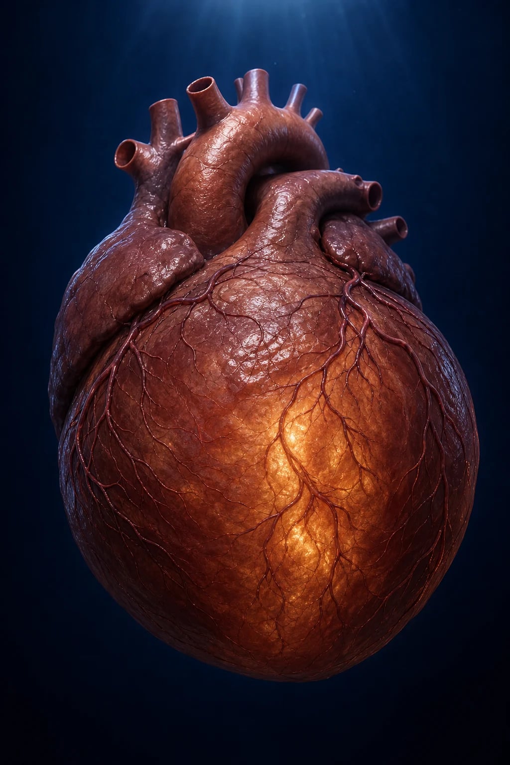
Overview & Definition
Dilated cardiomyopathy (DCM) is a disease of the heart muscle characterised by dilatation and systolic impairment of the left (or both) ventricle(s) that is not explained by abnormal loading conditions (hypertension, valve disease, congenital heart disease) or coronary artery disease sufficient to cause global systolic dysfunction.[1][2]
The 2016 ESC revised definition (Pinto et al.) makes the diagnosis objective:[2]
- LV end-diastolic dimension (LVEDD) corrected for body surface area and sex that is greater than 117% of the predicted value (i.e. more than 2 standard deviations above the mean, equivalent to a Z-score over 2), AND
- LV ejection fraction (LVEF) under 45% (or fractional shortening under 25%). [1]
A new category — hypokinetic non-dilated cardiomyopathy (HNDC) — was introduced for reduced EF (under 45%) without dilatation, an early or pre-dilated phenotype frequently seen in relatives of DCM patients; it predicts progression to overt DCM and is itself a trigger for family screening.[2]
The clinical importance of DCM is that it is the commonest cardiomyopathy in adults, a leading cause of heart failure with reduced ejection fraction (HFrEF) in the young and middle-aged, the most frequent indication for heart transplantation worldwide, and a major cause of sudden cardiac death (SCD) before age 50. The exam skill lies in (1) recognising the syndrome is not ischaemic despite resembling it, (2) working through the structured aetiological differential — because many causes (alcohol, tachycardia, hypothyroidism, anthracycline) are reversible — and (3) applying device therapy (ICD/CRT), family screening and genetic testing that are specific to DCM and differ from generic HFrEF management.[1]
Classification
DCM is classified by aetiology (the most clinically useful axis — it determines reversibility), by inheritance pattern, and by echocardiographic/CMR phenotype. [1]
Primary (genetic / familial)
- 30 to 50% of DCM; autosomal-dominant in 80 to 90%
- Single largest gene: TTN truncating variants (TTnTV) in roughly 15 to 25% of familial and 8 to 18% of sporadic DCM
- Other major genes: LMNA (high SCD risk, conduction disease), MYH7, TPM1, TNNT2, BAG3, RBM20, FLNC, PLN, DES
- Presents with family history of DCM, SCD under 50, conduction disease (LMNA) or arrhythmia out of proportion to EF
- Genetic testing + cascade screening of first-degree relatives is recommended
Myocarditis-DCM (post-inflammatory)
- Acute or chronic; viral (coxsackie B, parvovirus B19, adenovirus, echovirus, HIV, SARS-CoV-2, hepatitis C)
- Autoimmune (systemic lupus, rheumatoid, giant-cell myocarditis — worst prognosis)
- Echo: dilated LV, may have regional wall-motion abnormality
- CMR: subepicardial or mid-wall LGE (often inferolateral); oedema on T2
- Endomyocardial biopsy if giant-cell suspected; immunosuppression for biopsy-proven autoimmune / virus-negative forms
Toxic — alcohol & drugs
- Alcohol: heavy use over 5 to 10 years (over 80 g/day); reversible with abstinence in up to one-third
- Anthracyclines: doxorubicin cumulative dose over 400 mg/m2 — dose-dependent free-radical injury (Type 1, irreversible)
- Trastuzumab: Type 2, reversible on stopping (HER2 blockade, not free-radical)
- Cocaine, methamphetamines, chloroquine, clozapine, anabolic steroids
- Manage: cessation, then standard GDMT
Peripartum cardiomyopathy (PPCM)
- HF with EF under 45% in last month of pregnancy to first 5 months postpartum, in absence of another cause
- Shares genetic predisposition with DCM (titin truncations in up to 15%)
- 16-kDa prolactin fragment (anti-angiogenic) pathogenic — rationale for bromocriptine
- High recovery rate (50 to 70%) with guideline therapy; high recurrence in subsequent pregnancy
Tachycardia-induced (TICMP)
- Persistent tachyarrhythmia (AF, flutter, ectopic atrial tachycardia, PJRT, frequent PVCs over 10 to 15% burden)
- Reversible: rate or rhythm control typically restores EF within weeks to months
- Hallmark: normalised EF after ablation; 'PVC cardiomyopathy'
Infiltrative / metabolic / endocrine
- Haemochromatosis: iron deposition in myocardium; check ferritin, transferrin saturation, HFE gene
- Sarcoidosis: granulomatous infiltration; CMR patchy LGE, FDG-PET; corticosteroids
- Hypothyroidism / hyperthyroidism, acromegaly, phaeochromocytoma
- Thiamine (beriberi), selenium (Keshan), carnitine deficiency
- Chagas disease (Trypanosoma cruzi) — leading cause in Latin America; apical aneurysm
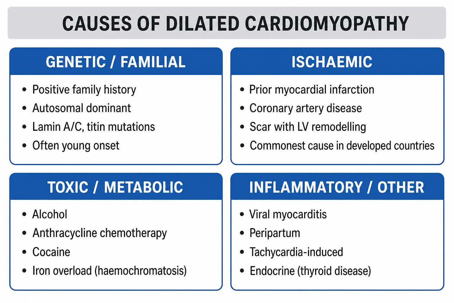
By inheritance pattern, familial DCM is autosomal dominant in the great majority (sarcomeric protein genes, cytoskeletal genes, nuclear envelope — LMNA, desmosomal — DES, DSP, FLNC); X-linked (DMD, Becker; G4.5/TAZ — Barth syndrome) is rarer but distinctive; mitochondrial inheritance and recessive forms (e.g. due to TAZ, some desminopathies) occur. A three-generation family history (with SCD, pacemaker/ICD, transplant, unexplained HF before age 60) is mandatory and is the single most cost-effective piece of the diagnostic work-up.[1]
By cardiac-MRI phenotype, the late-gadolinium-enhancement (LGE) pattern separates aetiologies: mid-wall septal LGE is classical for non-ischaemic DCM and predicts SCD; subepicardial inferolateral LGE suggests myocarditis or sarcoid; patchy multifocal LGE with FDG uptake suggests sarcoidosis; diffuse LGE with low T1 and T2* suggests amyloid (now classified separately under restrictive cardiomyopathy); no LGE is the commonest pattern in DCM and predicts better prognosis and recovery.[5]
Epidemiology & Risk Factors
DCM has an estimated prevalence of roughly 1 in 250 adults and an annual incidence of about 7 per 100,000, accounting for around one-third of all heart failure cases in those under 50 and the commonest indication for cardiac transplantation.[1]
Demographic risk: men more than women (2:1 for alcoholic, idiopathic); mean age at diagnosis in the 40s to 50s; African ancestry is associated with earlier onset, more severe HF and worse outcomes (BAG3 and TTN variants and higher hypertension burden contribute). [1]
Aetiological risk factors (the high-yield list): [1]
- Family history of DCM, SCD under age 50, pacemaker/ICD, or unexplained heart failure — first-degree relatives have a 20 to 35% chance of carrying the disease; genetic/familial DCM is the largest single aetiology.[1]
- Heavy alcohol use (chronic, more than 7 to 14 standard drinks/day for over 5 to 10 years) — historically the largest reversible cause in India.
- Cancer chemotherapy — anthracyclines (doxorubicin cumulative dose over 400 mg/m2, daunorubicin, epirubicin) cause dose-dependent irreversible cardiotoxicity via topoisomerase-IIbeta-mediated free-radial injury; trastuzumab (HER2 antibody) causes reversible Type 2 dysfunction; cyclophosphamide, 5-fluorouracil/capecitabine (vasospasm), checkpoint inhibitors (myocarditis).
- Pregnancy — peripartum cardiomyopathy, risk factors multiparity, age over 30, African ancestry, pre-eclampsia, tocolysis, prolonged tocolysis.
- Persistent tachyarrhythmia — AF with uncontrolled rate, frequent PVC burden over 10 to 15%.
- Prior viral illness or myocarditis — coxsackie B, parvovirus B19, HIV, SARS-CoV-2.
- Endocrine / metabolic — poorly controlled diabetes, hypothyroidism and hyperthyroidism, phaeochromocytoma, acromegaly, haemochromatosis.
- Nutritional — thiamine deficiency (beriberi) in alcoholics and polished-rice diets, selenium (Keshan disease) in endemic regions of China, carnitine deficiency in children.
- Autoimmune disease — systemic lupus erythematosus, dermatomyositis, scleroderma, rheumatoid arthritis.
- Infections endemic to a region — Chagas disease (Trypanosoma cruzi) in Latin America (the leading cause of DCM in those regions); HIV directly and via antiretrovirals; Lyme disease (more typically conduction block and myocarditis).
- Drugs of abuse — cocaine, methamphetamines, anabolic steroids.
- Toxins — cobalt (historic "Queensbeer" beer-drinkers' cardiomyopathy), lead, arsenic.
- Neuromuscular disease — Duchenne and Becker muscular dystrophy (X-linked dystrophin gene) cause subclinical DCM in up to 90%.
Pathophysiology
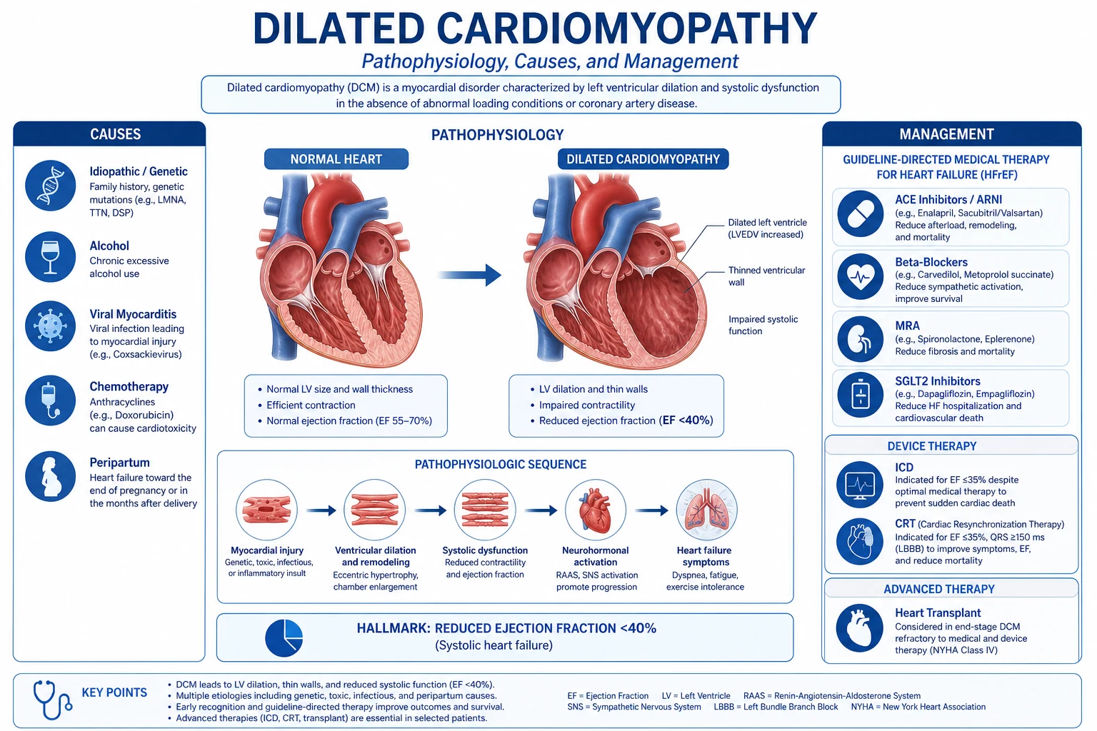
DCM is a final common pathway — many insults converge on a small set of cellular events. Three interlocking mechanisms drive the dilated, failing phenotype:[1][3]
1. Loss of cardiomyocytes and impaired contractility. In genetic DCM, mutations in sarcomeric (TTN, MYH7, ACTC, TPM1, TNNT2), Z-disc / cytoskeletal (DES, LDB3, MYPN, BAG3), nuclear-envelope (LMNA, EMD) and ion-channel / desmosomal (FLNC, PLN, SCN5A) proteins produce a structurally defective contractile apparatus that cannot withstand the cyclical mechanical load, leading to myocyte disarray, dropout and apoptosis. Titin-truncating variants (TTnTV) are the single commonest — the giant titin filament spans the sarcomere and truncations in its A-band region (most pathogenic) disrupt passive-tension sensing and signal transduction, eventually triggering necrosis and replacement fibrosis. In anthracycline cardiotoxicity, doxorubicin intercalates DNA and inhibits topoisomerase-IIbeta in cardiomyocyte mitochondria, generating reactive oxygen species that damage cardiolipin, the mitochondrial iron-sulphur clusters, and trigger ferroptosis; the dose-cumulative injury is Type 1, irreversible because cardiomyocytes do not regenerate. In alcoholic cardiomyopathy, ethanol and its metabolite acetaldehyde impair mitochondrial oxidative phosphorylation, reduce contractile protein synthesis, deplete myocardial selenium and B-vitamins, and increase apoptosis. In peripartum cardiomyopathy, oxidative stress in late pregnancy generates a cleaved 16-kDa prolactin fragment that is anti-angiogenic and pro-apoptotic, driving capillary rarefaction and myocyte death — the rationale for bromocriptine.[8][9]
2. Adverse remodelling. Loss of functioning myocardium triggers wall stress rises (Laplace's law: wall stress is proportional to (pressure × radius) / (2 × wall thickness)), which stretches the surviving myocytes, causes myocyte slippage and eccentric hypertrophy, and progressively thins and dilates the ventricle. As the chamber dilates, the mitral and tricuspid annuli stretch, producing functional (secondary) mitral and tricuspid regurgitation, which feeds back into more volume overload and more dilatation — a vicious cycle. [1]
3. Neurohormonal activation. Reduced cardiac output is interpreted as underperfusion: the sympathetic nervous system releases catecholamines (chronically cardiotoxic — myocyte apoptosis, arrhythmia, hypokalaemia); the renin-angiotensin-aldosterone system (RAAS) activates (angiotensin II vasoconstricts and drives hypertrophy/fibrosis; aldosterone retains sodium and promotes fibrosis); vasopressin (ADH) rises (water retention, dilutional hyponatraemia). These initially compensate but chronically drive further adverse remodelling. The counter-regulatory natriuretic peptides (ANP, BNP) released from stretched myocardium are overwhelmed.[3]
4. Fibrosis and arrhythmogenesis. Replacement fibrosis (visible as late gadolinium enhancement on cardiac MRI) replaces lost myocytes with non-conducting scar, which slows conduction and creates re-entry circuits — the substrate for ventricular arrhythmias and SCD. Diffuse interstitial fibrosis (measurable by T1 mapping and extracellular volume) further impairs compliance and contractility. Mid-wall septal LGE is the strongest CMR predictor of SCD in DCM.[5]
5. Stasis and thromboembolism. The dilated, poorly contracting ventricle, particularly the apex, becomes a low-flow chamber. Combined with atrial fibrillation (from atrial stretch) and endothelial dysfunction, this predisposes to apical mural thrombus and systemic embolism (stroke, mesenteric, limb) — the rationale for anticoagulation when EF is low, AF is present, or LV thrombus is demonstrated. [1]
Clinical Presentation
The presentation of DCM overlaps completely with heart failure with reduced ejection fraction (HFrEF) — DCM is, after all, the most common substrate for HFrEF in younger adults. The four classical modes of presentation are:[1][3]
- Symptomatic heart failure (most common) — progressive exertional dyspnoea, orthopnoea, paroxysmal nocturnal dyspnoea, fatigue, ankle swelling, abdominal fullness (hepatic congestion), nocturia. The trajectory is usually weeks to months; a more abrupt course suggests superimposed acute decompensation (arrhythmia, ischaemia, infection, non-adherence).
- Arrhythmia / sudden cardiac death — atrial fibrillation (atrial stretch, very common at presentation, drives further decompensation), ventricular ectopics / non-sustained VT, or syncope / aborted SCD as the first manifestation — particularly in LMNA, FLNC, PLN, DES, RBM20 mutations where arrhythmia often precedes contractile failure.
- Thromboembolism — stroke, TIA, mesenteric or limb ischaemia from an apical LV thrombus or AF.
- Incidental / asymptomatic — abnormal ECG, cardiomegaly on chest X-ray, abnormal echo found at health screening or during family screening of an affected relative. [1]
Examination reflects biventricular failure: displaced, diffuse (volume-overloaded) apex beat laterally and inferiorly; elevated JVP with prominent v wave if functional TR; S3 gallop (the auscultatory hallmark of systolic dysfunction — low-pitched early-diastolic sound at the apex with the bell); bibasal inspiratory crackles; pleural effusions (right greater); hepatomegaly with pulsatile liver if functional TR; ascites and pitting ankle/sacral oedema; cool peripheries, low-volume pulse, narrow pulse pressure in low-output states. A mitral regurgitation murmur (functional, apical, systolic) and tricuspid regurgitation murmur (functional, lower left sternal, louder on inspiration) are common. [1]
Atypical presentations (high-yield): [1]
- Elderly: falls, confusion, anorexia, fatigue rather than dyspnoea — easily misattributed to "old age".
- Diabetic: silent or blunted dyspnoea; may present late with cardiogenic shock.
- Pregnancy (peripartum): dyspnoea attributed to normal pregnancy; oedema misattributed to pre-eclampsia; lower threshold for echocardiography in late pregnancy and puerperium.
- Children / young adults: failure to thrive, recurrent chest infections, exercise intolerance; family history usually positive.
- Athletes: "the ill athlete" — fatigue, reduced performance; electrocardiographic changes overlap with athletic remodelling, making diagnosis hard — CMR and deconditioning are key.
- Neuromuscular disease (Duchenne/Becker): wheelchair-bound, may present in respiratory failure; cardiac involvement precedes skeletal muscle symptoms. [1]
Differential Diagnosis
The first task is to exclude the mimics that look like DCM but require different management — especially ischaemic heart disease, which is by far the commonest cause of a dilated, poorly contracting LV. A dedicated aetiological work-up (coronary angiography or CTCA, CMR, genetic testing, endocrine and autoimmune screen) is mandatory before the label "idiopathic DCM" is accepted.[1][2]
- Ischaemic cardiomyopathy (CAD) — the single most important exclusion. Distinguish by: risk factors (diabetes, smoking, dyslipidaemia, hypertension, family history of premature CAD), history (prior MI, angina, revascularisation), ECG (Q waves, regional wall-motion abnormality), regional (vs global) wall-motion abnormalities on echo, subendocardial LGE in a coronary distribution on CMR, and definitive coronary angiography/CTCA showing obstructive disease. The distinction matters: ischaemic DCM may be treatable by revascularisation; DCM management (genetic testing, family screening, ICD criteria) differs.
- Hypertensive heart disease — long-standing hypertension causing LV dilatation and failure; distinguish by history of hypertension, LV hypertrophy (echo: increased wall thickness with dilatation), and absence of family history of DCM. Management centres on aggressive BP control plus the same HFrEF pillars.
- Valvular heart disease — chronic severe aortic or mitral regurgitation (volume overload), or chronic mitral regurgitation primary (myxomatous, rheumatic) can cause a dilated LV with reduced EF; distinguish by the characteristic murmur and valve pathology on echo. The distinction matters because surgical correction (AVR/MVR) is definitive if EF is not yet severely depressed.
- Hypertrophic cardiomyopathy (HCM) — "burnt-out" phase — late-stage HCM with systolic dysfunction can mimic DCM; distinguish by family history of HCM, prior history of asymmetrical septal hypertrophy, systolic anterior motion of mitral valve, and genetic testing (sarcomeric variants, often different genes).
- Arrhythmogenic right-ventricular cardiomyopathy (ARVC) — desmosomal gene mutations; classically right-heart-predominant with epsilon waves, T-wave inversion in V1 to V3, late potentials on signal-averaged ECG, regional RV dyskinesia on echo/CMR. Left-dominant ARVC (LDAC) can mimic DCM; CMR shows subepicardial / mid-wall LGE of the LV lateral wall.
- Cardiac amyloidosis (now classified separately as restrictive) — biventricular thickening with low voltages on ECG ("discordance"), low-flow / low-gradient aortic stenosis coexistence, bilateral carpal tunnel, wild-type ATTR in elderly men or hereditary ATTR with neuropathy; CMR shows diffuse subendocardial LGE, low T1, high ECV; confirm with 99mTc-PYP/DPD scintigraphy and serum/urine free-light-chain. Distinguish from DCM because of tafamidis therapy (ATTR).
- Athlete's heart — physiological remodelling with mild LV dilatation, increased wall thickness, bradycardia, normal diastolic function, and normal/high VO2 max on CPET — no LGE on CMR; deconditioning normalises the phenotype.
- Pericardial constriction — distinguish by pericardial knock, Kussmaul's sign, pericardial calcification, tubular (non-dilated) ventricles, bi-atrial enlargement, and septal bounce on echo/CMR. [1]
A normal BNP/NT-proBNP with normal EF and no LGE effectively excludes clinically significant DCM. The single most powerful discriminator for ischaemic vs non-ischaemic aetiology is the LGE pattern on CMR — subendocardial (ischaemic) vs mid-wall/subepicardial (DCM/myocarditis).[5]
Clinical & Bedside Assessment
DCM cannot be diagnosed clinically, but the bedside examination localises the disease, gauges severity, and identifies complications. [1]
Vital signs: tachycardia (compensatory or AF), tachypnoea, low pulse pressure (reduced stroke volume), hypotension in advanced disease or cardiogenic shock, low SpO2 in pulmonary oedema. Body mass, alcohol history, stigmata of chronic liver disease, thyroid enlargement, hyperpigmentation (haemochromatosis), bruising (amyloid), systemic features of autoimmune disease, and peripheral oedema all yield aetiological clues. [1]
Jugular venous pressure — measured at 45°; elevated in right-heart congestion. A prominent cv wave suggests functional TR; a steep y descent with a pericardial knock points to constriction. Hepatojugular reflux (sustained abdominal pressure raising JVP more than 4 cm for over 15 seconds) confirms right-heart congestion. [1]
Apex beat — classically displaced laterally and inferiorly (volume overload) and diffuse / hyperdynamic; a sustained heave points to pressure overload (hypertrophy, HCM). [1]
Auscultation — S3 gallop is the auscultatory hallmark of systolic dysfunction (low-pitched, just after S2, heard with the bell at the apex, left lateral position). An S4 is more typical of diastolic dysfunction or HCM. Functional mitral regurgitation (apical, pansystolic, soft, radiating to axilla) and functional tricuspid regurgitation (left lower sternal, pansystolic, louder on inspiration) are common. A gallop rhythm of S3 plus tachycardia is called the "gallop triad" and signals decompensation. [1]
Precordial examination for RV heave (pulmonary hypertension), pleural effusion (dullness, reduced breath sounds at bases), and signs of systemic embolism (focal neurology, cold limb, abdominal pain). [1]
Severity is graded functionally by NYHA class (I no limitation; II slight; III marked; IV at rest) — a powerful prognostic marker that drives device and transplant decisions. [1]
Investigations
The diagnostic strategy is (a) confirm DCM and grade severity (echo, CMR), (b) define the aetiology (coronary imaging, genetic testing, biopsy, endocrine and autoimmune screen), (c) stratify risk (Holter, biomarkers, exercise testing), and (d) screen first-degree relatives.[1][3]
First-line in suspected DCM: [1]
- 12-lead ECG — almost always abnormal but non-specific: sinus tachycardia, left bundle branch block (common and prognostically adverse), left anterior fascicular block, premature ventricular complexes, non-sustained VT, T-wave inversion (lateral leads), poor R-wave progression, atrial fibrillation. Low voltages with hypertrophy on echo suggest amyloid. Epsilon waves and TWI V1 to V3 suggest ARVC. AV block with a DCM picture strongly suggests LMNA, EMD (Emery-Dreifuss), or sarcoidosis and is an indication for endomyocardial biopsy / FDG-PET.
- Chest X-ray — global cardiomegaly (cardiothoracic ratio over 0.5), pulmonary venous congestion, Kerley B lines, interstitial/alveolar oedema, pleural effusions.
- Transthoracic echocardiography (TTE) — the pivotal test. Confirms: dilated LV (LVEDD over 117% predicted or Z-score over 2), EF under 45% (Simpson's biplane), global hypokinesis, increased LV end-diastolic and end-systolic volumes, thin-walled LV, functional MR and TR (annular dilatation, leaflet tethering), dilated left atrium, possible LV apical thrombus (use contrast / echocardiographic contrast if suspicion high), right-ventricular involvement (biventricular DCM), and pericardial effusion (in myocarditis). Speckle-tracking strain (reduced global longitudinal strain) detects early dysfunction before EF falls.
- BNP / NT-proBNP — elevated; high negative predictive value (a normal level excludes significant DCM).
- Bloods — FBC (anaemia, iron studies — iron deficiency is common in HF and independently worsens prognosis), U&E (renal dysfunction — a prognostic marker and a constraint on RAAS/MRA use), LFTs (hepatic congestion), TFTs (exclude thyroid disease), fasting glucose / HbA1c, lipid profile, ferritin and transferrin saturation (haemochromatosis), calcium, troponin (chronically mildly raised in advanced HF; acutely raised in myocarditis or ACS), uric acid, D-dimer.
- Aetiological screen — viral serology (HIV, hepatitis B and C, coxsackie, parvovirus B19 — limited clinical yield except HIV, which is treatable), autoimmune screen (ANA, anti-dsDNA, rheumatoid factor, anti-CCP), serum/urine protein electrophoresis and free light chains (amyloid), iron studies and HFE gene (haemochromatosis), serum ACE and 24-h urinary calcium (sarcoidosis), TSH, free T4 (thyroid), 24-h urine metanephrines if phaeochromocytoma suspected. [1]
Advanced investigations: [1]
- Coronary angiography or CT coronary angiography (CTCA) — mandatory in essentially all patients with new DCM to exclude occult ischaemic heart disease (the commonest DCM mimic); CTCA is preferred in younger patients with low pre-test probability of CAD.
- Cardiac MRI (CMR) — the gold standard for tissue characterisation. Confirms ventricular volumes and EF; late-gadolinium-enhancement (LGE) pattern localises the disease and predicts SCD: mid-wall septal LGE (classical DCM, predicts SCD — Gulati et al.), subepicardial inferolateral LGE (myocarditis / sarcoid / Chagas), patchy LGE with FDG uptake (sarcoidosis), subendocardial LGE in a coronary distribution (ischaemia — exclude CAD). T1 mapping and extracellular volume (ECV) quantify diffuse fibrosis; T2 mapping / STIR detects oedema (active myocarditis).
- Endomyocardial biopsy (EMB) — restricted to (a) suspected giant-cell myocarditis (which demands urgent immunosuppression and transplant assessment) — typically young, fulminant HF, ventricular arrhythmia, high troponin; (b) suspected cardiac sarcoidosis (Dallas-like granulomas); (c) fulminant lymphocytic myocarditis. Sensitivity is patchy because DCM involvement is focal — CMR-targeted biopsy improves yield.
- FDG-PET / CT — for suspected cardiac sarcoidosis (patchy uptake with perfusion defect — "reverse mismatch") and amyloid (low uptake).
- 24 to 48-hour Holter monitor — to detect non-sustained VT (drives ICD discussion), AF, and quantify PVC burden (tachycardiomyopathy).
- Cardiopulmonary exercise testing (CPET) — peak VO2 and VE/VCO2 slope for transplant work-up (peak VO2 under 12 to 14 mL/kg/min with limitations despite optimal therapy is a transplant criterion).
- Genetic testing — targeted next-generation sequencing panel of established DCM genes recommended in: any DCM with family history, conduction disease (LMNA, DES), sudden death in family, onset under 50, echocardiographic features suggesting specific genotype (e.g. RV involvement → desmosomal; conduction disease → LMNA). A pathogenic / likely-pathogenic variant is found in roughly 25 to 40% of familial DCM and 10 to 20% of apparently sporadic DCM. Cascade genetic testing of first-degree relatives follows.
- Family screening — clinical screening (ECG, echo ± CMR) of all first-degree relatives every 1 to 3 years from age 10 is standard; if a pathogenic variant is identified in the proband, targeted cascade genetic testing is offered. This is the single most under-applied life-saving intervention in DCM.[1]
Dilated cardiomyopathy — key numbers
Management — Resuscitation
DCM with acute decompensated heart failure or cardiogenic shock is an emergency. ABCDE first. [1]
Acute pulmonary oedema — sit the patient upright, high-flow oxygen to target SpO2 94 to 98% (88 to 92% in COPD/CO2 retainers), IV loop diuretic (furosemide 40 to 80 mg IV bolus, or a continuous infusion 5 to 20 mg/hour for larger doses), IV nitrates (GTN infusion) if BP over 110 mmHg systolic — venodilator effect reduces preload; non-invasive ventilation (CPAP or BiPAP) for respiratory distress (reduces work of breathing, recruits alveoli, lowers preload and afterload); morphine 2.5 to 5 mg IV only if distressed (evidence of possible harm with routine use). Identify and treat the precipitant (AF, ischaemia, infection, anaemia, non-adherence, NSAIDs).[3][4]
Cardiogenic shock — hypotension (SBP under 90), cold peripheries, oliguria, confusion, lactate rise. The failing dilated ventricle is not helped by more diuretic. Use inotropic support: dobutamine 2 to 20 micrograms/kg/min (beta-1 agonist, positive inotrope and chronotrope — may worsen ischaemia and arrhythmia), or milrinone 0.125 to 0.75 micrograms/kg/min (PDE-3 inhibitor, inodilator — preferred if pulmonary hypertension, but vasodilator effect may worsen hypotension), or levosimendan (calcium sensitisier, inodilator) in refractory cases. Noradrenaline for vasoplegic shock on top of low-output. Consider mechanical circulatory support (IABP, Impella, VA-ECMO) as a bridge to recovery, transplant, or decision. Do not start a beta-blocker in a decompensated patient — wait until euvolaemic and stable. [1]
Arrhythmia management — AF with rapid rate: IV amiodarone 300 mg over 1 hour then 900 mg over 24 hours if haemodynamically stable; DC cardioversion if unstable. Sustained VT / VF: DC shock (200 J biphasic); amiodarone 300 mg IV for refractory; beta-blockade long-term. Complete heart block in sarcoidosis, LMNA, Chagas: temporary then permanent pacing, with consideration of ICD/CRT-D if EF under 35%. [1]
LV thrombus / systemic embolism — anticoagulate immediately with low-molecular-weight heparin (enoxaparin 1 mg/kg SC twice daily) then warfarin or DOAC, while planning long-term anticoagulation (see below). [1]
Management — Definitive & Stepwise
DCM management has four layers: (1) treat the underlying cause (reversible DCMs improve dramatically — alcohol cessation, tachycardia control, thyroid replacement, anthracycline cessation, immune suppression for biopsy-proven autoimmune myocarditis), (2) the four pillars of HFrEF (GDMT), (3) device therapy (ICD, CRT), (4) advanced therapies (transplant, LVAD).[1][3][4]
Layer 1 — Treat the cause
- Alcohol abstinence — mandatory; up to one-third of alcoholic DCMs recover substantial EF with abstinence. Refer to alcohol-liaison services.
- Tachycardia-induced — rate control (beta-blocker, non-dihydropyridine CCB if EF preserved — avoid in HFrEF) or rhythm control (cardioversion, ablation for atrial tachycardia/PVCs). EF typically recovers within 1 to 6 months.
- Thyroid disease — levothyroxine for hypothyroidism; carbimazole / PTU for thyrotoxicosis; both can produce dramatic recovery.
- Iron deficiency — IV ferric carboxymaltose (or ferric derisomaltose) repletes stores and improves symptoms and exercise capacity in HFrEF irrespective of anaemia.
- Haemochromatosis — therapeutic phlebotomy (target ferritin under 50 to 100) or iron chelation (deferasirox); can halt progression.
- Sarcoidosis — corticosteroids (prednisolone 0.5 to 1 mg/kg/day tapering) for biopsy-proven or FDG-PET-positive cardiac sarcoid; methotrexate if steroid-sparing needed.
- Autoimmune myocarditis (virus-negative, biopsy-proven) — immunosuppression (prednisolone plus azathiomycine or mycophenolate); giant-cell myocarditis demands urgent combination immunosuppression and transplant evaluation.
- Peripartum cardiomyopathy — standard GDMT compatible with pregnancy/breastfeeding (hydralazine-nitrate for afterload reduction, beta-blocker [metoprolol, bisoprolol; avoid ACEi/ARB/MRA in pregnancy — teratogenic — but start postpartum if not breastfeeding]), bromocriptine 2.5 mg twice daily for 8 weeks (suppresses the pathogenic 16-kDa prolactin fragment), and anticoagulation if EF under 30%. Recovery of EF (to above 50%) in 50 to 70%.[8][9]
- Cancer therapy-related — early cardioprotective therapy (ACEi/ARB + beta-blocker), consider dexrazoxane (iron chelator) before high-dose anthracyclines; trastuzumab typically reversible on cessation; checkpoint-inhibitor myocarditis is a medical emergency (high-dose steroids).
Layer 2 — Four pillars of HFrEF (GDMT)
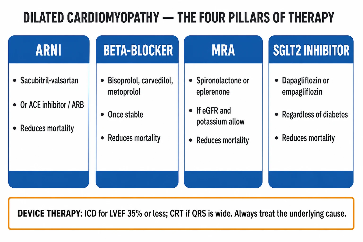
Each pillar has an independent mortality benefit demonstrated in landmark trials. Start all four together at low dose and titrate to target, rather than sequencing.[3][4]
- ARNI (sacubitril/valsartan) OR ACE-inhibitor — sacubitril/valsartan target dose 97/103 mg twice daily (PARADIGM-HF); enalapril 10 mg twice daily; ramipril 10 mg daily; lisinopril 20 to 40 mg daily. ACE-inhibitor-to-ARNI switch needs a 36-hour washout to prevent angioedema. Benefit: blocks RAAS, reduces mortality, hospitalisation, and progressive remodelling.
- Beta-blocker (one of the four proven: carvedilol target 25 mg twice daily [50 mg twice daily if over 85 kg], bisoprolol 10 mg daily, metoprolol succinate 200 mg daily, nebivolol 10 mg daily in those over 70). Carvedilol had the strongest evidence in non-ischaemic DCM specifically (US Carvedilol HF Study, NEJM 1996).[10] Start only when euvolaemic and haemodynamically stable; never in decompensated HF.
- Mineralocorticoid receptor antagonist (MRA) — spironolactone 12.5 to 50 mg daily (RALES in severe HFrEF; EMPHASIS-HF in mild); eplerenone 25 to 50 mg daily if gynaecomastia. Monitor potassium and creatinine at 1, 4, 8 weeks then 3-monthly.
- SGLT2 inhibitor — dapagliflozin 10 mg daily (DAPA-HF) or empagliflozin 10 mg daily (EMPEROR-Reduced). Benefit independent of diabetes; first-line in all HFrEF including non-ischaemic DCM.
Additional pharmacotherapy: [1]
- Diuretic (loop) — furosemide 20 to 80 mg orally daily (or bumetanide, torsemide) for symptomatic congestion; no mortality benefit — purely symptom control. Add thiazide (metolazone 2.5 to 5 mg) for diuretic resistance ("sequential nephron blockade") — close electrolyte monitoring.
- Ivabradine — for sinus rhythm with heart rate 70 or more despite maximally tolerated beta-blocker (SHIFT trial). Inhibits the SA-node If current; no benefit in AF (and may cause 1:1 conduction of atrial tachyarrhythmias).
- Hydralazine-nitrate — for African ancestry, or those intolerant of ACEi/ARNI (renal dysfunction, hyperkalaemia). Hydralazine 37.5 mg + isosorbide dinitrate 20 mg three times daily titrating up (A-HeFT).
- Digoxin — for symptom control or rate control in AF; narrow therapeutic window (0.5 to 0.9 ng/mL), no mortality benefit (DIG trial), avoid in renal failure and conduction disease. [1]
Layer 3 — Device therapy
- Implantable cardioverter-defibrillator (ICD) for primary prevention of SCD — indicated for LVEF under 35%, NYHA II to III, expected survival over 1 year with meaningful quality of life, after at least 3 months of optimal medical therapy (the medical therapy must be in place because EF can recover, particularly in non-ischaemic DCM).[3][4] DANISH trial (NEJM 2016) showed ICD did not reduce all-cause mortality in non-ischaemic DCM — current guidelines still recommend ICD in this group, but with shared decision-making and careful attention to age, comorbidity and life-expectancy.[7] In LMNA / FLNC / PLN / RBM20 mutation carriers, ICD may be indicated at higher EF because arrhythmia often precedes contractile failure. ICD is contraindicated within 40 days of MI, in NYHA IV refractory, in incessant VT, in significant psychiatric burden, or in those with life expectancy under 1 year.
- Cardiac resynchronisation therapy (CRT) — for QRS duration over 150 ms with LBBB morphology and EF under 35% in sinus rhythm (Class I); benefit less certain for QRS 120 to 149 ms or non-LBBB. CRT-D (with defibrillator) preferred in DCM unless contraindicated. Improves symptoms, EF, hospitalisation, and mortality.
- Conventional pacing — for symptomatic bradycardia, complete heart block (sarcoid, LMNA, Chagas), but avoid RV apical pacing in low EF (worsens HF) — prefer CRT if significant pacing need.
Layer 4 — Anticoagulation
- Indications: atrial fibrillation (CHA2DS2-VASc), LV apical thrombus, prior thromboembolism, severe LV dysfunction (EF under 30%) — the latter is debated; consider individual risk (Daring-DCM data conflicting).[1]
- DOAC (apixaban 5 mg twice daily, rivaroxaban 20 mg daily, dabigatran 150 mg twice daily) preferred over warfarin in non-valvular AF; warfarin (INR 2 to 3) for LV thrombus (limited DOAC evidence).
Layer 5 — Advanced therapies
- Cardiac transplantation — gold-standard for end-stage DCM refractory to optimal therapy. Indications: peak VO2 under 12 to 14 mL/kg/min, recurrent hospitalisation, progressive end-organ dysfunction, refractory ventricular arrhythmia, cardiogenic shock requiring inotropes/MCS. Absolute contraindications: active malignancy or infection, severe irreversible pulmonary hypertension (PVR over 5 Wood), severe obesity/BMI over 35, active substance use, poor adherence.
- Durable LVAD (left-ventricular assist device) — bridge to transplant or destination therapy in non-transplant candidates; modern continuous-flow devices (HeartMate 3) have substantially improved outcomes.
- Palliative care — for those not candidates for advanced therapy; symptom-focused (opioids for dyspnoea, diuretics, low-dose sedatives); align with patient priorities; deactivation of ICD shocks at end of life (turn off therapies). [1]
Specific Subtypes & Scenarios
Peripartum cardiomyopathy (PPCM) — defined as HF with EF under 45% in last month of pregnancy to 5 months postpartum, no other cause. Pathophysiology: oxidative stress generates a cleaved 16-kDa prolactin fragment that is anti-angiogenic — rationale for bromocriptine. Risk factors: multiparity, age over 30, African ancestry, pre-eclampsia, prolonged tocolysis. Shared genetic predisposition with DCM (titin truncations in 6 to 15%).[8] Management: standard GDMT modified for pregnancy — avoid ACEi/ARB/MRA in pregnancy (teratogenic), use hydralazine-nitrate for afterload reduction; beta-blocker (metoprolol, bisoprolol — avoid atenolol in pregnancy: IUGR); start ACEi/ARB/MRA postpartum if not breastfeeding; bromocriptine 2.5 mg twice daily for 8 weeks (stop breastfeeding); anticoagulation if EF under 30%. Recovery of EF in 50 to 70%; recurrence in subsequent pregnancy up to 30 to 50% if EF has not normalised — counsel against further pregnancy.[9]
Anthracycline cardiotoxicity — Type 1 (anthracyclines, classical chemotherapeutics): dose-dependent, irreversible, free-radical and topoisomerase-IIbeta-mediated myocyte injury; cumulative doxorubicin dose over 400 mg/m2 markedly increases risk. Type 2 (trastuzumab): not dose-dependent, reversible on cessation, HER2 blockade without myocyte loss. Surveillance: baseline echo + troponin, repeat at 3, 6, 9, 12 months (high-risk regimens), and annually thereafter; global longitudinal strain is more sensitive than EF. Prevention: dexrazoxane (iron chelator) before high-dose anthracyclines; liposomal doxorubicin (lower cardiotoxicity); avoid concomitant trastuzumab during anthracycline. Treatment: early ACEi + beta-blocker; EF recovery more likely if started early. [1]
Alcoholic cardiomyopathy — chronic heavy alcohol (over 7 to 14 standard drinks/day for 5 to 10 years); thiamine co-deficiency common. Recovery in up to one-third with sustained abstinence. Continued drinking predicts death. Thiamine 100 mg daily IV/oral for beriberi. [1]
Tachycardiomyopathy (TICMP) — sustained tachyarrhythmia (AF with uncontrolled rate, atrial tachycardia, PJRT, very frequent PVCs over 10 to 15% burden, permanent junctional reciprocating tachycardia) over weeks to months. Reversible with rate or rhythm control; EF typically recovers within 1 to 6 months. PVC-ablation if a single dominant focus. Always consider in young patients with unexplained LV dysfunction and a tachyarrhythmia. [1]
Familial / genetic DCM — autosomal-dominant in 80 to 90%. High-risk genotypes: LMNA (high SCD risk, conduction disease, need ICD at higher EF, partial lipodystrophy), FLNC (filamin C — high SCD risk), PLN (phospholamban — R14del founder variant, arrhythmia, calcification), DES / DSP (desmin / desmoplakin — overlap with ARVC), RBM20 (severe, young onset, AF), BAG3, TTN (largest single gene; intermediate SCD risk; the "titin myopathy"). Cascade screening of first-degree relatives is mandatory.[1]
Chagas cardiomyopathy — Trypanosoma cruzi infection (endemic Latin America); apical aneurysm (classic), RBBB + LAHB ECG, GI megacolon/mega-oesophagus, biventricular HF, high thromboembolic risk. Antitrypanosomal therapy (benznidazole, nifurtimox) in acute / early indeterminate phase. Anticoagulate regardless of EF. [1]
Sarcoid cardiomyopathy — non-caseating granulomas in myocardium; patchy LGE on CMR (basal, septal, perivascular), FDG-PET positive; AV block in young patients is a red flag; ventricular arrhythmia; coexisting pulmonary / ocular / cutaneous sarcoid. Corticosteroids (prednisolone 0.5 to 1 mg/kg/day tapering); methotrexate / mycophenolate as steroid-sparing; ICD for sustained VT or EF under 35%. [1]
Iron-overload (haemochromatosis) cardiomyopathy — restrictive early, dilated late; HFE gene (C282Y homozygote), transferrin saturation over 45%, ferritin over 1000. Phlebotomy (target ferritin under 100), chelation (deferasirox). Reversible if caught early. [1]
Complications & Pitfalls
Cardiac: progressive heart failure (the natural history untreated), atrial fibrillation (atrial stretch — drives further decompensation and is the commonest arrhythmia), ventricular arrhythmia (NSVT, sustained VT, VF) and SCD (the leading mode of death in younger patients), LV apical thrombus and systemic embolism (stroke, mesenteric, limb), functional MR and TR (annular dilatation), right-heart failure (advanced disease, portends poor prognosis), cardiogenic shock. [1]
Systemic: cardiorenal syndrome (the tension between decongestion and renal perfusion), cardiac cachexia (sarcopenia, poor prognosis), hepatic congestion → cardiac cirrhosis (chronic right-heart failure), renal dysfunction, anaemia of chronic disease / iron deficiency. [1]
Complications of therapy: hyperkalaemia and renal dysfunction on RAAS / MRA, hypotension on SGLT2 / ARNI, beta-blocker-induced decompensation if started in congested patient, diuretic-induced hyponatraemia and hypokalaemia, euglycaemic ketoacidosis on SGLT2, gynaecomastia with spironolactone, device infection / lead failure / inappropriate shocks. [1]
Classic pitfalls: [1]
- Labelling DCM "idiopathic" without a structured work-up — most "idiopathic" cases are genetic, myocarditis-related, or have a missed reversible cause (alcohol, tachycardia, thyroid).
- Not screening first-degree relatives — the single most under-applied life-saving intervention; family screening identifies presymptomatic disease where treatment can prevent progression.
- Applying ICD criteria from ischaemic cardiomyopathy to non-ischaemic DCM — the DANISH trial showed no all-cause-mortality benefit of ICD in non-ischaemic DCM, especially over age 70.[7]
- Starting a beta-blocker in a decompensated, congested patient — wait until euvolaemic and stable.
- Missing the ACE-inhibitor-to-ARNI washout — 36 hours to avoid angioedema.
- Failing to consider giant-cell myocarditis in a young patient with fulminant HF and ventricular arrhythmia — demands urgent endomyocardial biopsy, immunosuppression, transplant evaluation.
- Not recognising LMNA / FLNC / PLN mutation carriers who need ICD at higher EF — arrhythmia precedes contractile failure.
- Forgetting anticoagulation in AF, LV thrombus, or severe LV dysfunction.
- Misattributing pregnancy dyspnoea / oedema to "normal pregnancy" — peripartum cardiomyopathy.
- Sending a patient for transplant without optimising GDMT first — many "refractory" cases improve once four pillars at target doses are achieved.
Prognosis & Disposition
Untreated DCM has a 5-year mortality of 50% historically; modern GDMT has reduced this to roughly 20 to 30% at 5 years in compliant patients, with substantial improvement in functional class and quality of life. Predictors of poor outcome: older age, lower EF, higher NYHA class, raised BNP/NT-proBNP, renal dysfunction, hyponatraemia, anaemia/iron deficiency, right-ventricular dysfunction, mid-wall LGE on CMR, NSVT on monitoring, LMNA / FLNC / RBM20 / PLN mutations, and non-recovery of EF.[1][5]
Recovery of EF (the "recovered DCM" phenotype) is possible in 20 to 40% with intensive GDMT and treatable cause, particularly peripartum (50 to 70%), tachycardia-induced, alcoholic, thyroid, iron deficiency. The decision to stop GDMT after recovery is debated — relapse is common; ESC recommends continuation with shared decision-making. [1]
Disposition depends on haemodynamic stability and response to therapy: [1]
- Admit: acute decompensation, cardiogenic shock, sustained VT, syncope with EF under 35%, new LV thrombus, fulminant myocarditis.
- Outpatient with HF specialist: stable chronic DCM — uptitrate GDMT to target, surveillance for arrhythmia, periodic echo (every 6 to 12 months), family screening, genetic counselling.
- ICU/CCU: cardiogenic shock, sustained VT/VF, fulminant myocarditis (giant-cell until disproven), pre-transplant mechanical support.
- Transplant centre referral: peak VO2 under 12 to 14 mL/kg/min, recurrent hospitalisation, inotrope dependence, refractory arrhythmia. [1]
A DCM patient needs lifelong cardiology follow-up, even after recovery, because relapse and late arrhythmia occur. [1]
Special Populations
- Children / paediatric DCM — most commonly idiopathic, inborn errors of metabolism, neuromuscular (Duchenne/Becker), anomalous coronary, myocarditis. Weight-based dosing of all drugs. Heart transplantation if refractory.
- Pregnancy / peripartum — covered above (PPCM); in known DCM, pregnancy is contraindicated if EF under 40% or NYHA III to IV (high maternal mortality); ACEi/ARB/MRA contraindicated; hydralazine-nitrate and selected beta-blockers (metoprolol, bisoprolol) are safe.
- Elderly — atypical presentation; balance GDMT benefit against hypotension, renal dysfunction, polypharmacy; titrate slowly; DANISH trial tempers ICD use over age 70.[7]
- Diabetic — SGLT2 inhibitors are doubly beneficial (heart failure and renal protection); avoid thiazolidinediones (fluid retention).
- Chronic kidney disease — monitor potassium and creatinine closely on RAAS / MRA; SGLT2 inhibitors renoprotective down to eGFR 20; transplant candidacy reduced at advanced CKD.
- Athletes — differentiating athlete's heart from DCM is hard: deconditioning + CMR (no LGE) + CPET (normal/high VO2 max) help; "return to play" decisions for athletes with DCM are individualised.
- Neuromuscular disease (Duchenne/Becker, myotonic dystrophy, Emery-Dreifuss) — surveillance echo from diagnosis; ACEi early delays cardiomyopathy; palliative support.
Evidence, Guidelines & Regional Differences
The two governing guidelines are the 2021 ESC / 2023 ESC Focused Update on Heart Failure (McDonagh et al.)[3] and the 2022 AHA / ACC / HFSA Heart Failure Guideline (Heidenreich et al.).[4] Both endorse the four-pillar framework for HFrEF and explicitly include non-ischaemic DCM. DCM-specific guidance lives in the AHA Scientific Statement on Specific Dilated Cardiomyopathies (Bozkurt et al., 2016)[1] and the ESC revised definition of DCM / HNDC (Pinto et al., 2016).[2]
Landmark trials and statements every exam candidate must know: [1]
- US Carvedilol Heart Failure Study (Packer et al., NEJM 1996) — carvedilol reduced mortality 65% in chronic HFrEF, with particular benefit in non-ischaemic DCM.[10]
- PARADIGM-HF (McMurray, NEJM 2014) — sacubitril/valsartan superior to enalapril.
- DAPA-HF (McMurray, NEJM 2019) / EMPEROR-Reduced (Packer, NEJM 2020) — SGLT2 inhibitors reduce mortality and hospitalisation in HFrEF.
- DANISH trial (Køber et al., NEJM 2016) — ICD did not reduce all-cause mortality in non-ischaemic systolic HF.[7]
- Gulati et al., JAMA 2013 — mid-wall LGE in non-ischaemic DCM independently predicts SCD and mortality.[5]
- Ware et al., NEJM 2016 — shared titin truncating variants in peripartum and familial DCM.[8]
- Caforio et al., Eur Heart J 2013 — ESC position statement on myocarditis (Lake Louise CMR criteria, EMB indications).[6]
Regional deltas: [1]
- India (NEET-PG / INICET context) — alcohol remains a leading cause; peripartum DCM common; Chagas is not endemic; sarcoidosis under-recognised; access limits the use of ARNI, SGLT2i, transplant; genetic testing largely unavailable outside research centres, so family screening by clinical phenotype predominates.
- Latin America — Chagas cardiomyopathy is the leading cause of non-ischaemic DCM; apical aneurysm, RBBB+LAHB, antitrypanosomal therapy, high thromboembolic risk (anticoagulate regardless of EF).
- Sub-Saharan Africa — peripartum cardiomyopathy particularly common (high incidence); HIV-related DCM (viral + antiretroviral toxicity); endomyocardial fibrosis in equatorial regions (a separate restrictive cardiomyopathy, but presents with right-heart failure).
- USA — full guideline framework with ARNI, SGLT2i, transplant, LVAD widely available; genetic testing standard in familial DCM.
- UK — NICE NG106 (chronic HF) broadly aligned with ESC; specialist inherited cardiac conditions services for cascade screening; DANISH trial influence on ICD decision-making. [1]
Current controversies: (1) the role of ICD in non-ischaemic DCM after DANISH — risk stratification with CMR LGE, genetic subtype, GLS is increasingly used to refine selection; (2) the optimal withdrawal of GDMT after EF recovery; (3) the role of gene therapy (still experimental); (4) the defib versus pace-only CRT decision in non-ischaemic DCM over age 70. [1]
Exam Pearls
- DCM = dilated LV (LVEDD over 117% predicted or Z over 2) with EF under 45%, not explained by CAD, hypertension, valve disease or congenital heart disease.[2]
- Commonest cardiomyopathy in adults; commonest indication for cardiac transplantation.
- Aetiology: genetic / familial (30 to 50%, titin the largest single gene), myocarditis, alcohol, anthracycline, peripartum, tachycardia-induced, sarcoidosis, haemochromatosis, hypothyroidism, Chagas. Always search for a treatable cause before "idiopathic".[1]
- Investigations: echo (dilated thin-walled LV, EF under 45%), CMR (mid-wall LGE = DCM; subepicardial inferolateral = myocarditis / sarcoid; subendocardial in coronary distribution = ischaemia), coronary angiography / CTCA (exclude CAD), genetic testing + family screening.[5]
- Four pillars of HFrEF — ARNI/ACEi + beta-blocker + MRA + SGLT2i — each with independent mortality benefit; carvedilol has the strongest evidence in non-ischaemic DCM.[10]
- ICD for primary prevention if EF under 35% after at least 3 months of optimal medical therapy. DANISH trial (NEJM 2016) — no all-cause-mortality benefit of ICD in non-ischaemic DCM, especially over 70.[7]
- CRT if QRS over 150 ms with LBBB and EF under 35%.
- Anticoagulate if AF, LV thrombus, or prior thromboembolism; consider if EF under 30%.
- Mid-wall LGE on CMR independently predicts SCD (Gulati, JAMA 2013).[5]
- Peripartum cardiomyopathy — last month of pregnancy to 5 months postpartum, EF under 45%; bromocriptine (suppresses 16-kDa prolactin fragment); shared titin genetics with DCM; 50 to 70% recovery; high recurrence in subsequent pregnancy.[8][9]
- Anthracycline = Type 1 (dose-dependent, irreversible, free-radical; doxorubicin over 400 mg/m2). Trastuzumab = Type 2 (reversible). Dexrazoxane protects.[1]
- Tachycardiomyopathy is reversible with rate/rhythm control.[1]
- Family screening of all first-degree relatives every 1 to 3 years from age 10; cascade genetic testing if pathogenic variant identified.[1]
- LMNA mutation — high SCD risk, conduction disease — ICD at higher EF.
- Transplant for end-stage DCM; peak VO2 under 12 to 14 mL/kg/min a key criterion.
Exam application bank (NEET-PG / INICET)
One-line answer
Dilated cardiomyopathy (DCM) is a disease of the heart muscle defined by dilatation and systolic impairment of one or both ventricles (LV end-diastolic dimension corrected for body surface area and sex, more than 117% of predicted; or Z-score over 2; with ejection fraction under 45%) unexplained by abnormal loading (hypertension, valve disease) or coronary artery disease sufficient to cause the impairment. It is the commonest cardiomyopathy and a leading cause of heart failure with reduced ejection fraction (HFrEF), sudden cardiac death (SCD) and heart transplantation in the young. Aetiology is genetic in 30 to 50% (titin-truncating variants TTnTV in roughly 15 to 25%), but also myocarditis (viral: coxsackie, parvovirus B19, SARS-CoV-2), alcohol, anthracycline chemotherapy (doxorubicin), peripartum, tachycardia-induced, haemochromatosis, sarcoidosis, hypothyroidism and Chagas disease (La
Worked stems (answer without another resource)
Stem 1 — Classic presentation. Map symptoms to mechanism; name the first investigation and first treatment step with dose/route if drug therapy is standard. [1]
Stem 2 — Unstable / complicated. List red flags that force immediate resuscitation, theatre, ICU, antidote, or reperfusion — and what you do in the first 15 minutes. [1]
Stem 3 — Atypical group. Elderly, pregnancy, child, or immunocompromised: how presentation and thresholds change. [1]
Stem 4 — Differential trap. Name the three closest mimics and one discriminator for each. [1]
Stem 5 — Disposition. Who goes home with safety-netting, who is admitted, who needs HDU/ICU/theatre, and what follow-up is mandatory. [1]
Rapid viva checklist
- Definition + classification
- Pathophysiology chain
- Bedside signs / criteria
- Score with exact components (if any)
- Emergency bundle
- Definitive therapy with doses
- Complications of disease and of treatment
- Special populations
- Guideline/trial name if classic
- Three exam traps
Coverage self-check
If you cannot answer any stem above from this page alone, re-read the matching section — the page is intended to be self-sufficient for final-prof and NEET-PG/INICET questions on Dilated Cardiomyopathy.
DCM causes — mnemonic
DILATED
Alcohol, anthracycline (doxorubicin over 400 mg/m2), trastuzumab, cocaine, cobalt, chloroquine, steroids
Genetic 30 to 50% — titin (TTN) the largest single gene; also LMNA, MYH7, BAG3, FLNC, PLN, RBM20
Tachycardia-induced (AF, atrial tachycardia, frequent PVCs) — reversible with rate/rhythm control
Myocarditis (coxsackie, parvovirus B19, HIV, SARS-CoV-2), sarcoidosis, SLE, giant-cell myocarditis
Last month of pregnancy to 5 months postpartum — 16-kDa prolactin fragment; bromocriptine; high recovery rate
Haemochromatosis (HFE C282Y), transfusion-dependent thalassaemia — phlebotomy / chelation
Hypothyroidism, hyperthyroidism, phaeochromocytoma, acromegaly, thiamine (beriberi), selenium (Keshan)
Duchenne/Becker dystrophin, myotonic dystrophy, Emery-Dreifuss (LMNA), Chagas (Trypanosoma cruzi)
References
- [1]Bozkurt B, Colvin M, Cook J, et al. Current Diagnostic and Treatment Strategies for Specific Dilated Cardiomyopathies: A Scientific Statement From the American Heart Association Circulation, 2016.PMID 27832612
- [2]Pinto YM, Elliott PM, Arbustini E, et al. Proposal for a revised definition of dilated cardiomyopathy, hypokinetic non-dilated cardiomyopathy, and its implications for clinical practice: a position statement of the ESC working group on myocardial and pericardial diseases Eur Heart J, 2016.PMID 26792875
- [3]McDonagh TA, Metra M, Adamo M, et al. 2023 Focused Update of the 2021 ESC Guidelines for the diagnosis and treatment of acute and chronic heart failure Eur Heart J, 2023.PMID 37622666
- [4]Heidenreich PA, Bozkurt B, Aguilar D, et al. 2022 AHA/ACC/HFSA Guideline for the Management of Heart Failure: Executive Summary: A Report of the American College of Cardiology/American Heart Association Joint Committee on Clinical Practice Guidelines J Am Coll Cardiol, 2022.PMID 35379504
- [5]Gulati A, Jabbour A, Ismail TF, et al. Association of fibrosis with mortality and sudden cardiac death in patients with nonischemic dilated cardiomyopathy JAMA, 2013.PMID 23462786
- [6]Caforio AL, Pankuweit S, Arbustini E, et al. Current state of knowledge on aetiology, diagnosis, management, and therapy of myocarditis: a position statement of the European Society of Cardiology Working Group on Myocardial and Pericardial Diseases Eur Heart J, 2013.PMID 23824828
- [7]Køber L, Thune JJ, Nielsen JC, et al. Defibrillator Implantation in Patients with Nonischemic Systolic Heart Failure N Engl J Med, 2016.PMID 27571011
- [8]Ware JS, Li J, Mazaika E, et al. Shared Genetic Predisposition in Peripartum and Dilated Cardiomyopathies N Engl J Med, 2016.PMID 26735901
- [9]Hilfiker-Kleiner D, Haghikia A, Masuko D, et al. Long-term follow-up in peripartum cardiomyopathy patients with contemporary treatment: low mortality, high cardiac recovery, but significant cardiovascular co-morbidities Eur J Heart Fail, 2019.PMID 31724271
- [10]Packer M, Bristow MR, Cohn JN, et al. Carvedilol inhibits clinical progression in patients with mild symptoms of heart failure. US Carvedilol Heart Failure Study Group Circulation, 1996.PMID 8941105