Cardiology · Cardiology
Heart Failure
Also known as Congestive cardiac failure · CCF · HFrEF · HFpEF · Cardiac failure
Heart failure is a clinical syndrome — symptoms plus signs plus an objective structural or functional cardiac abnormality — split first by ejection fraction: HFrEF (EF ≤40%), HFmrEF (41–49%), HFpEF (EF ≥50%). NYHA I–IV grades current symptoms. HFrEF is treated with four pillars (ARNI/ACE-inhibitor, beta-blocker, MRA, SGLT2 inhibitor), each with independent mortality benefit. Acute pulmonary oedema: sit up, oxygen, IV furosemide, nitrates if BP permits, CPAP if needed.
On this page & tools
Your progress
Saved locally on this device.
Practise this topic
Exam tags
Red flags
Overview & Definition
Heart failure is a clinical syndrome, not a single disease. It is defined by the combination of (1) typical symptoms — breathlessness, fatigue, ankle swelling; (2) typical signs — elevated jugular venous pressure (JVP), pulmonary crackles, peripheral oedema; and (3) objective evidence of a structural or functional cardiac abnormality — most often a reduced or preserved ejection fraction on echocardiography, or a raised natriuretic peptide.[1] Any cardiac disease severe enough can produce it; the heart ultimately fails through one of two mechanical routes — it cannot contract forcefully enough to eject (systolic / pump failure), or it cannot relax and fill adequately at normal pressures (diastolic / filling failure), and in most patients both elements coexist.
The defining physiological problem is that cardiac output (CO = stroke volume × heart rate) falls short of the body's metabolic demand, or is maintained only at the cost of raised filling pressures. This shortfall triggers the compensatory neurohormonal cascades that dominate the pathophysiology and that every modern therapy is designed to interrupt. Heart failure is therefore best understood as a progressive, neurohormonally driven remodelling disease whose symptoms are the surface of a deeper structural and molecular problem — which is why a strategy of merely relieving symptoms (diuretics alone) does nothing to change the natural history, while drugs that interrupt remodelling (the "four pillars") do.[1]
Classification
Classification by ejection fraction
The ejection fraction (EF) split is the single most important classification in heart failure, because it determines which disease-modifying therapies work and which do not. The current universal definition divides patients into three groups:[1][2]
HFrEF — reduced EF
- Ejection fraction ≤40%
- Impaired contractility, dilated (eccentric) remodelling
- S3 gallop the auscultatory hallmark
- All four pillars of GDMT have proven mortality benefit
- Typical causes: ischaemia, dilated cardiomyopathy, hypertension
HFmrEF — mildly reduced
- Ejection fraction 41–49%
- An intermediate, evolving category
- SGLT2 inhibitors have proven outcome benefit
- Other pillars considered case-by-case, extrapolating from HFrEF
- Some patients recover toward HFpEF or HFrEF over time
HFpEF — preserved EF
- Ejection fraction ≥50%
- Impaired relaxation, stiff non-compliant ventricle, raised filling pressure
- S4 more typical than S3
- Only SGLT2 inhibitors have proven outcome benefit
- Typical patient: elderly, female, hypertensive, diabetic, obese
The reason this split matters so much is mechanistic: HFrEF is a disease of impaired contractility driven by neurohormonal activation, so blocking RAAS, sympathetic overdrive, and (with SGLT2 inhibitors) other maladaptive axes changes the disease course. HFpEF is fundamentally different — a stiff, poorly relaxing ventricle in a systemically inflamed, comorbid patient — and the HFrEF toolkit largely fails in it, with the single exception of SGLT2 inhibitors which work across the whole EF spectrum.[1]
The NYHA functional classification
The New York Heart Association (NYHA) classification grades the patient's current functional symptoms — it is subjective, describes how the patient feels right now, and can move in either direction with treatment or decompensation:[1]
NYHA functional classification
ACC/AHA stages A–D — the objective disease trajectory
The ACC/AHA staging system is a different axis entirely: it describes where the patient sits in the objective disease trajectory, and crucially it never regresses — a Stage C patient who becomes asymptomatic on therapy is still Stage C.[2]
[1]Other classifications
Heart failure is further classified by temporal pattern: new-onset (first presentation), acute decompensation of chronic (most admissions), and end-stage/advanced. By clinical picture: congestive (volume overload dominant) versus low-output (underperfusion dominant). And by which chambers dominate: left-sided (pulmonary congestion — dyspnoea, orthopnoea, crackles), right-sided (systemic congestion — raised JVP, hepatomegaly, oedema), or biventricular — though by the time most patients present both circuits are involved. Pure right heart failure (cor pulmonale) usually reflects pulmonary hypertension or chronic lung disease. [1]
Epidemiology & Risk Factors
Heart failure is a disease of ageing populations. It affects roughly 1–2% of all adults, rising to over 10% of those older than 70, and its prevalence is growing as survival from myocardial infarction improves and populations age. Despite enormous therapeutic advances, heart failure remains life-limiting: five-year survival historically rivalled many cancers, and a hospitalisation for heart failure is itself one of the strongest prognostic markers in all of cardiology.[1]
The leading underlying causes differ by EF group. In HFrEF, ischaemic heart disease is the single commonest cause (around half of all cases), followed by hypertension, dilated cardiomyopathy, and valvular disease. In HFpEF, the drivers are hypertension, diabetes, obesity, atrial fibrillation, and age — the typical HFpEF patient is an elderly, hypertensive, diabetic woman. The conditions that cause HFpEF are exactly the conditions that produce a stiff, hypertrophied, poorly relaxing ventricle over decades. [1]
Heart failure by the numbers
The precipitants of acute decompensation are a distinct, high-yield list that examiners expect verbatim — identifying and treating the precipitant is as important as decongesting the patient: [1]
FAILURE
Non-adherence / withdrawal of therapy — the commonest single precipitant
New atrial fibrillation or fast AF / VT above all
Acute coronary syndrome
Excessive dietary salt or fluid intake
Infection (pneumonia, UTI, sepsis)
Worsening function, low haemoglobin
Thyroid disease, NSAIDs, calcium-channel blockers, negative inotropes
A prior heart failure hospitalisation is itself a powerful prognostic marker — it predicts further admissions and death, not merely an isolated bad event. Each admission marks a step-down in the patient's long-term trajectory. [1]
Pathophysiology
The failing pump and neurohormonal activation
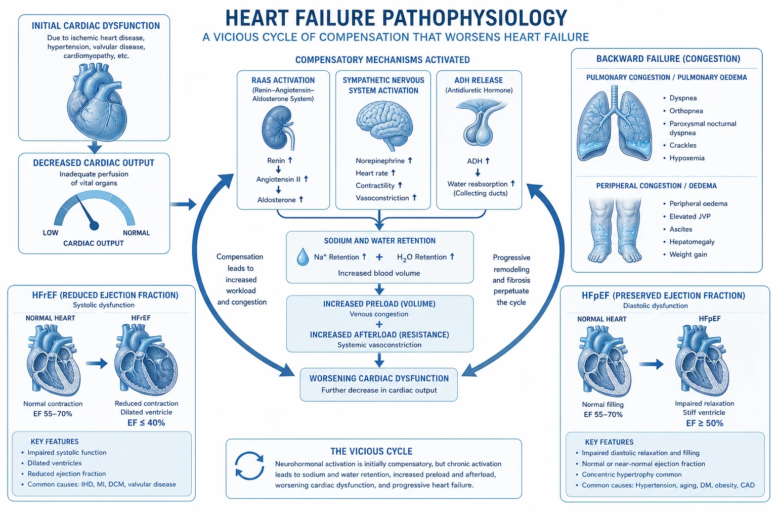
Heart failure begins with a failing pump — reduced cardiac output from impaired contractility (HFrEF) or impaired filling at acceptable pressures (HFpEF). The body interprets the falling output as underperfusion and mounts a compensatory neurohormonal response. The four key axes are the renin–angiotensin–aldosterone system (RAAS), the sympathetic nervous system, arginine vasopressin (ADH), and the counter-regulatory natriuretic peptides.[1]
RAAS — renin, angiotensin II, aldosterone
Reduced renal perfusion (and sympathetic input to the juxtaglomerular cells) releases renin, which cleaves angiotensinogen to angiotensin I, which angiotensin-converting enzyme (ACE) converts to angiotensin II. Angiotensin II is a potent vasoconstrictor (raising afterload), stimulates aldosterone release from the adrenal cortex (sodium and water retention at the distal nephron, potassium loss), promotes ADH release, drives cardiac myocyte hypertrophy and fibroblast collagen deposition, and is directly cardiotoxic in the long term. Aldosterone itself, independently of angiotensin II, causes myocardial fibrosis and is a key driver of adverse remodelling — which is why mineralocorticoid receptor antagonists (MRAs) earn their place as an independent pillar rather than being redundant with ACE-inhibitors.[1]
Sympathetic nervous system
Falling cardiac output triggers baroreceptor-mediated sympathetic discharge. Catecholamines (noradrenaline, adrenaline) initially support stroke volume and heart rate — the rationale for the old, now-abandoned "inotrope-first" approach — but chronically they are directly cardiotoxic: they drive myocyte apoptosis, provoke arrhythmia, increase myocardial oxygen demand (worsening ischaemia), downregise beta-1 receptors, and accelerate adverse remodelling. Plasma noradrenaline correlates with mortality in heart failure.[1]
Vasopressin (ADH) and the natriuretic peptides
Arginine vasopressin (ADH) rises via non-osmotic release, driving free-water reabsorption through V2 receptors in the collecting duct — the mechanism behind the dilutional hyponatraemia characteristic of advanced disease and a marker of poor prognosis. Against all this, the natriuretic peptides — ANP from the atria and BNP (B-type) from the ventricles, both released in response to myocardial stretch — promote natriuresis, vasodilation, and counter-regulation of RAAS and sympathetic activity. They are the body's own brake on the system, but in chronic heart failure they are ultimately overwhelmed by the three maladaptive axes above. Their measurement (BNP / NT-proBNP) is the basis of the diagnostic biomarker.[1]
Cardiac remodelling and ventricular interdependence
Cardiac remodelling is the structural signature of chronic neurohormonal injury: myocyte hypertrophy and elongation, interstitial fibrosis, chamber dilation (in HFrEF) or wall thickening with reduced compliance (in HFpEF), and a shift toward a more spherical, mechanically inefficient left ventricle. Remodelling is reversible, and reverse remodelling (shrinking chamber size, recovering function) is the therapeutic goal of disease-modifying therapy — it tracks with better outcomes. The two ventricles share a septum and a pericardium, so dysfunction in one soon compromises the other (ventricular interdependence): a failing right ventricle shifts the septum leftward and impairs LV filling, while LV dilation and pulmonary congestion raise pulmonary pressures and load the RV — the basis of biventricular failure.[1]
How the four pillars map onto this model
Each pillar of HFrEF therapy exists to interrupt a specific maladaptive axis: [1]
FOUR PILLARS
ACE-inhibitor / ARB / ARNI blocks angiotensin II
Beta-blocker blocks catecholamine cardiotoxicity
Blocks aldosterone's direct profibrotic effect on the myocardium
SGLT2 inhibitor (mechanism in HF still incompletely understood)
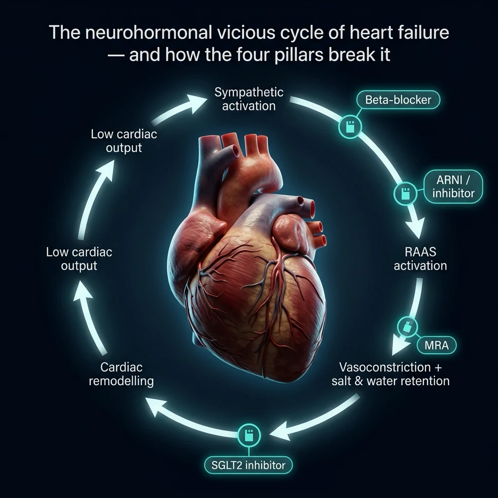
The SGLT2 inhibitor mechanism in HF is independent of glucose-lowering and probably involves a combination of mild diuresis (preferential proximal tubular sodium-glucose co-transport blockade lowering preload), improved myocardial energetics (ketone body utilisation), reduced inflammation, and lowered blood pressure.[8][9]
HFpEF pathophysiology — fundamentally different
HFpEF is not a contractility problem. The ventricle is stiff and non-compliant, with impaired active relaxation and elevated filling pressures; symptoms arise because the left atrium must generate high pressures to fill a stiff ventricle, which transmits back to the pulmonary veins and causes pulmonary congestion. The underlying driver is multi-system comorbidity — obesity, diabetes, hypertension, chronic kidney disease — producing systemic microvascular inflammation, coronary endothelial dysfunction, and myocardial fibrosis. This is why therapies built around interrupting the RAAS/sympathetic response to poor contractivity largely fail in HFpEF: the contractility axis was never the problem.[1]
Clinical Presentation
Symptoms
The symptom cluster reflects which circulation is congested and how far forward flow has fallen: [1]
- Exertional dyspnoea — the earliest and commonest symptom; falling cardiac output cannot rise with exertion.
- Orthopnoea — breathlessness within minutes of lying flat. Lying flat redistributes intravascular volume from the splanchnic beds and legs into the central circulation, raising pulmonary venous pressure and provoking pulmonary congestion. Relief on sitting up (or sleeping on extra pillows) is the defining feature. Two-pillow orthopnoea suggests moderate disease.
- Paroxysmal nocturnal dyspnoea (PND) — waking gasping 1–2 hours after falling asleep. As with orthopnoea, recumbency reabsorbs interstitial oedema back into the circulation faster than a failing LV can clear it; the patient sits upright and may seek fresh air. PND usually indicates more advanced disease than orthopnoea alone.
- Fatigue and effort intolerance — low cardiac output; the patient tires on minimal activity.
- Ankle swelling and abdominal fullness — right-sided congestion; ascites and hepatic congestion give bloating, anorexia, and right-upper-quadrant aching.
- Nocturia — renal perfusion improves when the patient lies down, so sodium and water excretion shifts to the night.
- Cachexia and muscle wasting — advanced, end-stage HF; the cardiac cachexia syndrome carries a poor prognosis. [1]
Signs
The sign inventory on focused cardiovascular and respiratory examination: [1]
- Elevated JVP — the single most reliable bedside marker of volume overload and right heart congestion.
- Displaced apex beat — laterally displaced and diffuse in a dilated LV (HFrEF); sustained and heaving in a hypertrophied LV (HFpEF or pressure overload).
- Third heart sound (S3 gallop) — the auscultatory hallmark of systolic/HFrEF: a low-pitched sound just after S2, produced by rapid ventricular filling into a non-compliant or volume-overloaded ventricle.
- Fourth heart sound (S4) — more typical of diastolic dysfunction/HFpEF, produced by atrial contraction into a stiff ventricle.
- Bibasal fine crackles — pulmonary oedema (may be absent if decongested, or in chronic HF with lymphatic compensation).
- Pleural effusion — usually bilateral, right larger than left (greater pleural surface area on the right).
- Hepatomegaly, ascites, peripheral and sacral pitting oedema — right-sided congestion.
- Tachycardia, cool peripheries, narrow pulse pressure, low systolic BP — low-output / advanced HF.
- Pulsus alternans — alternating strong and weak pulses in advanced LV failure. [1]
Left- versus right-sided, and biventricular
Left-sided signs (dyspnoea, orthopnoea, PND, crackles, pleural effusion) reflect pulmonary venous congestion behind a failing left ventricle. Right-sided signs (raised JVP, hepatomegaly, dependent oedema, ascites) reflect systemic venous congestion behind a failing right ventricle. In practice, by the time most patients present they have biventricular failure — isolated left heart failure tends to progress to right heart failure as pulmonary pressures rise. Atypical presentations are deliberately tested: the elderly patient may present with confusion, falls, fatigue, or anorexia rather than classical dyspnoea; the diabetic patient may have a relatively silent presentation due to autonomic neuropathy. [1]
Differential Diagnosis
The differential of breathlessness and oedema is broad. Each competitor is distinguished on specific grounds — and examiners expect the distinguishing features, not just a list of names. [1]
COPD / asthma
- Wheeze, prolonged expiration, smoking history
- Spirometry shows obstruction (FEV1/FVC reduced)
- Can coexist with HF — the two together are common
- BNP often borderline; echo usually preserved EF unless overlap
Pulmonary embolism
- Sudden-onset pleuritic dyspnoea, risk factors for VTE
- Hypoxia out of proportion to CXR findings
- D-dimer, CTPA for diagnosis
- Echo may show RV strain, raised RVSP — mimics right HF
Pneumonia
- Fever, purulent productive cough, focal consolidation
- Acute, infective tempo with raised inflammatory markers
- Can itself precipitate acute HF decompensation
Anaemia
- Fatigue and exertional dyspnoea with normal cardiac exam
- FBC confirms low haemoglobin
- High-output state — can worsen existing HF
Renal failure / nephrotic
- Generalised oedema with proteinuria or deranged renal function
- Normal cardiac imaging
- Natriuretic peptides may be falsely elevated in CKD
Cirrhosis
- Ascites and oedema with stigmata of chronic liver disease
- JVP normal (unless concurrent cardiac disease) — the key discriminator from right HF
- Low albumin, deranged LFTs, portal hypertension signs
Chronic venous insufficiency
- Bilateral ankle oedema, varicose veins, haemosiderin staining
- Normal JVP, normal heart, normal BNP
- Worse after standing, relieved by elevation
A normal BNP/NT-proBNP together with a normal echocardiogram is decisive in excluding a cardiac cause of dyspnoea — the negative predictive value is very high. HFpEF specifically must be separated from other causes of dyspnoea with a preserved EF (obesity, deconditioning, primary lung disease, anaemia, chronic thromboembolic disease), where the clinical picture, natriuretic peptide level, and diastolic function assessment on echo (E/e′ ratio, left atrial size) together make the diagnosis; the HFA-PEFF score and H2FPEF score formalise this clinical-biomarker-imaging approach.[1]
Clinical & Bedside Assessment
The focused bedside examination in suspected or established HF yields a remarkable amount of information. [1]
Jugular venous pressure — measured with the patient reclined at 45°, the internal jugular vein (not the carotid — the JVP is biphasic, non-pulsatile, occlusible, and varies with respiration). The vertical height above the sternal angle is read in centimetres of water; greater than 3 cm above the sternal angle (i.e. a JVP palpable more than 8 cm above the right atrium) is elevated. An elevated JVP is the most specific bedside sign of right-sided volume overload and correlates with prognosis. The hepatojugular reflux (sustained firm pressure over the liver for 15–30 seconds producing a sustained JVP rise of more than 4 cm) indicates that the right heart cannot accommodate the venous return — a sign of right HF or biventricular failure. [1]
Apex beat — position (5th intercostal space, mid-clavicular line is normal; lateral displacement indicates a dilated LV; a hyperdynamic, volume-loaded apex suggests volume overload such as severe regurgitation; a sustained heaving apex suggests pressure overload / hypertrophy / HFpEF). [1]
Auscultation — listen specifically for an S3 (low-pitched, just after S2, best heard at the apex with the bell in left lateral position; the hallmark of systolic HF), an S4 (before S1, indicating a stiff non-compliant ventricle — HFpEF or hypertension), gallop rhythm (tachycardia plus S3), and murmurs of underlying valvular disease (aortic stenosis, mitral regurgitation) which may be the cause rather than the consequence. A bibasal crackle pattern suggests pulmonary oedema, although chronic HF patients may have clear lungs thanks to lymphatic compensation despite raised filling pressures. [1]
Severity and perfusion — blood pressure, peripheral temperature, capillary refill, urine output, and mentation gauge end-organ perfusion and the boundary with cardiogenic shock (hypotension plus signs of hypoperfusion). The presence of a narrow pulse pressure, cool peripheries, and oliguria signal a low-output state that may need inotropic support rather than further diuresis. [1]
Investigations
First-line work-up
First-line investigations in suspected heart failure are the echocardiogram, natriuretic peptides, 12-lead ECG, chest X-ray, and a blood panel (U&E, eGFR, FBC, LFT, TFT, ferritin, fasting glucose/HbA1c, lipid profile, and urinalysis).[1]
Natriuretic peptides — the rule-out biomarker
BNP and NT-proBNP are powerful rule-out tests — a normal level makes HF highly unlikely. The exact threshold depends on the clinical setting. NICE NG106 (UK) uses: [1]
BNP / NT-proBNP thresholds (NICE NG106)
BNP is degraded by neprilysin — so patients on an ARNI (sacubitril-valsartan) have a falsely elevated BNP (the drug blocks the enzyme that clears it); NT-proBNP is the preferred marker in ARNI-treated patients. Both peptides can be elevated by anything that stretches the heart (AF, PE, RV failure, renal failure, sepsis) and lowered by obesity — interpret in context. [1]
Echocardiography — the pivotal test
The echocardiogram is the single most informative investigation in HF. It measures: [1]
- Left ventricular ejection fraction (LVEF) — the basis of the HFrEF/HFmrEF/HFpEF split.
- Chamber size and wall thickness — dilated LV in HFrEF; concentric LVH in HFpEF or hypertension; dilated left atrium reflecting chronically raised filling pressure.
- Diastolic function — mitral inflow E and A waves, tissue Doppler e′ velocity, and the E/e′ ratio (a surrogate for LV filling pressure; E/e′ greater than 14 suggests raised pressure), left atrial volume index, and tricuspid regurgitant velocity. Together these grade diastolic dysfunction.
- Valvular structure and function — underlying valvular disease as a cause (aortic stenosis, mitral regurgitation) or consequence (functional MR from LV dilation, tricuspid regurgitation from RV dilation).
- Right ventricular size and function, and estimated right ventricular systolic pressure (RVSP) — pulmonary hypertension and right heart involvement.
- Regional wall motion abnormalities — suggesting ischaemic aetiology.
- Pericardial effusion and structural anomalies (e.g. thrombus in a dilated LV, apical ballooning). [1]
Chest X-ray
Classic CXR signs of heart failure form a familiar sequence of increasing severity: cardiomegaly (cardiothoracic ratio greater than 0.5); upper-lobe blood diversion (cephalisation, from raised pulmonary venous pressure); Kerley B lines (short horizontal linear opacities at the lung bases — interstitial oedema and distended lymphatics); perihilar "bat-wing" alveolar pulmonary oedema (frank alveolar flooding, a late sign); and pleural effusions (usually bilateral, right often larger).[1]
ECG
The 12-lead ECG is abnormal in the great majority of HF patients (a completely normal ECG makes HF unlikely). Look for: prior Q-wave or ischaemic changes (aetiology), LV hypertrophy with strain (hypertensive/HFpEF), left bundle branch block (relevant for CRT — wide QRS with LBBB morphology), atrial fibrillation (precipitant or consequence), brady- and tachy-arrhythmias, and conduction disease. [1]
Second-line and aetiology-defining tests
- Coronary angiography when an ischaemic aetiology needs defining — especially before revascularisation or in new-onset HF where ischaemia is suspected.
- Cardiac MRI the gold standard for tissue characterisation — infiltrative (amyloid, sarcoid), inflammatory (myocarditis), hypertrophic, and arrhythmogenic cardiomyopathies; gives precise LV/RV volumes and LVEF, and late gadolinium enhancement patterns diagnose the underlying substrate.
- Cardiopulmonary exercise testing (CPET) — peak oxygen consumption (peak VO2) is central to transplant and LVAD candidacy work-up.
- Cardiac biopsy — rarely, in suspected giant-cell myocarditis or fulminant myocarditis.
- Iron studies (ferritin, transferrin saturation) — iron deficiency is common in HF, worsens symptoms, and is independently treated with intravenous iron (ferric carboxymaltose) irrespective of anaemia.
- Troponin, D-dimer, HIV and hepatitis serology, autoimmune screen, 24-hour urine for catecholamines / metanephrines (phaeochromocytomy) — where specific aetiologies are suspected. [1]
Management — Resuscitation
Acute cardiogenic pulmonary oedema
Acute pulmonary oedema with respiratory distress is a medical emergency. The first ten minutes follow a familiar bundle:[1]
Acute pulmonary oedema — first 10 minutes
Position
Sit the patient upright to pool fluid in the lung bases and reduce venous return
Oxygen
High-flow oxygen (target SpO2 at least 92%) via mask or nasal cannulae
IV loop diuretic
Furosemide 40 to 80 mg IV bolus (higher doses 80 to 120 mg in severe/larger patients; an IV infusion may follow)
Vasodilator
IV nitrate (e.g. glyceryl trinitrate infusion) if systolic BP is at least 110 mmHg; nitrates reduce preload and afterload and relieve pulmonary congestion
Non-invasive ventilation
CPAP or BiPAP for ongoing respiratory distress — reduces work of breathing, recruits alveoli, lowers preload and afterload, improves oxygenation
Find the precipitant
Identify and treat the precipitant (arrhythmia, ischaemia, infection, non-adherence)
Escalate
Transfer to intensive care if respiratory failure persists or shock develops
Oxygen is given to correct hypoxia; in the breathless but not hypoxic patient, NIV is the more effective intervention. Morphine (2.5 to 5 mg IV) was once routine for distress and preload reduction but is now used cautiously, if at all, because routine use has been associated with harm (hypoventilation, ICU admission, mechanical ventilation). CPAP/BiPAP is indicated for cardiogenic pulmonary oedema with respiratory distress and a respiratory rate over 25 — it reduces intubation rates and (in some meta-analyses) mortality; BiPAP is acceptable in hypercapnic overlap with COPD.[1]
Cardiogenic shock
Cardiogenic shock is hypotension (systolic BP less than 90 mmHg) with signs of hypoperfusion (cold clammy skin, oliguria, confusion, lactic acidosis) due to a primary cardiac cause. The principles differ from pulmonary oedema: the patient needs inotropic and/or vasopressor support and cause-directed therapy, not more diuretic. Inotropes include dobutamine (beta-1 agonist; 2 to 20 microgram/kg/min) and milrinone (phosphodiesterase-3 inhibitor, combined inotropic and vasodilator — "inodilator"); a vasopressor such as noradrenaline is added if vasodilatory shock dominates. Cause-directed therapy is essential — urgent reperfusion for the ischaemic cause, valve intervention for mechanical catastrophe, mechanical circulatory support (IABP, Impella, VA-ECMO) for refractory shock as a bridge to recovery, decision, or transplant. (See the dedicated cardiogenic shock topic.)[1]
Management — Definitive & Stepwise
HFrEF — the four pillars (GDMT)
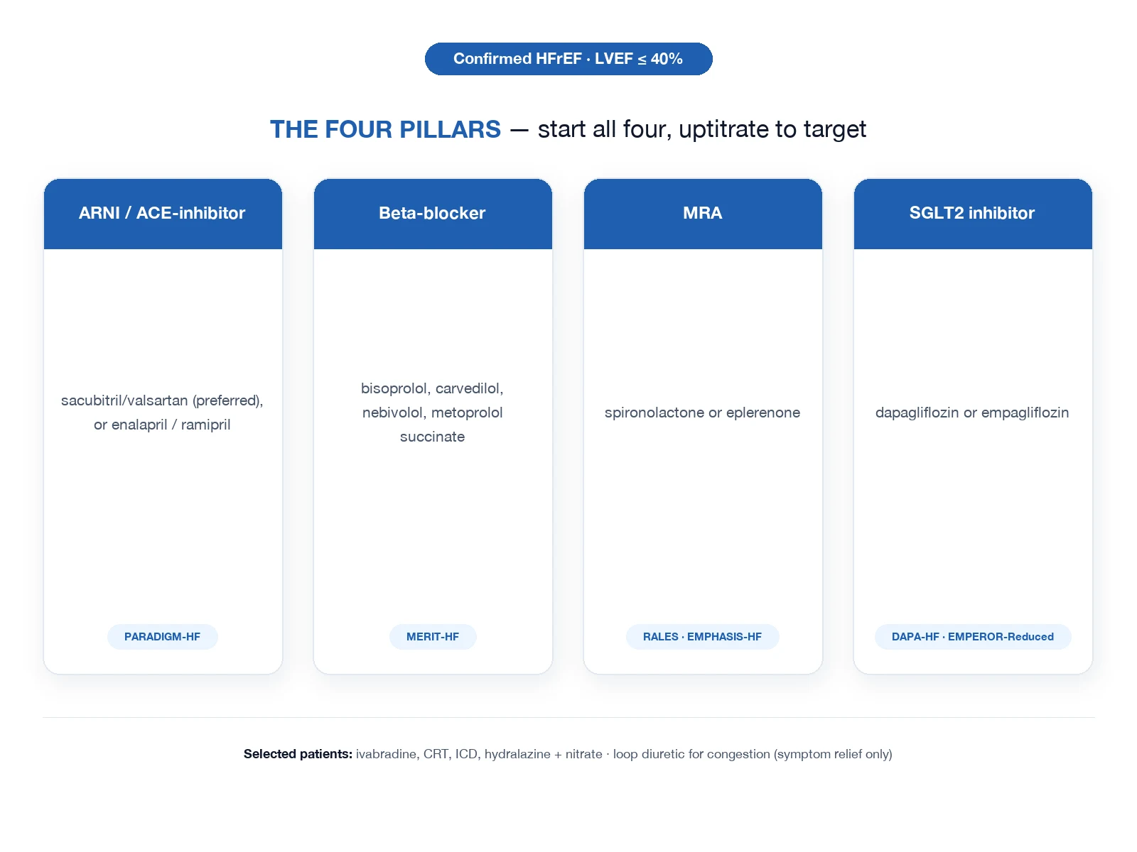
For HFrEF, guideline-directed medical therapy (GDMT) rests on four foundational drug classes, each with an independent mortality benefit demonstrated in a landmark trial. Where tolerated, all four are now started (sequenced by tolerability rather than one class fully titrated before the next begins) at low dose and uptitrated to target over weeks.[1]
Pillar 1 — RAAS blockade: ARNI or ACE-inhibitor
The angiotensin receptor-neprilysin inhibitor (ARNI) sacubitril-valsartan is preferred over an ACE-inhibitor on the basis of PARADIGM-HF, in which sacubitril-valsartan 97/103 mg twice daily reduced cardiovascular death and HF hospitalisation compared with enalapril.[4] If an ACE-inhibitor is used instead, enalapril 10 to 20 mg twice daily (the CONSENSUS regimen, which first proved an ACE-inhibitor improved survival in severe HF)[3] or ramipril 10 mg once daily. Starting dose for sacubitril-valsartan is 49/51 mg twice daily (24/26 mg twice daily if on low-dose ACE-i), uptitrated to the 97/103 mg twice daily target. Switching from an ACE-inhibitor to ARNI requires a 36-hour washout to avoid angioedema (neprilysin inhibition plus residual ACE inhibition dangerously raises bradykinin). For patients intolerant of ACE-inhibitor or ARNI on renal or hyperkalaemia grounds, an angiotensin receptor blocker (ARB) such as valsartan 160 mg twice daily or candesartan 32 mg once daily is reasonable.[1]
Pillar 2 — Beta-blocker
One of three beta-blockers proven in HF — bisoprolol 10 mg once daily (CIBIS-II), carvedilol 25 mg twice daily (or up to 50 mg twice daily in patients over 85 kg; COPERNICUS/COMET), or metoprolol succinate 200 mg once daily (MERIT-HF)[5]; nebivolol 10 mg once daily is acceptable in patients aged 70 or older (SENIORS). The cardinal rule is "start low, go slow" — initiate at a low dose (bisoprolol 1.25 mg once daily; carvedilol 3.125 mg twice daily; metoprolol succinate 12.5 to 25 mg once daily) only once the patient is euvolaemic and haemodynamically stable, and uptitrate every two to four weeks toward target. A beta-blocker must never be started in an acutely decompensated, congested patient — it is a negative inotrope and can precipitate collapse — and must never be stopped abruptly (acute adrenergic withdrawal can precipitate decompensation or arrhythmia).[1]
Pillar 3 — Mineralocorticoid receptor antagonist (MRA)
Spironolactone 25 to 50 mg once daily in moderate-to-severe HF (RALES)[6] or mild symptomatic HFrEF (EMPHASIS-HF)[7], or eplerenone 25 to 50 mg once daily post-MI with LV dysfunction. The MRA earns its place as an independent pillar because it blocks the direct profibrotic effect of aldosterone on the myocardium, an effect not fully suppressed by ACE-inhibitors (aldosterone "escape"). Monitor potassium and creatinine at baseline, after initiation (1 to 2 weeks), after every uptitration, and routinely — hyperkalaemia and a rise in creatinine are the dose-limiting toxicities. Spironolactone also causes gynaecomastia and breast tenderness (eplerenone much less so), a reason to switch to eplerenone if troublesome.[1]
Pillar 4 — SGLT2 inhibitor
Dapagliflozin 10 mg once daily (DAPA-HF)[8] or empagliflozin 10 mg once daily (EMPEROR-Reduced)[9] — benefit regardless of diabetes status. SGLT2 inhibitors are uniquely the only pillar with proven outcome benefit across the entire ejection-fraction spectrum, including HFpEF (EMPEROR-Preserved and DELIVER)[10][11]. They are well tolerated, need no dose titration, and additionally slow diabetic kidney disease. They cause volume depletion (an advantage for congested patients, a caution when initiating in hypovolaemic ones) and a small genital infection risk.
HFrEF four pillars — representative drugs and target doses
Symptomatic therapy — loop diuretic
A loop diuretic — furosemide 40 mg once daily, uptitrating as needed (up to 240 mg daily in severe congestion; 1:1 conversion to bumetanide, and roughly 2:1 conversion to torsemide) — is given for symptom relief and decongestion but does not improve survival. It is the first drug in the acutely congested patient and is continued at the lowest dose that keeps the patient euvolaemic. Diuretic resistance may respond to combination with a thiazide (e.g. metolazone) for sequential nephron blockade — but watch for over-diuresis, hypokalaemia, and renal impairment. [1]
Additional disease-modifying and symptomatic therapies
- Ivabradine 5 to 7.5 mg twice daily (SHIFT) for patients in sinus rhythm with heart rate at least 70 bpm despite maximally tolerated beta-blocker, and LVEF ≤35% — it selectively inhibits the SA node If current, lowering heart rate without affecting contractility. It works only in sinus rhythm and is useless in atrial fibrillation (the AV node, not the If current, controls rate in AF).[12]
- Hydralazine plus nitrate (A-HeFT) — hydralazine 37.5 to 75 mg three times daily plus isosorbide dinitrate 20 to 40 mg three times daily — particularly for self-identified African-American patients with HFrEF already on optimal therapy, or for patients intolerant of RAAS blockade.[13]
- Digoxin 62.5 to 250 microgram once daily — for symptom control, especially in HFrEF with concomitant atrial fibrillation (rate control); it does not improve mortality but reduces hospitalisations. Monitor for toxicity (nausea, visual disturbance, arrhythmia), especially with hypokalaemia and renal impairment.
- Intravenous iron (ferric carboxymaltose) for iron deficiency (ferritin less than 100 microgram/L, or 100 to 299 with transferrin saturation under 20%), which improves symptoms and exercise capacity irrespective of anaemia.
- Anticoagulation is reserved for HF with concomitant AF, a thrombus in the LV, or another standard indication — routine anticoagulation in HF in sinus rhythm is not recommended.
Device therapy — CRT and ICD
CRT (resynchronisation)
- QRS at least 130 ms with LBBB morphology
- LVEF ≤35% despite at least 3 months of optimal medical therapy
- NYHA class II to IV (or class I in ischaemic cardiomyopathy)
- Improves symptoms, LVEF, and survival; reduces hospitalisation
ICD (sudden-death prevention)
- Primary prevention: LVEF ≤35% after at least 3 months of optimal medical therapy, NYHA II to III, expected survival beyond 1 year
- Reduces sudden cardiac death from ventricular arrhythmia — the leading mode of death in HFrEF
- Secondary prevention after surviving VT/VF arrest
- Combined with CRT (CRT-D) when both criteria met
Cardiac resynchronisation therapy (CRT) is indicated for HFrEF with a wide QRS (at least 130 ms) and LBBB morphology, an LVEF ≤35% despite at least three months of optimal medical therapy, and symptoms (NYHA II to IV, or NYHA I in ischaemic cardiomyopathy). An implantable cardioverter-defibrillator (ICD) is offered for primary prevention of sudden cardiac death when LVEF remains ≤35% despite optimised GDMT (allowing at least three months for reverse remodelling), in NYHA II–III, with reasonable expected survival. Sudden cardiac death from ventricular arrhythmia is a leading mode of death in HFrEF, and the ICD is the specific therapy that addresses it.[1]
HFpEF management
HFpEF management is comparatively limited: SGLT2 inhibitors are the only class with proven outcome benefit (EMPEROR-Preserved and DELIVER reduced HF hospitalisation and cardiovascular death)[10][11]. The remainder of management is diuretics for congestion (furosemide or torsemide) plus aggressive control of the comorbidities that drive the syndrome — hypertension (target below 130/80), atrial fibrillation (rate and rhythm control), diabetes (SGLT2 inhibitors first-line; avoid thiazolidinediones which cause fluid retention), obesity, and ischaemia. Classic HFrEF GDMT does not work in HFpEF and should not be reflexively applied.[1]
HFmrEF management
In HFmrEF (EF 41 to 49%), SGLT2 inhibitor benefit is established; the other three pillars are considered case-by-case, extrapolating from HFrEF evidence. The closer the EF to 40%, the stronger the case for full HFrEF-style GDMT.[1]
Specific Subtypes & Scenarios
Acute decompensated heart failure
The framework for any acute decompensation: identify and treat the precipitant (use the FAILURE mnemonic), decongest with IV loop diuretic (furosemide 40 to 80 mg IV bolus, or a continuous infusion; a daily urine output target and weight monitoring guide dosing), optimise haemodynamics (vasodilator if hypertensive, inotrope if hypoperfusing), monitor renal function and electrolytes (diuresis-induced AKI and hypokalaemia), and transition back to optimised oral GDMT before discharge — every admission is an opportunity to start or uptitrate the four pillars.[1]
HFpEF, HFmrEF
(Covered above — the central teaching point is that only the SGLT2 inhibitor class has cross-spectrum evidence.) [1]
Cardiogenic shock
Inotropic and/or mechanical circulatory support plus cause-directed therapy (see Management — Resuscitation and the dedicated cardiogenic shock topic). [1]
Right heart failure / cor pulmonale
Right heart failure is usually secondary to pulmonary hypertension (Group 2, from left heart disease, is the commonest) or chronic lung disease (cor pulmonale, Group 3). Management targets the underlying cause — optimising left-sided filling pressures, treating COPD or interstitial lung disease, and where appropriate treating pulmonary arterial hypertension with specialised therapy (PDE-5 inhibitors, endothelin receptor antagonists, riociguat). Diurese congestion; the RV tolerates excessive afterload poorly. [1]
Specific reversible cardiomyopathies
Several aetiologies deserve explicit naming because they are reversible or partially reversible with cause-directed therapy: [1]
- Tachycardiomyopathy — chronic tachycardia (persistent AF, inappropriate sinus tachycardia, ectopic atrial tachycardia) produces a dilated, failing LV that recovers substantially once rate or rhythm is controlled.
- Peripartum cardiomyopathy — HF with LVEF ≤45% towards the end of pregnancy or in the months postpartum; treated with standard HFrEF GDMT (with pregnancy-specific drug caveats — avoid ACE-inhibitor/ARB/MRA in pregnancy; bromocriptine has emerging evidence), and may recover fully. Recurrence in subsequent pregnancy is a real risk.
- Alcoholic cardiomyopathy — a dilated cardiomyopathy in chronic heavy drinkers; abstinence leads to substantial recovery in many.
- Anthracycline-induced cardiotoxicity (doxorubicin) and anti-HER2 therapy (trastuzumab) — important and increasingly common drug causes; anthracycline damage is cumulative and largely irreversible, trastuzumab is often reversible on cessation. Baseline and surveillance echocardiography are standard in oncology patients receiving cardiotoxic therapy.
- Iron overload cardiomyopathy (haemochromatosis, repeated transfusions), thyroid dysfunction (both hyper- and hypothyroidism), selenium deficiency, thiamine deficiency (beriberi), and HIV-related cardiomyopathy are all treatable aetiologies to actively seek. [1]
Cardiomyopathies — structural pattern recognition
The four cardiomyopathy patterns an examiner expects: dilated (DCM — dilated LV, thin walls, commonest, often idiopathic or familial or post-viral), hypertrophic (HCM — asymmetric septal hypertrophy, dynamic LVOT obstruction, diastolic dysfunction, sudden death risk), restrictive (RCMP — stiff non-compliant ventricles, e.g. amyloidosis, eosinophilic), and arrhythmogenic right ventricular cardiomyopathy (ARVC — fibrofatty RV replacement, ventricular arrhythmia, sudden death). HCM and amyloidosis predominantly cause HFpEF-like physiology; DCM predominantly causes HFrEF. Cardiac MRI with late gadolinium enhancement is the discriminating test. [1]
Complications & Pitfalls
Complications
The complications of chronic heart failure include: [1]
- Atrial fibrillation — common, both a precipitant and a consequence; loss of atrial contribution to filling and a fast ventricular rate worsen HF.
- Ventricular arrhythmia and sudden cardiac death — a leading mode of death in HFrEF, addressed by primary-prevention ICD.
- Recurrent hospitalisation — each admission worsens the long-term trajectory and is itself a poor prognostic marker.
- Cardiogenic shock — end-stage decompensation.
- Cardiorenal syndrome — the tension between decongestion (which needs diuresis) and renal perfusion (which diuresis can worsen); rising creatinine on diuretic therapy must be balanced against the harm of persistent congestion.
- Cardiac cachexia — skeletal muscle wasting; a poor prognostic sign in advanced HF.
- Hepatic congestion (cardiac cirrhosis) — chronic right HF produces a congestive hepatopathy with raised bilirubin and (late) coagulopathy.
- Thromboembolism — from a dilated, hypokinetic LV or AF.
- Hyponatraemia, anaemia, iron deficiency, renal dysfunction — common comorbidities that worsen symptoms and prognosis. [1]
Pitfalls — the classic errors
- Applying all four HFrEF pillars to a patient with HFpEF — only the SGLT2 inhibitor helps; classic GDMT has not shown outcome benefit in HFpEF.
- Failing to uptitrate to target doses — many patients are left indefinitely on sub-therapeutic starting doses; the mortality benefit accrues to target dosing.
- Starting a beta-blocker in a still-congested, decompensated patient — it is a negative inotrope and can precipitate collapse.
- Ignoring rising potassium or creatinine on RAAS blockade / MRA — reassess dose, volume status, and contributing drugs (NSAIDs) rather than reflexively stopping; small rises are expected and acceptable, marked rises need action.
- Missing the ACE-inhibitor-to-ARNI washout — a 36-hour gap is mandatory to prevent angioedema.
- Failing to identify and treat the precipitant of an acute decompensation — inviting early relapse and re-admission.
- Interpreting an elevated BNP in a patient on ARNI — BNP is falsely elevated; use NT-proBNP.
- Underusing SGLT2 inhibitors — they work in HFrEF, HFmrEF, and HFpEF, and in patients with or without diabetes; over-conservative prescribing denies patients benefit. [1]
Prognosis & Disposition
Despite modern guideline-directed medical therapy (GDMT), heart failure remains life-limiting. Overall five-year mortality remains roughly 50%, worse with advanced NYHA class, repeated hospitalisations, hyponatraemia, rising natriuretic peptides, declining eGFR, and right-ventricular failure. A single HF hospitalisation is a major prognostic event — one-year mortality after discharge commonly approaches 20–30% in older cohorts, which is why discharge planning, GDMT optimisation, and early follow-up are examinable management steps, not afterthoughts.[1]
Prognostic markers examiners expect
| Marker | Direction of risk | Clinical use |
|---|---|---|
| LVEF | Lower → worse (HFrEF continuum) | Device eligibility (ICD/CRT), GDMT intensity |
| NYHA class | Higher → worse | Symptom burden, transplant/device referral |
| BNP / NT-proBNP | Higher → worse | Diagnosis, prognosis, pre-discharge trajectory |
| Prior HF hospitalisation | Strong adverse marker | Triggers aggressive optimisation |
| Hyponatraemia | Independent adverse marker | Reflects ADH drive / advanced disease |
| eGFR / rising creatinine | Worse renal function → worse outcomes | Limits RAAS/MRA titration |
| QRS width / LBBB | Wide LBBB → consider CRT if HFrEF | Device selection |
| Peak VO2 / haemodynamics | Low peak VO2 → advanced HF | Transplant/LVAD evaluation |
NYHA functional class (reproduce)
- I — no limitation of ordinary physical activity
- II — slight limitation; comfortable at rest; ordinary activity causes symptoms
- III — marked limitation; less-than-ordinary activity causes symptoms
- IV — symptoms at rest; any activity increases discomfort
ACC/AHA stages (complement NYHA — structure vs symptoms)
- A — at risk (hypertension, diabetes, CAD) without structural disease or symptoms
- B — pre-HF: structural disease / raised NP / abnormal filling without current/prior symptoms
- C — symptomatic HF (current or prior)
- D — advanced HF requiring specialised interventions
Disposition rules
Acute decompensated HF / pulmonary oedema
- Ward vs HDU/ICU: ICU/HDU if respiratory failure needing NIV/intubation, cardiogenic shock, life-threatening arrhythmia, or need for inotropes/vasopressors/MCS.
- Sit up, oxygen, IV loop diuretic, nitrates if BP allows, NIV for respiratory distress with oedema.
Safe discharge checklist
- Near euvolaemia (stable weight, resolving oedema, no resting dyspnoea)
- Stable vital signs off oxygen (or stable home O2 plan)
- Transitioned to oral diuretic with a clear flexible dosing plan
- GDMT started/continued (do not stop prognostic drugs for mild creatinine/K rises without cause)
- Electrolytes and renal function checked after diuretic/RAAS changes
- Early follow-up within 7–14 days (phone or clinic) — the highest-risk window for readmission
- Self-care education: daily weights, salt/fluid advice individualised, red-flag dyspnoea/oedema, medication adherence
- Vaccinations (influenza, pneumococcal, COVID-19) and comorbidity optimisation (iron deficiency, sleep apnoea, thyroid, anaemia) [1]
Advanced HF triggers for specialist referral
Recurrent hospitalisations, persistent NYHA III–IV despite optimised GDMT, intolerance of GDMT from hypotension/renal limits, peak VO2 markedly reduced, progressive end-organ dysfunction, or dependence on inotropes → evaluate for transplant, LVAD, palliative care in parallel, not sequentially late.
Special Populations
Elderly and frail
Balance prognostic drug benefit against hypotension, falls, CKD, cognitive impairment, and polypharmacy. Prefer once-daily regimens when possible. Start low, titrate slow, but do not deny SGLT2 inhibitors, beta-blockers, or MRAs solely for age. HFpEF physiology dominates in older women with hypertension and AF. Involve geriatric assessment when frailty is clear.
Chronic kidney disease
- Expect up to ~30% creatinine rise after ACE-I/ARB/ARNI start if euvolaemic — often acceptable; larger rises → check obstruction, overdiuresis, bilateral RAS, NSAIDs.
- Hyperkalaemia limits MRA/ARNI — use dietary measures, diuretic adjustment, potassium binders in selected patients rather than abandoning GDMT reflexively.
- SGLT2 inhibitors slow CKD progression (DAPA-CKD, EMPA-KIDNEY logic) and are foundational in HF across EF spectrum when eGFR allows initiation.
- Avoid NSAIDs; dose-adjust renally cleared drugs; watch digoxin levels if used.
Atrial fibrillation and HF
AF both causes and results from HF. Rate control (beta-blocker first line) is foundational; consider digoxin as adjunct for rate in HFrEF. Rhythm control (including early AF ablation) may improve outcomes in selected HFrEF patients with AF (CASTLE-AF logic). Anticoagulate by CHA2DS2-VASc unless contraindicated — HF itself scores a point.
Diabetes and HF
SGLT2 inhibitors are first-line cardio-renal drugs in this overlap (DAPA-HF, EMPEROR-Reduced/Preserved, SOLOIST-like evidence streams). Avoid thiazolidinediones (fluid retention). Metformin is generally safe in stable HF with adequate eGFR. Screen for silent ischaemia when presentation is atypical.
Pregnancy and peripartum cardiomyopathy (PPCM)
- New HF in the last month of pregnancy or within 5 months postpartum without other cause → consider PPCM.
- Management in a cardio-obstetric team: afterload reduction with hydralazine + nitrate (ACE-I/ARB/ARNI contraindicated in pregnancy); beta-blocker (e.g. metoprolol) if needed; diuretics for congestion; anticoagulation if thrombus/AF.
- Bromocriptine is used in some protocols as disease-modifying therapy for PPCM (region-dependent evidence).
- Subsequent pregnancy risk is high if LVEF does not recover — counselling is mandatory.
Chemotherapy / cancer therapy-related cardiac dysfunction
Anthracyclines and HER2-targeted therapy (trastuzumab) are classic. Monitor LVEF; hold/adjust oncologic therapy with cardio-oncology input; treat along HFrEF lines when systolic dysfunction appears. Dexrazoxane and liposomal anthracyclines are preventive strategies in selected oncology protocols.
Iron deficiency (even without anaemia)
IV ferric carboxymaltose improves symptoms and reduces HF hospitalisations in iron-deficient HFrEF (AFFIRM-AHF / IRONMAN-style evidence). Check ferritin and transferrin saturation in all symptomatic HF patients.
Right heart failure and pulmonary hypertension
Treat the cause (left heart disease, PE, lung disease, CTEPH). Diuretics for congestion; avoid excessive afterload reduction that drops coronary perfusion. Specialist PH pathways for precapillary disease.
Devices and special decisions
- ICD for primary prevention: typically LVEF ≤35% despite ≥3 months GDMT, NYHA II–III, expected survival >1 year (details vary by ischaemic vs non-ischaemic aetiology and guideline edition).
- CRT: LVEF ≤35%, sinus rhythm, LBBB with QRS ≥150 ms (strongest evidence), NYHA II–IV on GDMT; weaker evidence for non-LBBB or QRS 130–149 ms.
- Ivabradine: sinus rhythm, HR ≥70 on maximally tolerated beta-blocker, HFrEF — reduces hospitalisation (SHIFT).
Evidence, Guidelines & Regional Differences
The two foundational guidelines
The framework used here rests on the 2023 Focused Update of the 2021 ESC Guidelines[1] and the 2022 AHA/ACC/HFSA Guideline for the Management of Heart Failure[2]. Both converge on the same core: four foundational HFrEF therapies initiated together (rather than rigidly sequenced) where tolerated, SGLT2 inhibitors recommended across the entire ejection-fraction spectrum, and diuretics reserved for symptom control.
Landmark trials behind each pillar
RAAS / ARNI
- CONSENSUS 1987 — enalapril reduced mortality in severe HF (PMID 2883575)
- PARADIGM-HF 2014 — ARNI superior to enalapril (PMID 25176015)
Beta-blockade
- MERIT-HF 1999 — metoprolol succinate reduced mortality (PMID 10376614)
- CIBIS-II (bisoprolol) and COPERNICUS (carvedilol) corroborated
MRA
- RALES 1999 — spironolactone in severe HF (PMID 10471456)
- EMPHASIS-HF 2011 — eplerenone in mild symptomatic HF (PMID 21073363)
SGLT2 inhibitor
- DAPA-HF 2019 — dapagliflozin in HFrEF (PMID 31535829)
- EMPEROR-Reduced 2020 — empagliflozin in HFrEF (PMID 32865377)
- EMPEROR-Preserved 2021 and DELIVER 2022 — across the EF spectrum (PMID 34449189, 36027570)
Adjuncts
- SHIFT 2010 — ivabradine in SR with HR at least 70 (PMID 20801500)
- A-HeFT 2004 — hydralazine-nitrate in African-Americans (PMID 15533851)
Regional deltas
[1] [1]The overall four-pillar framework is globally consistent across ESC, AHA/ACC/HFSA, and NICE — but access drives sequencing. In India (the NEET-PG/INICET context), cost typically drives initial use of generic ACE-inhibitor/ARB and beta-blocker, with ARNI and SGLT2 inhibitors added where affordable, rather than the "all four together" ideal. The Cardiological Society of India (CSI) endorses the four-pillar framework with these access-driven sequencing caveats.
Current controversies
Active areas of debate include: the optimal sequencing of the four pillars when all cannot be started simultaneously (the four-pillar "sequencing" literature); whether vericiguat, omecamtiv mecarbil, and finerenone add incremental benefit; the persistent difficulty of treating HFpEF beyond SGLT2 inhibitors and comorbidity control; the role of SGLT2 inhibitors across the whole EF spectrum and in HFmrEF; and whether LVEF is a continuous or categorical variable (the recovery group — patients whose EF recovers above 40% — sits ambiguously between HFrEF and HFpEF). [1]
Worked Exam Stems — GDMT Targets & Acute Decisions
Target doses of foundational HFrEF therapy (high-yield table)
| Class | Example agents and target doses (adult) | Key exam caveats |
|---|---|---|
| ARNI | Sacubitril/valsartan up-titrate toward 97/103 mg BD (start 24/26 or 49/51 mg BD) | Stop ACE-I 36 h before starting ARNI; ARB can switch more directly |
| ACE-I | Ramipril 10 mg daily; enalapril 10–20 mg BD; perindopril 8 mg daily | Cough, hyperK, creatinine rise, angioedema |
| ARB | Candesartan 32 mg daily; valsartan 160 mg BD | If ACE-I cough/angioedema history (angioedema still caution) |
| Evidence-based beta-blocker | Bisoprolol 10 mg daily; carvedilol 25 mg BD (50 mg BD if >85 kg); nebivolol 10 mg; metoprolol succinate 200 mg daily | Not all beta-blockers equal in HF — use evidence-based agents |
| MRA | Spironolactone 25–50 mg daily; eplerenone 50 mg daily | K+ and renal monitor; gynaecomastia → eplerenone |
| SGLT2i | Dapagliflozin 10 mg daily; empagliflozin 10 mg daily | Genital thrush, volume depletion; sick-day rules |
| Loop diuretic (symptoms) | Furosemide oral 20–80+ mg; bumetanide 1–5 mg; torasemide where available | For congestion only — no mortality benefit alone |
| SGLT2 / ARNI / BB / MRA | Start in parallel at low dose rather than endless sequential delay when BP/renal function allow | Four pillars for HFrEF |
Worked stem — warm-and-wet vs cold-and-wet
A 70-year-old with known HFrEF presents dyspnoeic, BP 150/90, warm extremities, extensive crepitations and oedema (warm and wet). First moves: sit up, oxygen as needed, IV furosemide 40–80 mg, consider nitrate infusion if hypertensive/ischaemic pain free of RV infarct concerns, NIV if acidotic/distressed. A different patient with BP 80/50, cool mottled skin, lactate 4 (cold and wet/cold and dry) needs ICU, inotrope/vasopressor, urgent echo, stop pure vasodilators, consider cardiogenic shock pathway — not more high-dose nitrate. [1]
Worked stem — "creatinine rose 20% after enalapril"
If euvolaemic without hyperkalaemia and rise <30%, continue and recheck — RAAS blockade saves lives. If rise >30% or K+ ≥5.5, hold, reassess volume, obstruction, NSAIDs, bilateral renal artery stenosis; restart at lower dose when safe.
Worked NEET-PG Stems — Heart Failure
- Warm and wet hypertensive pulmonary oedema → sit up, O2, IV furosemide 40–80 mg, nitrates if BP allows, NIV.
- Cold and wet, BP 80 systolic → shock pathway; stop pure vasodilators; inotrope/ICU; urgent echo.
- HFrEF discharge meds → ARNI/ACE-I + evidence BB + MRA + SGLT2i ± diuretic; do not withhold all GDMT for mild creatinine rise.
- LVEF 30%, LBBB QRS 160 ms, NYHA III on GDMT → CRT-D evaluation.
- Iron deficiency in HFrEF even without anaemia → IV iron improves symptoms/hospitalisations.
- Peripartum dyspnoea + dilated LV → PPCM; no ACE-I in pregnancy; hydralazine/nitrate strategy. [1]
Exam Pearls
- The four pillars of HFrEF — ARNI/ACE-inhibitor, beta-blocker, MRA, SGLT2 inhibitor — each with its own landmark trial; know all four and the evidence.
- NYHA I–IV (functional, current symptoms) is not the same axis as ACC/AHA stages A–D (objective disease trajectory, never regresses).
- The ACE-inhibitor-to-ARNI switch needs a 36-hour washout to avoid angioedema.
- Ivabradine works only in sinus rhythm — it blocks the SA node If current and does nothing in atrial fibrillation.
- HFpEF: only SGLT2 inhibitors (EMPEROR-Preserved, DELIVER) improve outcomes — classic GDMT fails.
- S3 = systolic HF (HFrEF); S4 = diastolic/HFpEF. Kerley B lines and bat-wing oedema are the classic CXR pattern.
- Beta-blockers in HF: start low, go slow, never stop abruptly. Monitor potassium and creatinine on RAAS/MRA.
- Hyponatraemia in advanced HF reflects ADH-driven water retention and signals poor prognosis; a prior HF hospitalisation is itself a high-risk marker.
- NT-proBNP, not BNP, in patients on ARNI (BNP falsely elevated).
- CRT needs QRS at least 130 ms with LBBB; ICD primary prevention needs LVEF ≤35% after at least three months of optimal medical therapy.
- Tachycardiomyopathy, peripartum cardiomyopathy, alcoholic cardiomyopathy — three reversible aetiologies an examiner rewards you for naming.
- Iron deficiency in HF is treated with intravenous iron regardless of anaemia. [1]
Exam application bank (NEET-PG / INICET)
One-line answer
Heart failure is a clinical syndrome — symptoms plus signs plus an objective structural or functional cardiac abnormality — split first by ejection fraction: HFrEF (EF ≤40%), HFmrEF (41–49%), HFpEF (EF ≥50%). NYHA I–IV grades current symptoms. HFrEF is treated with four pillars (ARNI/ACE-inhibitor, beta-blocker, MRA, SGLT2 inhibitor), each with independent mortality benefit. Acute pulmonary oedema: sit up, oxygen, IV furosemide, nitrates if BP permits, CPAP if needed.
Worked stems (answer without another resource)
Stem 1 — Classic presentation. Map symptoms to mechanism; name the first investigation and first treatment step with dose/route if drug therapy is standard. [1]
Stem 2 — Unstable / complicated. List red flags that force immediate resuscitation, theatre, ICU, antidote, or reperfusion — and what you do in the first 15 minutes. [1]
Stem 3 — Atypical group. Elderly, pregnancy, child, or immunocompromised: how presentation and thresholds change. [1]
Stem 4 — Differential trap. Name the three closest mimics and one discriminator for each. [1]
Stem 5 — Disposition. Who goes home with safety-netting, who is admitted, who needs HDU/ICU/theatre, and what follow-up is mandatory. [1]
Rapid viva checklist
- Definition + classification
- Pathophysiology chain
- Bedside signs / criteria
- Score with exact components (if any)
- Emergency bundle
- Definitive therapy with doses
- Complications of disease and of treatment
- Special populations
- Guideline/trial name if classic
- Three exam traps
Coverage self-check
If you cannot answer any stem above from this page alone, re-read the matching section — the page is intended to be self-sufficient for final-prof and NEET-PG/INICET questions on Heart Failure.
References
- [1]McDonagh TA, Metra M, Adamo M, et al. 2023 Focused Update of the 2021 ESC Guidelines for the diagnosis and treatment of acute and chronic heart failure Eur Heart J, 2023.PMID 37622666
- [2]Heidenreich PA, Bozkurt B, Aguilar D, et al. 2022 AHA/ACC/HFSA Guideline for the Management of Heart Failure: A Report of the American College of Cardiology/American Heart Association Joint Committee on Clinical Practice Guidelines J Am Coll Cardiol, 2022.PMID 35379503
- [3]CONSENSUS Trial Study Group Effects of enalapril on mortality in severe congestive heart failure. Results of the Cooperative North Scandinavian Enalapril Survival Study (CONSENSUS) N Engl J Med, 1987.PMID 2883575
- [4]McMurray JJ, Packer M, Desai AS, et al. Angiotensin-neprilysin inhibition versus enalapril in heart failure N Engl J Med, 2014.PMID 25176015
- [5]MERIT-HF Study Group Effect of metoprolol CR/XL in chronic heart failure: Metoprolol CR/XL Randomised Intervention Trial in Congestive Heart Failure (MERIT-HF) Lancet, 1999.PMID 10376614
- [6]Pitt B, Zannad F, Remme WJ, et al. The effect of spironolactone on morbidity and mortality in patients with severe heart failure. Randomized Aldactone Evaluation Study Investigators N Engl J Med, 1999.PMID 10471456
- [7]Zannad F, McMurray JJ, Krum H, et al. Eplerenone in patients with systolic heart failure and mild symptoms N Engl J Med, 2011.PMID 21073363
- [8]McMurray JJV, Solomon SD, Inzucchi SE, et al. Dapagliflozin in Patients with Heart Failure and Reduced Ejection Fraction N Engl J Med, 2019.PMID 31535829
- [9]Packer M, Anker SD, Butler J, et al. Cardiovascular and Renal Outcomes with Empagliflozin in Heart Failure N Engl J Med, 2020.PMID 32865377
- [10]Anker SD, Butler J, Filippatos G, et al. Empagliflozin in Heart Failure with a Preserved Ejection Fraction N Engl J Med, 2021.PMID 34449189
- [11]Solomon SD, McMurray JJV, Claggett B, et al. Dapagliflozin in Heart Failure with Mildly Reduced or Preserved Ejection Fraction N Engl J Med, 2022.PMID 36027570
- [12]Swedberg K, Komajda M, Bohm M, et al. Ivabradine and outcomes in chronic heart failure (SHIFT): a randomised placebo-controlled study Lancet, 2010.PMID 20801500
- [13]Taylor AL, Ziesche S, Yancy C, et al. Combination of isosorbide dinitrate and hydralazine in blacks with heart failure N Engl J Med, 2004.PMID 15533851