Cardiology · Cardiology
Hypertrophic Cardiomyopathy
Also known as Hypertrophic cardiomyopathy · HCM · HOCM · Hypertrophic obstructive cardiomyopathy · Idiopathic hypertrophic subaortic stenosis · IHSS · ASH (asymmetric septal hypertrophy)
Hypertrophic cardiomyopathy (HCM) is an autosomal dominant sarcomere-protein mutation disease producing unexplained left ventricular hypertrophy, classically asymmetric septal. Histology shows myocyte disarray, interstitial fibrosis and intramural small-vessel disease. Dynamic left ventricular outflow tract (LVOT) obstruction with systolic anterior motion (SAM) of the mitral valve and secondary mitral regurgitation occurs in about two-thirds. The cardinal clinical threats are sudden cardiac death (SCD) in the young, diastolic heart failure, atrial fibrillation and angina from microvascular ischaemia. The bedside signature is an ejection systolic murmur at the left sternal edge that increases with Valsalva and standing and decreases with squatting — the opposite of fixed aortic stenosis. Diagnosis is by echocardiography (LV wall thickness 15 mm or more in adults, or a lower threshold in relatives), with cardiac MRI and genetic testing as core adjuncts. Beta-blockers are first-line; disopyramide or non-dihydropyridine calcium-channel blockers are second-line; septal reduction therapy (Morrow surgical myectomy or alcohol septal ablation) is reserved for drug-refractory obstructive disease; and an implantable cardioverter-defibrillator (ICD) is given to patients at high risk of SCD. The cardiac myosin inhibitor mavacamten is a new disease-modifying option for symptomatic obstructive HCM.
On this page & tools
Your progress
Saved locally on this device.
Exam tags
Red flags
Overview & Definition
Hypertrophic cardiomyopathy (HCM) is defined as unexplained left ventricular hypertrophy in a non-dilated ventricle, in the absence of another cardiac or systemic disease (hypertension, aortic stenosis, athletic remodelling, amyloidosis) capable of producing the magnitude of hypertrophy observed.[1][4]
The diagnostic wall-thickness threshold is 15 mm or more in any LV myocardial segment in an adult, measured by any imaging modality (echo, cardiac MRI, CT). In a first-degree relative of a known HCM patient, a lower threshold of 13 mm or more applies, reflecting the genetic context. In children, the diagnosis is made when wall thickness is two or more standard deviations above the body-surface-area-predicted mean (a z-score of 2 or more).[1][2]
HCM was previously named hypertrophic obstructive cardiomyopathy (HOCM), idiopathic hypertrophic subaortic stenosis (IHSS) and asymmetric septal hypertrophy (ASH) — historical terms still encountered in MCQ stems and older texts. The modern unifying term is HCM, with the qualifier obstructive or non-obstructive added because prognosis and management diverge sharply between the two.[1]
The clinical skill in HCM is not naming the disease — it is (1) distinguishing HCM from the mimics that cause LVH (athlete's heart, hypertensive heart disease, amyloidosis), (2) risk-stratifying for sudden cardiac death, and (3) choosing between medical therapy, septal reduction and ICD for the individual patient. [1]
Classification
HCM is classified along four axes — obstruction, hypertrophy distribution, genotype, and clinical phase — because each drives management differently.[1]
By obstruction (the axis that drives therapy)
Obstructive HCM
- LVOT gradient 30 mmHg or more at rest (about one-third of patients)
- Includes latent/provocable obstruction: gradient under 30 mmHg at rest but 30 mmHg or more with Valsalva, standing, amyl nitrite, or exercise (about a further one-third)
- SAM of mitral valve, dynamic ejection murmur, secondary mitral regurgitation
- Murmur increases with Valsalva and standing, decreases with squatting and handgrip
Non-obstructive HCM
- LVOT gradient under 30 mmHg at rest and with provocation (about one-third)
- No SAM, no dynamic murmur; may have mid-cavitary or apical obliteration
- Symptoms (dyspnoea, angina) arise from diastolic dysfunction and microvascular ischaemia rather than obstruction
- Septal reduction therapy is NOT indicated; treat with beta-blockade, heart-failure therapy, and consider myosin inhibitors
By hypertrophy distribution (morphology)
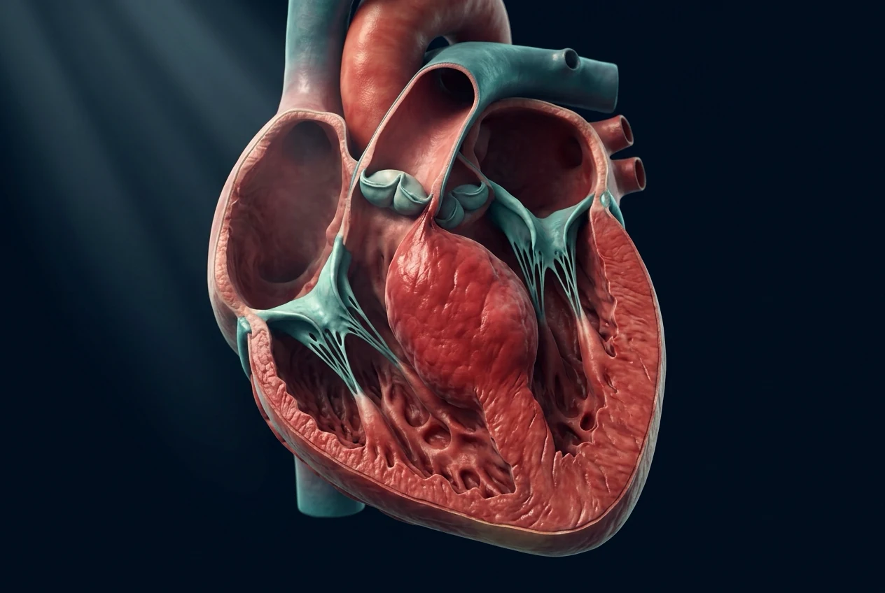
- Reverse-curve (asymmetric septal) — the classic phenotype; basal septum dominant; highest yield of sarcomere mutations and the form most associated with obstruction and SAM.
- Neutral (concentric) — symmetric LVH; broad differential (hypertensive, amyloid).
- Sigmoid septum — curved, septal bulge; commonest morphology in the elderly, often non-obstructive, lower sarcomere yield.
- Apical HCM — hypertrophy confined to the LV apex with cavity obliteration; common in Japan; giant negative T waves in precordial leads; spade-shaped LV cavity on imaging.
- Mid-cavitary obstruction — hypertrophy in mid-LV cavity with apical aneurysm in a minority. [1]
By genotype
- Sarcomere-positive HCM — pathogenic variant in a sarcomere gene identified (about 40 to 60 percent of unselected cases; up to 70 percent in family studies). Onset younger, more fibrosis, higher arrhythmia risk.
- Sarcomere-negative (genotype-negative) HCM — no variant identified; often milder phenotype, later onset. [1]
By clinical phase
- Hyperdynamic/obstructive phase — the classic phase of most symptomatic young and middle-aged patients.
- Burned-out (dilated) phase — about 5 to 10 percent progress over decades to LV dilation, wall thinning, and systolic dysfunction (EF under 50 percent) resembling DCM; managed with guideline-directed heart-failure therapy. [1]
Epidemiology & Risk Factors
HCM has a population prevalence of about 1 in 500 (0.2 percent) — the commonest monogenic cardiac disorder and the commonest inherited heart-muscle disease.[4] Prevalence is similar across sexes and ethnicities; women are diagnosed later and present more often with heart failure and symptoms (a recognised care-gap).[1]
HCM is the commonest medical cause of sudden cardiac death (SCD) in young athletes and competitive sportspeople in the United States and many other regions, although the absolute annual risk in the overall HCM population is modest (see Prognosis).[4]
Risk modifiers for penetrance, severity and SCD:[1]
- Family history of HCM or of sudden death before age 50 in a first-degree relative.
- Pathogenic sarcomere mutation (genotype-positive), particularly compound/double heterozygotes and certain high-risk alleles (e.g., some MYH7 and TNNT2 variants — historically TNNT2 associated with mild hypertrophy but high SCD risk).
- Degree of LVH — maximal wall thickness over 30 mm markedly increases SCD risk.
- Age at diagnosis — childhood onset and adolescent presentation carry higher SCD rates.
- Male sex and intense competitive sport — surface latent disease. [1]
Pathophysiology
Genetic and molecular basis
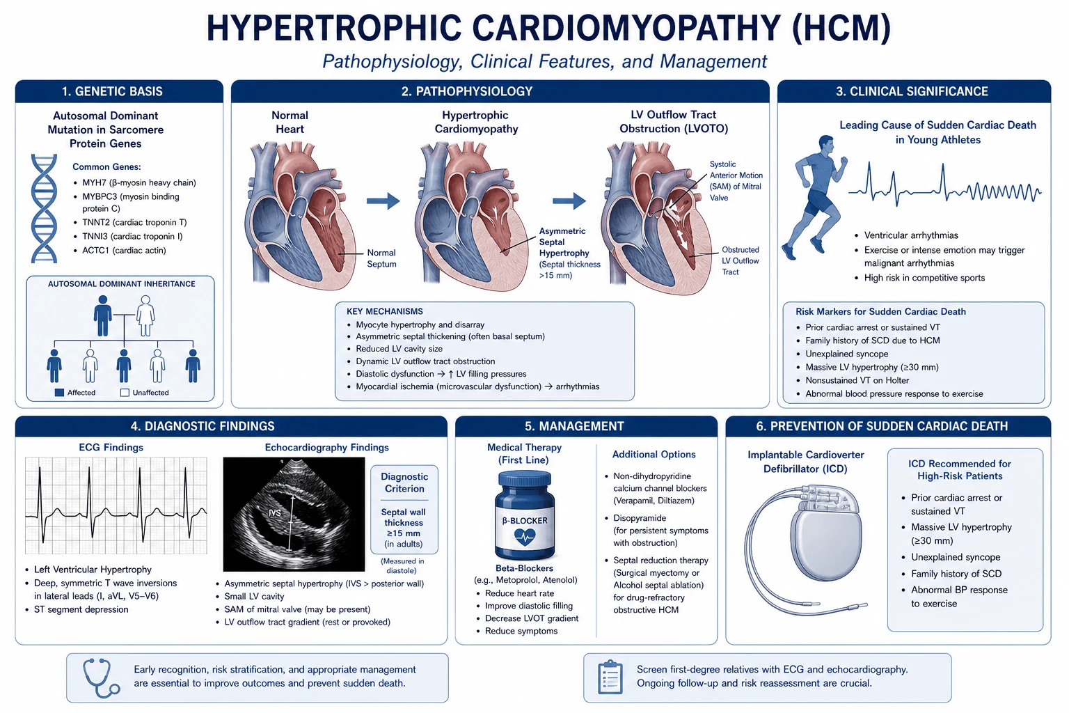
HCM is inherited as an autosomal dominant trait with variable penetrance and age-dependent expression (hypertrophy typically manifests in adolescence through mid-adulthood). About 40 to 60 percent of unselected patients carry a pathogenic variant in one of the sarcomere protein genes; in family studies this rises to about 70 percent.[1][4]
Sarcomere genes in HCM — SARC
SARCOMERE
HCM is a disease of the sarcomere contractile apparatus
beta-myosin heavy chain; ~30 to 35 percent of cases
myosin-binding protein C; ~20 to 30 percent; often later onset
cardiac troponin T, troponin I, alpha-tropomyosin — less common but classic high-SCD-risk alleles
cardiac alpha-actin, regulatory and essential myosin light chains — rarer
MYH7 (encoding beta-myosin heavy chain) and MYBPC3 (encoding myosin-binding protein C) together account for the majority of positively genotyped cases. TNNT2 mutations are comparatively rare but are classically associated with mild hypertrophy and disproportionate SCD risk — a high-yield viva point. [1]
The histological triad and its clinical consequences
Whatever the gene, the HCM heart shares a stereotyped histology:[4]
Histological triad of HCM and its consequences
- Myocyte disarray — the histological hallmark: myocytes oriented obliquely and chaotically with abnormal intercellular connections, rather than the parallel array of normal myocardium. Disarray is the structural substrate for re-entry and ventricular arrhythmia.
- Interstitial fibrosis — increased collagen deposition that stiffens the myocardium (diastolic dysfunction) and is visible as late gadolinium enhancement (LGE) on cardiac MRI. Extensive LGE (especially exceeding 15 percent of LV mass) predicts SCD and HF.
- Intramural small-vessel disease — medial thickening of intramural coronary arterioles produces a mismatch between demand and supply, causing microvascular ischaemia. This drives angina in the absence of epicardial CAD, contributes to fibrosis, and is the substrate for the scar that causes arrhythmia. [1]
Mechanism of dynamic LVOT obstruction and SAM
In obstructive HCM the hypertrophied basal septum narrows the LVOT. As ejection accelerates through the narrowed tract, two coupled mechanisms draw the anterior mitral leaflet toward the septum during systole (systolic anterior motion, SAM):[1]
- The Venturi effect — high-velocity flow through the narrowed LVOT generates a lateral suction force on the leaflet.
- Flow-drag (pushing) forces — the ejected blood stream directly pushes the leaflet toward the septum; modern mechanical modelling emphasises this contribution. [1]
SAM has three consequences: (1) it further narrows the LVOT (dynamic obstruction); (2) it produces a posteriorly directed jet of mitral regurgitation (the leaflet cannot coapt); and (3) it makes the obstruction dynamic — varying beat-to-beat with preload, afterload and contractility. This is why the murmur increases with anything that reduces preload or afterload (Valsalva strain phase, standing, nitrates, dehydration) and decreases with anything that increases them (squatting, passive leg raise, handgrip). [1]
Diastolic dysfunction, microvascular ischaemia and the arrhythmia substrate
- Diastolic dysfunction is near-universal. Hypertrophy, fibrosis and abnormal calcium handling slow relaxation and reduce compliance, raising LV end-diastolic pressure. The left atrium hypertrophies; left atrial enlargement predisposes to atrial fibrillation. Symptoms (dyspnoea, orthopnoea, raised JVP) occur despite a normal or hyperdynamic ejection fraction.
- Microvascular ischaemia — the thickened myocardium outstrips its capillary supply and the arterioles themselves are diseased, producing exertional chest pain, dynamic ST changes, and progressive replacement fibrosis.
- Arrhythmia substrate — the combination of disarray, fibrosis, ischaemia and dispersion of repolarisation creates the substrate for ventricular tachycardia and fibrillation and hence sudden cardiac death. [1]
The haemodynamic equation that explains the murmur
The instantaneous LVOT gradient is governed by the simplification of the Bernoulli equation used in Doppler echo — peak gradient (mmHg) = 4 x (peak velocity) squared. A velocity of 3 m/s therefore gives a gradient of 36 mmHg. This is also why anything that increases contractility (digoxin, catecholamines, exercise) or decreases preload/afterload worsens the gradient and the murmur, and why beta-blockade and volume loading reduce it. [1]
Clinical Presentation
Classical symptom triad
Most HCM patients are asymptomatic at diagnosis (detected by family screening, murmur, or incidental ECG/echo). When symptomatic, three symptom clusters dominate:[1][4]
- Dyspnoea (the commonest symptom) — from raised LV end-diastolic pressure and diastolic dysfunction; worsened by atrial fibrillation (loss of atrial kick is poorly tolerated in the stiff ventricle).
- Angina — from microvascular ischaemia despite angiographically normal epicardial coronaries in many patients.
- Syncope or pre-syncope — from a combination of LVOT obstruction (especially provoked by exertion or arrhythmia), neurally mediated reflex vasodilation, and arrhythmia (VT/VF or AF with rapid ventricular response). [1]
Sudden cardiac death as the first manifestation
In young patients HCM may first present as sudden cardiac death during or just after exertion (competitive sport is the classic context). This is the single most feared presentation and the rationale for pre-participation screening and sport restriction in diagnosed patients.[4]
Atypical and special presentations
- Elderly / late-onset HCM — often sigmoid septum, milder course, hypertension overlap; presents with heart failure, AF, or exertional dyspnoea. Cardiac amyloidosis is the critical mimic to exclude (low ECG voltages, discordant echo/ECG, carpal tunnel history, raised cardiac biomarkers).
- Apical HCM — atypical chest pain; giant negative T waves in V2 to V6; spade-shaped LV cavity; excellent prognosis for SCD but can cause apical aneurysm and thrombus.
- Burned-out / dilated phase — late progression to systolic HF (EF under 50 percent) resembling DCM; treated with standard HFrEF therapy.
- Women — diagnosed later; more often present with heart failure; under-represented in trials.
- Children and adolescents — may present with syncope or SCD; wall thickness z-score of 2 or more defines disease; SCD rates are higher. [1]
Differential Diagnosis
A systolic murmur with LVH on ECG is not always HCM. The high-yield differentials:[1][4]
Bedside differentials of the murmur
HCM (HOCM)
- Ejection systolic murmur at LEFT sternal edge
- Murmur INCREASES with Valsalva (strain), standing, amyl nitrite
- Murmur DECREASES with squatting, passive leg raise, handgrip
- No radiation to carotids (carotid pulse may be **bisferiens**)
Aortic stenosis
- Ejection systolic murmur at RIGHT second intercostal space, radiating to carotids
- Murmur DECREASES (or unchanged) with Valsalva
- Murmur INCREASES with squatting, INCREASES (slow rising) carotid pulse (pulsus parvus et tardus)
- Ejection click, soft/absent A2
Mitral valve prolapse
- Late-systolic murmur +/- mid-systolic click at apex
- Click and murmur MOVE EARLIER with Valsalva and standing (longer murmur), later with squatting
- No LVH on ECG
VSD (small)
- Pansystolic murmur at lower left sternal edge
- Murmur INCREASES with handgrip (afterload), unchanged by Valsalva
- Thrill often present
Differentials of unexplained LVH
- Athlete's heart — physiological LVH from endurance training; the grey zone is 12 to 13 mm wall thickness. Discriminators: in athlete's heart the LV is mildly enlarged (LV end-diastolic diameter over 54 mm in men), thickness regresses with deconditioning (6 to 12 weeks), LA size is normal, ECG shows bradycardia and early repolarisation (not pathological T inversion), no LGE on MRI, no family history of HCM/SCD, and the wall is symmetric. HCM: wall often over 15 mm, LA enlarged, abnormal ECG, LGE may be present, does not regress.
- Hypertensive heart disease — long-standing hypertension with concentric LVH; HCM excluded if hypertrophy regresses with BP control and there is no family history.
- Cardiac amyloidosis (AL or ATTR/wild-type) — the great mimic in older patients; concentric LVH with low voltages on ECG, bilateral carpal tunnel history, disproportionate raised troponin/BNP, apical sparing pattern of LGE, abnormal 99mTc-PYP scan (ATTR). Diagnose early because digoxin and calcium-channel blockers are dangerous in amyloidosis.
- Aortic stenosis — pressure-overload concentric LVH; distinguished by the murmur and a gradient across a stenotic valve (not a dynamic subaortic gradient).
- Restrictive cardiomyopathy, glycogen storage diseases (Danon, Fabry, PRKAG2) — Fabry disease is X-linked and treatable with enzyme replacement; consider in males with concentric LVH and negative T waves in inferolateral leads. [1]
Clinical & Bedside Assessment
The murmur and its manoeuvre behaviour
The classic murmur of HOCM is an ejection systolic murmur heard best at the left sternal edge (and apex), rough, crescendo-decrescendo, that does not radiate to the carotids (the obstruction is dynamic and subvalvular). The companion mitral regurgitation murmur is a separate pansystolic, blowing murmur at the apex radiating to the axilla.[1]
The manoeuvre behaviour is the single most testable bedside fact in HCM — memorise the full table:[1]
Manoeuvre
- Valsalva (strain phase)
- Standing from squat
- Squatting from standing
- Passive leg raise
- Handgrip (sustained)
- Amyl nitrite inhalation
HOCM
- INCREASES (decreased preload)
- INCREASES (decreased preload)
- DECREASES (increased preload + afterload)
- DECREASES (increased preload)
- DECREASES (increased afterload)
- INCREASES (vasodilation)
AS
- Decreases / unchanged
- Decreases / unchanged
- Increases
- Increases
- Increases
- Increases
MVP
- Click-murmur earlier and longer
- Click-murmur earlier and longer
- Click-murmur later and shorter
- Click-murmur later
- Click-murmur later and shorter
- Click-murmur earlier
Pulse, apex and heart sounds
- Pulse — jerky, brisk upstroke; in obstructive disease a bisferiens pulse (double systolic impulse) may be felt. Volume is normal — distinct from the pulsus parvus et tardus of aortic stenosis.
- Apex beat — displaced in late disease or non-obstructive forms; in HOCM classically a double (or triple) apical impulse — a pre-systolic outward movement (vigorous atrial contraction into the stiff ventricle, the palpable equivalent of an S4) followed by the systolic thrust.
- Heart sounds — S4 gallop is common (stiff, non-compliant ventricle); a paradoxically split S2 may occur from prolonged LV ejection in severe obstruction.
- Thrill — a systolic thrill may be palpable at the left sternal edge in severe obstruction. [1]
Investigations
ECG
The ECG is abnormal in about 90 percent of HCM patients and may be the first clue in an asymptomatic athlete. Findings include:[1][4]
- Left ventricular hypertrophy with voltage criteria (Sokolow-Lyon SV1 + RV5/RV6 over 35 mm; Cornell criteria).
- LV strain pattern — ST depression and deep T-wave inversions in I, aVL, V5 to V6.
- Lateral or precordial pathological Q waves (septal depolarisation through hypertrophied septum) — a mimic of old MI.
- Apical HCM — giant (> 10 mm) deep T-wave inversions in V2 to V6 with high R-wave amplitude — virtually pathognomonic.
- Left atrial enlargement (bifid P in V1, widened P in II), PR shortening, and conduction disease.
- A normal ECG does not exclude HCM, but a normal ECG plus normal echo in a relative makes disease unlikely. [1]
Chest X-ray
Non-specific: may be normal; cardiomegaly, left atrial enlargement (double shadow, splaying of carina), and in heart failure pulmonary venous congestion. Useful to exclude alternative diagnoses. [1]
Echocardiography (transthoracic — first line and central)
TTE answers five questions: (1) is hypertrophy present and where, (2) is there LVOT obstruction and what is the gradient, (3) is there SAM and mitral regurgitation, (4) what is the diastolic function, and (5) are there associated lesions.[1]
Diagnostic and quantification thresholds reproduced verbatim (AHA/ACC 2020):[1]
| Parameter | Diagnostic / threshold |
|---|---|
| Maximal LV wall thickness (adult, any segment) | 15 mm or more |
| Maximal LV wall thickness (first-degree relative) | 13 mm or more (or z-score over 2 in a child) |
| LVOT gradient — non-obstructive | under 30 mmHg (rest and provocation) |
| LVOT gradient — obstructive | 30 mmHg or more at rest, or on provocation (Valsalva, amyl nitrite, exercise) |
| Continuous-wave Doppler peak velocity | gradient by Bernoulli = 4 x v squared (v in m/s) |
| Septal-to-posterior-wall ratio (asymmetric) | 1.3 or more |
SAM is graded semi-quantitatively (1 to 3 by length of septal contact) and predicts the posteriorly directed MR jet. Diastolic function (E/A, e prime, E/e prime ratio, LA volume index) is impaired in nearly all symptomatic patients. [1]
Cardiac MRI
The reference standard for wall thickness in every segment (especially the apex, missed on TTE), LV mass, and — critically — late gadolinium enhancement (LGE), which detects focal replacement fibrosis. LGE exceeding 15 percent of LV mass independently predicts SCD and is increasingly used to tip a borderline risk decision toward ICD.[1]
Holter / ambulatory monitoring
A 24 to 48-hour (ideally 7-day) Holter is mandatory in the SCD risk evaluation — non-sustained VT (NSVT) is a major risk modifier and may be silent. [1]
Exercise testing
A symptom-limited treadmill or bicycle test with continuous BP measurement identifies (1) provocable LVOT gradients, (2) exercise-induced symptoms, and (3) the blood-pressure response — an abnormal BP response (a fall of over 10 mmHg, or failure to rise by over 20 mmHg during effort) is a SCD risk modifier in patients under 40. [1]
Genetic testing
Cascade genetic testing of the index case and first-degree relatives is recommended. If the index has a pathogenic variant, relatives can be genotyped; genotype-positive but phenotype-negative relatives need serial clinical surveillance (echo/ECG/MRI annually in children/adolescents, every 2 to 5 years in adults). Genotype-negative relatives are discharged from surveillance.[1]
Coronary angiography / CT coronary
Reserved for patients with angina whose pre-test probability of epicardial CAD is significant, or before septal reduction therapy. Microvascular angina is common with angiographically normal coronaries. [1]
Management — Resuscitation
HCM does not usually present as a resuscitation emergency in the way an MI does, but two scenarios are time-critical:[1]
Cardiac arrest (VF / pulseless VT)
- Immediate defibrillation (200 J biphasic) and standard advanced life support.
- After recovery, an ICD is indicated (secondary prevention) regardless of risk-score results. [1]
Haemodynamic collapse with severe LVOT obstruction
This is the scenario that traps the unwary. The obstructed HCM ventricle is hypercontractile, underfilled and highly load-dependent — the wrong drug kills the patient. [1]
[1]Management — Definitive & Stepwise
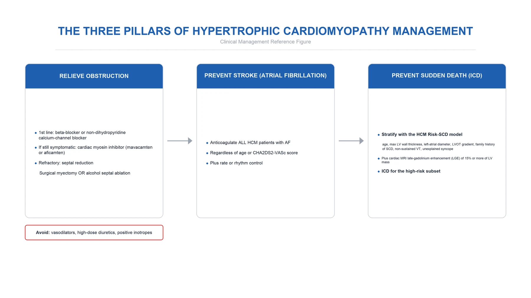
The strategy has four parallel tracks running together: (1) lifestyle and family screening, (2) symptom control, (3) septal reduction for drug-refractory obstruction, and (4) SCD risk management (ICD).[1][2]
Track 1 — Lifestyle and family screening
- Competitive sport is contraindicated in clinically diagnosed HCM (Class 3 in the AHA/ACC 2020 — i.e., should not participate). Recreational, low-intensity aerobic activity is encouraged. The 36th Bethesda Conference (US) and ESC recommendations converge on this.[1]
- Avoid dehydration and hot environments (preload-dependent obstruction worsens).
- First-degree relatives screened with ECG + echocardiography +/- cardiac MRI and genetic testing — every 1 to 2 years in children/adolescents, every 2 to 5 years in adults.
- Endocarditis prophylaxis is reserved for prosthetic valves, prior endocarditis, and certain congenital disease — not routine for native HCM valves.
Track 2 — Symptomatic drug therapy
Step 1 — Beta-blocker (first-line):[1][2]
- Agents — propranolol (long-acting, 160 to 240 mg orally daily in divided doses, or up to 320 mg/day), metoprolol succinate (100 to 200 mg orally daily), bisoprolol (5 to 10 mg orally daily), atenolol (50 to 100 mg orally daily), nadolol (40 to 80 mg orally daily). In acute settings, esmolol intravenously (loading 500 micrograms/kg over 1 minute, then 50 to 200 micrograms/kg/minute) is preferred for its short half-life.
- Target — resting heart rate 60 to 65 per minute; titrate to symptoms and gradient.
- Mechanism — reduces heart rate (longer diastole, better filling), reduces contractility (less SAM and gradient), blunts exertional surges.
- Monitoring — heart rate, BP, symptoms, side effects (fatigue, depression, erectile dysfunction, bronchospasm). [1]
Step 2 — Add disopyramide (Class IA), or switch to a non-dihydropyridine CCB:[1][2]
- Disopyramide — 100 to 200 mg orally, three to four times daily (controlled-release 250 mg twice daily where available), titrated. Mechanism: negative inotropy reduces SAM and gradient without reducing preload. Often combined with a beta-blocker for synergistic effect (beta-blocker for rate control, disopyramide for inotropy). Monitor QTc (risk of proarrhythmia); avoid in long QT and combine cautiously with other QT-prolonging drugs.
- Verapamil — 240 to 480 mg orally daily (sustained release). Useful as monotherapy or in addition when beta-blockers are not tolerated. Avoid in patients with obstruction plus conduction disease, severe heart failure, hypotension, or in combination with disopyramide (combined negative dromotropy and inotropy can cause complete heart block and shock). Diltiazem (180 to 360 mg/day) is an alternative. [1]
Step 3 — Cardiac myosin inhibitors (new class):[3][5]
- Mavacamten — the first-in-class cardiac myosin inhibitor, FDA-approved for symptomatic obstructive HCM (NYHA class II to III). It reduces the number of actin-myosin cross-bridges, lowering hypercontractility, LVOT gradient, and LV filling pressures. Dose 2.5 to 15 mg orally once daily, titrated to LVOT gradient and LVEF (treatment interrupted if LVEF falls below 50 percent). In the EXPLORER-HCM trial it improved peak VO2, NYHA class, gradient, and quality of life versus placebo.[3]
- Aficamten — a next-in-class myosin inhibitor with a shorter half-life; in the MAPLE-HCM trial it improved exercise performance versus metoprolol in obstructive HCM.[5]
- Place in algorithm — symptomatic obstructive HCM before or instead of invasive septal reduction; the role relative to myectomy/ASA continues to evolve.
Step 4 — Septal reduction therapy (refractory obstructive disease):[1]
Indicated for NYHA class III to IV symptoms (or class II with exertional syncope) with LVOT gradient 50 mmHg or more despite maximally tolerated drug therapy. Two techniques: [1]
Surgical septal myectomy (Morrow)
- Extended Morrow subaortic myectomy — the **gold standard**, especially in younger patients, in centres of excellence
- Removes a trough of basal septal myocardium via aorta; can address concurrent lesions (mitral repair, CABG)
- Permanent complete heart block in about 2 to 5 percent; peri-procedural mortality under 1 percent in expert centres
- Preferred when other cardiac surgery is needed, or anatomy unfavourable for ablation
Alcohol septal ablation (ASA)
- Percutaneous — selective injection of **pure alcohol 1 to 2 mL** into the **first septal perforator** branch of the LAD to induce a controlled septal infarct
- Preferred in older patients, those with comorbidities precluding surgery, or patient preference
- **Peri-procedural complete heart block in 10 to 20 percent** (transient AV block even higher) — pre-procedural pacemaker planning
- Creates a septal scar; long-term theoretical arrhythmia concern (debated)
Track 3 — SCD risk management (ICD)
ICD is the only therapy proven to prevent sudden cardiac death in HCM. Indications:[1][2]
- Secondary prevention — prior cardiac arrest, spontaneous sustained VT, or spontaneous sustained VF — ICD indicated regardless of risk score.
- Primary prevention — risk-stratified: [1]
ESC 2014 — HCM Risk-SCD equation computes a 5-year SCD probability from age, maximal LV wall thickness, left atrial diameter, LVOT gradient, family history of SCD, NSVT, and unexplained syncope. ICD recommended when 5-year risk is 6 percent or more (considered when 4 to under 6 percent; generally not indicated below 4 percent).[2]
AHA/ACC 2020 — major SCD risk modifiers approach (presence of one or more supports ICD discussion, with the magnitude and number of modifiers determining strength of recommendation):[1]
AHA/ACC 2020 SCD risk modifiers
SCD risk modifiers in HCM — SCD
SCD
unexplained, especially exertional
family history of SCD under 50
non-sustained VT on Holter
Add: massive LVH (over 30 mm), abnormal BP response to exercise, extensive LGE. The ESC quantifies these into the HCM Risk-SCD 5-year score with the 6 percent cut-off. [1]
Track 4 — Specific comorbidities
- Atrial fibrillation — common in HCM and poorly tolerated (loss of atrial kick in the stiff ventricle). Rhythm control is preferred (amiodarone 200 mg orally daily is the usual first-line agent; sotalol as an alternative; disopyramide/quinidine if needed). Anticoagulation is recommended for all HCM patients with AF regardless of CHA2DS2-VASc score (warfarin traditionally preferred; DOACs now acceptable based on observational data). Acute haemodynamic instability warrants urgent electrical cardioversion. Consider atrial fibrillation ablation or surgical left-atrial appendage exclusion at the time of myectomy.[1]
- Burned-out / dilated phase (EF under 50 percent) — switch to guideline-directed HFrEF therapy: ACE inhibitor/ARB/ARNI, beta-blocker (avoid inotropic sensitisation; bisoprolol/carvedilol/metoprolol succinate), mineralocorticoid receptor antagonist, SGLT2 inhibitor, and diuretics as needed (preload dependence is no longer a concern once EF has fallen). Consider transplant evaluation in advanced cases.
- End-stage and refractory — heart transplantation for drug- and device-refractory HF; mechanical circulatory support as a bridge.
Specific Subtypes & Scenarios
- Apical HCM — spade-shaped cavity, giant T inversions; no LVOT obstruction so beta-blockade for symptoms, screen for apical aneurysm and thrombus (anticoagulate if present), SCD risk is generally lower.
- Mid-cavitary HCM — gradient is in the mid-ventricle; apical aneurysm in a minority; treat the gradient (beta-blocker +/- myosin inhibitor; surgical apical myectomy in rare cases).
- Burned-out / dilated phase — see above.
- HCM in athletes — differentiate from athlete's heart (see Differential). If HCM confirmed, disqualify from competitive sport (Bethesda/ESC consensus).
- HCM in children — wall thickness z-score of 2 or more defines disease; higher SCD rates; the paediatric SCD risk model differs from the adult HCM Risk-SCD.
- HCM in the elderly / late-onset — sigmoid septum, hypertension overlap; exclude cardiac amyloidosis (99mTc-PYP scan, MRI with LGE pattern, serum/urine free light chains).
- HCM in pregnancy — see Special Populations. [1]
Complications & Pitfalls
- Sudden cardiac death (VF) — the cardinal and most feared, especially in the young and during exertion.
- Atrial fibrillation and its consequences — embolic stroke, haemodynamic deterioration, HF.
- Heart failure — diastolic in the classic phase; systolic in the burned-out phase.
- Syncope and injury from obstruction or arrhythmia.
- Infective endocarditis on the SAM-damaged mitral apparatus (small absolute risk).
- Thromboembolism from AF or from apical aneurysm in apical HCM. [1]
Complications of septal reduction therapy: [1]
- Complete heart block requiring permanent pacemaker (more common after ASA, 10 to 20 percent, vs myectomy 2 to 5 percent).
- Ventricular septal defect (myectomy, rare).
- Coronary dissection, LAD injury, large anterior infarct (ASA).
- Residual or recurrent obstruction requiring repeat intervention. [1]
Classic pitfalls (examiner favourites): [1]
- Mislabelling an athlete — decondition and use MRI before ending a career.
- Missing cardiac amyloidosis in the elderly HCM-look-alike — different management, dangerous drugs.
- Giving nitrates for "chest pain" in undiagnosed HOCM — collapses the patient.
- Withholding a beta-blocker because of mild symptoms — first-line for a reason.
- Missing non-sustained VT by failing to do a Holter in the SCD risk work-up.
- Combining disopyramide with verapamil — heart block and shock.
- Anticoagulating with the CHA2DS2-VASc score — HCM mandates anticoagulation in AF regardless of score.
- Using digoxin in obstructive HCM — increases contractility and worsens the gradient. [1]
Prognosis & Disposition
The overall annual HCM-related mortality in a specialist clinic is about 1 to 2 percent, dramatically lower than the 4 to 6 percent reported in older tertiary-referral series — the difference reflects ascertainment bias (modern cohorts include many mild, family-screened cases) rather than a change in the disease.[4]
- SCD annual incidence — about 1 percent overall in adults, but stratified from under 0.5 percent (low risk) to over 3 to 5 percent (high risk, multiple modifiers). Most common in adolescents and young adults.
- Heart-failure progression — about 5 to 10 percent progress to the burned-out/dilated phase over decades; HF death and stroke together account for substantial non-SCD mortality in older patients.
- After ICD — appropriate shock rates of 3 to 5 percent per year in primary-prevention HCM implants; quality of life generally good but shocks and device complications are real.
- After septal reduction — symptom relief is excellent and durable; myectomy and ASA give comparable symptom improvement and survival in matched cohorts. [1]
Disposition: asymptomatic genotype-positive/phenotype-negative relatives are managed in a specialist HCM clinic with serial surveillance; symptomatic obstructive disease is managed medically with escalation to a centre of excellence for myosin-inhibitor, myectomy or ASA evaluation; high-risk patients are referred for ICD; the burned-out phase is managed in a heart-failure/transplant pathway. Acute collapse with obstruction is admitted to a critical-care environment with HCM-aware anaesthesia. [1]
Special Populations
- Pregnancy — chronic stable HCM is generally well tolerated in pregnancy because the increased heart rate and reduced systemic vascular resistance partly offset the gradient. Counsel pre-pregnancy: assess symptoms, gradient, SCD risk, and review drugs. Continue beta-blockers through pregnancy and labour if previously needed (fetal bradycardia and growth restriction are monitored). Avoid inferior vena cava compression (left lateral position in labour). Vaginal delivery is preferred; caesarean for obstetric indications or severe symptoms. Regional anaesthesia (low-dose epidural) is preferred for operative delivery — but avoid sudden drops in afterload; sympathomimetics with beta-agonist activity are avoided (use phenylephrine). Avoid ergometrine. High-risk patients (recent syncope, severe obstruction, EF under 50 percent) are counselled against pregnancy.[1]
- Elderly — sigmoid morphology, hypertension overlap; watch for amyloidosis; lower threshold to evaluate comorbid CAD; tolerates beta-blockade less well (start low).
- Children and adolescents — z-score-based diagnosis; serial screening through the growth years; higher SCD rate; the HCM Risk-Kids calculator (not the adult HCM Risk-SCD) is used.
- Non-cardiac surgery — the obstructed HCM patient is high-risk for anaesthetic-related collapse. Principles: maintain preload (avoid hypovolaemia, careful fasting, intravenous fluids), maintain sinus rhythm and a controlled heart rate (beta-blockade continued peri-operatively), maintain afterload (avoid vasodilating agents and regional sympathetic blockade that cause abrupt vasodilation; favour phenylephrine over ephedrine for vasopressor support), avoid hypercontractility (no ketamine in high doses, no pure beta-agonists). Spinal anaesthesia can precipitate collapse from sudden afterload reduction — caution or avoid.
- Anticoagulated HCM — anticoagulation is common (AF). No routine bridging is needed for most procedures; balance bleeding against stroke per standard protocols.
Evidence, Guidelines & Regional Differences
- 2020 AHA/ACC Guideline for the Diagnosis and Treatment of Patients With Hypertrophic Cardiomyopathy (Ommen et al., Circulation 2020) — the current North American standard; introduced mavacamten, emphasised a shared-decision approach to ICD and sport, and codified the major SCD risk-modifier approach (rather than a single equation).
- 2014 ESC Guidelines on Diagnosis and Management of HCM (Elliott et al., European Heart Journal 2014) — the European standard; the HCM Risk-SCD 5-year equation with the 6 percent cut-off for ICD originates here. [1]
- EXPLORER-HCM (Olivotto et al., Lancet 2020) — mavacamten improved peak VO2, NYHA class, LVOT gradient and quality of life vs placebo in symptomatic obstructive HCM. PMID 32871100.[3]
- MAPLE-HCM (Lewis et al., JAMA Cardiology 2026) — aficamten improved exercise performance vs metoprolol in obstructive HCM. PMID 42307914.[5]
Key thresholds examiners reward: wall thickness 15 mm (adult), 13 mm (relative), z-score 2 (child); LVOT gradient 30 mmHg (obstruction), 50 mmHg (intervention); HCM Risk-SCD 6 percent 5-year cut-off; massive LVH over 30 mm. [1]
[1]Regional deltas: the AHA/ACC major-modifier approach vs the ESC HCM Risk-SCD equation for ICD decisions diverge for borderline cases (a patient with two modifiers but a low equation score is treated differently in the US vs Europe). Genetic testing uptake and the availability of mavacamten/aficamten vary by health-system funding; in resource-limited settings rheumatic and hypertensive heart disease dominate and HCM is under-diagnosed. [1]
Exam Pearls
- Murmur: ejection systolic at the LEFT sternal edge, increases with Valsalva and standing, decreases with squatting, handgrip and passive leg raise — the opposite of aortic stenosis.
- Genetics: autosomal dominant; MYH7 (beta-myosin heavy chain) and MYBPC3 (myosin-binding protein C) cause the majority; TNNT2 = mild hypertrophy, high SCD risk.
- Histology triad: myocyte disarray + interstitial fibrosis + intramural small-vessel disease — substrate for arrhythmia, diastolic dysfunction and angina.
- SAM (systolic anterior motion) of the anterior mitral leaflet — Venturi + drag; produces dynamic obstruction and posteriorly directed mitral regurgitation.
- Diagnosis: echo with LV wall thickness 15 mm or more in adults (13 mm or more in relatives, z-score 2 or more in children); ECG abnormal in 90 percent; apical HCM gives giant T inversions.
- Athlete's heart grey zone: 12 to 13 mm; differentiate by LV size, LA size, ECG, MRI LGE, deconditioning, family history.
- Drugs to AVOID in obstructive HCM: nitrates, diuretics, vasodilators (ACE-i/ARBs, hydralazine), digoxin and other inotropes, pure beta-agonists, and IABP.
- First-line: beta-blocker (propranolol/metoprolol/bisoprolol/nadolol); second-line: disopyramide (Class IA) — never combine with verapamil; alternative: verapamil/diltiazem.
- Septal reduction: Morrow myectomy (gold standard, low pacemaker rate) vs alcohol septal ablation (higher pacemaker rate, no surgery).
- Mavacamten — first-in-class cardiac myosin inhibitor, FDA-approved for symptomatic obstructive HCM (EXPLORER-HCM); aficamten is next-in-class.
- AF in HCM: rhythm control + anticoagulate ALL regardless of CHA2DS2-VASc.
- SCD risk: family history of SCD, massive LVH over 30 mm, NSVT, unexplained syncope, abnormal BP response on exercise, extensive LGE on MRI — ICD if high (HCM Risk-SCD 6 percent over 5 years).
- Screen first-degree relatives with ECG, echo, MRI and genetic testing; competitive sport contraindicated. [1]
Exam application bank (NEET-PG / INICET)
One-line answer
Hypertrophic cardiomyopathy (HCM) is an autosomal dominant sarcomere-protein mutation disease producing unexplained left ventricular hypertrophy, classically asymmetric septal. Histology shows myocyte disarray, interstitial fibrosis and intramural small-vessel disease. Dynamic left ventricular outflow tract (LVOT) obstruction with systolic anterior motion (SAM) of the mitral valve and secondary mitral regurgitation occurs in about two-thirds. The cardinal clinical threats are sudden cardiac death (SCD) in the young, diastolic heart failure, atrial fibrillation and angina from microvascular ischaemia. The bedside signature is an ejection systolic murmur at the left sternal edge that increases with Valsalva and standing and decreases with squatting — the opposite of fixed aortic stenosis. Diagnosis is by echocardiography (LV wall thickness 15 mm or more in adults, or a lower threshold in r
Worked stems (answer without another resource)
Stem 1 — Classic presentation. Map symptoms to mechanism; name the first investigation and first treatment step with dose/route if drug therapy is standard. [1]
Stem 2 — Unstable / complicated. List red flags that force immediate resuscitation, theatre, ICU, antidote, or reperfusion — and what you do in the first 15 minutes. [1]
Stem 3 — Atypical group. Elderly, pregnancy, child, or immunocompromised: how presentation and thresholds change. [1]
Stem 4 — Differential trap. Name the three closest mimics and one discriminator for each. [1]
Stem 5 — Disposition. Who goes home with safety-netting, who is admitted, who needs HDU/ICU/theatre, and what follow-up is mandatory. [1]
Rapid viva checklist
- Definition + classification
- Pathophysiology chain
- Bedside signs / criteria
- Score with exact components (if any)
- Emergency bundle
- Definitive therapy with doses
- Complications of disease and of treatment
- Special populations
- Guideline/trial name if classic
- Three exam traps
Coverage self-check
If you cannot answer any stem above from this page alone, re-read the matching section — the page is intended to be self-sufficient for final-prof and NEET-PG/INICET questions on Hypertrophic Cardiomyopathy.
[1]References
- [1]Ommen SR, Mital S, Burke MA, et al. 2020 AHA/ACC Guideline for the Diagnosis and Treatment of Patients With Hypertrophic Cardiomyopathy: Executive Summary: A Report of the American College of Cardiology/American Heart Association Joint Committee on Clinical Practice Guidelines Circulation, 2020.PMID 33215938
- [2]Authors/Task Force members, Elliott PM, Anastasakis A, et al. 2014 ESC Guidelines on diagnosis and management of hypertrophic cardiomyopathy: the Task Force for the Diagnosis and Management of Hypertrophic Cardiomyopathy of the European Society of Cardiology (ESC) Eur Heart J, 2014.PMID 25173338
- [3]Olivotto I, Oreziak A, Barriales-Villa R, et al. Mavacamten for treatment of symptomatic obstructive hypertrophic cardiomyopathy (EXPLORER-HCM): a randomised, double-blind, placebo-controlled, phase 3 trial Lancet, 2020.PMID 32871100
- [4]Maron BJ. Hypertrophic cardiomyopathy: a systematic review JAMA, 2002.PMID 11886323
- [5]Lewis GD, Garcia-Pavia P, Masri A, et al. Exercise Performance With Aficamten vs Metoprolol in Obstructive Hypertrophic Cardiomyopathy: The MAPLE-HCM Randomized Clinical Trial JAMA Cardiol, 2026.PMID 42307914