Cardiology · General Medicine
Infective Endocarditis
Also known as IE · Infective endocarditis · Subacute bacterial endocarditis · SBE
Infective endocarditis (IE) is a microbial infection of the endocardial surface, usually a heart valve, forming friable vegetations of platelets and fibrin that entrap organisms. It classically presents with fever, a new or changing murmur, and embolic, immunologic or septic phenomena. Diagnosis rests on blood cultures and echocardiography integrated through the Duke criteria. Management combines prolonged bactericidal antibiotics (benzylpenicillin/ceftriaxone ± gentamicin for streptococci; flucloxacillin for MSSA; vancomycin for MRSA) with early surgery for heart failure, uncontrolled infection or the prevention of embolism.
On this page & tools
Your progress
Saved locally on this device.
Practise this topic
Exam tags
Red flags
Overview & Definition
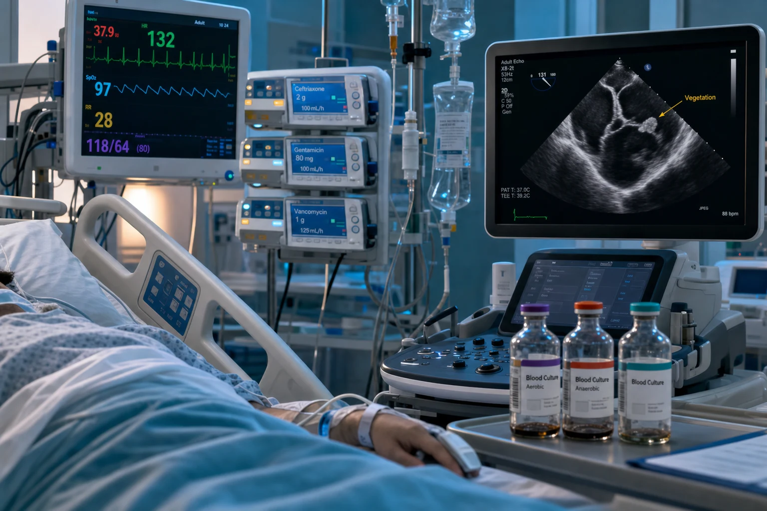
Infective endocarditis (IE) is a microbial infection of the endocardial surface of the heart — most often a heart-valve leaflet, but also the chordae tendineae, mural endocardium, or the lining of a prosthetic valve or intracardiac device. The pathological hallmark is the vegetation, a mass of platelets and fibrin colonised by microorganisms, usually bacteria and rarely fungi. Once uniformly fatal, IE now kills roughly one in six patients during the index admission despite antibiotics and surgery, so early recognition and disciplined use of the Duke criteria matter.[3]
The central clinical skill in IE is integration, not a single test: the diagnosis is made by combining clinical features, blood cultures and echocardiography through the structured Duke criteria, first proposed in 1994, modified in 2000 and updated again in 2023.[4][5][6] The therapeutic pivot is prolonged, high-dose, bactericidal, intravenous antibiotics — because the vegetation shelters organisms from phagocytes and bacteriostatic drugs — plus, in roughly half of patients, early surgery to deal with the mechanical and uncontrolled-infective complications that antibiotics alone cannot reverse.[1]
[3]Classification
IE is classified along three axes that together predict the likely organism and the empiric regimen.[1][3]
By tempo
- Acute — days, aggressive, destroys a normal valve; S. aureus
- Subacute — weeks, indolent, on an abnormal valve; viridans streptococci
- Boundaries blurred as disease shifts to older, prosthetic, healthcare-exposed patients
By valve substrate
- Native-valve IE (NVE) — about 72% of cases
- Prosthetic-valve IE (PVE) — early <60 d vs late >60 d (ESC); <1 yr vs >1 yr (older texts)
- Device-related (CIED) — pocket or lead infection seeding the tricuspid valve
By portal
- Community-acquired — viridans strep, classic dental/RHD
- Healthcare-associated (HAIE) — nosocomial or non-nosocomial (lines, haemodialysis)
- IV-drug-use (IVDU) — S. aureus, tricuspid/right heart
A few non-infectious mimics must be separated at the outset because they are culture-negative and demand entirely different management. Non-bacterial thrombotic endocarditis (NBTE, marantic) forms sterile vegetations in malignancy or a hypercoagulable state (Trousseau), and Libman-Sacks endocarditis forms sterile verrucous vegetations in systemic lupus erythematosus. Both produce systemic emboli and murmurs but negative blood cultures and no systemic sepsis — they are mimics of IE, not IE itself.[3]
Epidemiology & Risk Factors
The incidence of IE is 3-10 cases per 100,000 person-years in industrialised populations, with a clear upward drift over the last two decades driven by ageing, prosthetic material and healthcare exposure. In the United States the hospitalisation rate for IE roughly doubled between 1998 and 2009 in a nationwide analysis, with Staphylococcus aureus the dominant organism.[14] The incidence peaks in two groups: older adults with degenerative or prosthetic valves, and younger people who inject drugs.[1]
The modern face of the disease was drawn by the International Collaboration on Endocarditis–Prospective Cohort Study (ICE-PCS), which prospectively enrolled 2,781 adults with definite IE across 58 hospitals in 25 countries. It documented a median age of about 58 years, three-quarters native-valve IE, and S. aureus as the single commonest organism, with an in-hospital mortality near 18% and about half of patients needing surgery.[11]
Native-valve predisposition
- Rheumatic heart disease (still dominant where RHD persists)
- Bicuspid aortic valve, mitral valve prolapse, degenerative/calcific valve disease
- Congenital: VSD, PDA, coarctation, unrepaired cyanotic CHD
- Previous IE — highest single-condition recurrence risk
Prosthetic & device
- Prosthetic valves — lifelong risk; ~1-6% at 5 yr
- CIED (pacemaker/ICD) leads — tricuspid seeding
- Intracardiac patches, occluder devices — highest in first 6 months
Portal & host
- Intravenous drug use — repeated S. aureus bacteraemia; tricuspid
- Indwelling venous catheters, haemodialysis — coag-neg staph, S. aureus
- Poor dental hygiene — viridans streptococci
- Diabetes, immunosuppression, malignancy
The rise of prosthetic material, indwelling lines and S. aureus explains why the disease has shifted from young rheumatic-heart patients (where viridans streptococci dominated) to older, healthcare-exposed and prosthetic-valve patients, where S. aureus and coagulase-negative staphylococci now dominate.[1][11]
Pathophysiology
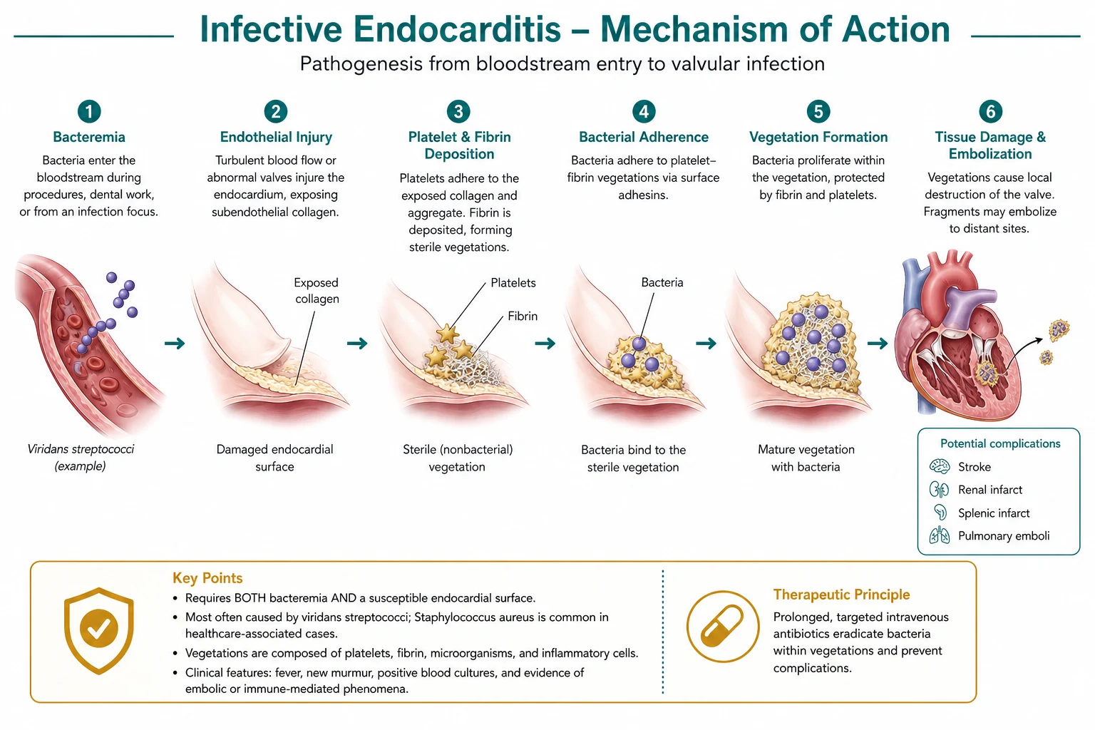
Endocarditis begins where turbulent or high-velocity blood flow injures the endocardium. A jet from a stenotic, regurgitant or shunted lesion strikes the downstream endothelium and denudes it; platelets and fibrin deposit on the raw surface to form a sterile vegetation (non-bacterial thrombotic endocarditis, NBTE).[3] During a transient bacteraemia — from tooth-brushing, chewing, a healthcare procedure, an intravenous line or injection drug use — circulating organisms seed this nidus, adhere through surface adhesins (bacterial MSCRAMMs in staphylococci), multiply within the fibrin mesh sheltered from phagocytes, and an infected vegetation is established. Two cardinal rules follow:
- Vegetations form on the low-pressure, downstream surface where the jet lands. In aortic regurgitation they sit on the atrial face of the mitral valve (the jet of AR strikes the anterior mitral leaflet — the "jet lesion"); in mitral regurgitation on the atrial surface of the mitral leaflets; in a VSD on the right (low-pressure) side of the defect and on the tricuspid leaflets opposite the jet; in aortic stenosis on the ventricular side of the aortic valve. Knowing where the jet lands predicts where to look on echocardiography.[3]
- The right heart is seeded first by bolus IV injection. A bolus of S. aureus injected into a vein passes through the right heart before the lungs filter it, so the tricuspid valve is the IV-drug-user's valve, and right-sided lesions shed septic pulmonary emboli to the lungs rather than systemic emboli to the brain and viscera.
As the vegetation grows it may destroy valve tissue (leaflet perforation, chordal rupture, abscess extending into the annulus or septum producing fistulae or atrioventricular conduction block) or fragment into the circulation, causing systemic embolisation. Left-sided lesions embolise to the brain (stroke), spleen, kidneys, mesentery and limbs; right-sided lesions seed the lungs as septic pulmonary emboli, infarcts and empyema.[3]
A parallel immune-complex response drives the immunologic phenomena — glomerulonephritis, Osler nodes, Roth spots and a positive rheumatoid factor — and a sustained inflammatory response produces the fever, anaemia and raised inflammatory markers that are the laboratory signature of the disease.[5]
The pathogenesis cascade — from sterile nidus to infected vegetation
This six-step cascade explains every preventive and therapeutic principle in IE: antibiotic prophylaxis targets the transient bacteraemia of step 3; bactericidal, prolonged, IV therapy overcomes the protected sanctuary of step 5; early surgery reverses the mechanical destruction and uncontrolled infection of step 6. It also explains the organisms' tropism: species with strong dextran-mediated adhesion (Streptococcus mutans, sanguinis) and fibronectin-binding staphylococci are over-represented because they adhere far more efficiently than transient contaminants such as E. coli or Pseudomonas, which rarely cause native-valve IE despite frequent bacteraemia.[3]
Causative organisms — by substrate and context
Knowing the likely organism from the clinical context drives empiric therapy. The table below is the working microbiology of IE every candidate must carry.[11][3]
Native-valve (community)
- Viridans streptococci (~20-30%) — #1 historically; dental source; *S. sanguinis, S. mutans, S. mitis, S. salivarius*
- S. aureus (~30%) — now the commonest single organism; acute, destructive
- Enterococci (~10%) — GU/GI source; *E. faecalis, E. faecium*
- HACEK (~1-3%) — fastidious Gram-negatives
- S. gallolyticus (S. bovis) — screen the colon
Prosthetic valve
- Early (<60 d / <1 yr): coagulase-negative staph (*S. epidermidis*) — nosocomial acquisition during surgery; S. aureus; Gram-negatives
- Late (>60 d / >1 yr): resembles native-valve — streptococci, S. aureus, enterococci; also coag-neg staph
- Fungi (*Candida*) — early prosthetic, immunosuppressed
- Always add rifampicin (after bacteraemia clears) — biofilm penetration
IV-drug-use
- S. aureus >60% (often MSSA, watch MRSA)
- Tricuspid valve (right-sided); septic pulmonary emboli
- Coag-neg staph, streptococci, Gram-negatives, Candida (esp. heroin + contaminated lemon juice)
- Polymicrobial or fungal raises the index for injection-drug use
Culture-negative
- Prior antibiotics — the commonest cause (organisms suppressed, not killed)
- Coxiella burnetii (Q fever) — anti-phase-I IgG >1:800; treat with doxycycline + hydroxychloroquine
- Bartonella spp. (henselae, quintana) — serology/PCR; homelessness, alcoholism, cat exposure
- Tropheryma whipplei, Brucella, Legionella, fungi — all need serology or PCR, not culture
The single most useful rule: the valve substrate and the clinical portal predict the organism, and the organism predicts the empiric regimen. A young dental-source native-valve case points to viridans streptococci and penicillin; a febrile prosthetic-valve patient three weeks after surgery points to coagulase-negative staph and vancomycin-plus-rifampicin; a febrile intravenous-drug user with pulmonary infiltrates points to S. aureus and the tricuspid valve.[11]
Clinical Presentation
Presentation spans a fulminant septic illness to an indolent wasting disease, but the central thread is always fever plus a murmur plus evidence of embolic, immunologic or septic phenomena.[11]
History
The history is built from systemic, septic and embolic threads. Fever (often low-grade with chills, occasionally hectic) is present in about 90% of patients; an afebrile patient already on antibiotics does not exclude IE. Constitutional symptoms — malaise, anorexia, weight loss, night sweats, fatigue, myalgia and arthralgia — dominate the subacute presentation and may precede the murmur by weeks. Ask directly about new neurological symptoms (weakness, visual disturbance, headache — embolic stroke or mycotic aneurysm), chest/pleuritic pain and haemoptysis (septic pulmonary emboli in right-sided IE), abdominal/flank pain (splenic or renal infarct), and cold, painful extremities (limb embolus). A focused risk-factor history is essential: known valve disease or prosthetic valve, previous IE, congenital heart disease, intravenous drug use, indwelling lines and haemodialysis, recent dental or healthcare procedures, and intracardiac devices.[3]
Examination — the peripheral stigmata
Examination looks for the new or changing murmur (the single most important bedside sign; a new regurgitant murmur suggests acute valve destruction) and the peripheral stigmata of embolisation and immune-complex deposition. Fever plus a murmur is the minimum diagnostic cluster, but a constellation of characteristic peripheral signs rewards the thorough examiner:[3]
- Petechiae (commonest sign) — conjunctival, oral mucosa, palate, extremities.
- Splinter haemorrhages — linear subungual haemorrhages (non-specific; also trauma).
- Osler nodes — tender pea-sized nodules on finger and toe pulps.
- Janeway lesions — painless macules on palms and soles.
- Roth spots — boat-shaped haemorrhages with pale centres on fundoscopy.
- Clubbing — late, in subacute disease.
- Splenomegaly, arthralgia, heart failure, embolic stroke, acute limb ischaemia, renal infarct, mesenteric ischaemia.[5]
The two examinable "lesions on the extremities" are contrasted below — examiners love this distinction.[5]
Osler nodes
- Painful / tender
- Finger & toe pulp nodules
- Immunologic (immune-complex vasculitis)
- A minor Duke criterion
Janeway lesions
- Painless
- Macules on palms & soles
- Vascular (septic microemboli)
- A minor Duke criterion
Atypical presentation is the rule, not the exception, in vulnerable groups. The elderly may be afebrile or present with confusion, heart failure or stroke; the immunocompromised have blunted inflammatory signs; prosthetic-valve IE may present only as low-grade fever with a new regurgitant murmur or valve dehiscence; and right-sided (IVDU) IE may present with septic pulmonary emboli mimicking pneumonia rather than a classic systemic embolic illness.[1]
Tempo and clinical course
The tempo of IE splits the disease into two clinical archetypes, though modern practice blurs them. Recognising the tempo predicts the organism, the urgency and the prognosis.[11]
Acute vs subacute IE — the two archetypes
Differential Diagnosis
The diagnostic challenge runs in two directions: IE masquerading as other febrile or embolic illnesses, and non-infectious vegetations mimicking IE. A structured differential with a discriminator for each is below.[1][3]
Marantic / NBTE
- Sterile vegetations in malignancy or hypercoagulability
- Cultures negative; no systemic sepsis
- Look for underlying adenocarcinoma / Trousseau
Libman-Sacks (SLE)
- Sterile verrucous vegetations on mitral/aortic valve
- ANA/dsDNA positive, other SLE features
- Cultures negative; immune-mediated
Atrial myxoma
- Left atrial mass mimics mitral stenosis + emboli
- Systemic febrile illness, positional murmur
- Echo resolves; cultures negative
FUO / sepsis mimic
- Lymphoma, TB, abscess, autoimmune
- Echo negative; no typical organisms
- Persistent fever without vegetation
Pulmonary embolism
- Right-sided IE mimics PE — pleuritic pain, infiltrates
- Cultures + echo distinguish
- Septic emboli are cavitating, peripheral
Persistently negative cultures with a convincing syndrome raise culture-negative IE (prior antibiotics — the commonest cause — or fastidious/intracellular organisms: Coxiella burnetii, Bartonella spp., Tropheryma whipplei, Brucella) rather than excluding the diagnosis. These need serology and PCR rather than more culture bottles.[1][6]
Clinical & Bedside Assessment
Bedside assessment confirms the diagnostic cluster and hunts for complications. The single most important investigation is three sets of blood cultures drawn from separate venepuncture sites, spaced in time, before antibiotics wherever possible — because the aetiologic agent and its susceptibility, which dictate weeks of therapy, come from these cultures. In a patient with suspected subacute IE who is haemodynamically stable, it is worth waiting the few hours for three cultures drawn over a day before starting antibiotics. In sepsis or septic shock, however, do not delay empiric antibiotics to wait for cultures: draw them and treat immediately.[3]
Examine for the new or changing murmur (a new regurgitant murmur suggests acute valve destruction and impending heart failure) and the peripheral stigmata above. Examine the fundus for Roth spots, the abdomen for splenomegaly and tenderness (splenic infarct), the urine for microscopic haematuria, and perform a focused neurological examination for an embolic stroke.[1]
A new atrioventricular block — PR prolongation or higher — is a near-pathognomonic bedside clue to a perivalvular aortic-root abscess and mandates transoesophageal echocardiography and urgent surgical referral. A serial ECG showing progressive PR prolongation in a febrile patient with IE is, for practical purposes, an abscess until proven otherwise.[1]
Echocardiography is the imaging cornerstone. A transthoracic echo (TTE) is first-line to detect vegetations and grade regurgitation and ventricular function, but its sensitivity for vegetations is only about 60%, and it is especially limited for prosthetic valves, small vegetations and posterior structures. Transoesophageal echo (TOE) — sensitivity above 90% for vegetations and abscess — is mandatory whenever TTE is negative but suspicion persists, for all prosthetic-valve IE, to detect perivalvular abscess/fistula, and to characterise vegetations before surgery.[3][1]
Transthoracic echo (TTE)
- First-line, non-invasive, bedside
- Sensitivity for vegetations ~60% (40-75%); lower for prosthetic / small / posterior
- Specificity >90% when a vegetation is seen
- Adequate in many native-valve cases with good windows
Transoesophageal echo (TOE)
- Sensitivity >90% for vegetations and abscess
- Mandatory for all prosthetic valves, suspected abscess/fistula, negative TTE with high suspicion
- Required pre-operatively and to monitor therapy when indicated
- Detects perivalvular complications TTE misses
The choice between TTE and TOE follows a simple rule: start with TTE in every suspected case, but if the clinical probability remains moderate-to-high (positive blood cultures, prosthetic valve, new murmur, staphylococcal bacteraemia, persistent fever) proceed to TOE regardless of the TTE result — a negative TTE never excludes IE, and a positive TTE may reveal abscess or fistula that changes management from medical to surgical.[3]
Blood-culture technique deserves the same rigour as the imaging. Draw three sets (aerobic and anaerobic bottles per set) from three separate venepuncture sites, spaced at least 30-60 minutes apart (or, in urgent sepsis, drawn sequentially without undue delay), from different sites — never from existing lines unless a line-draw is specifically being compared. Use rigorous skin antisepsis (chlorhexidine-alcohol, allow to dry) to minimise contamination with coagulase-negative staphylococci, whose single positive bottle is a contaminant, not a diagnosis. The yield falls sharply after antibiotics begin — by 40-50% within hours — which is why cultures before antibiotics are paramount; if antibiotics have already been given, delay repeat cultures for 2-3 days if clinically safe, and request prolonged incubation and cast your net for fastidious organisms with serology.[3][6]
Investigations
| Test | Why it matters |
|---|---|
| Blood cultures (3 sets, separate sites, before antibiotics) | The keystone — identifies organism and susceptibility, drives weeks of therapy |
| Echocardiography — TTE then TOE | Visualises the vegetation, abscess, dehiscence, regurgitation; major Duke criterion |
| FBC | Normocytic anaemia, neutrophil leucocytosis (acute), thrombocytopenia |
| CRP / ESR | Raised in >90%; tracks response to therapy |
| U&E, LFTs | Baseline renal/hepatic function; AKI from sepsis, emboli or immune-complex GN |
| ECG | Baseline; new PR prolongation / AV block = aortic-root abscess |
| Urinalysis | Microscopic haematuria (and red-cell casts if glomerulonephritis) — a minor criterion |
| Serology / PCR | For culture-negative IE: Coxiella (anti-phase-I IgG >1:800), Bartonella, Brucella, Tropheryma |
| [18F]FDG PET/CT | Major criterion in 2023 Duke-ISCVID for abnormal uptake around prosthetic valves / grafts (ESC) |
Bloods usually show a normocytic, normochromic anaemia of chronic disease, a leucocytosis (marked in S. aureus, modest or absent in subacute disease), raised CRP and ESR, and often deranged renal function. Urinalysis showing microscopic haematuria supports an immunologic process (glomerulonephritis). The rheumatoid factor may be positive (an immunologic minor criterion).[5]
The Duke criteria
Because no single finding confirms IE, the diagnosis integrates blood-culture and echocardiographic findings (major criteria) with predisposition, fever, vascular phenomena, immunologic phenomena and supporting microbiology (minor criteria) into the categories of definite, possible and rejected endocarditis. The criteria were proposed by the Duke Endocarditis Service in 1994 (two major, six minor; definite/possible/rejected),[4] modified in 2000 (redefining "possible" as one major + one minor or three minor; promoting S. aureus bacteraemia to a major criterion),[5] and updated again in 2023 by Duke-ISCVID, adding [18F]FDG PET/CT and SPECT/CT abnormal uptake, cardiac CT detecting perivalvular complications, intraoperative inspection, and new molecular microbiology (16S/18S rRNA PCR, metagenomic sequencing of excised tissue) as major/imaging/microbiologic criteria.[6]
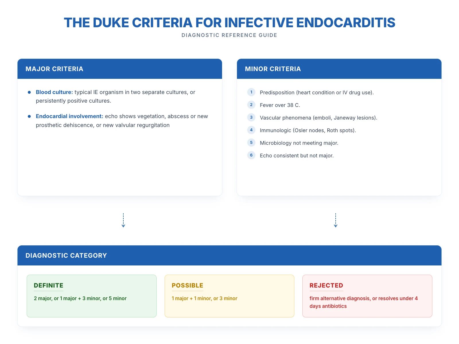
MAJOR criteria (two):[4]
- A typical positive blood culture for IE:
- (i) two separate blood cultures positive for typical microorganisms — viridans streptococci, S. aureus, HACEK group, or community-acquired enterococci — in the absence of a primary focus; OR
- (ii) persistently positive blood cultures — at least two positive cultures drawn more than 12 hours apart, or a majority of three or more separate cultures with the first and last drawn at least one hour apart; OR
- (iii) a single positive blood culture for Coxiella burnetii or an anti-phase-I IgG antibody titre greater than 1:800.
- Endocardial involvement on echo:
- an oscillating intracardiac mass (vegetation) on a valve or supporting structures, in the path of regurgitant jets, or on implanted material; OR
- an abscess; OR
- new partial dehiscence of a prosthetic valve; OR
- new valvular regurgitation (worsening or changing of a pre-existing murmur is insufficient).[4][5]
MINOR criteria (five):[5]
- Predisposition — a predisposing heart condition or intravenous drug use.
- Fever — temperature 38 °C or higher.
- Vascular phenomena — arterial emboli, septic pulmonary infarcts, mycotic aneurysm, intracranial haemorrhage, conjunctival haemorrhages, Janeway lesions.
- Immunologic phenomena — glomerulonephritis, Osler nodes, Roth spots, rheumatoid factor.
- Microbiological evidence — a positive blood culture not meeting a major criterion, or serologic evidence of active infection with an organism consistent with IE.[4][5]
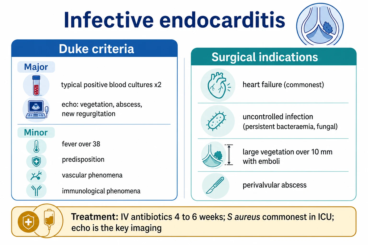
A case is definite (modified Duke) if it meets two major criteria, OR one major + three minor, OR five minor — or if there is pathologic evidence of IE on excised tissue or a positive culture of an embolic specimen. Possible IE meets one major + one minor, or three minor. IE is rejected if there is a firm alternative diagnosis, resolution with less than four days of antibiotics, or no pathologic evidence at surgery or autopsy.[4][5]
Management — Resuscitation
Resuscitation addresses the sepsis and the heart failure that dominate the first hours, while securing the microbiological diagnosis.[1][3]
- Septic / septic-shock IE: give oxygen and insert two large-bore IV cannulae, draw three sets of blood cultures from separate sites before antibiotics, then start empiric IV antibiotics immediately — do not delay therapy in sepsis. Give crystalloid fluids cautiously and use vasopressors (noradrenaline first-line) for shock; correct electrolytes; monitor urine output, lactate and renal function. Follow the sepsis-six bundle and escalate to critical care early.
- Acute severe regurgitation and pulmonary oedema: this is the commonest surgical emergency in IE. Sit the patient up, give high-flow oxygen, IV furosemide, a vasodilator (glyceryl trinitrate infusion if blood pressure permits) and non-invasive ventilation for respiratory failure — but these measures only buy time. Definitive treatment is urgent valve surgery, and surgical referral must be made at the moment the diagnosis of acute severe regurgitation is made, without waiting for culture completion.[1]
- Embolic stroke: maintain perfusion and oxygenation, avoid hypotension, and involve neurology and cardiothoracic surgery; the question of early surgery after stroke is complex and time-dependent (see Evidence section).
The non-negotiable rule: three sets of blood cultures before antibiotics, every time — the whole of definitive therapy hangs on what these cultures grow.[3]
Management — Definitive & Stepwise
Definitive therapy rests on three pillars: bactericidal antibiotic therapy directed at the organism and its MIC, early surgery for mechanical or uncontrolled complications, and prevention of recurrence through prophylaxis.[3]
Empiric antibiotic therapy (before culture results)
Empiric therapy is dictated by the valve substrate and the clinical context. Every regimen below combines a cell-wall-active bactericidal agent (beta-lactam or glycopeptide) with cover for the most likely organisms; doses assume normal renal function and must be adjusted to MIC and renal function once cultures return.[1][3]
| Scenario | Empiric regimen (drug + dose + route) |
|---|---|
| Native-valve, community-acquired | Ampicillin-sulbactam 3 g IV 6-hourly (or benzylpenicillin 1.2 g IV 4-hourly) + gentamicin 1 mg/kg IV 8-hourly; add flucloxacillin 2 g IV 6-hourly if S. aureus suspected (e.g. cellulitis, IVDU). ESC: ampicillin + gentamicin + flucloxacillin |
| Native-valve, broad/unknown (severe sepsis, healthcare exposure) | Vancomycin 15 mg/kg IV 12-hourly (target trough 15-20) + gentamicin 1 mg/kg IV 8-hourly + cefepime 2 g IV 8-hourly |
| Prosthetic valve (early <1 yr, or any PVE pending cultures) | Vancomycin 15 mg/kg IV 12-hourly + gentamicin 1 mg/kg IV 8-hourly + cefepime 2 g IV 8-hourly + rifampicin 300-450 mg IV/PO 12-hourly (add rifampicin only once bacteraemia has cleared, typically after 3-5 days, to avoid resistance) |
| IV-drug-use (tricuspid/right-sided) | Flucloxacillin 2 g IV 6-hourly (MSSA likely); vancomycin 15 mg/kg IV 12-hourly if MRSA risk (nosocomial, known colonisation). Gentamicin is generally not needed in uncomplicated right-sided MSSA IE |
| Culture-negative / fastidious suspected | Vancomycin + gentamicin + cefepime (with serology for Coxiella, Bartonella); consider doxycycline + hydroxychloroquine if Coxiella confirmed |
Definitive (organism-directed) therapy
Once the organism and MIC are known, therapy is narrowed and prolonged. Total duration is counted from the first day of effective antibiotics (the first negative culture day for some guidelines).[3][9]
- Penicillin-susceptible viridans streptococci (MIC ≤0.12 mg/L): benzylpenicillin (penicillin G) 1.2 g (2 MU) IV 4-hourly (total 12-18 MU/day) — or ceftriaxone 2 g IV once daily for 4 weeks. A 2-week course adding gentamicin 1 mg/kg IV 8-hourly for the first 2 weeks is acceptable in selected uncomplicated cases (young, native valve, MIC ≤0.12, no emboli, prompt response).[3]
- Penicillin-relatively-resistant streptococci (MIC >0.12 to ≤0.5 mg/L): benzylpenicillin/ceftriaxone 4 weeks + gentamicin 1 mg/kg IV 8-hourly for the first 2 weeks.[3]
- MSSA native-valve IE: flucloxacillin (or nafcillin/oxacillin) 2 g IV 4- to 6-hourly for 6 weeks; gentamicin 1 mg/kg IV 8-hourly for the first 3-5 days is optional and no longer routine. For uncomplicated right-sided MSSA IE in IVDU, a 2-week course of flucloxacillin + gentamicin may suffice.[3]
- MRSA IE: vancomycin 15 mg/kg IV 12-hourly (or 25-30 mg/kg/day continuous/intermittent, trough 15-20) for 6 weeks; daptomycin 6-10 mg/kg IV daily is an alternative. Gentamicin is not routinely added (nephrotoxicity, no benefit).[9]
- Enterococcal IE (ampicillin-sensitive): ampicillin 2 g IV 4-hourly + gentamicin 1 mg/kg IV 8-hourly for 4-6 weeks; for aminoglycoside-resistant or HLAR strains use ampicillin + ceftriaxone (synergistic double-beta-lactam) for 6 weeks. Vancomycin replaces ampicillin for ampicillin-resistant strains.[3]
- HACEK IE: ceftriaxone 2 g IV daily for 4 weeks (ampicillin-sulbactam is an alternative).[13]
- Culture-negative IE: treat the underlying organism once identified — doxycycline + hydroxychloroquine for Coxiella (≥18 months), gentamicin (or ceftriaxone) + doxycycline for Bartonella, trimethoprim-sulfamethoxazole for Tropheryma whipplei.[6]
Surgery — the three ESC indications
Early surgery (during the initial antibiotic course) is needed in roughly half of patients, driven by three ESC-defined indications.[1][2]
Heart failure
- The commonest indication (~60-70% of operated cases)
- Acute severe regurgitation from valve destruction/rupture
- Urgent surgery regardless of organism or culture status
Uncontrolled infection
- Persistent bacteraemia/fever > 5-7 days on correct antibiotics
- Perivalvular extension — abscess, fistula, heart block
- Fungal or multi-resistant organisms not responding medically
Prevention of embolism
- Large (>10 mm), mobile vegetations with embolic risk
- Prior embolic event with a persisting large vegetation
- Highest embolic risk in the first days of therapy
Surgery is also indicated for prosthetic-valve IE with dehiscence or obstruction, large vegetations from organisms difficult to eradicate (fungi), and CIED infection (requiring complete device and lead extraction). The operation is usually valve replacement with a mechanical or bioprosthetic prosthesis, but valve repair is increasingly used for isolated mitral or tricuspid lesions where feasible, avoiding long-term anticoagulation.[1]
The clearest evidence for surgery as embolic prevention is Kang and colleagues' randomised trial of early surgery (within 48 hours) versus conventional treatment in left-sided IE with large vegetations: early surgery reduced the composite of in-hospital death and embolic events (3% vs 23%, hazard ratio 0.10) — a benefit driven entirely by fewer embolic events, since all-cause mortality alone was not significantly different (3% vs 5%).[7]
The decision to operate, and how urgently, follows a structured assessment of the indication and the operative risk.[1]
Deciding on and timing surgery in IE
Is there heart failure from acute severe regurgitation?
If yes — URGENT surgery, regardless of organism, cultures or duration of antibiotics. This is the commonest and most lethal indication; do not delay for medical stabilisation.
Is there uncontrolled infection?
Persistent bacteraemia or fever beyond 5-7 days of correct antibiotics, perivalvular abscess/fistula, new AV block, or a fungal/multiresistant organism — surgery (urgent or early) is usually needed.
Is there a high embolic risk?
Large (>10 mm) mobile vegetation, prior embolus with residual vegetation, or rapidly growing vegetation in the first week — early surgery within days, while the embolic-risk window is open.
What is the operative risk?
Age, comorbidity, prosthetic vs native, neurological status. Recent stroke requires balancing haemorrhagic-transformation risk against the cardiac indication; delay 1-2 weeks post-ischaemic stroke if feasible.
Replace vs repair
Valve replacement (mechanical or bioprosthesis) is standard; mitral/tricuspid repair is increasingly used for localised destructive lesions where feasible, sparing lifelong anticoagulation.
Prophylaxis
Specific Subtypes & Scenarios
Native-valve (community)
- Viridans streptococci, S. aureus, enterococci, HACEK
- Empiric: benzylpenicillin/ampicillin + gentamicin ± flucloxacillin
- Often subacute unless S. aureus
Prosthetic valve
- Early (<60 d / <1 yr): coag-neg staph (S. epidermidis), S. aureus, Gram-negatives
- Late (>60 d / >1 yr): resembles native-valve (strep, staph, enterococcus)
- Empiric: vancomycin + gentamicin + cefepime + rifampicin (after bacteraemia clears)
IV drug use
- S. aureus (>60%); often MSSA, watch MRSA
- Tricuspid valve (right-sided); septic pulmonary emboli
- Empiric: flucloxacillin (MSSA) or vancomycin (MRSA); often curable medically
Healthcare-associated
- S. aureus (incl. MRSA), coag-neg staph, enterococcus, Candida
- Lines, haemodialysis, recent procedures
- Empiric: vancomycin-based + Gram-negative cover
Culture-negative IE follows prior antibiotics (the commonest reason — organisms suppressed, not killed) or fastidious/intracellular organisms (Coxiella, Bartonella, Tropheryma, Brucella, Legionella) and fungi (Candida, Aspergillus), which need serology or PCR rather than more culture bottles. The 2023 Duke-ISCVID update explicitly adds 16S/18S rRNA PCR and metagenomic sequencing of excised tissue as microbiologic criteria, reflecting this reality.[6]
Streptococcus gallolyticus (formerly S. bovis) bacteraemia/IE mandates colonoscopy, because of its strong (~30%) association with advanced colonic neoplasia — finding the organism in a blood culture is itself an indication to screen the colon. Fungal IE (Candida, Aspergillus) occurs with immunosuppression, long-term indwelling lines, total parenteral nutrition and IV drug use, produces large friable vegetations with major embolic risk, and has a poor prognosis — it almost always needs surgery combined with prolonged antifungal therapy (liposomal amphotericin B or an echinocandin, step-down to fluconazole).[1]
Cardiac-implantable electronic device (CIED) infections of the pocket or lead may seed the tricuspid valve; they usually require complete percutaneous or surgical device and lead extraction in addition to targeted antibiotics, as infection of the insulated lead is essentially impossible to sterilise medically.[1]
HACEK
The HACEK group are slow-growing, fastidious Gram-negative oropharyngeal commensals that need prolonged incubation (now mostly recovered in modern automated blood-culture systems within 5 days). They cause about 1-3% of IE, typically subacute on native valves, and are uniformly susceptible to ceftriaxone 2 g IV daily for 4 weeks.[13]
Complications & Pitfalls
Complications fall into cardiac (the valve and surrounding structures), neurologic, embolic/systemic, renal, and infectious/septic. The commonest cause of death is heart failure from acute valvular regurgitation, which alone accounts for most surgical indications and the bulk of mortality.[3]
| Complication | Notes |
|---|---|
| Heart failure | Commonest cause of death; acute severe regurgitation from leaflet perforation/rupture — surgical emergency |
| Perivalvular extension | Abscess, fistula, prosthetic dehiscence; new AV block = aortic-root abscess; urgent surgery |
| Embolic stroke | Up to 20-40% of left-sided IE; may be the presenting event; risk highest in first days |
| Mycotic aneurysm | Arterial wall infection from septic emboli; intracranial risk of catastrophic haemorrhage |
| Splenic/renal/mesenteric infarct | Left-sided embolisation; flank pain, haematuria, abdominal pain |
| Septic pulmonary emboli | Right-sided (IVDU) IE; cavitating peripheral infiltrates, empyema |
| Acute kidney injury | Sepsis, embolic renal infarct, immune-complex glomerulonephritis, nephrotoxic antibiotics (gentamicin, vancomycin) |
| Conduction disease | AV block from septal/annular abscess; transient with oedema or permanent from destruction |
| Septic shock / metastatic infection | Overwhelming S. aureus; seeding to spine, joints, retina |
Neurological complications — the most feared embolic target
The brain is the commonest and most dangerous systemic embolic target. Embolic stroke occurs in 20-40% of left-sided IE and may be the first presentation; risk is highest in the first week of symptoms and falls sharply once effective antibiotics begin, with most events occurring before or within the first few days of therapy. Beyond stroke, neurological complications include intracranial mycotic aneurysms (which may rupture catastrophically), haemorrhagic transformation of an infarct, brain abscess, meningitis, encephalopathy and seizures. A new headache, meningism or a declining conscious level in IE demands urgent imaging (CT then MRI) and, for mycotic aneurysm, CT or digital-subtraction angiography.[3]
The interaction between embolic stroke and cardiac surgery is a recurring exam question. Ischaemic stroke is not an absolute contraindication to early surgery, but surgery within the first few days after a large stroke carries a high risk of haemorrhagic transformation once the patient is on cardiopulmonary bypass; the ESC therefore suggests, where possible, delaying non-emergency surgery for one to two weeks after an ischaemic stroke and for three to four weeks after a haemorrhagic stroke, balanced against the urgency of the cardiac indication. Haemorrhagic stroke, by contrast, generally precludes early surgery.[1]
Embolic risk — when it peaks and falls
Embolic risk is not constant: it is highest before and within the first few days of starting antibiotics (15-25% of patients embolise before or at diagnosis) and falls to about 1-2% per week thereafter as the vegetation organisation and sterilisation proceed. The predictors of ongoing embolism are a large vegetation (over 10 mm), mobile or pedunculated morphology, increasing size on serial echo, mitral-valve location (higher than aortic), and S. aureus or Candida as the organism. This time-course is the biological basis for the surgical indication of early valve surgery for large mobile vegetations in the first week — the window of benefit closes as the embolic risk falls.[7][1]
The principal pitfall is treating S. aureus bacteraemia as a simple line infection without echocardiographic exclusion of IE — significant S. aureus bacteraemia carries a high probability of endocarditis, and suboptimal evaluation (omitting echocardiography, too-short antibiotic course) is associated with worse outcome. Involve an infectious-diseases specialist early and image the heart.[12]
A second classic pitfall is over-diagnosing IE from a single positive culture — a single bottle growing a coagulase-negative staphylococcus is far more likely to be a skin contaminant than PVE; the Duke criteria require persistence (multiple bottles, multiple sites) precisely to exclude contamination.[5]
Prognosis & Disposition
Despite modern antibiotics and surgery, IE remains lethal, with in-hospital mortality of about 15-20% for native-valve IE, 20-40% for prosthetic-valve IE, and 40-60% when S. aureus is the organism. One-year mortality approaches 30%.[11][3] Adverse prognostic features are the same factors that predict death and drive surgery: S. aureus infection, prosthetic-valve involvement, heart failure, older age, perivalvular complications (abscess, conduction block), embolic events, large vegetations, and healthcare-associated acquisition.[11]
Conversely, viridans-streptococcal infection is comparatively favourable, and indicated surgery is itself protective — a reminder that the threshold to operate in IE is appropriately low, and that delaying surgery for heart failure in the hope of medical stabilisation is a recurring and lethal error.[11]
Disposition requires 4-6 weeks of intravenous therapy (often via a peripherally inserted central catheter, with outpatient parenteral antibiotic therapy once the patient is stable, afebrile, culture-negative and surgically cleared), serial inflammatory markers and repeat echocardiography to document shrinking vegetations and stable valve function, dental clearance (eliminate the oral source of bacteraemia), infectious-diseases, cardiology and (where relevant) cardiothoracic-surgery follow-up, and patient education on the recurring risk and the need for prophylaxis. In S. aureus bacteraemia, adherence to infectious-diseases specialist recommendations (including echocardiographic evaluation) measurably improves outcome.[12]
Special Populations
| Group | Considerations |
|---|---|
| Pregnancy | Rare but dangerous; right-sided IVDU IE more common. Penicillins, cephalosporins safe; vancomycin acceptable; avoid aminoglycosides (fetal ototoxicity) and tetracyclines. Cardiopulmonary bypass in pregnancy is feasible if heart failure mandates urgent surgery |
| Prosthetic valve | Lifelong risk; early (<60 d) coag-neg staph and nosocomial organisms, late resembles native-valve. Add rifampicin after bacteraemia clears; TOE mandatory; surgery for dehiscence/obstruction |
| Elderly | Often afebrile; present with confusion, heart failure or stroke; more comorbidity and prosthetic/degenerative valves; higher mortality; lower threshold to image |
| Immunocompromised / lines | Consider fungal (Candida, Aspergillus) and resistant organisms; remove indwelling lines; broaden empiric cover |
| IV drug use | Right-sided (tricuspid) IE, younger, often curable medically with 2-6 weeks; high recurrence if injection continues; address addiction |
| Paediatric / CHD | Often on congenital substrate (VSD, tetralogy, post-surgical repair); surgery and anticoagulation considerations differ |
Evidence, Guidelines & Regional Differences
The evidence base for IE rests on a small number of landmark studies and a set of converging guidelines that now agree on most points but diverge sharply on prophylaxis.[1][3]
- ESC 2023 (Delgado) vs AHA/IDSA 2015 (Baddour): both endorse the Duke framework and the three surgical indications (heart failure, uncontrolled infection, prevention of embolism). The ESC 2023 guideline is the most current and adds [18F]FDG PET/CT and cardiac CT as major diagnostic criteria, mirroring the 2023 Duke-ISCVID update; the AHA statement is older but its antibiotic recommendations remain standard.[1][3][6]
- Kang 2012 RCT is routinely (and wrongly) summarised as "early surgery saves lives" — it did not show a significant mortality reduction. Its benefit was a drop in embolic events within a composite endpoint (death + emboli), which is exactly how it should be cited. Subsequent observational data and the 2023 ESC guidance accept early surgery for large (>10 mm) mobile vegetations with high embolic risk, especially after a first embolic event.[7]
- Prophylaxis — the great divergence: the AHA (US) restricts prophylaxis to high-risk cardiac lesions for dental procedures only; NICE (UK) went further and recommends no routine antibiotic prophylaxis at all, emphasising oral hygiene instead; ESC (Europe) takes a middle position, recommending prophylaxis for the highest-risk group for dental procedures. India (ICMR/NCDC) does not mandate routine dental prophylaxis but recognises the high-risk groups; empiric antibiotic choices in India must account for local resistance (high community MRSA, ESBL Gram-negatives).[8]
- IDSA MRSA guidance (Liu 2011) remains the reference for vancomycin dosing (trough 15-20 mg/L), duration (6 weeks for native, ≥6 weeks for prosthetic), and the option of daptomycin for vancomycin failures or intolerance.[9]
- Controversy: routine surgery for large vegetations >10 mm without embolism remains debated; the ENTER trial and observational data suggest benefit is concentrated in the first week after presentation, before the embolic risk falls.
Exam Pearls
- Osler vs Janeway: Osler = tender, immune-complex, on the pulps; Janeway = painless, septic emboli, on palms and soles. The painless lesion is the infected one.[5]
- S. bovis (gallolyticus) bacteraemia mandates colonoscopy — strong association with colonic neoplasia.[3]
- S. aureus bacteraemia = endocarditis until proven otherwise — image the heart (TOE if needed), involve ID.[12]
- Duke definite: 2 major OR 1 major + 3 minor OR 5 minor. Possible: 1 major + 1 minor, or 3 minor.[4][5]
- New PR prolongation in IE = aortic-root abscess — call the surgeon.[1]
- Three sets of blood cultures before antibiotics, every time — spaced, from separate sites.[3]
- Vegetations form on the low-pressure, downstream surface of a jet — atrial surface in MR, ventricular surface in AS, right side of a VSD.[3]
- Heart failure is the commonest indication for surgery and the commonest cause of death — operate early, do not wait for culture completion.[1]
- Rifampicin is added in prosthetic-valve IE — but only after bacteraemia clears (3-5 days) to prevent rapid emergence of resistance.[1]
- Viridans streptococci = dental source; enterococci = GU/GI source; S. aureus = skin/IVDU/lines; HACEK = oropharyngeal; Coxiella/Bartonella = culture-negative, need serology.[13]
Self-test: name the Duke category
A 45-year-old with a bicuspid aortic valve (predisposition) has fever 39 °C, three blood cultures growing viridans streptococci, and an echocardiographic vegetation. Predisposition (minor) + fever (minor) + typical positive blood culture (major) + vegetation on echo (major).
Answer: Definite IE — two major criteria (typical culture + endocardial involvement) alone suffice; the two minor criteria do not change the category but support it.
Self-test: which organism, which valve, which drug?
A 29-year-old man who injects heroin presents with fever, pleuritic chest pain and cavitating peripheral lung infiltrates; blood cultures grow Staphylococcus aureus.
Answer: right-sided (tricuspid) IE in an IVDU, seeded because the bolus of organisms passes through the right heart first; embolise to the lungs as septic pulmonary emboli. Empiric/definitive therapy for MSSA is flucloxacillin 2 g IV 6-hourly (vancomycin if MRSA); uncomplicated right-sided MSSA IE may be curable in 2 weeks.
Red Flags
Exam application bank (NEET-PG / INICET)
One-line answer
Infective endocarditis (IE) is a microbial infection of the endocardial surface, usually a heart valve, forming friable vegetations of platelets and fibrin that entrap organisms. It classically presents with fever, a new or changing murmur, and embolic, immunologic or septic phenomena. Diagnosis rests on blood cultures and echocardiography integrated through the Duke criteria. Management combines prolonged bactericidal antibiotics (benzylpenicillin/ceftriaxone ± gentamicin for streptococci; flucloxacillin for MSSA; vancomycin for MRSA) with early surgery for heart failure, uncontrolled infection or the prevention of embolism.
Worked stems (answer without another resource)
Stem 1 — Classic presentation. Map symptoms to mechanism; name the first investigation and first treatment step with dose/route if drug therapy is standard. [1]
Stem 2 — Unstable / complicated. List red flags that force immediate resuscitation, theatre, ICU, antidote, or reperfusion — and what you do in the first 15 minutes. [1]
Stem 3 — Atypical group. Elderly, pregnancy, child, or immunocompromised: how presentation and thresholds change. [1]
Stem 4 — Differential trap. Name the three closest mimics and one discriminator for each. [1]
Stem 5 — Disposition. Who goes home with safety-netting, who is admitted, who needs HDU/ICU/theatre, and what follow-up is mandatory. [1]
Rapid viva checklist
- Definition + classification
- Pathophysiology chain
- Bedside signs / criteria
- Score with exact components (if any)
- Emergency bundle
- Definitive therapy with doses
- Complications of disease and of treatment
- Special populations
- Guideline/trial name if classic
- Three exam traps
Coverage self-check
If you cannot answer any stem above from this page alone, re-read the matching section — the page is intended to be self-sufficient for final-prof and NEET-PG/INICET questions on Infective Endocarditis.
References
- [1]Delgado V, Ajmone Marsan N, et al. 2023 ESC Guidelines for the management of endocarditis Eur Heart J, 2023.PMID 37622656
- [2]Habib G, Lancellotti P, et al. 2015 ESC Guidelines for the management of infective endocarditis: The Task Force for the Management of Infective Endocarditis of the European Society of Cardiology (ESC). Endorsed by: European Association for Cardio-Thoracic Surgery (EACTS), the European Association of Nuclear Medicine (EANM) Eur Heart J, 2015.PMID 26320109
- [3]Baddour LM, Wilson WR, et al. Infective Endocarditis in Adults: Diagnosis, Antimicrobial Therapy, and Management of Complications: A Scientific Statement for Healthcare Professionals From the American Heart Association Circulation, 2015.PMID 26373316
- [4]Durack DT, Lukes AS, et al. New criteria for diagnosis of infective endocarditis: utilization of specific echocardiographic findings. Duke Endocarditis Service Am J Med, 1994.PMID 8154507
- [5]Li JS, Sexton DJ, et al. Proposed modifications to the Duke criteria for the diagnosis of infective endocarditis Clin Infect Dis, 2000.PMID 10770721
- [6]Fowler VG, Durack DT, et al. The 2023 Duke-International Society for Cardiovascular Infectious Diseases Criteria for Infective Endocarditis: Updating the Modified Duke Criteria Clin Infect Dis, 2023.PMID 37138445
- [7]Kang DH, Kim YJ, et al. Early surgery versus conventional treatment for infective endocarditis N Engl J Med, 2012.PMID 22738096
- [8]Wilson W, Taubert KA, et al. Prevention of infective endocarditis: guidelines from the American Heart Association: a guideline from the American Heart Association Rheumatic Fever, Endocarditis, and Kawasaki Disease Committee, Council on Cardiovascular Disease in the Young, and the Council on Clinical Cardiology, Council on Cardiovascular Surgery and Anesthesia, and the Quality of Care and Outcomes Research Interdisciplinary Working Group Circulation, 2007.PMID 17446442
- [9]Liu C, Bayer A, et al. Clinical practice guidelines by the infectious diseases society of america for the treatment of methicillin-resistant Staphylococcus aureus infections in adults and children Clin Infect Dis, 2011.PMID 21208910
- [10]Baddour LM, Wilson WR, et al. Infective endocarditis: diagnosis, antimicrobial therapy, and management of complications: a statement for healthcare professionals from the Committee on Rheumatic Fever, Endocarditis, and Kawasaki Disease, Council on Cardiovascular Disease in the Young, and the Councils on Clinical Cardiology, Stroke, and Cardiovascular Surgery and Anesthesia, American Heart Association: endorsed by the Infectious Diseases Society of America Circulation, 2005.PMID 15956145
- [11]Murdoch DR, Corey GR, et al. Clinical presentation, etiology, and outcome of infective endocarditis in the 21st century: the International Collaboration on Endocarditis-Prospective Cohort Study Arch Intern Med, 2009.PMID 19273776
- [12]Fowler VG Jr, Sanders LL, et al. Outcome of Staphylococcus aureus bacteremia according to compliance with recommendations of infectious diseases specialists: experience with 244 patients Clin Infect Dis, 1998.PMID 9770144
- [13]Sharara SL, Tayyar R, et al. HACEK endocarditis: a review Expert Rev Anti Infect Ther, 2016.PMID 27124204
- [14]Bor DH, Woolhandler S, Nardin R, Brusch J Infective endocarditis in the U.S., 1998-2009: a nationwide study PLoS One, 2013.PMID 23527296