Cardiology · Cardiology
Long QT and Channelopathies
Long QT syndrome and the related cardiac channelopathies (Brugada, CPVT, short QT, early repolarisation) are inherited arrhythmia syndromes caused by mutations in cardiac ion-channel genes that predispose young, structurally normal hearts to syncope, torsades de pointes, ventricular fibrillation and sudden cardiac death. The dominant therapy is beta-blockade (nadolol or propranolol for LQTS and CPVT), avoidance of QT-prolonging drugs, lifestyle modification, and ICD implantation for secondary prevention or high-risk primary prevention. Acute torsades de pointes is treated with IV magnesium 2 g, defibrillation if sustained, and overdrive pacing or isoprenaline for pause-dependent forms.
On this page & tools
Your progress
Saved locally on this device.
Exam tags
Red flags
Overview & Definition
The cardiac channelopathies are a group of inherited disorders caused by mutations in the genes encoding cardiac ion channels or their accessory regulatory proteins. They share a paradoxical and clinically dangerous phenotype: a heart that is structurally normal on echocardiography yet electrically unstable, predisposing to syncope, torsades de pointes (TdP), ventricular fibrillation (VF), and sudden cardiac death (SCD) — most often in children, adolescents, and young adults. The five clinically important syndromes are congenital long QT syndrome (LQTS), Brugada syndrome, catecholaminergic polymorphic ventricular tachycardia (CPVT), short QT syndrome (SQTS), and early repolarisation syndrome (ERS).[1]
LQTS has a prevalence of approximately 1 in 2,000 and is the most common of the group. The others are rarer — Brugada 1 in 2,000 to 5,000 (with a strikingly higher prevalence in Southeast Asia), CPVT 1 in 10,000, and SQTS exceptionally rare. Because the channelopathies leave no scar at autopsy, they are the prototypical cause of autopsy-negative sudden cardiac death in the young, which is why post-mortem genetic testing (the "molecular autopsy") and cascade screening of first-degree relatives are now standard practice after any unexplained death in a young person.[1][4]
The clinically important distinction that frames this entire topic is congenital versus acquired long QT. Congenital LQTS is genetic and life-long; acquired LQTS from QT-prolonging drugs, electrolyte disturbance, bradycardia, or hypothyroidism is far more common in routine practice and is the single most preventable cause of drug-induced TdP. Every prescriber must therefore understand both the inherited forms and the drug-electrolyte interactions that produce the same ECG signature. [1]
Classification
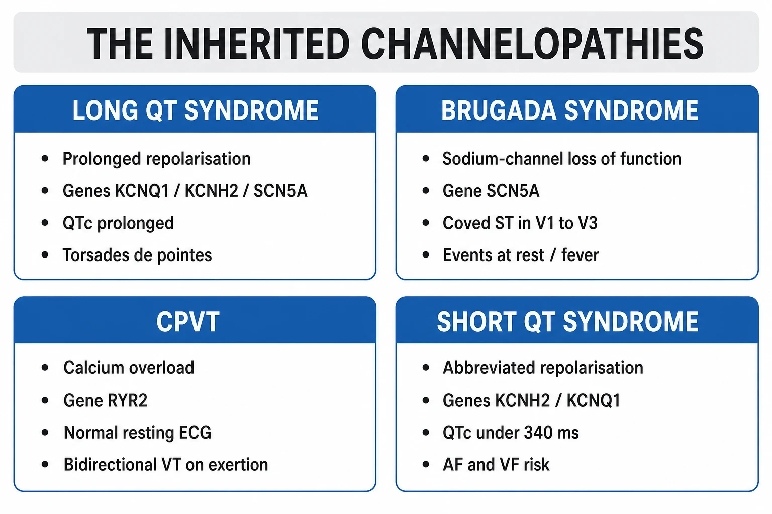
The 2013 HRS/EHRA/APHRS expert consensus divides the inherited primary arrhythmia syndromes by gene and dominant phenotype.[1] The three common LQTS genotypes account for over 90 percent of genotype-positive cases and have gene-specific triggers that examiners always probe.
LQT1 (KCNQ1, IKs LOF)
- Most common genotype, 35 to 40 percent of genotype-positive LQTS
- Trigger: exercise (especially swimming) and emotional stress
- Broad-based T wave; QTc 470 to 700 ms
- Beta-blocker highly effective; events reduced over 70 percent
LQT2 (KCNH2/hERG, IKr LOF)
- 25 to 30 percent of LQTS
- Trigger: auditory stimuli (alarm clock, doorbell, telephone), emotional stress, postpartum period
- Low-amplitude notched T wave in limb and left precordial leads
- Beta-blocker effective; keep serum K+ over 4.5 mmol/L
LQT3 (SCN5A, late INa GOF)
- 5 to 10 percent of LQTS
- Trigger: rest, sleep, slow heart rate (bradycardia-dependent)
- Long isoelectric ST segment followed by late-onset T wave
- Beta-blocker less effective; mexiletine or ranolazine shortens QT; ICD often needed
The rare LQTS genotypes are recognised but rarely the discriminator at MBBS level. LQT4 (ANK2, ankyrin-B), LQT5 (KCNE1), LQT6 (KCNE2), LQT7 (Andersen-Tawil syndrome, KCNJ2, with periodic paralysis and dysmorphism), LQT8 (Timothy syndrome, CACNA1C gain-of-function, with syndactyly and autism), and LQT9 to 15 are individually uncommon. Jervell and Lange-Nielsen syndrome is the recessive form caused by homozygous or compound heterozygous KCNQ1 or KCNE1 mutations, characterised by congenital profound sensorineural deafness and a particularly severe cardiac phenotype with early-onset SCD.[1]
Brugada syndrome
- SCN5A loss-of-function in ~30 percent of cases (most common)
- Coved type 1 ST elevation in V1-V3 at baseline or with provocation
- Syncope or VF at rest, sleep, or febrile illness
- Only proven therapy = ICD; quinidine and isoproterenol adjunctive; avoid sodium-channel blockers
CPVT
- RYR2 (autosomal dominant, ~60 percent) or CASQ2 (recessive)
- Bidirectional or polymorphic VT triggered by exercise or emotion
- Resting ECG normal — diagnosis by exercise stress test
- Nadolol first-line; flecainide adjunct; left cardiac sympathetic denervation; ICD for breakthrough
Short QT syndrome
- Gain-of-function in KCNH2 (SQT1), KCNQ1 (SQT2), or KCNJ2 (SQT3)
- QTc under 340 ms (or under 360 ms with family history or arrhythmia)
- Atrial fibrillation, VF, SCD at young age
- ICD only proven therapy; quinidine may prolong QT and reduce events
Early repolarisation syndrome
- J-point elevation over 0.1 mV in inferior (II, III, aVF) or lateral (I, aVL, V4-V6) leads
- Association with idiopathic VF; QTc normal
- ICD for survivors of cardiac arrest; quinidine adjunctive
- Most early repolarisation patterns are benign — syndrome requires symptoms or family history of SCD
Epidemiology & Risk Factors
The dominant epidemiological clue across all five syndromes is a family history of sudden cardiac death in a relative under 40, particularly unexplained drowning (a classic LQT1 presentation during swimming), a single-vehicle night-time car crash (often a syncopal event at the wheel), or sudden infant death syndrome. Autosomal dominant inheritance (Romano-Ward) is the typical pattern in LQTS, CPVT, and most Brugada and SQTS kindreds, conferring a 50 percent risk to first-degree relatives; Jervell-Lange-Nielsen is the recessive exception.[1]
Several acquired factors amplify the risk of an event in a genetically predisposed individual. Female sex lengthens the baseline QTc and confers higher risk, particularly in LQT2 post-puberty and through the postpartum period. Hypokalaemia, hypomagnesaemia, and hypocalcaemia all prolong repolarisation. Bradycardia (sleep, AV block, drugs) is arrhythmogenic in LQT3. QT-prolonging drugs are the single most important iatrogenic precipitant of TdP and are responsible for most acquired LQTS in clinical practice.[1][4]
Pathophysiology
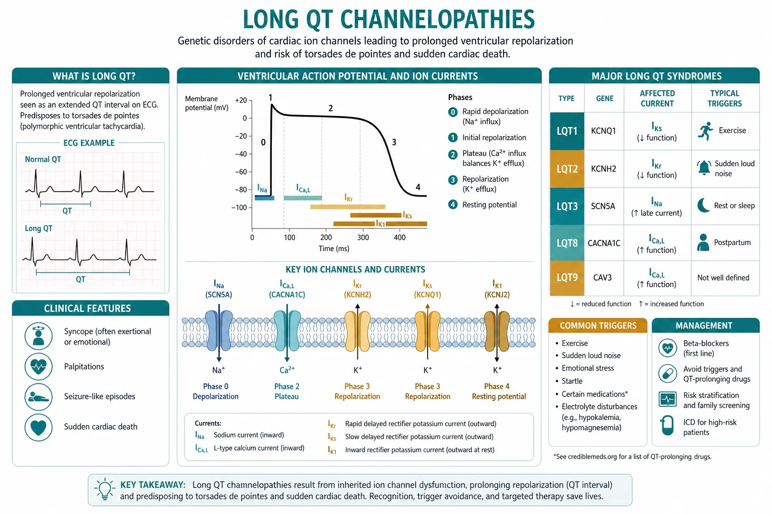
The unifying mechanism across the LQTS genotypes is prolongation of the ventricular action potential, which manifests on the surface ECG as a lengthened QT interval. The mechanism differs by genotype. In LQT1, a loss-of-function mutation in KCNQ1 reduces the slowly activating delayed rectifier potassium current (IKs), shortening repolarising outward current. In LQT2, a loss-of-function mutation in KCNH2 (hERG) reduces the rapidly activating delayed rectifier potassium current (IKr) — and because hERG is the channel most commonly blocked by pharmaceuticals, this same current is responsible for the vast majority of drug-induced LQTS. In LQT3, a gain-of-function mutation in SCN5A enhances the late sustained sodium current (late INa), which keeps depolarising current flowing long after the upstroke and preferentially prolongs the action potential at slow heart rates — explaining why LQT3 events cluster during sleep and rest.[1]
Whatever the ionic mechanism, prolonged repolarisation produces two downstream consequences that drive arrhythmogenesis. The first is increased dispersion of repolarisation — different layers of myocardium (notably mid-myocardial M cells) repolarise at markedly different times, creating a substrate for re-entry. The second is the appearance of early afterdepolarisations (EADs), oscillations of the membrane potential during the prolonged plateau phase that can trigger a premature ventricular beat, which then degenerates into polymorphic VT (TdP). The hallmark ECG of TdP is polymorphic VT with twisting of the QRS axis around the baseline, typically initiated by a short-long-short R-R sequence and often self-terminating but recurrent, with the potential to degenerate into VF. [1]
CPVT has a fundamentally different mechanism. A mutation in the cardiac ryanodine receptor (RYR2) or in calsequestrin (CASQ2) causes spontaneous diastolic calcium leak from the sarcoplasmic reticulum under adrenergic stimulation. The resultant intracellular calcium overload generates delayed afterdepolarisations (DADs) — different from the EADs of LQTS — that fire premature beats in bigeminal patterns and the characteristic bidirectional VT (alternating QRS axis beat-to-beat, superficially resembling supraventricular tachycardia with aberrancy). Because the resting membrane is normal, the resting ECG is normal, and the diagnosis is made by provoking the arrhythmia with exercise or adrenaline.[6]
Brugada syndrome results from a loss-of-function mutation in SCN5A (or, less commonly, in CACNA1C, GPD1-L, or other sodium-calcium channel subunits). The reduced inward sodium current disproportionately abbreviates the action potential in the right ventricular epicardium, abolishing the action potential dome and creating a transmural voltage gradient that produces the characteristic coved ST elevation in V1 to V3. The same substrate supports phase-2 re-entry and polymorphic VT/VF, which typically occurs at rest or during sleep, when vagal tone is high and sodium channel availability is reduced, and is often precipitated by fever (which further worsens sodium-channel trafficking).[7]
Short QT syndrome is caused by gain-of-function mutations in the same channels that are lost in LQTS (KCNH2, KCNQ1, KCNJ2), producing abbreviated action potentials, a very short QTc, and marked shortening of atrial and ventricular refractoriness that predisposes to both atrial fibrillation and VF.[10] Early repolarisation syndrome is mechanistically related: an accentuated outward potassium current (mediated by IKATP, IKr, or IKs) generates a J-point notch or slur that, in the inferior or lateral leads, creates a substrate for phase-2 re-entry and idiopathic VF.[11][12]
Clinical Presentation
The dominant clinical manifestations across all the channelopathies are syncope, seizure-like events, cardiac arrest, and sudden cardiac death in a young, otherwise healthy individual. The single most diagnostically powerful clue is the circumstance of the event, which is why the history alone often points to the genotype.[1]
In LQT1, syncope is classically triggered by exercise — especially swimming — and by emotional stress; the swimming phenotype is so characteristic that any unexplained drowning in a competent swimmer should prompt LQTS evaluation of the family. In LQT2, the trigger is auditory — a sudden loud noise such as an alarm clock, a doorbell, a ringing telephone, or an infant's cry — and emotional stress, with a marked increase in risk during the 9 months postpartum. In LQT3, events occur at rest or during sleep, and the patient may give a history of nocturnal syncope or a parent may describe a child found pulseless in the cot.[1][8]
The syncope of LQTS is often misdiagnosed as epilepsy for years before the correct diagnosis is made, because the cerebral hypoperfusion of TdP can produce brief tonic-clonic movements indistinguishable from a generalised seizure. The discriminating features are: syncope during exercise or with a recognisable trigger (rather than the typical aura and post-ictal confusion of true epilepsy), a family history of SCD, no post-ictal state, and a prolonged or atypical QTc on ECG.[1]
In CPVT, syncope or cardiac arrest is provoked by exercise or strong emotion in a child whose resting ECG is entirely normal; the bidirectional VT is reproducibly induced by exercise testing. In Brugada, syncope or VF typically occurs at rest, during sleep, after a large meal, or during a febrile illness; agonal nocturnal respiration is a recognised presentation. In SQTS, the patient may present with atrial fibrillation at a young age, syncope, or sudden death. In ERS, the presentation is idiopathic VF — typically in a young male with a J-point pattern in the inferior or lateral leads — and a family history of early SCD.[7][10][11]
Examination between events is almost always normal, which is itself a clue. Clues to syndromic forms include syndactyly (Timothy syndrome, LQT8), dysmorphic facies with periodic paralysis (Andersen-Tawil, LQT7), and congenital sensorineural deafness (Jervell-Lange-Nielsen). [1]
Differential Diagnosis
The differential of QT prolongation and an unexplained syncopal event in a young person is wide, and several diagnoses are dangerous to miss. The most important differentials for clinical practice are:[1][3]
Acquired LQTS (drugs)
- Class Ia and III antiarrhythmics: quinidine, procainamide, disopyramide, sotalol, amiodarone, dofetilide, ibutilide
- Macrolides: erythromycin, clarithromycin, azithromycin (less)
- Fluoroquinolones: moxifloxacin, ciprofloxacin, levofloxacin
- Antipsychotics: haloperidol, droperidol, ziprasidone, thioridazine, quetiapine
- Methadone; high-dose ondansetron; citalopram/escitalopram; terfenadine; tacrolimus
Acquired LQTS (electrolytes and metabolic)
- Hypokalaemia — the most common electrolyte precipitant
- Hypomagnesaemia
- Hypocalcaemia
- Hypothyroidism; anorexia nervosa; starvation; cirrhosis
- Bradycardia and AV block (pause-dependent TdP)
Other syncope mimics
- Vasovagal syncope: postural trigger, prodrome of warmth and nausea, rapid recovery
- Epilepsy: post-ictal confusion, tongue biting, urinary incontinence, EEG abnormality
- Aortic stenosis and HCM: exertional syncope with murmur
- Structural disease: ARVC, myocarditis, anomalous coronary artery
Brugada phenocopies
- Right bundle branch block, LVH, early repolarisation (normal variants)
- Acute ischaemia, pulmonary embolism, pericarditis, electrolyte disturbance
- Pectus excavatum, mechanical RV compression
- Resolve the underlying cause and re-evaluate the ECG
The cardinal rule is that any patient with QT prolongation must have acquired causes actively excluded before the diagnosis of congenital LQTS is made: review every drug against the CredibleMeds list, check potassium, magnesium, calcium, and thyroid function, and consider structural disease with echocardiography. Drug-induced QT prolongation is overwhelmingly an hERG-channel blockade phenomenon — the IKr current carried by the KCNH2/hERG channel is exquisitely sensitive to a structurally diverse range of pharmaceuticals because the channel's inner cavity is large and aromatic, accommodating many drug classes. The CredibleMeds registry classifies QT risk into three tiers that should be memorised: Known Risk of TdP (avoid entirely in LQTS — sotalol, quinidine, procainamide, dofetilide, ibutilide, erythromycin, clarithromycin, halofantrine, methadone, ondansetron IV high dose, thioridazine, droperidol), Possible Risk of TdP (use with caution and ECG monitoring — moxifloxacin, citalopram/escitalopram, quetiapine, ziprasidone), and Conditional Risk (TdP only in setting of overdose or other risk factor — azithromycin, ciprofloxacin, olanzapine). When a patient with LQTS or borderline QTc requires an antiemetic, prefer metoclopramide or domperidone; for an antibiotic in a penicillin-allergic patient, a beta-lactam or doxycycline is preferable to a macrolide or fluoroquinolone; for an antipsychotic, the atypical aripiprazole has minimal QT effect.[1]
A separate category of acquired LQTS that examiners test is the pause-dependent form, in which TdP is initiated by a short-long-short R-R sequence — a premature ventricular beat, a compensatory pause, then another premature beat that falls on the prolonged QT of the post-pause beat and degenerates into TdP. Pause-dependent TdP is the typical mechanism in bradycardia, high-grade AV block, and after a long asystolic pause, and is the form most responsive to isoprenaline or overdrive pacing. In contrast, pause-independent TdP (typical of congenital LQTS with adrenergic surges) is more responsive to beta-blockade. Recognising the short-long-short initiating sequence on a telemetry strip is a high-yield exam pearl and predicts the response to pacing versus beta-blockade. [1]
Clinical & Bedside Assessment
The focused history in a suspected channelopathy must capture four elements. First, the circumstance of the event — exercise versus auditory trigger versus rest versus sleep, the timing relative to emotional stress or the postpartum period, and any prodrome (true LQTS events often have little or no prodrome). Second, the family history — explicitly ask about sudden death in relatives under 40, unexplained drowning, cot death, sudden infant death syndrome, and single-vehicle accidents, and construct a three-generation pedigree. Third, the medication history, including over-the-counter and recreational drugs (cocaine, methadone). Fourth, the past medical history — deafness (JLN), periodic paralysis (Andersen-Tawil), syndactyly or autism (Timothy), seizures mislabelled as epilepsy.[1]
Bedside examination is usually normal. Look specifically for syndromic features: measure blood pressure ( exclude HCM with murmur, aortic stenosis), palpate the precordium (structurally normal in all channelopathies), and inspect for dysmorphism, syndactyly, scoliosis, and low-set ears. Perform a complete neurological examination if the presentation was a seizure-like event. [1]
The ECG is the single most important bedside test and is covered in detail in the next section. Manual QTc measurement is mandatory — automated readings are unreliable, particularly in the presence of T-wave morphology changes. [1]
Investigations
The cornerstone of investigation is the 12-lead ECG with manual QTc measurement. Use Bazett's formula (QTc = QT divided by the square root of the RR interval in seconds) for heart rates 60 to 90; Fridericia's (QTc = QT divided by RR to the power of one-third) is more accurate at extremes of heart rate. Measure in lead II or V5 to V6 across 3 to 5 beats and use the teach-the-tangent method to identify the T-wave end. The diagnostic thresholds are QTc over 470 ms in males and over 480 ms in females; QTc over 500 ms (or over 480 ms in confirmed genotype-positive LQTS) is high risk for TdP.[1][2]
T-wave morphology is a powerful genotype clue. LQT1 produces a broad-based T wave, LQT2 a low-amplitude bifid or notched T wave, and LQT3 a long isoelectric ST segment followed by a late-onset T wave. For Brugada, look for the three patterns in V1 to V3: type 1 (coved ST elevation over 2 mm followed by a negative T wave — diagnostic when spontaneous), type 2 (saddle-back ST elevation over 2 mm with a high take-off), and type 3 (either morphology under 2 mm). For ERS, look for J-point elevation over 0.1 mV with a notched or slurred terminal QRS in the inferior or lateral leads. For SQTS, the QTc is under 340 ms.[7][10][12]
The Schwartz score (1993, updated) is a formal diagnostic instrument that integrates ECG, clinical history, and family history to assign probability of LQTS. It is the standard framework for diagnosing LQTS when the genotype is unknown and should be reproduced verbatim.[2]
Schwartz diagnostic score for long QT syndrome
Provocative testing is used when the resting ECG is non-diagnostic. The exercise stress test has two roles: it can reveal paradoxical QT prolongation during early recovery in LQT1, and it is the diagnostic test for CPVT — provoking bidirectional or polymorphic VT reproducibly at a heart rate of 100 to 130 bpm. Adrenaline infusion (a graded epinephrine challenge) unmasks concealed LQT1 by paradoxically prolonging the QTc. Ajmaline or flecainide challenge (a sodium-channel blocker infusion) is used to unmask a Brugada pattern in patients with type 2 or 3 ECGs or a family history — but is reserved for specialised units because it can provoke VF. Holter monitoring (24 to 48 hours) captures paroxysmal arrhythmia and quantifies heart rate response to exercise. Echocardiography is performed in every case to exclude structural heart disease (HCM, ARVC, myocarditis) and to confirm the structurally normal heart that defines the channelopathies.[1][7]
Genetic testing is now standard and uses a panel covering KCNQ1, KCNH2, SCN5A, KCNE1, KCNE2, KCNJ2, CACNA1C, ANK2, RYR2, CASQ2, and others. The yield is approximately 75 percent in clinically definite LQTS, 60 percent in CPVT, and 30 percent in Brugada. A positive result enables cascade screening of relatives (cascade testing of first-degree relatives with ECG, exercise test, and targeted genetic testing for the proband's mutation). A negative result does not exclude the diagnosis — phenotype-negative/genotype-negative patients still require clinical follow-up.[4]
Management — Resuscitation
The acute management of torsades de pointes or ventricular fibrillation in a channelopathy patient follows a standardised bundle that combines arrhythmia termination with correction of the precipitant.[3]
Assess rhythm and pulse
Give IV magnesium
Correct electrolytes
Stop offending drug
Treat pause-dependent TdP (LQT3)
Avoid harmful drugs
Management — Definitive & Stepwise
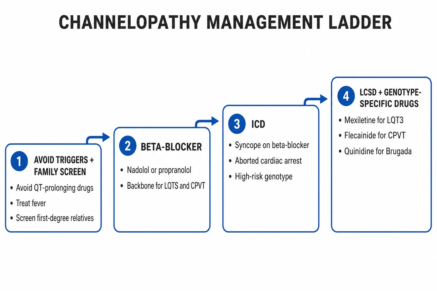
Definitive management combines lifestyle modification, beta-blockade, device therapy, and adjunctive pharmacology, individualised by syndrome and risk.[1][5]
Lifestyle modification applies to every channelopathy patient. Avoid all QT-prolonging drugs (consult CredibleMeds before any new prescription). Avoid competitive and high-intensity sport — especially swimming in LQT1, all competitive sport in CPVT and symptomatic Brugada. Avoid dehydration and electrolyte depletion. Avoid sudden loud auditory stimuli (turn off alarm clocks, telephones, and doorbells at night) in LQT2. Treat fever aggressively (paracetamol, cooling) in Brugada. Avoid alcohol excess and large meals in Brugada. Provide genetic counselling and family planning. [1]
Beta-blockade is first-line for LQTS and CPVT. Nadolol at 1 to 2 mg/kg/day once daily is preferred for LQT1, LQT2, and CPVT because it is long-acting, non-selective, and produces reliable 24-hour coverage. Propranolol at 2 to 4 mg/kg/day in 3 to 4 divided doses is an effective alternative. Metoprolol is less effective (selective beta-1) and should generally be avoided in LQTS. Titrate to the maximum tolerated dose and verify adequacy with an exercise test (target heart rate under 130 bpm at stage 2 of the Bruce protocol for CPVT). Beta-blockade reduces cardiac events by over 70 percent in LQT1 and LQT2 but is significantly less effective in LQT3.[5]
Nadolol
Dose
1 to 2 mg/kg/day orally
ICD therapy is indicated for secondary prevention in every patient who has survived a cardiac arrest (class I), and for primary prevention in high-risk phenotypes: LQTS with recurrent syncope despite beta-blocker, QTc over 500 ms, two or more gene-positive/phenotype-positive features; symptomatic spontaneous type 1 Brugada (syncope or VF); CPVT with breakthrough events on beta-blocker plus flecainide; and SQTS with symptoms or family history of SCD. The ICD does not prevent the arrhythmia — it terminates it — and is reserved for those in whom pharmacological and lifestyle measures are inadequate.[1][3]
Adjunctive and alternative therapies are used when beta-blocker plus ICD are inadequate or inappropriate: [1]
TREAT
- Left cardiac sympathetic denervation (LCSD) — a thoracoscopic or open surgical denervation of the lower half of the left stellate ganglion and the first 4 thoracic ganglia, lowering adrenergic input to the heart. Reserved for LQTS or CPVT with breakthrough events on beta-blocker plus ICD, or for patients in whom ICD is refused or inappropriate (children).
- Sodium-channel blockers — mexiletine 5 mg/kg/day in 3 divided doses (shortens QT in LQT3), ranolazine, or flecainide 100 to 300 mg/day (adjunctive in CPVT, reduces bidirectional VT burden by 70 to 80 percent in refractory cases).
- Quinidine — normalises the Brugada ECG in some patients and reduces VF episodes; adjunctive in Brugada and SQTS. Isoproterenol is used acutely for Brugada electrical storm.
- Verapamil — adjunct in CPVT (suppresses DADs); generally combined with nadolol.
- Potassium supplementation — keep serum K+ over 4.5 mmol/L in LQT2 (IKr is K+-sensitive). [1]
Specific Subtypes & Scenarios
Each channelopathy has its own high-yield clinical pearl set. [1]
LQT1 — exercise and swimming triggered; broad-based T wave; beta-blocker highly effective (events reduced over 70 percent); ICD for syncope despite beta-blocker or QTc over 500 ms. The most common genotype and the one most reliably controlled by beta-blockade. [1]
LQT2 — auditory and emotional triggered, postpartum heightened risk; low-amplitude notched T wave; beta-blocker effective; potassium supplementation to keep K+ over 4.5 mmol/L; advise the patient to disable alarm clocks and telephones at night. The 9-month postpartum period is the highest-risk window in any LQTS genotype.[8]
LQT3 — rest and sleep triggered, bradycardia-dependent; long isoelectric ST; beta-blocker less effective (and may be harmful if it slows the rate excessively); mexiletine shortens QT; ranolazine is an emerging option; ICD often required. The QT paradoxically shortens with exercise, so high-intensity sport is less restricted than in LQT1, but competitive sport is still avoided. [1]
Jervell-Lange-Nielsen syndrome (recessive KCNQ1 or KCNE1) — congenital sensorineural deafness; very long QTc; high risk of early SCD; ICD often required in childhood. Andersen-Tawil (LQT7, KCNJ2) — periodic paralysis, dysmorphic facies, bidirectional VT, mild QT prolongation with prominent U waves. Timothy (LQT8, CACNA1C) — syndactyly, autism, congenital heart disease, very long QT; high mortality in infancy.[1]
Brugada syndrome — only proven therapy is ICD. Quinidine and isoproterenol are adjunctive for arrhythmia storm. Treat fever aggressively (fever unmasks and worsens the ECG and triggers VF). Avoid all sodium-channel blockers (check brugadadrugs.org). No competitive sport if symptomatic. Asymptomatic type 1 patients with a negative electrophysiology study have a low event rate (under 1 percent per year).[7]
CPVT — nadolol is first-line and reduces events dramatically; flecainide is added for breakthrough (reduces bidirectional VT burden); left cardiac sympathetic denervation for breakthrough on combination therapy; ICD for survivors of cardiac arrest or sustained VT despite maximal therapy. All competitive sport is contraindicated. The diagnosis is exercise-induced; the resting ECG is normal, so the exercise test is mandatory in any child with exercise-induced syncope.[6]
SQTS — ICD is the only proven therapy; quinidine may be used to prolong QT and reduce events. Atrial fibrillation is common and may be the presentation. Programming the ICD is technically challenging because the short QT can be misinterpreted as a T wave and lead to inappropriate shocks.[10]
ERS — most early repolarisation patterns are benign and require no treatment. The syndrome is defined by idiopathic VF or family history of SCD plus J-point elevation in the inferior or lateral leads. Survivors of cardiac arrest receive an ICD; quinidine and isoproterenol are adjunctive.[11][12] ERS subtypes and risk stratification — the Antzelevitch 2016 J-wave consensus distinguishes type 1 ERS (J-point elevation localised to the lateral precordial leads, generally benign), type 2 (inferior or inferolateral J-point elevation, intermediate risk), and type 3 (global early repolarisation, highest risk). High-risk ERS features include J-point elevation over 0.2 mV, horizontal ST segment (rather than ascending), dynamic changes, and a history of syncope or family SCD. The high-risk inferolateral pattern with horizontal ST carries approximately a 3-fold increased risk of arrhythmic death over the benign ascending-variant pattern.[12]
Complications & Pitfalls
Disease-related complications include recurrent torsades de pointes and ventricular fibrillation despite therapy, cardiac arrest, and sudden cardiac death. With appropriate therapy (beta-blocker plus ICD where indicated), the annual event rate falls below 1 percent in LQTS, but untreated disease carries a 5-year mortality of around 20 percent in symptomatic patients.[5]
ICD-related complications are common and important: pocket infection, lead fracture or displacement, inappropriate shocks (often from T-wave oversensing in LQTS or sinus tachycardia in CPVT), device malfunction, and the psychological burden of living with a device (anxiety, depression, body-image concerns, driving restrictions). In children, lead failure is the dominant long-term complication and may require extraction. Left cardiac sympathetic denervation can cause Horner syndrome (usually mild and transient) and post-syndetic neuralgia. [1]
The classic diagnostic pitfalls are: (1) misdiagnosing LQTS syncope as epilepsy for years before an ECG is checked; (2) failing to measure QTc manually — automated reports are unreliable, particularly in LQT2 with notched T waves; (3) prescribing a QT-prolonging drug (especially a macrolide, ondansetron, or antipsychotic) to an LQTS patient without checking CredibleMeds; (4) diagnosing Brugada on a type 2 or 3 ECG without provocation testing or family evaluation; (5) treating CPVT with a beta-1 selective agent (metoprolol) instead of nadolol — metoprolol is insufficient; (6) missing the diagnosis of CPVT because the resting ECG is normal and an exercise test was not done.[1][6]
Prognosis & Disposition
Prognosis depends on genotype, QTc duration, history of syncope or cardiac arrest, age at first event, and response to therapy. LQT1 has the best prognosis on beta-blocker. LQT3 carries the highest SCD risk before age 40, and events at slow heart rates are less responsive to beta-blockade. LQT2 confers increased risk in the postpartum period; Brugada has a 7 to 10 percent annual recurrence of VF after cardiac arrest but under 1 percent per year in asymptomatic patients. CPVT has high mortality if untreated but is well controlled on nadolol plus flecainide; SQTS carries an early SCD risk and ICD is the only proven therapy.[8][9]
Disposition is outpatient for asymptomatic patients on stable therapy (annual cardiology review with ECG, exercise test, and ICD interrogation as appropriate), inpatient for a new diagnosis with syncope or after cardiac arrest (for risk stratification, ICD implantation, or LCSD), and ITU/CCU for ongoing TdP or VF, electrical storm, or post-arrest stabilisation. Genetic counselling and cascade screening of first-degree relatives (ECG, exercise test, targeted genetic testing) must be initiated from the index case.[4]
Special Populations
Pregnancy: continue beta-blocker throughout pregnancy — never discontinue. LQT2 has the highest risk in the 9-month postpartum period, so peripartum monitoring and beta-blocker dose optimisation are essential. Vaginal delivery is generally preferred; epidural anaesthesia is acceptable but avoid hypotension and electrolyte shifts. Genetic counselling before pregnancy is recommended.[1]
Paediatric: the Schwartz score is valid in children; therapy is identical with weight-based dosing. LQT1 males carry the highest risk in the pre-pubertal years; LQT2 females in the post-pubertal years. Activity restriction must be balanced against the developmental importance of play. ICD implantation in small children is technically challenging and LCSD may be preferred as a temporising strategy.[9]
Athletes: competitive sport is contraindicated in CPVT (all sport) and symptomatic Brugada, and restricted in genotype-positive LQTS (especially LQT1 swimming). Recreational low-to-moderate intensity activity is usually safe. The 2017 AHA/ACC/HRS eligibility recommendations for athletes with channelopathies generally preclude competitive sport regardless of treatment — the restriction is phenotype- and gene-based, not symptoms-based. [1]
Elderly: the elderly more often have acquired than congenital LQTS — focus on drug review (especially polypharmacy with antibiotics, antipsychotics, and antiarrhythmics) and electrolyte disturbance. Baseline QTc is mildly longer in older women. [1]
Family: cascade screening is mandatory. Offer ECG and exercise testing to all first-degree relatives of a confirmed case; if the proband has an identified mutation, offer targeted genetic testing. Genetic counselling before reproductive decisions is standard.[4]
Evidence, Guidelines & Regional Differences
The 2013 HRS/EHRA/APHRS expert consensus on inherited primary arrhythmia syndromes (Priori et al., PMID 24011539) is the unifying international document and the most frequently cited reference for diagnosis and management. It is supplemented by the 2015 ESC guidelines on ventricular arrhythmias and SCD, the 2017 AHA/ACC/HRS ventricular arrhythmia guideline, and the 2006 ACC/AHA/ESC guidelines (Zipes et al., PMID 16935995) which still carry class recommendations for ICD implantation.[1][3]
The Schwartz diagnostic score (1993, PMID 8339437) remains the standard diagnostic instrument for clinically suspected LQTS. The Moss 2000 cohort (PMID 10673253) established that beta-blockade reduces cardiac events by over 70 percent in LQT1 and LQT2. The Priori 2003 risk-stratification paper (PMID 12736279) quantified genotype-specific risk and informed the modern ICD strategy. The Goldenberg 2008 paediatric study (PMID 18427136) identified QTc over 470 ms and syncope as the dominant risk factors in children. The Antzelevitch 2005 second consensus on Brugada (PMID 15898165) and the 2016 J-wave syndromes consensus (PMID 27423412) frame ERS. The Hayashi 2009 CPVT cohort (PMID 19398665) defined the natural history and risk factors.[2][5][6][7][8][9][12]
Regional deltas are small but worth knowing: the 2017 AHA/ACC/HRS guideline restricts competitive sport more aggressively than the European guidelines; brugadadrugs.org (Antzelevitch group) and CredibleMeds (Arizona CERT) are the practical international references for drug avoidance; in Southeast Asia, Brugada and Lai Tai (sudden unexplained nocturnal death syndrome) are recognised as a single entity with high population prevalence and the SCN5A mutation screening is sometimes offered as a population screen in endemic regions. [1]
Exam Pearls
- LQT1 (most common, 35 to 40 percent): exercise/swimming triggered, broad T wave, beta-blocker highly effective.
- LQT2: auditory triggers (alarm, doorbell, telephone), emotional stress, postpartum; notched T wave; keep K+ over 4.5 mmol/L.
- LQT3: rest/sleep, bradycardia-dependent; beta-blocker less effective; mexiletine and ranolazine shorten QT; ICD often needed.
- CPVT: normal resting ECG, exercise-induced bidirectional VT, RYR2 mutation — nadolol first-line, flecainide add-on.
- Brugada: coved type 1 ST V1-V2 with symptoms; only proven therapy = ICD; avoid sodium-channel blockers; treat fever.
- SQTS: QTc under 340 ms, AF and VF at young age; ICD only proven therapy.
- QTc over 500 ms = high risk for TdP — stop QT-prolonging drugs, correct electrolytes, refer to electrophysiology.
- Acute TdP: IV magnesium 2 g (regardless of serum level) + defibrillation if sustained + isoprenaline or overdrive pacing for pause-dependent (LQT3).
- Always check CredibleMeds before prescribing to any LQTS or borderline-QTc patient.
- Cascade screen first-degree relatives of any case with ECG, exercise test, targeted genetic testing. [1]
Exam application bank (NEET-PG / INICET)
One-line answer
Long QT syndrome and the related cardiac channelopathies (Brugada, CPVT, short QT, early repolarisation) are inherited arrhythmia syndromes caused by mutations in cardiac ion-channel genes that predispose young, structurally normal hearts to syncope, torsades de pointes, ventricular fibrillation and sudden cardiac death. The dominant therapy is beta-blockade (nadolol or propranolol for LQTS and CPVT), avoidance of QT-prolonging drugs, lifestyle modification, and ICD implantation for secondary prevention or high-risk primary prevention. Acute torsades de pointes is treated with IV magnesium 2 g, defibrillation if sustained, and overdrive pacing or isoprenaline for pause-dependent forms. [1]
Worked stems (answer without another resource)
Stem 1 — Classic presentation. Map symptoms to mechanism; name the first investigation and first treatment step with dose/route if drug therapy is standard. [1]
Stem 2 — Unstable / complicated. List red flags that force immediate resuscitation, theatre, ICU, antidote, or reperfusion — and what you do in the first 15 minutes. [1]
Stem 3 — Atypical group. Elderly, pregnancy, child, or immunocompromised: how presentation and thresholds change. [1]
Stem 4 — Differential trap. Name the three closest mimics and one discriminator for each. [1]
Stem 5 — Disposition. Who goes home with safety-netting, who is admitted, who needs HDU/ICU/theatre, and what follow-up is mandatory. [1]
Rapid viva checklist
- Definition + classification
- Pathophysiology chain
- Bedside signs / criteria
- Score with exact components (if any)
- Emergency bundle
- Definitive therapy with doses
- Complications of disease and of treatment
- Special populations
- Guideline/trial name if classic
- Three exam traps
Coverage self-check
If you cannot answer any stem above from this page alone, re-read the matching section — the page is intended to be self-sufficient for final-prof and NEET-PG/INICET questions on Long QT and Channelopathies.
References
- [1]Priori SG, Wilde AA, Horie M, et al. HRS/EHRA/APHRS expert consensus statement on the diagnosis and management of patients with inherited primary arrhythmia syndromes: document endorsed by HRS, EHRA, and APHRS in May 2013 and by ACCF, AHA, PACES, and AEPC in June 2013 Heart Rhythm, 2013.PMID 24011539
- [2]Schwartz PJ, Moss AJ, Vincent GM, Crampton RS Diagnostic criteria for the long QT syndrome. An update Circulation, 1993.PMID 8339437
- [3]Zipes DP, Camm AJ, Borggrefe M, et al. ACC/AHA/ESC 2006 Guidelines for Management of Patients With Ventricular Arrhythmias and the Prevention of Sudden Cardiac Death: a report of the American College of Cardiology/American Heart Association Task Force and the European Society of Cardiology Committee for Practice Guidelines (writing committee to develop Guidelines for Management of Patients With Ventricular Arrhythmias and the Prevention of Sudden Cardiac Death): developed in collaboration with the European Heart Rhythm Association and the Heart Rhythm Society Circulation, 2006.PMID 16935995
- [4]Ackerman MJ, Priori SG, Willems S, et al. HRS/EHRA expert consensus statement on the state of genetic testing for the channelopathies and cardiomyopathies: this document was developed as a partnership between the Heart Rhythm Society (HRS) and the European Heart Rhythm Association (EHRA) Europace, 2011.PMID 21810866
- [5]Moss AJ, Zareba W, Hall WJ, et al. Effectiveness and limitations of beta-blocker therapy in congenital long-QT syndrome Circulation, 2000.PMID 10673253
- [6]Hayashi M, Denjoy I, Extramiana F, et al. Incidence and risk factors of arrhythmic events in catecholaminergic polymorphic ventricular tachycardia Circulation, 2009.PMID 19398665
- [7]Antzelevitch C, Brugada P, Borggrefe M, et al. Brugada syndrome: report of the second consensus conference Heart Rhythm, 2005.PMID 15898165
- [8]Priori SG, Schwartz PJ, Napolitano C, et al. Risk stratification in the long-QT syndrome N Engl J Med, 2003.PMID 12736279
- [9]Goldenberg I, Moss AJ, Zareba W, et al. Risk factors for aborted cardiac arrest and sudden cardiac death in children with the congenital long-QT syndrome Circulation, 2008.PMID 18427136
- [10]Brugada R, Hong K, Dumaine R, et al. Sudden death associated with short-QT syndrome linked to mutations in HERG Circulation, 2004.PMID 14676148
- [11]Haissaguerre M, Derval N, Sacher F, et al. Sudden cardiac arrest associated with early repolarization N Engl J Med, 2008.PMID 18463377
- [12]Antzelevitch C, Yan GX, Ackerman MJ, et al. J-Wave syndromes expert consensus conference report: Emerging concepts and gaps in knowledge Heart Rhythm, 2016.PMID 27423412