Cardiology · Cardiology
Mitral Regurgitation
Also known as Mitral incompetence · Mitral insufficiency · MR · Chronic primary MR · Functional (secondary) MR · Acute mitral regurgitation
Mitral regurgitation (MR) is systolic backflow of blood from the left ventricle (LV) into the left atrium (LA) through an incompetent mitral valve. Chronic MR runs an asymptomatic compensated phase (LA and LV dilate, eccentric hypertrophy) before decompensation with dyspnoea, fatigue and atrial fibrillation. Acute MR (papillary-muscle or chordal rupture post-MI) presents as fulminant pulmonary oedema and cardiogenic shock. Cardinal sign: pansystolic murmur at the apex radiating to the axilla, soft S1, and a third heart sound. Diagnosis and grading are by echocardiography (EROA, regurgitant volume/fraction, vena contracta). Definitive treatment is mitral valve repair (preferred) or replacement; surgery timing rests on symptoms or LV dysfunction (EF under 60% or LV end-systolic diameter at least 40 mm). Transcatheter edge-to-edge repair (MitraClip) is an option for inoperable functional MR (COAPT).
On this page & tools
Your progress
Saved locally on this device.
Exam tags
Red flags
Overview & Definition
Mitral regurgitation (MR) is the backward flow of blood from the left ventricle into the left atrium during systole through an incompetent mitral valve. It is the commonest organic valvular lesion in adults in the developed world after aortic sclerosis, and its prevalence rises steeply with age as degenerative and functional disease accumulate.[1]
The clinical and conceptual skill in MR is to recognise that the same murmur and the same echo grading describe two very different diseases:[2]
- Chronic MR — a slow, compensated remodelling illness in which the LA and LV dilate for years before symptoms. The danger is operating too late (irreversible LV dysfunction) — hence surgery is recommended for asymptomatic severe primary MR once LV size or function crosses thresholds.
- Acute MR — a haemodynamic emergency (papillary muscle/chordal rupture) with no time for compensation: a small, stiff LA is suddenly overloaded, LA pressure spikes, and the patient presents in cardiogenic shock and pulmonary oedema. [1]
The mitral valve is not just two leaflets — it is an apparatus of leaflets (anterior and posterior, each with three scallops — anterolateral A1/A2/A3, posterolateral P1/P2/P3), the annulus (a dynamic saddle-shaped fibrous ring), chordae tendineae (primary/secondary/tertiary), papillary muscles (anterolateral and posteromedial) and the underlying LV myocardium. Failure of any one component can produce MR, which is why the differential of causes is so broad and why a structured mechanistic classification (Carpentier) is essential.[1]
[1]Classification
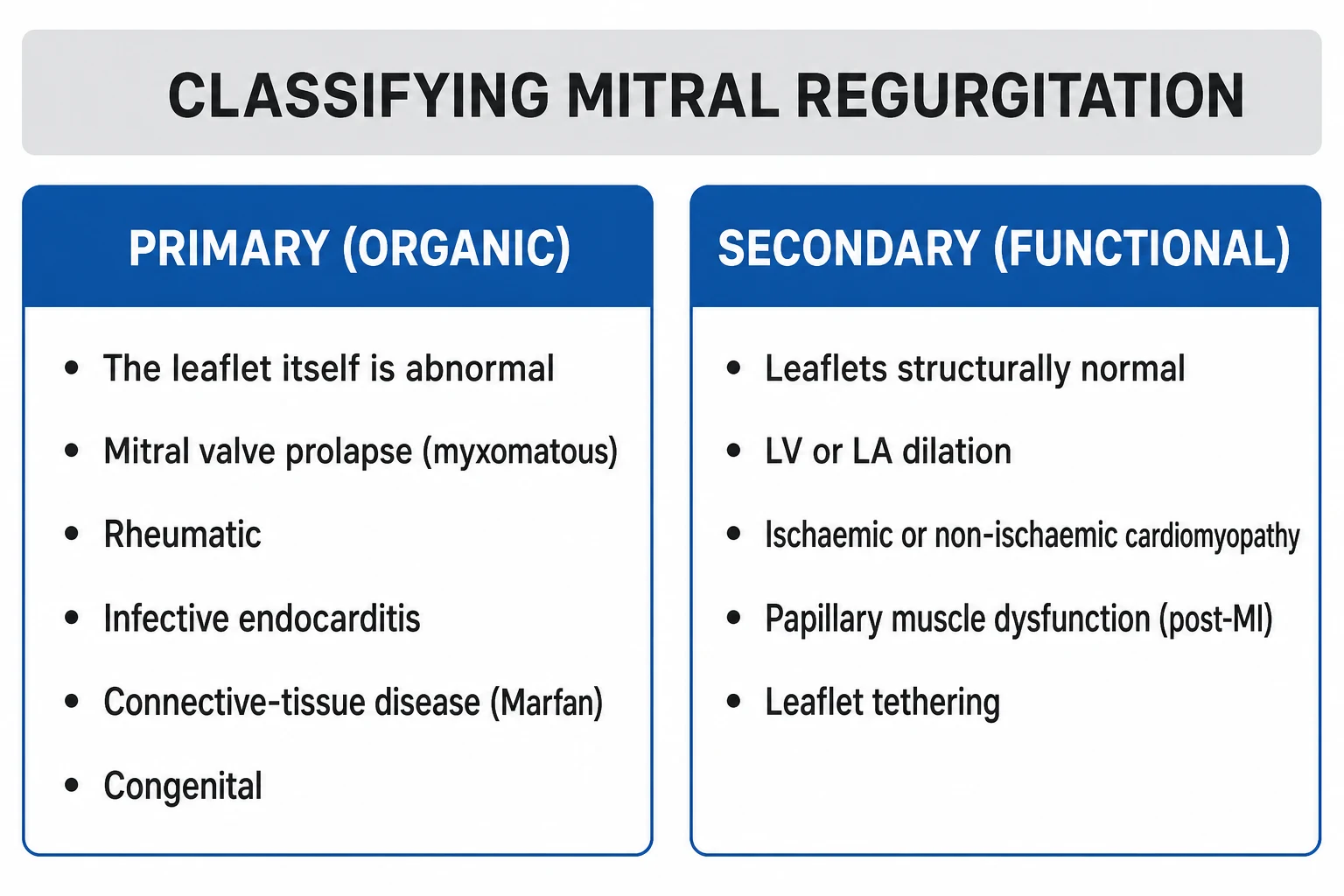
MR is classified three overlapping ways — by mechanism (Carpentier), by aetiology (primary vs secondary), and by time-course (acute vs chronic). All three must be answerable in a viva.[1]
PRIMARY (organic/degenerative)
- Disease of the valve LEAFLETS or apparatus itself
- Causes: MVP (Barlow, fibroelastic deficiency) 60-70% in West, rheumatic #1 worldwide, endocarditis, congenital, collagen-vascular (Marfan, Ehlers-Danlos), drug-induced (fenfluramine, cabergoline, pergolide), annular calcification
- Valve is structurally ABNORMAL on echo
- Surgery = mitral valve REPAIR (preferred) when feasible — 95% repair rate for posterior leaflet
SECONDARY (functional)
- Valve leaflets are structurally NORMAL
- MR arises from LV/LA dilation and annular stretching, OR papillary muscle displacement
- Causes: ischaemic (post-MI wall motion, Type IIIb), non-ischaemic (DCM), HFrEF, hypertensive heart disease, chronic AF (LA enlargement)
- Treat the underlying cardiomyopathy FIRST (GDMT); TEER if persistent severe MR despite therapy (COAPT)
Carpentier functional classification (the mechanistic map examiners want verbatim):[1][2]
- Type I — normal leaflet motion. The leaflets move normally but leak because of annular dilation (functional, LV dilation), leaflet perforation (endocarditis) or a cleft (congenital).
- Type II — excess leaflet motion (prolapse/flail). The leaflet edge overrides the annular plane in systole. Classic of degenerative/MVP and chordal rupture. Usually affects the posterior leaflet (P2 scallop).
- Type IIIa — restricted leaflet motion in BOTH diastole and systole. Rheumatic (leaflet thickening, commissural fusion, chordal shortening), radiation, carcinoid, SLE (Libman-Sacks).
- Type IIIb — restricted leaflet motion in SYSTOLE only. The classic ischaemic/functional pattern — the leaflets are tethered by a dilated, spherical LV pulling the papillary muscles apically and laterally. [1]
Time-course: [1]
- Chronic — compensated (asymptomatic) for years, then decompensated. Causes: degenerative/MVP, rheumatic, functional, annular calcification.
- Acute — hours to days; no compensatory remodelling. Causes: papillary muscle rupture post-MI, chordal rupture (MVP, trauma, spontaneous), leaflet perforation (endocarditis), prosthetic valve dysfunction, blunt chest trauma, acute ischaemic papillary muscle dysfunction, acute rheumatic. [1]
Causes — Acute vs Chronic
The aetiology table is the single most tested item in an MR viva, because the tempo dictates the entire management pathway. Separate acute from chronic causes in your answer. [1]
Acute causes (hours to days — surgical emergency)
ACUTE MR — the six causes to name
Papillary muscle rupture post-MI is the archetype. It occurs in 1–2% of acute MIs, typically 2–7 days after infarction, and almost always involves the posteromedial papillary muscle (single PDA blood supply vs the dual LAD/LCx supply of the anterolateral muscle). Partial rupture produces severe but survivable MR; complete rupture is usually fatal within hours without surgery. Operative mortality is high (25–40%) but surgery is the only chance of survival.[1]
Chordae tendineae rupture occurs spontaneously in myxomatous MVP (especially Barlow disease with multiple flail segments), with trauma (deceleration, chest compressions), with infective endocarditis, and rarely as the presenting feature of a connective-tissue disorder. The result is an acute flail leaflet with sudden severe MR. [1]
Infective endocarditis causes MR through vegetation preventing coaptation, leaflet perforation, chordal rupture, or abscess with annular destruction. Acute severe MR in IE is an indication for emergency surgery. [1]
Prosthetic valve dysfunction — thrombosis of a mechanical valve, dehiscence of a suture ring, structural deterioration of a bioprosthesis, or a stuck leaflet in a bileaflet mechanical valve — produces acute or subacute MR and is a surgical/transcatheter emergency. [1]
Blunt chest trauma (road traffic collisions, falls, CPR) can avulse chordae or a papillary muscle head, producing acute MR days to weeks after the injury; a careful history is decisive. [1]
Chronic causes (years — watchful timing)
CHRONIC MR — aetiology by frequency and geography
Degenerative (myxomatous) MR is the commonest cause of chronic primary MR in the developed world (60–70%), and divides into two pathologies examiners contrast: Barlow disease (younger patients, diffuse myxomatous change, thick redundant billowing leaflets, multiple prolapsing segments, complex repair) and fibroelastic deficiency (older patients, thin translucent leaflets with a single flail segment, usually P2, straightforward durable repair). The distinction predicts surgical complexity and repairability.[1]
Rheumatic MR remains the commonest cause worldwide. Rheumatic carditis produces leaflet thickening, commissural fusion, chordal shortening and calcification — usually producing a mixed MS+MR picture. The mitral annulus and subvalvar apparatus are heavily involved, so repair is less feasible than in degenerative disease and mitral valve replacement is often required. Secondary prevention with benzathine penicillin G is part of management. [1]
Ischaemic (functional) MR arises from LV remodelling after myocardial infarction: regional wall motion abnormality (especially infero-basal) displaces the posteromedial papillary muscle, tethering the leaflets (Carpentier IIIb). It may be acute (papillary muscle dysfunction in stunned/infarcted myocardium) or chronic (progressive remodelling). [1]
Functional (non-ischaemic) MR occurs in dilated cardiomyopathy and HFrEF — LV spherical dilation stretches the annulus and tethers the leaflets, and chronic AF with LA enlargement further dilates the annulus. The valve is structurally normal; the MR is a geometric consequence of LV/LA disease. [1]
Mitral annular calcification (MAC) in the elderly (especially women, diabetics, renal failure, Paget disease) is a degenerative calcification of the mitral annulus (U-shaped ring) that restricts leaflet closure and may invade the conduction system producing heart block. Severe MAC is a surgical challenge (no annulus to sew to). [1]
Connective-tissue and systemic causes: Marfan syndrome and Ehlers-Danlos (leaflet prolapse from defective fibrillin/collagen); SLE (Libman-Sacks endocarditis — verrucous vegetations on the atrial surface of the mitral leaflets, often with antiphospholipid antibodies); hypertrophic cardiomyopathy (systolic anterior motion of the mitral leaflet + MR); prior mediastinal radiotherapy (decades later — fibrotic, calcified, restricted leaflets, Type IIIa); carcinoid (right-sided valves, but tricuspid plus pulmonary, rarely mitral with a patent foramen ovale). [1]
Drug-induced valvulopathy: fenfluramine-phentermine (fen-phen), dexfenfluramine, ergotamine/pergolide/cabergoline (serotonergic agonists) produce serotony-like lesions resembling carcinoid — thickened, restricted leaflets with a plaque-like fibrous coating, often regurgitant. [1]
Mitral valve prolapse (MVP) itself is the most common primary precursor in the West: prevalence roughly 2–3% of the population, female predominance in the young (Barlow myxomatous form), with a characteristic mid-systolic click ± late systolic murmur. Most MVP is benign; the minority with severe MR, flail leaflet, or marked myxomatous change are the ones who develop complications.[1]
Risk factors for progression / severe disease: age over 60, male sex (for degenerative flail), hypertension, ischaemic heart disease, prior rheumatic fever, bicuspid aortic valve, marfanoid habitus, prior mediastinal radiotherapy, drug exposure (fenfluramine, ergots, cabergoline), chronic kidney disease (annular calcification). [1]
Epidemiology & Risk Factors
MR is the commonest valve lesion of adults in developed countries — mild MR is detectable on echo in roughly 1–2% of the population, and the prevalence of moderate-to-severe MR rises sharply after age 65, driven by degenerative disease in the elderly and functional MR in the expanding heart-failure population.[1][2] The age-adjusted prevalence of at-least-moderate MR is approximately 2% of adults and approaches 10% in those over 75.
Cause by geography and setting (high-yield): [1]
| Setting / population | Likely cause |
|---|---|
| Developed world, chronic | Degenerative / mitral valve prolapse (Barlow disease, fibroelastic deficiency) |
| Developing world (India, sub-Saharan Africa, Indigenous Australia) | Rheumatic heart disease (often mixed MS+MR) |
| Post-MI patient | Ischaemic — papillary muscle dysfunction or rupture (functional Type IIIb, or acute rupture) |
| HFrEF / dilated LV | Functional (secondary) MR — annular dilation + tethering |
| IVDU, prosthetic valve, bacteraemia | Infective endocarditis (perforation, vegetation preventing coaptation) |
| Young, thin, hypermobile woman | MVP / connective tissue disorder (Marfan, Ehlers-Danlos) |
| Elderly woman with renal failure | Mitral annular calcification |
| Drug exposure | Fenfluramine-phentermine (fen-phen), ergotamines, cabergoline, pergolide (serotonergic valvulopathy) |
| Connective-tissue / systemic disease | SLE (Libman-Sacks), Marfan, Ehlers-Danlos, rheumatoid arthritis, carcinoid, radiation |
Mortality where it matters: untreated severe symptomatic chronic MR carries a 5-year mortality of roughly 40%, and severe secondary MR in HFrEF independently doubles mortality. Acute MR from papillary muscle rupture is near-100% fatal without surgery.[1]
Pathophysiology
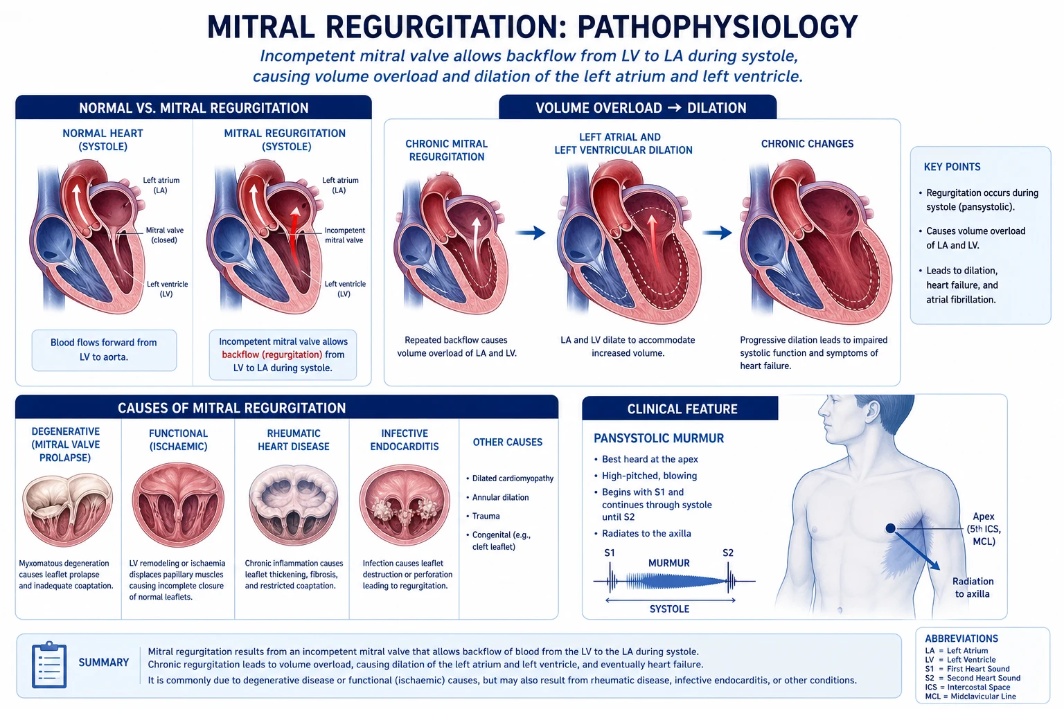
The fundamental lesion is a leak in systole: part of the LV stroke volume takes the low-resistance path back into the LA instead of the high-resistance path into the aorta. The consequences differ completely depending on time-course and on LA compliance.[1][3]
Chronic MR — the volume-overload model: [1]
- Regurgitant volume enters the LA each systole. The total LV stroke volume equals forward SV + regurgitant volume; the LV therefore ejects a larger total volume at a lower afterload (because blood escapes two ways — the regurgitant orifice acts as a second, low-resistance outflow). By the Laplace relationship (wall stress = P × r / 2h), the lower systolic pressure and eccentric hypertrophy keep wall stress near-normal despite dilation.
- The LA and LV dilate to accommodate the extra volume. LV mass increases by eccentric hypertrophy (sarcomeres added in series). EF is preserved or even supranormal because afterload is low — this is why EF overestimates true myocardial contractility in MR and why the threshold for operating (EF under 60%) is higher than for other lesions.
- Compensation can last years: LA compliance rises, LV filling pressure stays near-normal, and the patient is asymptomatic. Forward output is maintained by the increased total stroke volume.
- Decompensation: as the LV dilates further, contractile fibres overstretch, systolic function falls (often silently), filling pressure rises, LA pressure transmits to the pulmonary veins, and the patient develops exertional dyspnoea, orthopnoea, fatigue. LA enlargement predisposes to atrial fibrillation. Late disease brings pulmonary hypertension (from chronic LA hypertension), tricuspid regurgitation (secondary), and right heart failure. [1]
Acute MR — the pressure-overload emergency: [1]
- A sudden large regurgitant orifice (papillary muscle or chordal rupture) dumps volume into a small, non-compliant LA that has had no time to dilate.
- LA pressure and pulmonary venous pressure spike in systole (giant v waves on pulmonary capillary wedge tracing — often exceeding 50 mmHg).
- The result is acute pulmonary oedema and a fall in forward output, producing cardiogenic shock. The LV cannot compensate because the afterload fall cannot rescue a suddenly enormous regurgitant fraction; EF may even look normal while forward output collapses. [1]
Why ischaemic MR favours the posteromedial papillary muscle: the posteromedial papillary muscle has a single blood supply (from the posterior descending artery), whereas the anterolateral papillary muscle has dual supply (LAD + circumflex). Ischaemia or infarction of the posteromedial muscle (inferior MI) therefore causes dysfunction/rupture far more often.[1]
Functional (secondary) MR — geometry, not the valve: in HFrEF or ischaemic cardiomyopathy the LV dilates and becomes spherical; the papillary muscles are displaced apically and laterally, tethering the leaflets so they cannot coapt (Carpentier IIIb). The regurgitant orifice is dynamic — it worsens with ischaemia and with increased LV volume, and shrinks with diuretic/vasodilator therapy and resynchronisation (CRT). This dynamic behaviour explains why treatment of the underlying cardiomyopathy is the first step in secondary MR, and why a fixed surgical annuloplasty without addressing the failing LV often fails.[2]
Clinical Presentation
Chronic compensated MR is often asymptomatic for years, detected on a routine murmur examination or incidental echo. As decompensation begins:[1]
- Exertional dyspnoea → orthopnoea, paroxysmal nocturnal dyspnoea (pulmonary venous congestion).
- Fatigue (low forward cardiac output).
- Palpitations (atrial ectopics, then atrial fibrillation from LA enlargement).
- Less commonly: right-heart symptoms (ankle swelling, abdominal distension from hepatomegaly, ascites) once pulmonary hypertension and right-heart failure supervene.
- Haemoptysis, hoarseness (Ortner syndrome from LA compressing the recurrent laryngeal nerve), and embolic events are rarer but described in severe chronic MR. [1]
Acute MR (papillary muscle or chordal rupture) is dramatic:[1]
- Sudden severe dyspnoea, orthopnoea, frothy/pink sputum (acute pulmonary oedema).
- Hypotension, cool peripheries, oliguria, altered sensorium (cardiogenic shock).
- Often in the first 2–7 days post-MI (papillary muscle rupture), or after trauma, or spontaneously in severe MVP (chordal rupture).
- The murmur may be surprisingly soft or even absent in acute severe MR because LA and LV pressures equalise early in systole, abolishing the gradient that generates the murmur — a classic exam trap. Always echo a shocked post-MI patient with new pulmonary oedema. [1]
Mitral valve prolapse presentation: young, thin woman; atypical chest pain, palpitations, anxiety, autonomic symptoms (postural orthostatic tachycardia, panic-like episodes); classic auscultatory finding is a mid-systolic click that moves closer to S1 with Valsalva/standing (less preload → earlier prolapse) and later with squatting/handgrip (more preload). A late systolic murmur follows the click if MR is present.[1]
Atypical presentations (deliberately tested): [1]
- Elderly — fatigue, falls, functional decline, or new AF; the murmur may be missed or attributed to aortic stenosis; annular calcification is common.
- Post-MI — sudden pulmonary oedema 2–7 days after infarction equals papillary muscle rupture until proven otherwise.
- Endocarditis — MR with constitutional features (fever, night sweats, embolic phenomena, new murmur, splinter haemorrhages).
- Pregnancy — mild-moderate chronic MR is usually well tolerated (afterload falls in pregnancy); severe MR may decompensate from the gestational volume load.
- Diabetic/autonomic patient — symptoms may be blunted; the first sign may be decompensated HF. [1]
Differential Diagnosis
A systolic murmur is not always MR. The high-yield differentials with their distinguishing features:[1]
- Aortic stenosis (AS) — ejection systolic murmur at the right upper sternal border radiating to the carotids; crescendo-decrescendo; soft/absent A2; ejection click; slow-rising pulse; heaving (sustained) apex. Handgrip decreases AS (more afterload delays opening) and increases MR; Valsalva decreases AS and MR but increases HOCM.
- Hypertrophic obstructive cardiomyopathy (HOCM) — ejection systolic at the lower left sternal edge; increases with Valsalva and standing (less preload → more obstruction) and decreases with squatting/handgrip; bifid "triple ripple" apex; often a diastolic murmur from concomitant MR.
- Ventricular septal defect (VSD) — pansystolic murmur at the lower left sternal edge with a thrill; radiation is not to the axilla; increases with handgrip; sudden onset post-MI or congenital. Bundle-branch block on ECG.
- Tricuspid regurgitation (TR) — pansystolic at the lower left sternal edge; Carvallo's sign (murmur increases with inspiration); prominent cv waves in the JVP and pulsatile liver; inhalation augments right-sided murmurs, the opposite of left-sided.
- Pulmonary regurgitation (Graham Steell) — early diastolic decrescendo at the lower left sternal edge; pulmonary hypertension features; not a systolic murmur.
- Mitral stenosis — mid-diastolic rumble with presystolic accentuation, opening snap, loud S1; differentiation is by timing, not site.
- Aortic regurgitation — early diastolic decrescendo at the left sternal edge with Austin Flint mid-diastolic rumble (confused with MS); collapsing (water-hammer) pulse. [1]
Murmurs and manoeuvres — the viva crib
HAND-VALSALVA
raises afterload — INCREASES MR, AR, VSD; DECREASES AS, HOCM
drops afterload — DECREASES MR/AR/VSD; INCREASES AS; INCREASES HOCM
if the murmur softens with handgrip, it is NOT MR (think AS or HOCM)
drops preload — DECREASES MR/AS; INCREASES HOCM (only murmur that rises)
raises preload+afterload — INCREASES MR/AS; DECREASES HOCM
increases RIGHT-sided murmurs — Carvallo sign (TR)
Clinical & Bedside Assessment
General: look for marfanoid habitus (tall, arm span greater than height, arachnodactyly, high-arched palate, pectus — MVP/Marfan), stigmata of rheumatic fever (mitral facies, history of chorea/Sydenham), endocarditis (splinter haemorrhages, Janeway lesions, Osler nodes, Roth spots, conjunctival petechiae), right-heart failure (elevated JVP, hepatomegaly, ankle oedema, ascites), and signs of ischaemia (post-MI scars on ECG, prior revascularisation).[1]
Pulse: AF is common (LA enlargement); pulse may be small-volume in low-output states or bounding in chronic compensated high-volume MR. Pulsus alternans indicates advanced LV failure. [1]
Apex: displaced, hyperdynamic (thrusting), laterally displaced apex beat from LV dilation. An apical systolic thrill suggests severe MR. [1]
Auscultation (reproduce verbatim): [1]
- S1 — soft or absent (incomplete/delayed valve closure; the classic finding). In mild MR S1 may be normal.
- S2 — usually normal splitting; wide splitting if MR shortens LV ejection (early A2). Loud P2 if pulmonary hypertension has supervened.
- S3 — a third heart sound is common in significant chronic MR (rapid early filling of a volume-loaded LV) and does NOT imply heart failure in this context as it would in AS.
- Murmur — pansystolic (holosystolic), high-pitched, blowing, loudest at the apex, radiating to the axilla (posterolateral jet) or to the left sternal edge/base (anterior/anterior-leaflet jet). Intensity does not reliably correlate with severity — a soft murmur in acute MR is the classic trap.
- MVP — mid-systolic (non-ejection) click ± late systolic murmur. Click moves toward S1 with Valsalva/standing, away from S1 (later) with squatting/handgrip. [1]
Bedside manoeuvres (memorise the table): [1]
| Manoeuvre | Effect on preload/afterload | MR | AS | HOCM |
|---|---|---|---|---|
| Handgrip / phenylephrine (afterload up) | Increases MR | ↑ | ↓ | ↓ |
| Valsalva (strain) / standing (preload + afterload down) | Decreases MR | ↓ | ↓ | ↑↑ |
| Squatting / leg raise (preload + afterload up) | Increases MR | ↑ | ↑ | ↓ |
| Amyl nitrite (afterload down, venodilation) | Decreases MR | ↓ | ↑ | ↑ |
| Inspiration | Right-sided murmurs increase (Carvallo's sign — TR) | — | — | — |
Investigations
The goal of investigation is to confirm the diagnosis, define the mechanism (Carpentier type), grade severity, assess the LV (size and function) and the pulmonary circulation, and exclude concomitant CAD before surgery.[4]
Electrocardiogram (ECG): non-specific but supportive. LA enlargement (bifid P-wave in lead II — "mitral P", P-wave duration over 120 ms, terminal P-negative force in V1 greater than 40 ms·ms); atrial fibrillation; LV hypertrophy (voltage criteria) and strain; Q waves (prior infarction) suggesting ischaemic MR; right-axis deviation or RBBB if pulmonary hypertension has developed. [1]
Chest X-ray: cardiomegaly with LA enlargement (double shadow along the right heart border, splaying of the carina, left main bronchus elevation, straightening of the left heart border); in chronic MR, signs of pulmonary venous congestion (upper-lobe blood diversion, Kerley B lines, interstitial oedema); in acute MR, florid pulmonary oedema with a near-normal heart size (no time to dilate). [1]
Transthoracic echocardiography (TTE) — the cornerstone. TTE defines mechanism, severity, LV and LA size, pulmonary artery pressure, and excludes other valve disease.[4]
- Mechanism — leaflet morphology (myxomatous/prolapse, rheumatic thickening, vegetation, flail, restricted tethering), annular size, regional wall motion.
- Severity — integrated using colour Doppler jet area, vena contracta width, proximal isovelocity surface area (PISA)-derived effective regurgitant orifice area (EROA) and regurgitant volume/fraction, and pulmonary venous flow pattern (systolic blunting/reversal in severe MR). [1]
Severity grading of primary MR (ASE/EACVI, reproduced verbatim):[4]
| Parameter | Mild | Moderate | Severe (primary) |
|---|---|---|---|
| EROA (cm²) | under 0.20 | 0.20–0.39 | at least 0.40 |
| Regurgitant volume (mL) | under 30 | 30–59 | at least 60 |
| Regurgitant fraction (%) | under 30 | 30–49 | at least 50 |
| Vena contracta width (cm) | under 0.3 | 0.3–0.69 | at least 0.7 |
| Pulmonary venous flow | normal | blunted | systolic reversal |
For secondary MR the severe thresholds are lower (EROA at least 0.30 cm², regurgitant volume at least 45 mL) because the regurgitant orifice is dynamic and the LV is already failing.[2]
MR severity — the four numbers that define SEVERE primary MR
Stage 3
Transoesophageal echocardiography (TOE/TEE): reserved for inconclusive TTE, detailed preoperative anatomy (which scallop is flail — usually P2; feasibility of repair), intraoperative guidance, and to assess for LAA thrombus before cardioversion. 3D TOE is now standard in surgical planning and dramatically improves the surgeon's spatial map of the prolapsing segment.[1]
Other imaging: [1]
- Cardiac MRI — gold-standard for LV volumes, EF and mass, useful when echo windows are poor and for myocardial viability (late gadolinium enhancement to distinguish ischaemic from non-ischaemic functional MR, and to quantify MR volume directly by phase-contrast).
- Coronary angiography — performed before surgery in men over 40, postmenopausal women, and anyone with risk factors or ischaemic ECG, to exclude concomitant CAD needing CABG at the same operation.
- Exercise echocardiography — in symptomatic patients whose resting echo severity looks disproportionate, or to document a rise in pulmonary artery systolic pressure (PASP over 60 mmHg on exercise supports intervention).[9]
BNP/NT-proBNP — a rising level in asymptomatic severe MR is an adverse prognostic marker and a Class IIa trigger for closer surveillance or early surgery consideration.[1]
Management — Resuscitation
Acute severe MR is a cardiovascular emergency. The priorities are oxygenation, afterload reduction, inotropic support and mechanical support while arranging definitive (surgical) repair.[1][2]
- Airway/breathing — high-flow oxygen; non-invasive ventilation (CPAP/BiPAP) for pulmonary oedema (reduces work of breathing, improves oxygenation, drops preload/afterload); intubate if failing.
- Vasodilator therapy (the single most useful pharmacological lever) — IV sodium nitroprusside 0.3–3 mcg/kg/min (titrated), or IV glyceryl trinitrate 10–200 mcg/min, to reduce afterload and preferentially drive blood forward into the aorta. Avoid if hypotensive.
- Inotropes — dobutamine 2.5–20 mcg/kg/min or milrinone 0.125–0.75 mcg/kg/min for low-output/cardiogenic shock; milrinone also reduces afterload (inodilator).
- Mechanical circulatory support — intra-aortic balloon pump (IABP) reduces afterload and augments coronary perfusion, dramatically improving forward output in acute MR; consider VA-ECMO in refractory shock as a bridge to surgery.
- Diuretics — IV furosemide 20–80 mg bolus (or infusion) for pulmonary congestion.
- Avoid pure vasoconstrictors where possible (they worsen the regurgitant fraction by raising afterload); if vasopressors are unavoidable, noradrenaline is preferred to pure alpha-agonists.
- Emergency surgery — once stabilised, the definitive treatment is urgent mitral valve repair or replacement. In ischaemic acute MR (papillary muscle rupture) the operative mortality is high (25–40%) but surgery is the only chance of survival.[1]
Acute severe MR — the resuscitation bundle
Management — Definitive & Stepwise
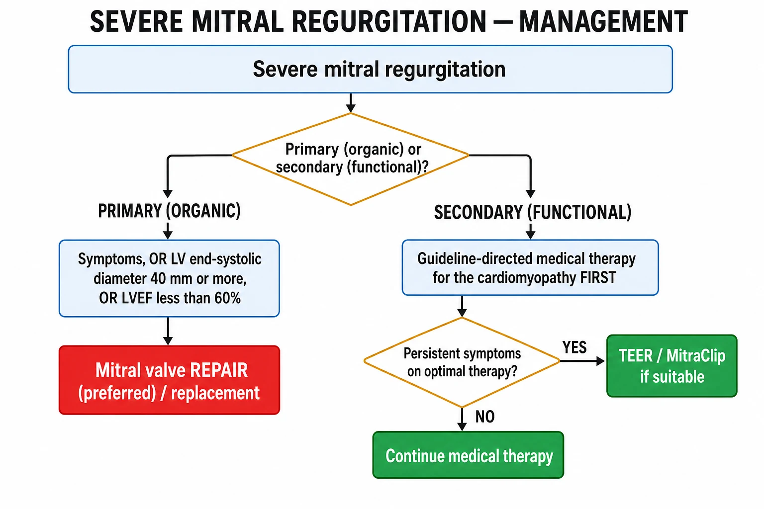
Management is fundamentally different for primary vs secondary MR and is driven by symptoms, LV size and function, and repairability.[1][2]
Primary (organic) MR — surgical triggers
Severe primary MR — when to operate (AHA/ACC 2020, ESC 2021)
Symptomatic (NYHA II-IV) + LVEF over 30%
Asymptomatic + LVEF under-or-equal 60% OR LVESD at least 40 mm
Asymptomatic + preserved LV (EF over 60%, LVESD under 40) + repairable, low surgical risk
Asymptomatic + preserved LV + new-onset AF OR PASP over 50 mmHg
Asymptomatic + preserved LV + flail leaflet + high repair success (over 95%)
LVEF under 30%
Symptomatic severe primary MR with LVEF over 30% — surgery is Class I (repair preferred).[1]
Repair vs replacement — the cornerstone decision. Mitral valve repair is strongly preferred (Class I) for posterior leaflet (especially P2) prolapse/flail, where durable repair rates at expert centres exceed 95%. Repair preserves the native subvalvar apparatus (better LV function), avoids lifelong anticoagulation (mechanical valves) and reoperation (bioprosthetic valves), and carries lower operative and long-term mortality than replacement. Replacement is reserved for extensive bileaflet disease, severe calcification, rheumatic destruction, endocarditis with destruction, or when repair is not feasible at the operating centre. When replacement is necessary, chordal-sparing techniques preserve LV function and reduce postoperative LV dysfunction.[1][2]
MITRAL VALVE REPAIR (preferred)
- Posterior leaflet (P2) prolapse/flail: 95% repair rate at expert centres
- Techniques: triangular/quadrangular leaflet resection, chordal replacement (neochordae, ePTFE), annuloplasty ring, edge-to-edge (Alfieri) stitch
- Preserves subvalvar apparatus + native leaflets → better LV function
- No lifelong anticoagulation; lower operative mortality (under 1%)
- 10-year freedom from reoperation over 90-95% in degenerative disease
MITRAL VALVE REPLACEMENT
- When repair infeasible: extensive bileaflet disease, severe calcification, rheumatic destruction, destructive endocarditis
- Mechanical: lifelong warfarin INR 3.0 (range 2.5-3.5); durable 20-30 yr; preferred under age 65 or already anticoagulated
- Bioprosthetic: warfarin 3 months then aspirin; structural deterioration 10-15 yr; preferred over 65 or when anticoagulation undesirable
- Always CHORDAL-SPARING when feasible to preserve LV function
- Operative mortality 2-6%
Surgical techniques (examiners reward naming these): [1]
- Annuloplasty ring — a prosthetic ring (complete or partial band) sewn around the mitral annulus to reduce its size and restore coaptation. Used alone in functional MR (restrictive/undersized annuloplasty) and added to nearly every degenerative repair to stabilise the annulus. Undersized annuloplasty is the classic operation for functional/ischaemic MR (often combined with CABG).
- Triangular / quadrangular resection — the standard operation for posterior (P2) prolapse: resect the prolapsing segment (triangular for small, quadrangular for large), reconstruct the leaflet, and reinforce with an annuloplasty ring. The most durable repair in all of valve surgery.
- Chordal replacement (neochordae) — synthetic polytetrafluoroethylene (ePTFE, Gore-Tex) chords are anchored to the papillary muscle and to the prolapsing leaflet edge, used especially for anterior leaflet and bileaflet prolapse.
- Edge-to-edge (Alfieri) stitch — approximates the free edges of the anterior and posterior leaflets at the site of the regurgitant jet, creating a double-orifice valve. The surgical basis of transcatheter MitraClip.
- Chordal-sparing MVR — preserving the native leaflets and subvalvar apparatus during replacement prevents catastrophic postoperative LV dysfunction. [1]
Anticoagulation after mechanical MVR: lifelong warfarin with INR target 3.0 (range 2.5–3.5) for a mechanical mitral valve (higher than the aortic target, because the mitral position is more thrombogenic — lower flow velocity); add aspirin 75–100 mg if additional risk factors. Bioprosthetic valves: warfarin INR 2.5 for 3 months then aspirin alone (unless another indication). Mechanical valves last 20–30 years; bioprosthetic valves deteriorate structurally over 10–15 years (faster in younger patients and renal failure).[1]
Secondary (functional) MR — treat the cardiomyopathy first
Treat the underlying cardiomyopathy first — guideline-directed medical therapy (GDMT) for HFrEF: beta-blocker + ACEi/ARNI + MRA + SGLT2 inhibitor at target doses, diuretics for congestion, digoxin/anticoagulation for AF, and cardiac resynchronisation therapy (CRT) if QRS over 150 ms with LBBB (CRT reduces functional MR by improving LV synchrony and reducing annular size).[2]
Surgery/TEER for secondary MR is adjunctive, reserved for symptoms refractory to optimal GDMT with persistent severe MR. Both surgical restrictive annuloplasty (often at the time of CABG in ischaemic MR) and transcatheter edge-to-edge repair (TEER, MitraClip) are options; TEER is preferred in high surgical-risk patients. The 2021 ESC guideline elevates TEER to a Class IIa recommendation in symptomatic patients with severe secondary MR despite optimal GDMT who are unsuitable for surgery.[2]
COAPT vs MITRA-FR — the two TEER trials that defined secondary MR
Anticoagulation for atrial fibrillation in MR (the commonest complication) — use CHA₂DS₂-VASc (score at least 2 in men / at least 3 in women → oral anticoagulation; the mitral valve disease itself no longer mandates anticoagulation unless it is rheumatic MS, which remains a high-risk condition). DOACs are acceptable in non-valvular AF (which includes non-rheumatic MR); for rheumatic MR with AF, warfarin is conventional.[1]
Specific Subtypes & Scenarios
- Mitral valve prolapse (MVP) — young women; mid-systolic click ± late systolic murmur; click moves toward S1 with Valsalva. Most are benign; beta-blockers for symptomatic palpitations/chest pain; surveillance echo for progressive MR; surgery for severe MR. High-risk features: flail leaflet, severe MR, marked LA enlargement, EF fall — these define the minority needing intervention. Risk stratify with echo: thickened (over 5 mm) redundant leaflets + severe MR carry higher risk of sudden death and endocarditis.[1]
- Ischaemic MR — papillary muscle dysfunction (Type IIIb, chronic) or rupture (acute, days 2–7 post-MI). The posteromedial papillary muscle (single PDA supply) ruptures more often than the anterolateral (dual LAD/LCx supply). Acute rupture equals surgical emergency with high mortality. Chronic ischaemic MR — revascularise (CABG) ± restrictive annuloplasty; outcome depends on LV viability and reversibility.
- Functional MR (non-ischaemic) — DCM/HFrEF; treat the cardiomyopathy, consider CRT, TEER if refractory. Dynamic — severity changes with volume status and ischaemia, hence exercise/stress echo can unmask severity.
- Rheumatic MR — commonest cause in the developing world; often mixed MS+MR; thickened leaflets, restricted motion (Type IIIa), calcified annulus; repair less feasible → often MVR. Antistreptococcal prophylaxis with benzathine penicillin G 1.2 MU IM every 3-4 weeks (or for at least 10 yr after last attack / until age 40).
- Infective endocarditis — acute or subacute MR from vegetation preventing coaptation, leaflet perforation, or chordal rupture; manage per endocarditis guidelines (cultures, targeted antibiotics for 4–6 weeks, surgery for heart failure, uncontrolled infection, large vegetation over 10 mm, embolism).
- Mitral annular calcification (MAC) — elderly women, diabetics, CKD; U-shaped calcific ring; restricts leaflet closure; may invade conduction system (heart block). Surgical challenge (no annulus to sew to); transcatheter options emerging.
- Acute severe MR (papillary muscle/chordal rupture) — emergency as above; afterload reduction (nitroprusside, IABP) + emergency surgery.
- Drug-induced valvulopathy (fenfluramine, cabergoline, pergolide) — withdraw the offending drug; leaflet changes may stabilise or regress; surgery if severe MR persists.
- Hypertrophic cardiomyopathy — systolic anterior motion (SAM) of the mitral leaflet produces an LVOT obstruction + MR; treat with beta-blockers/disopyramide, avoid vasodilators/inotropes; septal reduction therapy (myectomy or alcohol ablation) if refractory.
- Iatrogenic MR — post-AF ablation, post-MV surgery, post-myxoma resection.
Complications & Pitfalls
Cardiac: LV systolic dysfunction (often occult because EF overestimates contractility — the central pitfall), LV and LA dilation, atrial fibrillation (from LA enlargement — commonest arrhythmia, predisposes to thromboembolism), heart failure (left then biventricular once pulmonary hypertension develops), pulmonary hypertension, tricuspid regurgitation (secondary), right-heart failure, sudden cardiac death (rare, in severe MVP/flail with severe MR or marked myxomatous change).[1]
Procedural (after repair/replacement): residual MR, systolic anterior motion (SAM) causing LVOT obstruction after repair of redundant leaflets, posterior leaflet restriction (over-resection), haemolysis (especially after a rigid ring or a leaking prosthetic), prosthetic valve thrombosis, prosthetic endocarditis, dehiscence, heart block (MAC surgery), stroke. [1]
Other: infective endocarditis (MR is both a cause of and a complication of IE — patients with MR need antibiotic prophylaxis only for prosthetic valves/previous IE/certain congenital disease, NOT for native MR per current AHA guidance), thromboembolic stroke (from AF). [1]
Classic pitfalls: [1]
- Operating too late — waiting for symptoms in primary MR; by the time the patient is symptomatic, LV dysfunction may be irreversible. Hence the EF under 60% / LVESD at least 40 mm triggers.
- Trusting EF — EF overestimates contractility in MR because afterload is low; an EF of 55% in severe MR is already abnormal.
- Misreading acute MR — a soft or absent murmur in a shocked post-MI patient does NOT exclude acute severe MR; the LA and LV pressures equalise, abolishing the gradient. Always echo.
- Confusing MR with TR/VSD/HOCM — use location, radiation and manoeuvres (see Differential Diagnosis).
- Treating secondary MR like primary — surgery on functional MR without optimising GDMT fails; treat the cardiomyopathy first.
- Forgetting anticoagulation in AF — AF in MR carries stroke risk; assess CHA₂DS₂-VASc.
- Over-relying on jet colour — colour jet area is load-dependent and eccentric jets understate severity; integrate EROA, vena contracta and pulmonary venous flow. [1]
Prognosis & Disposition
- Mitral valve repair — operative mortality under 1% in expert centres for degenerative MR; 10-year survival over 90%, freedom from reoperation over 95%. Repair is the single biggest determinant of long-term outcome.[1]
- Mitral valve replacement — operative mortality 2–6% depending on comorbidity; mechanical valves durable (20–30 yr) but require lifelong warfarin; bioprosthetic valves avoid warfarin but have structural deterioration (10–15-year reoperation rates).
- Untreated severe chronic MR — progressive LV dilation and dysfunction; 5-year mortality of roughly 40% once symptomatic; once EF falls below 50% the outlook worsens markedly.[1]
- Acute MR (papillary muscle rupture) — mortality 25–40% with surgery, near-100% without; early diagnosis and IABP bridge to surgery are decisive.
- Functional/secondary MR — prognosis tracks the underlying cardiomyopathy; MR severity is an independent adverse marker in HFrEF (COAPT showed survival benefit of TEER in selected patients).[5]
- Asymptomatic severe primary MR — surveillance echo annually (or 6-monthly if borderline LV size or progressive MR); prompt reporting of new symptoms; refer to a heart valve centre when triggers met. Endocarditis prophylaxis only if prosthetic valve / prior IE / specific congenital disease.[1]
Special Populations
- Pregnancy — mild-to-moderate chronic MR is usually well tolerated (systemic vascular resistance falls in pregnancy, reducing regurgitant volume). Severe MR may decompensate from the gestational volume load (especially in the third trimester and labour); manage with beta-blockers (rate control), diuretics (avoid over-diuresis), serial echo, and avoid the supine position (aortocaval compression). Vaginal delivery with early epidural (avoid sudden BP swings) is preferred; caesarean for obstetric indications. ACEi/ARBs are CONTRAINDICATED in pregnancy (switch to labetalol/nifedipine/methyldopa). Anticoagulation for AF: LMWH in pregnancy, warfarin avoided in first trimester. Repair before pregnancy if severe primary MR is planned where possible.[1]
- Elderly — annular calcification common, functional MR from HFrEF/HFpEF, comorbidity raises surgical risk; TEER preferred for inoperable severe MR. Watch for coexistent AS and AF.
- Children — congenital causes (cleft mitral leaflet, atrioventricular septal defect, parachute mitral valve); repair preferred to preserve valve growth; weight-based drug dosing.
- Rheumatic / Indigenous populations — younger patients with mixed MS+MR, recurrent acute rheumatic fever; secondary prophylaxis with benzathine penicillin G is part of management; high recurrence rates.
- End-stage renal disease — heavy annular calcification, accelerated bioprosthetic deterioration, high surgical risk; TEER or medical management often preferred.
Evidence, Guidelines & Regional Differences
Landmark trials examiners name: [1]
- COAPT (2018, NEJM)[5] — in secondary MR with HFrEF, TEER (MitraClip) on top of maximal GDMT reduced heart-failure hospitalisations and all-cause mortality at 2 years in patients with disproportionate MR (MR severity greater than expected for LV size). Positive trial — redefined secondary MR management.
- MITRA-FR (2018, NEJM)[6] — in proportionate secondary MR (larger failing LVs, milder MR), TEER added to GDMT did not reduce death or HF hospitalisation. Negative trial — taught us to select COAPT-type patients.
- EVEREST II (2011, NEJM)[7] — randomised TEER vs surgery in predominantly primary MR; TEER safer but less effective (more residual MR and reoperation). Established surgery as gold standard for primary MR and TEER for inoperable/high-risk.
- DESERVE / Kang 2009 (Circulation)[8] — early mitral repair in asymptomatic severe degenerative MR reduced the composite of operative mortality, hospitalisation for HF, and progression to symptomatic MR — supporting an early-surgery strategy at expert centres.
- AHA/ACC 2020 — surgery triggers as above; "mitral valve repair" preferred; early surgery at a Primary or Comprehensive Valve Center with a mitral repair rate over 95% for degenerative posterior disease.
- ESC/EACTS 2021 — broadly aligned; sets a Class IIa for early surgery in asymptomatic severe primary MR with preserved LV function and a high likelihood of durable repair; elevates TEER to Class IIa in symptomatic severe secondary MR refractory to GDMT.
- Both guidelines agree: EF threshold for surgery is under 60% (not 50%) because EF overestimates contractility in MR; repair is preferred over replacement; TEER is not a substitute for surgery in operable primary MR. [1]
Controversies: the role of TEER in primary MR in high-risk surgical patients; optimal management of ischaemic MR at the time of CABG (repair vs no touch — the CTSN trials showed mild-moderer ischaemic MR does not benefit from additive annuloplasty at CABG in the short term); whether to intervene on asymptomatic moderate secondary MR at the time of other cardiac surgery; transcatheter mitral valve replacement (TMVR) vs TEER. [1]
Exam Pearls
- Pansystolic murmur at the apex radiating to the axilla, soft S1, often an S3 — the MR triad. Severity does NOT track murmur intensity (acute MR can be silent).
- Posteromedial papillary muscle ruptures more often post-MI — single PDA blood supply vs anterolateral (dual LAD + LCx).
- Why EF under 60%? EF overestimates contractility in MR (low afterload); an EF of 55% in severe MR is already abnormal.
- Carpentier IIIb is ischaemic/functional; IIIa is rheumatic/radiation. Type II is prolapse/flail.
- P2 scallop is the commonest prolapsing segment; posterior leaflet repair is the most durable valve operation (>95%).
- Mechanical mitral valve — warfarin INR 3.0 (not 2.5 — mitral position is more thrombogenic than aortic).
- DOACs are OK in non-rheumatic MR + AF; only rheumatic mitral stenosis mandates warfarin.
- COAPT positive, MITRA-FR negative — select DISPROPORTIONATE secondary MR (severe MR for a small LV).
- MVP click moves toward S1 with Valsalva (less preload → earlier prolapse); away with squatting/handgrip.
- Acute MR post-MI = papillary muscle rupture — echo, nitroprusside, IABP, emergency surgery; murmur may be soft. [1]
Carpentier classification — TYPES
CARPENTIER
annular dilation or leaflet perforation (endocarditis)
prolapse/flail — degenerative, chordal rupture
both diastole+systole — rheumatic, radiation, carcinoid
systole only — ischaemic/functional (LV tethering)
Causes of MR — the apparatus walk
MR-APPARATUS
degenerative MVP (Barlow, fibroelastic deficiency) — #1 in West
#1 worldwide — thickened leaflets, often mixed MS+MR
calcification (elderly), dilation (functional/AF)
ischaemic dysfunction or rupture (posteromedial)
infective endocarditis — vegetation destroys leaflet
SLE Libman-Sacks, Marfan, Ehlers-Danlos
mediastinal radiotherapy — decades later, restricted leaflets
chordal/papillary rupture, trauma, prosthetic dysfunction
fenfluramine (fen-phen), ergots, cabergoline/pergolide
functional/ischaemic — HFrEF, DCM, ischaemic remodelling
systolic anterior motion — LVOT obstruction + MR
Exam application bank (NEET-PG / INICET)
One-line answer
Mitral regurgitation (MR) is systolic backflow of blood from the left ventricle (LV) into the left atrium (LA) through an incompetent mitral valve. Chronic MR runs an asymptomatic compensated phase (LA and LV dilate, eccentric hypertrophy) before decompensation with dyspnoea, fatigue and atrial fibrillation. Acute MR (papillary-muscle or chordal rupture post-MI) presents as fulminant pulmonary oedema and cardiogenic shock. Cardinal sign: pansystolic murmur at the apex radiating to the axilla, soft S1, and a third heart sound. Diagnosis and grading are by echocardiography (EROA, regurgitant volume/fraction, vena contracta). Definitive treatment is mitral valve repair (preferred) or replacement; surgery timing rests on symptoms or LV dysfunction (EF under 60% or LV end-systolic diameter at least 40 mm). Transcatheter edge-to-edge repair (MitraClip) is an option for inoperable functional MR
Worked stems (answer without another resource)
Stem 1 — Classic presentation. Map symptoms to mechanism; name the first investigation and first treatment step with dose/route if drug therapy is standard. [1]
Stem 2 — Unstable / complicated. List red flags that force immediate resuscitation, theatre, ICU, antidote, or reperfusion — and what you do in the first 15 minutes. [1]
Stem 3 — Atypical group. Elderly, pregnancy, child, or immunocompromised: how presentation and thresholds change. [1]
Stem 4 — Differential trap. Name the three closest mimics and one discriminator for each. [1]
Stem 5 — Disposition. Who goes home with safety-netting, who is admitted, who needs HDU/ICU/theatre, and what follow-up is mandatory. [1]
Rapid viva checklist
- Definition + classification
- Pathophysiology chain
- Bedside signs / criteria
- Score with exact components (if any)
- Emergency bundle
- Definitive therapy with doses
- Complications of disease and of treatment
- Special populations
- Guideline/trial name if classic
- Three exam traps
Coverage self-check
If you cannot answer any stem above from this page alone, re-read the matching section — the page is intended to be self-sufficient for final-prof and NEET-PG/INICET questions on Mitral Regurgitation.
Quick self-test — cover the answer
Q. A 68-year-old man develops sudden pulmonary oedema 4 days after an inferior STEMI. The murmur is soft. What is the diagnosis, the structural reason it affects that muscle, and the immediate management?
Answer
Acute severe MR from posteromedial papillary muscle rupture. The posteromedial papillary muscle has a single blood supply (PDA), so it is preferentially infarcted in inferior MI. The murmur is soft because LA and LV pressures equalise. Immediate management: oxygen + NIV, IV sodium nitroprusside (afterload reduction), IV furosemide, IABP (bridge), and emergency surgical repair/replacement (operative mortality 25–40% but near-100% without surgery).[1]
References
- [1]Otto CM, Nishimura RA, Bonow RO, et al. 2020 ACC/AHA guideline for the management of patients with valvular heart disease: A report of the American College of Cardiology/American Heart Association Joint Committee on Clinical Practice Guidelines J Thorac Cardiovasc Surg, 2021.PMID 33972115
- [2]Vahanian A, Beyersdorf F, Praz F, et al. 2021 ESC/EACTS Guidelines for the management of valvular heart disease Eur Heart J, 2022.PMID 34453165
- [3]Nishimura RA, Otto CM, Bonow RO, et al. 2014 AHA/ACC Guideline for the Management of Patients With Valvular Heart Disease: a report of the American College of Cardiology/American Heart Association Task Force on Practice Guidelines Circulation, 2014.PMID 24589853
- [4]Zoghbi WA, Adams D, Bonow RO, et al. Recommendations for Noninvasive Evaluation of Native Valvular Regurgitation: A Report from the American Society of Echocardiography Developed in Collaboration with the Society for Cardiovascular Magnetic Resonance J Am Soc Echocardiogr, 2017.PMID 28314623
- [5]Stone GW, Lindenfeld J, Abraham WT, et al. Transcatheter Mitral-Valve Repair in Patients with Heart Failure N Engl J Med, 2018.PMID 30280640
- [6]Obadia JF, Messika-Zeitoun D, Leurent G, et al. Percutaneous Repair or Medical Treatment for Secondary Mitral Regurgitation N Engl J Med, 2018.PMID 30145927
- [7]Feldman T, Foster E, Glower DD, et al. Percutaneous repair or surgery for mitral regurgitation N Engl J Med, 2011.PMID 21463154
- [8]Kang DH, Kim JH, Rim JH, et al. Comparison of early surgery versus conventional treatment in asymptomatic severe mitral regurgitation Circulation, 2009.PMID 19188506
- [9]Lancellotti P, Magne J, Dulgheru R, et al. Stress echocardiography in patients with native valvular heart disease Heart, 2018.PMID 29217633