Cardiology · Cardiology
Mitral Stenosis
Also known as Rheumatic mitral stenosis · Mitral valve stenosis · Narrowed mitral valve · Rheumatic MS
Mitral stenosis (MS) is a narrowing of the mitral valve orifice that obstructs flow from the left atrium to the left ventricle during diastole. The normal mitral valve area (MVA) is 4 to 6 cm squared; symptoms emerge as the area falls (severe under 1.5, very severe under 1.0). The leading cause worldwide is rheumatic heart disease (RHD) — commissural fusion, leaflet thickening, subvalvular chordal shortening producing the fish-mouth valve. The classic triad of auscultation is a loud (tapping) S1, an opening snap after S2, and a low-pitched mid-diastolic rumble at the apex with presystolic accentuation in sinus rhythm. Pathophysiology is a rising transmitral gradient cascade: raised LA pressure transmits back through the pulmonary veins, causing pulmonary venous hypertension, reactive pulmonary arterial hypertension, right ventricular pressure overload, and right-heart failure. Atrial fibrillation develops in 40 to 60 percent and is dangerous because loss of atrial contraction and irregular ventricular rate both reduce diastolic filling and precipitate acute pulmonary oedema. Severity is graded by valve area, mean transmitral gradient, and pulmonary artery systolic pressure (PASP), not by murmur loudness. Management rests on three arms: medical (rate control with beta-blockers or digoxin, diuretic, anticoagulation), secondary rheumatic prophylaxis with benzathine penicillin G, and definitive mechanical relief by percutaneous mitral commissurotomy (PMC) or mitral valve replacement (MVR). Favourable anatomy (pliable non-calcified leaflets, no LA thrombus, no or mild MR, Wilkins score at or below 8) → PMC; unfavourable → MVR. The PMC-versus-surgery equivalence is established by the Ben Farhat (Circulation 1998) and Reyes (NEJM 1994) trials. MS with atrial fibrillation is valvular AF — warfarin INR 2 to 3; DOACs are not recommended in moderate-to-severe MS.
On this page & tools
Your progress
Saved locally on this device.
Exam tags
Red flags

Overview & Definition
Mitral stenosis (MS) is a narrowing of the mitral valve orifice that obstructs diastolic flow from the left atrium (LA) to the left ventricle (LV). The valve opens passively during early LV relaxation and is driven by the LA-to-LV pressure gradient; with stenosis, diastolic flow requires a sustained pressure gradient, and the left atrial pressure rises in proportion to the severity of narrowing.[1][2]
The normal mitral valve area (MVA) is 4 to 6 cm squared. Clinical impact appears as the area falls: MVA under 1.5 cm squared = severe; under 1.0 cm squared = very severe; symptoms at rest are uncommon until the area is well below 1.5 cm squared and the mean transmitral gradient exceeds about 10 mmHg, with secondary pulmonary hypertension.[1][2][10]
Why the diagnosis matters for the exam: [1]
- MS is a mechanical obstruction with a mechanical cure — the most satisfying valvular lesion because the natural history can be normalised by PMC or MVR, unlike aortic stenosis (almost always replaced) or MR (repair versus replacement depending on aetiology).[1][2]
- MS is the dominant remaining valvular disease of the developing world (rheumatic heart disease), so its epidemiology, presentation and prevention strategy are high-yield for international exams (NEET-PG, INICET).[9]
- The exam loves the mitral auscultatory triad and the ways the findings change with severity, posture, atrial rhythm and intervention.[1][2]
- Management requires a precise anatomy-driven decision — PMC versus MVR — that exercises the candidate's understanding of valve morphology (Wilkins score, Cormier classification), and an evidence-based anticoagulation strategy — warfarin for valvular AF, not DOACs.[10][11]
Classification
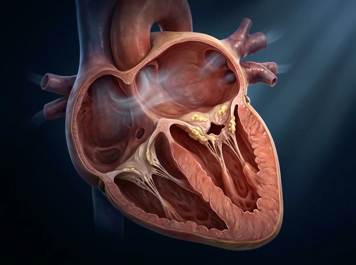
Severity is graded by valve area, mean gradient and pulmonary pressure (haemodynamic classification), and intervention candidacy is graded by anatomy (Wilkins and Cormier) and by aetiology. [1]
Severity (haemodynamic)
Mild
- MVA over 1.5 cm squared
- Mean transmitral gradient under 5 mmHg
- PASP usually under 30 mmHg
- Often asymptomatic; observe with serial echo
Moderate
- MVA 1.0 to 1.5 cm squared
- Mean transmitral gradient 5 to 10 mmHg
- PASP 30 to 50 mmHg
- Symptoms on moderate exertion; review for PMC candidacy
Severe
- MVA 1.0 to 1.5 cm squared with symptoms, OR
- Mean gradient 10 mmHg or above
- PASP above 50 mmHg
- Indicates intervention when symptomatic, when PASP rises, or for pregnancy
Very severe
- MVA under 1.0 cm squared
- Mean transmitral gradient above 15 mmHg
- PASP often above 60 to 70 mmHg with RV dysfunction
- Reflex syncope, severe pulmonary oedema with any tachycardia; mandate intervention
A pressure half-time (PHT) above 220 ms is supportive of severe MS on continuous-wave Doppler (PHT = MVA / 220 by empirical formula). The Wilkins echocardiographic score (Wilkins, Weyman, Abascal; Br Heart J 1988) sums four components graded 1 to 4 each: leaflet mobility, leaflet thickening, subvalvular thickening, calcification — total range 4 to 16. A score at or below 8 with pliable non-calcified leaflets is considered favourable for PMC. The Cormier classification groups anatomy by echocardiographic appearance into five classes (group 1: pliable non-calcified anterior leaflet with mild subvalvular disease — ideal; group 2: pliable non-calcified leaflet with severe subvalvular disease; group 3: calcified leaflet of any mobility).[1][2][8]
Classification by aetiology
Rheumatic
- **Dominant cause worldwide** — commissural fusion plus leaflet thickening plus subvalvular (chordal) shortening/fusion (the fish-mouth valve)
- Earliest diagnostic feature on echo is **restricted leaflet motion with hockey-stick anterior leaflet and diastolic doming**
- Almost always involves other valves (aortic, tricuspid) on systematic imaging
- Benzathine penicillin G prophylaxis after acute rheumatic fever reduces recurrence and disease progression
Degenerative (calcific)
- Anular calcification extending onto the leaflets
- Disease of elderly patients (over 70), often with chronic kidney disease or diabetes
- Leaflets are usually not mobile and not commissurally fused
- **Unfavourable for PMC** — usually requires MVR (often surgical or TAVR-style degenerative options)
Congenital
- Parachute mitral valve, double-orifice mitral valve (Shone complex)
- Often presents in childhood with associated left-heart obstruction
- Anatomy may be amenable to surgical repair or MVR rather than PMC
Other (mimics and rare)
- **Radiation** — prior mediastinal irradiation (Hodgkin lymphoma)
- **Connective tissue** — systemic lupus, mucopolysaccharidoses (rare)
- **Prosthetic valve dysfunction** — pannus or thrombosis
- **Inflammatory** — carcinoid (right-sided, sparing the mitral valve typically), methysergide
- **Functional** — left atrial tumour (myxoma) producing variable obstruction is a mimic, not a true stenosis
Epidemiology & Risk Factors
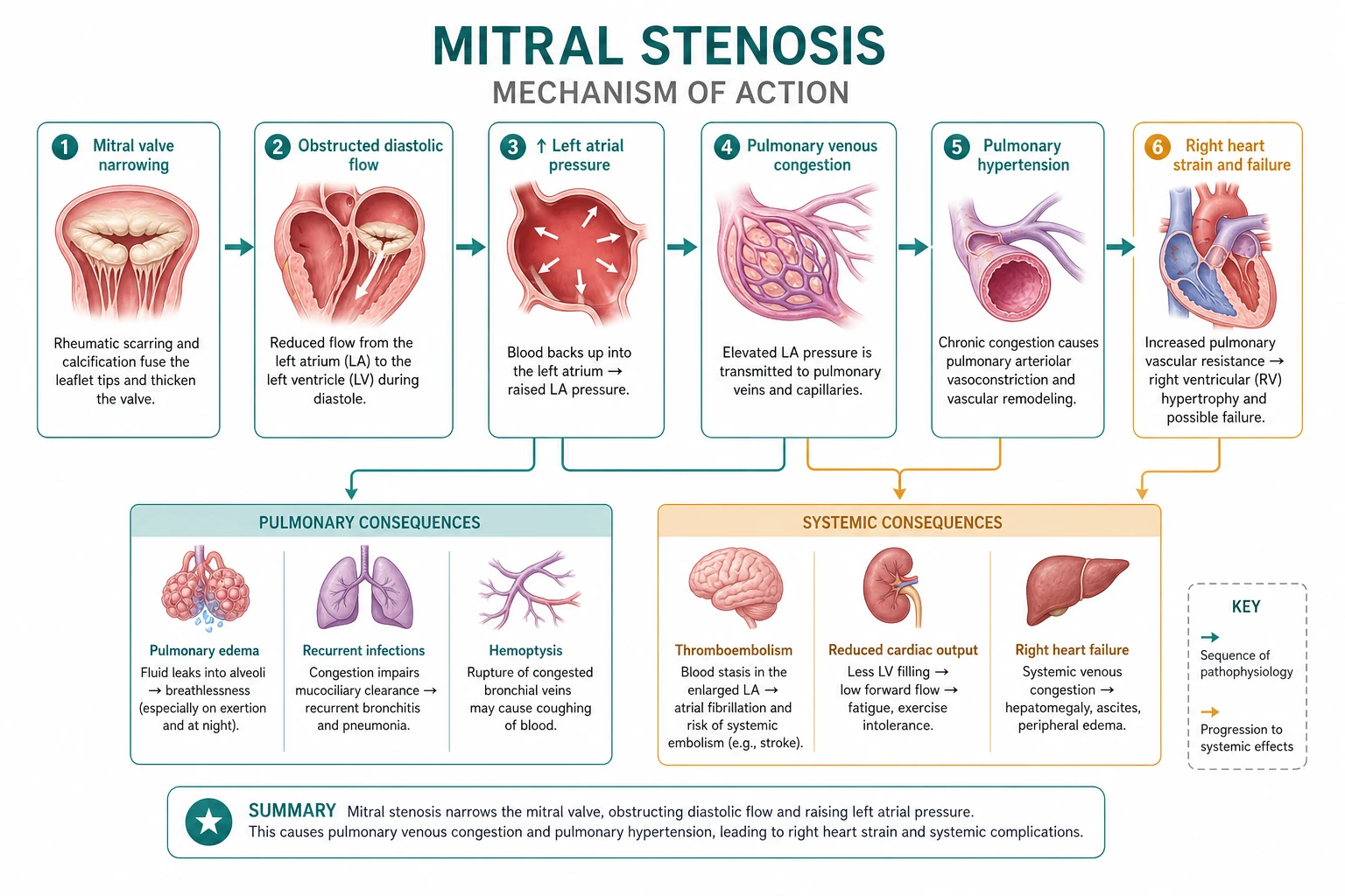
The dominant cause of MS worldwide is rheumatic heart disease (RHD) — a sequela of acute rheumatic fever (ARF) following inadequately treated group-A beta-haemolytic streptococcal pharyngitis (Streptococcus pyogenes). Although ARF and chronic RHD have all but disappeared from high-income countries with widespread penicillin treatment, RHD remains a leading cause of cardiovascular morbidity and mortality in low- and middle-income countries (South Asia, sub-Saharan Africa, Indigenous Australia, parts of Latin America).[1][5][9]
Key epidemiological facts: [1]
- Women are affected roughly twice as often as men — the female-to-male ratio is approximately 2:1.[9]
- Symptomatic MS typically presents in the third to fifth decade — a latent period of 2 to 3 decades from the initial ARF episode to symptomatic stenosis.[5][9]
- Genetic susceptibility to ARF has been confirmed (HLA-DR7, HLA-DR4 associations; familial clustering).[9]
- Socioeconomic risk factors: overcrowding, poverty, limited access to primary care and to benzathine penicillin G prophylaxis, recurrent streptococcal pharyngitis, and high background RHD prevalence.[5][9]
Risk-factor summary: [1]
- Prior acute rheumatic fever (the strongest clinical antecedent) — occurs at age 5 to 15 in the index attack.[5]
- Lack of or non-adherence to secondary prophylaxis (benzathine penicillin G 1.2 million units IM monthly).[5]
- Multiple recurrences of ARF — each recurrence accelerates valve damage.[5][9]
- Chronic kidney disease, diabetes, hyperparathyroidism — predispose to calcific/degenerative MS (a different aetiology, prominent in elderly patients).
- Age (the symptom-onset curve tracks time since ARF for rheumatic MS; tracks cardiovascular age and dialysis exposure for degenerative MS).
- Female sex for the rheumatic form.[9]
- Tachycardia of any cause (pregnancy, fever, atrial fibrillation, hyperthyroidism) precipitates decompensation by shortening diastolic filling time and raising the transmitral gradient.[1][5][9]
Mitral stenosis — key numbers
Pathophysiology
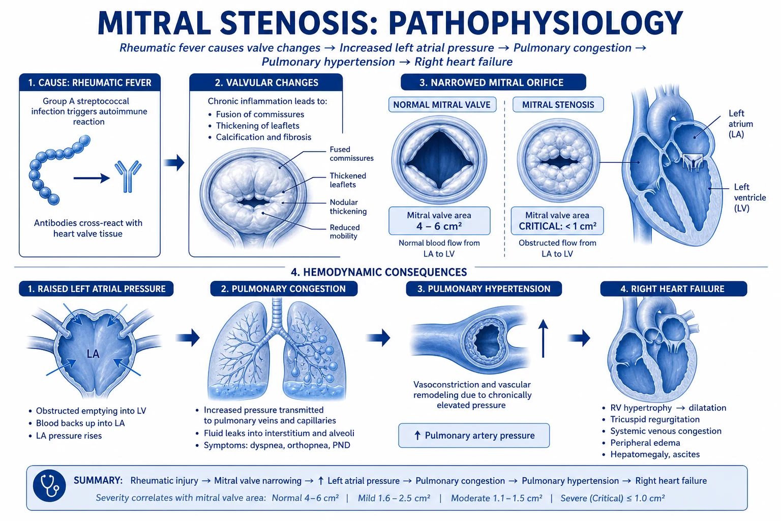
The pathological hallmark of rheumatic MS is commissural fusion accompanied by leaflet thickening, fibrosis and a variable degree of calcification, plus subvalvular chordal shortening and fusion — the so-called fish-mouth or buttonhole orifice when viewed from the LA in severe disease.[1][2][9]
The functional consequence is a fixed obstruction to diastolic flow — flow is now driven by a sustained pressure gradient across the valve, and the LA pressure rises in proportion to severity, modulated by the cardiac cycle and heart rate. [1]
The pressure cascade
1. Raised LA pressure. The LA-to-LV diastolic gradient rises from a few mmHg (normal) to 10 mmHg or above (severe MS) as the area falls. LA pressure elevation is the initiating event of every downstream consequence.[1][2]
2. Pulmonary venous congestion. The raised LA pressure transmits back through the pulmonary veins, raising pulmonary capillary wedge pressure and producing exertional dyspnoea, orthopnoea and paroxysmal nocturnal dyspnoea when the capillary hydrostatic pressure exceeds plasma oncotic reserve.[1]
3. Reactive pulmonary arterial hypertension (PAH). Sustained pulmonary venous hypertension triggers a reactive, vasoconstrictive component — intimal proliferation, medial hypertrophy and plexogenic arteriopathy in the pulmonary arterioles. Right ventricular pressure overload and right ventricular failure follow when the PAH becomes severe (PASP above 50 mmHg).[1][2][7]
4. Atrial enlargement and AF. The chronic pressure and volume load on the LA dilates it (giant LA — sometimes the whole left cardiac silhouette on chest X-ray), and atrial fibrillation develops in 40 to 60 percent of severe MS. AF is dangerous in MS for two reasons: (a) loss of the atrial kick reduces cardiac output by 20 to 30 percent because diastolic filling in MS depends on atrial contraction, and (b) the irregularly rapid ventricular response shortens diastole and elevates the transmitral gradient — a perfect storm for acute pulmonary oedema.[1][2][10][11]
5. Thromboembolism. Sluggish flow in the dilated LA, particularly in the left atrial appendage, predisposes to thrombus formation. Systemic embolism (cerebral, mesenteric, renal, peripheral) may be the first clinical manifestation of previously silent MS, occurring in 10 to 20 percent of patients with MS and AF if unanticoagulated.[1][11]
6. Haemoptysis. Rupture of pulmonary venules from raised pulmonary venous pressure produces pink frothy sputum or frank haemoptysis — an early symptom, frequently dismissed as "bronchitis" or "pulmonary tuberculosis" in endemic regions.[1]
7. Ortner syndrome. The enlarged LA or dilated pulmonary artery compresses the left recurrent laryngeal nerve, producing hoarseness — the Ortner (cardiovocal) syndrome, a high-yield clinical pearl unique to severe MS or other causes of giant LA / PA.[6]
8. Tachycardia precipitates decompensation. Any cause of tachycardia (AF with rapid rate, exertion, fever, pregnancy, hyperthyroidism) shortens diastolic filling time, raises the transmitral gradient (because flow per unit time must be pushed through a smaller area), and can precipitate acute pulmonary oedema even in a previously stable patient.[1][2][10]
Why MS kills: the preload-dependence paradox
The patient's forward cardiac output depends on adequate diastolic filling across the obstructed valve. Excessive preload reduction (large diuretic doses, nitrates) or excessive afterload reduction (ACE inhibitors, ARBs, nifedipine, hydralazine) may collapse transvalvular flow before pulmonary pressures fall, precipitating hypotension and shock. This is the single most important reason MS patients do not respond to standard heart-failure afterload reducers — the valve, not the LV, is the bottleneck.[1][2][10]
Loud S1 (tapping)
- Loud S1 reflects **abrupt tensing of the still-mitral-leaflets in early systole** — the cusps are forced widely apart by the high LA-to-LV gradient; closing them snaps them forcefully shut
Opening snap
- A high-frequency sound just after A2 (and P2 with prolonged PR) reflecting **sudden tensing of the stenotic but still-pliable leaflets** in early diastole
- An **absent opening snap suggests calcified rigid leaflets** and predicts lower PMC success
- **The A2-OS interval shortens with severity** — higher LA pressure pushes the leaflets open earlier
Mid-diastolic rumble
- Low-pitched rumbling diastolic murmur best heard at the **apex with the bell** in the **left lateral position**, after the opening snap
- **Length of the murmur reflects duration of turbulent flow** — a long rumbling murmur persisting to S1 implies severe MS
- **Presystolic accentuation** (crescendo before S1) is due to atrial contraction in sinus rhythm; **absent in AF**
Clinical Presentation
The classic presentation is a young woman in her 20s to 40s from an endemic region presenting with exertional dyspnoea, sometimes with haemoptysis, palpitations (AF), a history of stroke or transient ischaemic attack (systemic embolism), or hoarseness (Ortner syndrome).[1][2][9]
Symptoms (the high-yield list)
- Dyspnoea on exertion — the earliest and most common symptom; reflects pulmonary congestion as the gradient rises.[1][2]
- Orthopnoea and paroxysmal nocturnal dyspnoea (PND) — with progression of LA pressure rise at rest; presents as night-time cough or wheeze (often misdiagnosed as asthma).[1]
- Fatigue and effort intolerance — reduced cardiac output across a fixed obstruction.[2]
- Palpitations — the onset of atrial fibrillation, often new, frequently symptomatic with breathlessness at rest.[1][10]
- Haemoptysis — pink frothy sputum from pulmonary oedema, or frank red blood from ruptured pulmonary venules; an early symptom.[1]
- Hoarseness — Ortner syndrome from compression of the left recurrent laryngeal nerve by the enlarged LA or pulmonary artery.[6]
- Systemic embolism — stroke or peripheral ischaemia as the presenting feature of previously silent MS with AF.[1][10][11]
- Chest pain — atypical, possibly reflecting coexistent coronary disease or pulmonary hypertension (RV ischaemia).[2]
Signs — the bedside examination
General: mitral facies — a plum-coloured malar flush (cyanotic cheek blush from peripheral vasoconstriction in chronic low output); pulse may be irregularly irregular (AF) or regular; tapping apical impulse (felt but not displaced — the loud S1 is felt as a tap).[1][2]
Palpation: diastolic thrill at the apex in severe disease; right ventricular heave at the left sternal edge in pulmonary hypertension; palpable P2 in the pulmonary area.[2]
Auscultation (the mitral triad): [1]
- Loud (tapping) S1 — best heard at the apex; reflects high transmitral gradient tensing the leaflets shut in early systole; softens as the valve becomes calcified and rigid.[1]
- Opening snap (OS) — high-frequency sound after A2 (and P2 with prolonged PR) at the apex or just medial to it; A2-OS interval shortens with rising LA pressure and severity; absent OS in calcified immobile valves.[1][2]
- Mid-diastolic rumble — low-pitched rumble best heard at the apex with the bell in the left lateral position, beginning after the OS and lasting through to (or until) S1; length of the murmur is a marker of severity; presystolic accentuation in sinus rhythm.[1][2]
Signs of pulmonary hypertension and right-heart failure: loud P2, right ventricular heave, Graham Steell murmur — early diastolic murmur of functional pulmonary regurgitation at the left sternal edge; raised JVP with prominent a-waves (in sinus rhythm) or absent a-waves (in AF), tricuspid regurgitation, hepatomegaly, peripheral oedema, ascites.[1][2]
Dynamic findings: murmur lengthens with exertion and in the left lateral position (use both to bring out the murmur in a suspected case); the A2-OS interval shortens after exercise.[1][2]
Atypical presentation
- Late disease with soft S1 and absent OS — calcified rigid leaflets; severity is now read from haemodynamics, not auscultation.[1][2]
- Asymptomatic severe MS — found incidentally on echo; becomes symptomatic with pregnancy, AF, fever, hyperthyroidism or anaemia.[1][2]
- Pulmonary hypertension dominating the picture — RV heave, loud P2, peripheral oedema; the mitral findings may be subtle.[2]
- Embolic stroke as first presentation — silent MS presents with AF and cerebral embolism in a young patient under 40.[1][2][10][11]
Differential Diagnosis
The differential is best framed in two groups: (1) MS mimics (true diastolic obstruction of different cause) and (2) "pseudo-MS" (other causes of diastolic murmur at the apex or pulmonary congestion).[1][2][9]
Left atrial myxoma
- **Most important mimic** — pedunculated LA tumour producing variable obstruction; presents with positional dyspnoea, syncope, embolism and constitutional symptoms (fever, weight loss, raised ESR, anaemia)
- **Tumour 'plop'** — early diastolic sound replacing the opening snap
- Echo mass in the LA — usually attached by a stalk to the interatrial septum
- Surgical excision is curative; do not attempt PMC
Cor triatriatum
- Congenital membrane in the LA producing a proximal accessory chamber; rare
- Echo and TOE define membrane; CMR may be useful
- Surgical resection of the membrane is curative; PMC is not appropriate
Pulmonary vein stenosis
- Acquired (post-radiofrequency ablation) or congenital
- Variable presentation with exertional dyspnoea
- Echo and cardiac CT/MR imaging; management is catheter or surgical
Mitral stenosis of low output (low-flow)
- Apparent MVA-derived severity in a patient with severe LV dysfunction may **underestimate true valve area** (the gradient is low because flow is low)
- Confirmation with **low-dose dobutamine stress echo** to discriminate true severe from pseudo-severe MS
Austin Flint murmur (severe AR)
- Severe aortic regurgitation produces a low-pitched diastolic rumble at the apex as the regurgitant jet impinges on the anterior mitral leaflet (functional MS)
- Distinguish: **bounding pulse, wide pulse pressure, displaced apex, aortic diastolic murmur** at the left sternal edge
- Echo defines both lesions; the AR drives the murmur, not a stenotic MV
Tricuspid stenosis
- Mid-diastolic murmur at the lower left sternal edge, **louder on inspiration** (Carvallo sign), with **giant a-waves** on the JVP in sinus rhythm
- Often accompanies rheumatic MS (same aetiology); assess both at echo
The decisive single test for the MS differential is the transthoracic echocardiogram (TTE) — it confirms MS, excludes LA myxoma and cor triatriatum, grades severity, defines anatomy for intervention (leaflet mobility, calcification, MR, LA thrombus), and identifies pulmonary pressure.[1][2]
Clinical & Bedside Assessment
The bedside examination confirms suspected MS and quantifies severity before the echo arrives. [1]
- General inspection — mitral facies (malar cyanotic flush), nutritional status (cachexia in advanced disease), any Marfanoid habitus (consider alternative diagnosis).[1][2]
- Pulse — rate, rhythm (AF in 40 to 60 percent of severe MS), volume, pulsus parvus et tardus is NOT an MS finding (small late pulse is aortic stenosis).[2]
- JVP — raised with prominent a-waves in pulmonary hypertension and RV overload; giant a-waves with sinus rhythm suggest concomitant TS; absent a-waves with cannon waves suggest AF with high block.[2]
- Palpation — tapping apex (not displaced); diastolic thrill at the apex in severe MS; left parasternal heave (RV hypertrophy); palpable P2 at the pulmonary area.[1][2]
- Auscultation — full mitral triad at the apex in the left lateral position; loud P2 and any Graham Steell murmur at the left sternal edge.[1][2][7]
Dynamic manoeuvres to bring out a subtle murmur: exercise (lengthens the murmur), left lateral position with the bell (murmur longest), expiration (brings the LV closer to the chest wall).[1]
Severity clues at the bedside (haemodynamic, not just louder):
- Long murmur persisting close to S1 (in sinus rhythm) — more severe.[1][2]
- Short A2-OS interval (under 70 ms severe; over 110 ms mild) — more severe.[1]
- Soft S1 + absent OS — calcified rigid valve, often severe.[2]
- Pulmonary hypertension signs — loud P2, RV heave, raised JVP, peripheral oedema — the more severe the sequelae, the more severe the MS driving them.[1][7]
Investigations
Investigations establish the diagnosis, severity, anatomy, and comorbid status for intervention.[1][2]
First-line
1. 12-lead ECG — left atrial enlargement (broad, notched P wave in lead II — P mitrale; biphasic P with deep terminal negativity in V1 — Morris index over 0.04 sq mm); AF if irregular narrow-complex rhythm without P waves; right axis deviation, right ventricular hypertrophy in pulmonary hypertension.[1][2]
2. Chest X-ray — straightening of the left cardiac border (enlarged LA appendage), double density at the right cardiac border (enlarged LA), splaying of the carina (over 90 degrees), pulmonary venous congestion, Kerley B lines, interstitial oedema, and in advanced disease prominent pulmonary arteries and right heart enlargement.[1]
3. Transthoracic echocardiogram (TTE) — the pivotal test. Confirms the diagnosis, measures MVA by planimetry, pressure half-time (PHT) and continuity equation, mean and peak transmitral gradients by continuous-wave Doppler, pulmonary artery systolic pressure (PASP) from tricuspid regurgitation velocity, LA size, RV size and function, concomitant MR, mitral leaflet anatomy (mobility, thickening, subvalvular, calcification — the Wilkins components), and excludes LA myxoma and cor triatriatum.[1][2][8]
Pre-intervention
4. Transoesophageal echocardiogram (TOE) — mandatory before any percutaneous mitral commissurotomy (PMC) and before electrical cardioversion of AF in MS to exclude left atrial (LA) or left atrial appendage (LAA) thrombus; also used to refine anatomy when TTE is suboptimal.[1][2]
5. ECG-gated cardiac CT — for valve calcification quantification when echo is equivocal, and coronary CT angiography for surgical planning in patients over 40.[7]
6. Stress echocardiography — exercise echo in symptomatic patients with resting MVA 1.0 to 1.5 (mild at rest, but rising gradient with effort confirms hemodynamically severe MS); low-dose dobutamine stress echo in low-flow low-gradient states to discriminate true severe from pseudo-severe MS.[2]
7. Cardiac catheterisation — rarely needed for diagnosis; used when non-invasive tests are discordant, or for coronary assessment before surgical MVR (or in pre-operative assessment).[1][2]
8. Cardiac MR (CMR) — adjunct when echo is suboptimal or for RV quantification; not routine.[2]
Named scores / criteria
Wilkins echocardiographic score (Br Heart J 1988) — four components, each graded 1 to 4 (1 = normal/least, 4 = worst):[8]
| Component | 1 | 2 | 3 | 4 |
|---|---|---|---|---|
| Leaflet mobility | Highly mobile, only tip restricted | Mid-leaflet mobility, normal hinge | Base moving, mid-to-tip restricted | Minimal forward motion in diastole |
| Leaflet thickness | Near-normal (4 to 5 mm) | Mid-leaflet thickening, edges normal | Marked thickening across leaflet | Severe throughout |
| Subvalvular thickening | Minimal, just below leaflets | Mild chordal thickening extending to one third | Thickening extending to distal third | Extensive, extending to papillary muscles |
| Calcification | Single bright spot | Scattered bright spots at edges | Mid-leaflet brightness | Bright throughout |
Total 4 to 16; at or below 8 considered favourable for PMC. [1]
Cormier group 1
- Pliable non-calcified anterior leaflet, mild subvalvular disease
- **Most favourable for PMC**
Cormier group 2
- Pliable non-calcified leaflet with severe subvalvular disease
- PMC possible but higher risk of restenosis
Cormier group 3
- Calcified leaflet of any mobility
- **Unfavourable for PMC** — consider MVR
Management — Resuscitation
Acute decompensation in MS is usually driven by rate and rhythm (new fast AF), volume load (pregnancy, fluids), or failure of chronic management (missed prophylaxis, non-adherence). The pattern is typically acute pulmonary oedema with preserved or high blood pressure — the LV is fine, but the obstructed valve and rapid rate have precipitated congestion.[1][2]
First 30 minutes — the immediate priorities
- Position upright, give high-flow oxygen to target SpO2 94 to 98 percent.[1]
- Two large-bore IV cannulae, cardiac monitoring, 12-lead ECG (look for AF, new ischaemia).[1]
- IV loop diuretic — furosemide 20 to 40 mg IV (titrate up to 80 mg in diuretic-resistant cases) to relieve pulmonary congestion; titrate to relief of pulmonary oedema and adequate urine output (0.5 mL/kg/hr).[1][2]
- RATE CONTROL IS THE FIRST-LINE PRIORITY — slowing the heart rate lengthens diastole and reduces the transmitral gradient more effectively than pure preload reduction. Options:
- IV beta-blocker — metoprolol 2.5 to 5 mg IV bolus over 2 min (may repeat every 5 min up to 15 mg) — best for rate control in a non-asthmatic.[1][2]
- Diltiazem 0.25 mg/kg IV over 2 min then continuous infusion 5 to 15 mg/hr — if beta-blockers are contraindicated.[2]
- Digoxin — digoxin 250 mcg IV loading (stat, repeated to 500 to 750 mcg over 24 h) then 125 to 250 mcg daily oral — most useful in AF with rate control failure or left ventricular dysfunction; onset is 30 min to 2 h.[1][2]
- Cardioversion if the patient is haemodynamically unstable (syncope, hypotension, refractory pulmonary oedema) and AF is of recent onset (under 48 h) or LAA thrombus has been excluded by TOE.[1][2]
- Anticoagulate if AF, prior embolism or LA thrombus — heparin 80 units/kg IV bolus then 18 units/kg/hr infusion in the acute setting; convert to oral anticoagulation once stabilised.[1][11]
Specific ICU pearls (high-yield)
- Avoid aggressive afterload reduction (ACE inhibitors, ARBs, nitroprusside, nifedipine) — the LV is preload-dependent; reducing afterload without relieving the mitral obstruction can precipitate hypotension and shock. A small dose of nitroglycerin 10 to 20 mcg/min may help if BP permits and pulmonary oedema is severe, but rate control and diuresis do most of the work.[1][2][10]
- Bilevel positive airway pressure (BiPAP) is highly effective in MS pulmonary oedema — titrate IPAP/EPAP to relieve work of breathing and improve oxygenation while definitive rate control and diuresis take effect.[1]
- Avoid digoxin in WPW with AF — risk of degeneration to VF; use ibutilide or procainamide if electrical cardioversion unavailable; otherwise synchronised DCCV.[2]
Management — Definitive & Stepwise
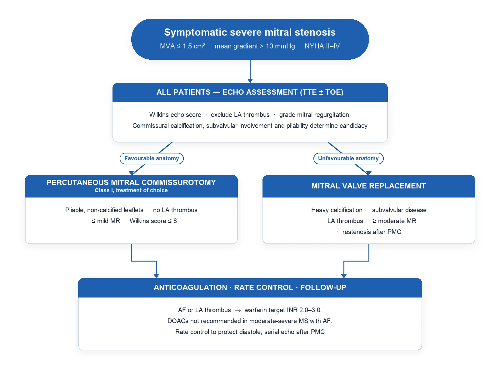
Definitive management is mechanical relief of the obstruction in patients who meet criteria; the decision is anatomy-driven (PMC versus MVR).[1][2][7]
Indications for intervention
Symptomatic severe MS (MVA at or below 1.5 cm squared or mean gradient above 10 mmHg, or symptoms on exertion); or asymptomatic severe MS with progressive pulmonary hypertension (PASP above 50 mmHg) or new-onset AF or planning pregnancy.[1][2][7]
Stepwise protocol
Step 1 — Medical optimisation (every patient). [1]
- Rate control — metoprolol 25 to 50 mg twice daily PO (titrate to HR 60 to 70 at rest) or digoxin as above; consider combination therapy in AF.[1][2]
- Diuretic for symptomatic congestion — furosemide 20 to 40 mg daily PO (or bumetanide 1 mg orally); combine with spironolactone 25 mg daily if persistent congestion.[1]
- Oral anticoagulation — warfarin target INR 2.0 to 3.0 for MS with AF, prior embolism, or LA thrombus; DOACs are NOT recommended in moderate-to-severe MS (defined as mitral valve area under 1.5 cm squared on echo) — this is valvular AF by trial definition; mechanical mitral prosthesis → INR 3.0 (range 2.5 to 3.5).[1][2][10][11]
- Secondary rheumatic prophylaxis — benzathine penicillin G 1.2 million units IM every 4 weeks for life (or at least until age 40 with stable disease); use erythromycin 250 mg twice daily orally in penicillin-allergic patients.[5][9]
- Lifestyle and triggers — avoid strenuous exertion if symptomatic; treat fever, anaemia, hyperthyroidism aggressively as each raises the gradient.[1]
Step 2 — Percutaneous mitral commissurotomy (PMC) — the treatment of choice when anatomy is favourable.[1][2][3][4]
- Eligibility: symptomatic severe MS, favourable valve anatomy (pliable, non-calcified leaflets, no LA thrombus, no more than mild MR, Wilkins at or below 8, Cormier 1 or 2).
- Technique — Inoue balloon PMC: transseptal puncture under fluoroscopy, balloon catheter (Inoue 26 to 30 mm) advanced across the mitral valve and inflated, splitting the fused commissures by mechanical force.[1]
- Evidence: Reyes (NEJM 1994) — randomised 60 patients with severe MS to balloon valvuloplasty versus open surgical commissurotomy; equivalent haemodynamic improvement at 7 days to 4 months[4]; Ben Farhat (Circulation 1998) — 7-year follow-up of balloon versus closed versus open commissurotomy, equivalent long-term outcome for balloon and open commissurotomy with lower cost and faster recovery[3]. Balloon commissurotomy is now the preferred option for favourable anatomy where the expertise is available.[3][4]
- Procedural risks: severe MR (4 to 5 percent — leaflet tear), residual atrial septal defect (transseptal), cardiac tamponade (under 1 percent), embolism, restenosis (~25 to 30 percent at 10 years).[1][7]
- Post-procedure care: bedrest, anticoagulation, telemetry for tamponade; lifelong warfarin if AF or LA thrombus; continue benzathine penicillin prophylaxis.[1][5]
Step 3 — Mitral valve replacement (MVR) when PMC is contraindicated or unfavourable.[1][2][7]
- Indications: severe MS with unfavourable anatomy (calcified rigid leaflets, Cormier 3, Wilkins above 8, significant MR, LA thrombus despite anticoagulation), restenosis after PMC, or symptom recurrence after failed intervention.[1][2]
- Options: mechanical prosthesis (lifelong warfarin INR 3.0, 2.5 to 3.5 — durable, preferred under 65) or bioprosthetic valve (no long-term warfarin but 10 to 15-year durability, preferred over 65 or when anticoagulation is contraindicated).[1][7]
- Surgical risk — operative mortality 2 to 5 percent; intra-operative transoesophageal echo to guide repair versus replacement.[2]
Step 4 — Adjunctive — anticoagulation, prophylaxis, surveillance.[5][9][11]
- Warfarin in MS plus AF, prior embolism or LA thrombus — INR 2.0 to 3.0.[1][10][11]
- Endocarditis prophylaxis is no longer recommended for native-valve MS (AHA/ESC 2021 update) — antibiotic prophylaxis is reserved for prosthetic valves and prior endocarditis.[7]
- Surveillance — clinical review 6 to 12 monthly; TTE 1 to 3 yearly for stable disease, sooner if symptoms or PASP rises.[1][2]
PMC — favourable anatomy
- Pliable non-calcified leaflets (Wilkins at or below 8, Cormier 1 or 2)
- No LA/LAA thrombus on TOE
- No more than mild MR
- **Treatment of choice** — Inoue balloon splits fused commissures
- Equivalent to open commissurotomy at 7 years (Ben Farhat 1998); less invasive, faster recovery
MVR — unfavourable anatomy
- Calcified rigid leaflets (Cormier 3, Wilkins above 8)
- Significant MR (moderate or worse) — PMC would worsen MR
- LA/LAA thrombus despite anticoagulation
- Mechanical valve → INR 3.0 lifelong (under 65)
- Bioprosthetic valve → no lifelong warfarin (over 65; contraindication to warfarin)
Specific Subtypes & Scenarios
- MS in pregnancy — the volume and rate load of pregnancy (plasma volume up 50 percent, cardiac output up 30 to 50 percent, resting heart rate up 10 to 20 bpm) decompensates MS in women who were previously asymptomatic; beta-blockers (metoprolol 25 to 50 mg twice daily) are the cornerstone, with furosemide 20 to 40 mg daily as needed. PMC is the procedure of choice when severe symptomatic MS does not respond to medical therapy in the second trimester (well-tolerated, no fluoroscopy contraindication with abdominal shielding); delivery should be planned at a centre with cardiac surgical backup.[1][2][7][9]
- New atrial fibrillation in severe MS — the highest-risk group for acute decompensation. Immediate rate control, heparin anticoagulation, and cardioversion if haemodynamically unstable or symptomatic; TOE-guided cardioversion if AF under 48 h is uncertain, to exclude LA thrombus.[1][10][11]
- Asymptomatic severe MS — surveillance strategy with annual TTE; intervention triggers: symptom onset, PASP above 50 mmHg, new AF, planning pregnancy, or progressive valve area reduction. PMC may be considered even in asymptomatic patients with very favourable anatomy and high operative risk for future intervention.[1][2][7]
- Calcific/degenerative MS in the elderly — usually unfavourable for PMC (lack of commissural fusion, heavy calcification); MVR with a bioprosthesis is the standard; transcatheter mitral valve implantation (TMVI) is an emerging option for select patients.[1][7]
- Mixed mitral disease (MS plus moderate MR) — moderate or worse MR pushes the decision to surgical MVR rather than PMC — the balloon would worsen MR by tearing leaflets; PMC is not appropriate.[1][7]
- Restenosis after PMC — re-intervention depends on anatomy; re-PMC if valve still favourable at repeat Wilkins assessment, otherwise MVR.[1][2]
- Mechanical mitral prosthesis during pregnancy — the highest valve-related pregnancy risk (warfarin teratogenicity in first trimester; maternal thromboembolism in any trimester); switch to low-molecular-weight heparin in the first 12 to 13 weeks (controversial — discuss with cardiac obstetric team) and resume warfarin mid-trimester; plan delivery by caesarean section after switching to UFH 24 to 36 h pre-delivery.[1][2][7]
Complications & Pitfalls
Cardiac: atrial fibrillation (40 to 60 percent of severe MS)[1][10], right-heart failure from pulmonary hypertension[2], pulmonary oedema and haemoptysis[1], systemic thromboembolism (stroke, mesenteric, renal, peripheral)[11], infective endocarditis (rare on native MS — prophylaxis no longer indicated)[7], left atrial appendage thrombus[10][11].
Procedural — PMC: severe MR (4 to 5 percent — leaflet or chordal tear), iatrogenic atrial septal defect (transseptal), cardiac tamponade (under 1 percent), stroke from embolised thrombus or air, restenosis at 5 to 10 years (~25 to 30 percent at 10 years).[1][7]
Procedural — MVR: prosthetic valve thrombosis and thromboembolism (if anticoagulation inadequate), structural valve deterioration (bioprosthetic, 10 to 15 years), paravalvular leak, infective endocarditis (1 to 2 percent per year), anticoagulant-related bleeding (mechanical valves require lifelong warfarin).[1][7]
Classic errors (what an examiner expects you to spot): [1]
- Treating the murmur not the valve area — symptom escalation is the trigger to intervene, but severity is graded by MVA, mean gradient, and PASP, not by murmur loudness; a soft murmur may accompany very severe MS if the valve is heavily calcified with low flow.[1][2]
- Offering PMC to an unfavourable valve (calcified, high Wilkins, with LA thrombus or significant MR) — fruitless balloon and increased procedural risk.[1][7]
- Prescribing a DOAC in MS + AF — these patients are NOT eligible for DOACs (the moderate-to-severe MS arm of valvular AF was excluded from the DOAC trials); warfarin only.[10][11]
- Failing to anticoagulate MS with AF even before intervention — embolic stroke in a young patient is the high-yield disaster scenario; anticoagulate at diagnosis.[1][10][11]
- Treating MS pulmonary oedema with vasodilators or nitrates as first-line — the LV is preload-dependent and the obstruction is fixed; rate control and careful diuresis are the answer; aggressive vasodilatation may precipitate hypotension.[1][2][10]
- Missing pregnancy in a young asymptomatic MS patient — counsel early; intervene before pregnancy if MVA is approaching 1.5 cm squared or plan PMC in the second trimester if severe MS becomes symptomatic.[1][7]
Prognosis & Disposition
Natural history of untreated MS is unfavourable once symptoms develop: 5-year survival without intervention drops from 80 to 90 percent in mild MS to below 30 percent in severe symptomatic MS with pulmonary hypertension.[1][9]
With PMC: in favourable anatomy, freedom from reintervention ~90 percent at 7 years and 65 to 75 percent at 10 years, with repeat PMC possible if restenosis occurs and valve remains favourable.[1][3][4]
With MVR: prosthetic valve survival is excellent, but lifelong anticoagulation risk (mechanical) or structural deterioration risk (bioprosthetic) frames long-term outcomes.[1][7]
Predictors of poor prognosis: older age at presentation, atrial fibrillation, severe pulmonary hypertension (PASP above 60 mmHg), heavily calcified valve, high Wilkins score, comorbidities limiting intervention, lack of access to benzathine penicillin prophylaxis with recurrent ARF.[1][2][5][9]
Disposition: [1]
- Asymptomatic mild/moderate MS — outpatient follow-up with serial echo every 1 to 3 years; lifestyle advice (avoid dehydration, treat AF onset promptly).[1]
- Symptomatic severe MS — refer to a structural heart / valve centre for anatomy assessment and PMC/MVR decision; anticoagulate and titrate rate control in the meantime.[1][7]
- Acute pulmonary oedema with AF — emergency admission to a monitored bed; HDU/CCU for oxygen, IV diuretic and rate control; structural heart team notification.[1][2]
Special Populations
- Pregnancy — increasing volume load and heart rate decompensate previously silent MS; beta-blockers (metoprolol 25 to 50 mg twice daily), diuretic as needed, and PMC in the second trimester for severe refractory symptoms; warfarin is teratogenic (avoid first trimester for valve indication); LMWH in the first trimester or INR-adjusted UFH for mechanical prostheses; multidisciplinary team (cardiologist, obstetrician, anaesthetist, neonatologist) for delivery and puerperium.[1][2][7]
- Paediatric / adolescent — usually congenital (parachute mitral valve, Shone complex) or early rheumatic; surgical repair preferred for paediatric anatomy; percutaneous options reserved for older children with rheumatic anatomy.[1]
- Elderly (calcific MS) — unfavourable anatomy for PMC; bioprosthetic MVR or TMVI for high-risk surgical candidates; medical management with rate control, diuretic and judicious anticoagulation is the realistic bridge.[1][7]
- Athletes — MS is incompatible with competitive sport once symptomatic or with PASP above 50 mmHg (or any echocardiographic evidence of right-heart dysfunction); asymptomatic mild MS may be eligible for recreational activity with appropriate rate control and avoidance of competitive endurance sports.[1]
- MS with AF — warfarin lifelong INR 2.0 to 3.0, rate control with beta-blocker plus digoxin as needed, address the valve (PMC or MVR if appropriate).[1][2][5][10][11]
- MS with mechanical prosthesis — INR 3.0 lifelong (range 2.5 to 3.5); avoid pregnancy in first trimester; consider self-monitoring INR; endocarditis prophylaxis for any dental/upper-respiratory procedure (AHA/ESC 2021 — restricted indication, individualised).[1][7]
- Indigenous / low-income populations — system-level prevention: improve housing, address overcrowding, register and treat streptococcal pharyngitis with penicillin, deliver benzathine penicillin G 1.2 million units IM monthly to ARF/RHD patients, train community health workers in screening echo.[5][9]
Evidence, Guidelines & Regional Differences
Key guidelines: [1]
- 2020 ACC/AHA Guideline for the Management of Patients With Valvular Heart Disease (Otto CM et al., Circulation 2021) — the contemporary US standard; class I indication for PMC in symptomatic severe MS with favourable anatomy (Wilkins at or below 8, no LA thrombus, no more than mild MR); warfarin (not DOAC) for MS plus AF.[1]
- 2014 AHA/ACC Guideline for the Management of Patients With Valvular Heart Disease (Nishimura RA et al., Circulation 2014) — historical foundation for severity grading and PMC indications; Wilkins scoring recommended for anatomy assessment.[2]
- 2021 ESC/EACTS Guidelines for the Management of Valvular Heart Disease (Vahanian A et al., Eur Heart J 2022) — European standard aligning with ACC/AHA on severity grading and PMC; emphasises heart-team decision-making for borderline anatomy.[7]
- 2015 AHA Revision of the Jones Criteria (Gewitz MH et al., Circulation 2015) — basis for ARF diagnosis and secondary prophylaxis with benzathine penicillin G 1.2 million units IM monthly.[5]
Landmark trials every candidate must know: [1]
- Reyes (NEJM 1994) — randomised 60 patients to PMC versus open surgical commissurotomy: equivalent haemodynamic improvement, lower cost, faster recovery with PMC.[4]
- Ben Farhat (Circulation 1998) — randomised 90 patients to balloon, closed surgical and open surgical commissurotomy with 7-year follow-up: balloon and open commissurotomy were equivalent; closed was inferior.[3]
- Wilkins score (Br Heart J 1988) — the score that predicts PMC outcome; at or below 8 favourable for PMC.[8]
- Marijon (Lancet 2012) — comprehensive review of the global burden, pathophysiology and prevention of rheumatic heart disease that frames the preventive and public-health approach.[9]
- Owens (Clinical Cardiology 2017) and Tanizawa (BMJ Open 2022) — modern evidence reviews of DOAC safety and stroke/embolism risk in MS with AF, underlying the warfarin only recommendation for valvular AF.[10][11]
Regional deltas: [1]
[1] [1]Current controversies: (1) DOAC safety in moderate MS with AF (post-hoc analyses of the original trials — limited evidence; guidelines remain against use)[10][11]; (2) opt-out DOAC strategy in MS with LA thrombus after 4 weeks of VKA — pending trials; (3) early PMC in asymptomatic severe MS[1][7]; (4) transcatheter MVR (TMVI) for high-risk surgical candidates[7]; (5) post-PMC antithrombotic strategy in sinus-rhythm PMC recipients without LA thrombus.
Exam Pearls
- The mitral auscultatory triad — loud tapping S1 + opening snap + low-pitched mid-diastolic rumble at the apex with presystolic accentuation (sinus rhythm) — is rheumatic MS until proven otherwise.[1][2]
- Severity is by valve area, not by murmur loudness — MVA 4 to 6 cm squared normal; severe under 1.5; very severe under 1.0. PASP above 50 mmHg indicates severe haemodynamic consequence.[1][2]
- Wilkins score 4 to 16, four components, each 1 to 4 — mobility, thickening, subvalvular thickening, calcification; at or below 8 favourable for PMC.[8]
- Favourable anatomy (pliable non-calcified leaflets, no LA thrombus, no/mild MR) → PMC; unfavourable → MVR. PMC and open commissurotomy are equivalent at 7 years (Ben Farhat).[3][4]
- MS + AF is valvular AF — warfarin INR 2 to 3, NOT a DOAC. Mechanical prosthesis → INR 3.0 (range 2.5 to 3.5).[1][10][11]
- Tachycardia (AF, exertion, pregnancy, fever) shortens diastole and spikes the gradient — rate control is the immediate priority in acute pulmonary oedema, not vasodilatation.[1][2]
- Ortner syndrome = hoarseness from left recurrent laryngeal nerve compression by enlarged LA/PA — a high-yield clinical pearl.[6]
- Graham Steell murmur = early diastolic murmur of functional pulmonary regurgitation in severe MS with pulmonary hypertension — at the left sternal edge, blowing quality.[1][2]
- Wilkins at or below 8 + favourable anatomy = PMC; bioprosthetic MVR if over 65 or pregnancy intent; mechanical MVR (warfarin INR 3.0 lifelong) under 65 unless contraindicated.[1][7]
- Mitral facies (malar flush) + tapping apex + diastolic thrill + mild MR (corrected to exclusion of LV failure) = rheumatic MS.[1][2]
- Acute pulmonary oedema in a young pregnant woman with new fast AF — was the valve previously silent MS? Rate control, TOE to exclude LAA thrombus, second-trimester PMC if refractory.[1][7]
- Systemic embolism in a young adult = silent MS with AF until proven otherwise — TTE or TOE now, and anticoagulation.[1][10][11]
- Secondary prevention of ARF with benzathine penicillin G 1.2 million units IM monthly reduces progression to severe MS — register, do not forget.[5][9]
Causes of mitral stenosis — mnemonic
RHEUM
Dominant cause worldwide — fish-mouth valve (95 to 98 percent of cases in endemic regions)
Tachycardia (AF, pregnancy, fever, hyperthyroidism) raises the transmitral gradient by shortening diastole
Stasis in dilated LA/LAA → systemic embolism (stroke, mesenteric, renal) especially with AF
Reactive PAH from chronic pulmonary venous congestion → right-heart failure
PMC if favourable (Wilkins at or below 8, no thrombus); MVR if unfavourable
Exam application bank (NEET-PG / INICET)
One-line answer
Mitral stenosis (MS) is a narrowing of the mitral valve orifice that obstructs flow from the left atrium to the left ventricle during diastole. The normal mitral valve area (MVA) is 4 to 6 cm squared; symptoms emerge as the area falls (severe under 1.5, very severe under 1.0). The leading cause worldwide is rheumatic heart disease (RHD) — commissural fusion, leaflet thickening, subvalvular chordal shortening producing the fish-mouth valve. The classic triad of auscultation is a loud (tapping) S1, an opening snap after S2, and a low-pitched mid-diastolic rumble at the apex with presystolic accentuation in sinus rhythm. Pathophysiology is a rising transmitral gradient cascade: raised LA pressure transmits back through the pulmonary veins, causing pulmonary venous hypertension, reactive pulmonary arterial hypertension, right ventricular pressure overload, and right-heart failure. Atrial fib
Worked stems (answer without another resource)
Stem 1 — Classic presentation. Map symptoms to mechanism; name the first investigation and first treatment step with dose/route if drug therapy is standard. [1]
Stem 2 — Unstable / complicated. List red flags that force immediate resuscitation, theatre, ICU, antidote, or reperfusion — and what you do in the first 15 minutes. [1]
Stem 3 — Atypical group. Elderly, pregnancy, child, or immunocompromised: how presentation and thresholds change. [1]
Stem 4 — Differential trap. Name the three closest mimics and one discriminator for each. [1]
Stem 5 — Disposition. Who goes home with safety-netting, who is admitted, who needs HDU/ICU/theatre, and what follow-up is mandatory. [1]
Rapid viva checklist
- Definition + classification
- Pathophysiology chain
- Bedside signs / criteria
- Score with exact components (if any)
- Emergency bundle
- Definitive therapy with doses
- Complications of disease and of treatment
- Special populations
- Guideline/trial name if classic
- Three exam traps
Coverage self-check
If you cannot answer any stem above from this page alone, re-read the matching section — the page is intended to be self-sufficient for final-prof and NEET-PG/INICET questions on Mitral Stenosis.
References
- [1]Otto CM, Nishimura RA, Bonow RO, et al. 2020 ACC/AHA Guideline for the Management of Patients With Valvular Heart Disease: A Report of the American College of Cardiology/American Heart Association Joint Committee on Clinical Practice Guidelines Circulation, 2021.PMID 33332150
- [2]Nishimura RA, Otto CM, Bonow RO, et al. 2014 AHA/ACC Guideline for the Management of Patients With Valvular Heart Disease: a report of the American College of Cardiology/American Heart Association Task Force on Practice Guidelines Circulation, 2014.PMID 24589853
- [3]Ben Farhat M, Ayari M, Maatouk F, et al. Percutaneous balloon versus surgical closed and open mitral commissurotomy: seven-year follow-up results of a randomized trial Circulation, 1998.PMID 9462525
- [4]Reyes VP, Raju BS, Wynne J, et al. Percutaneous balloon valvuloplasty compared with open surgical commissurotomy for mitral stenosis N Engl J Med, 1994.PMID 8084354
- [5]Gewitz MH, Baltimore RS, Tani LY, et al. Revision of the Jones Criteria for the diagnosis of acute rheumatic fever in the era of Doppler echocardiography: a scientific statement from the American Heart Association Circulation, 2015.PMID 25908771
- [6]Kalivoda I, Kryl J, Hodis J, et al. Ortner's syndrome in interdisciplinary collaboration Cas Lek Cesk, 2024.PMID 39772722
- [7]Vahanian A, Beyersdorf F, Praz F, et al. 2021 ESC/EACTS Guidelines for the management of valvular heart disease Eur Heart J, 2022.PMID 34453165
- [8]Wilkins GT, Weyman AE, Abascal VM, et al. Percutaneous balloon dilatation of the mitral valve: an analysis of echocardiographic variables related to outcome and the mechanism of dilatation Br Heart J, 1988.PMID 3190958
- [9]Marijon E, Mirabel M, Celermajer DS, et al. Rheumatic heart disease Lancet, 2012.PMID 22405798
- [10]Owens RE, Kabbani SS, Ramirez DA, et al. Direct oral anticoagulant use in nonvalvular atrial fibrillation with valvular heart disease: a systematic review Clin Cardiol, 2017.PMID 28004413
- [11]Tanizawa K, Harada N, Hida S, et al. Incidence of stroke, systemic embolism and bleeding events in patients without anticoagulation based on real-world data in Japan: a retrospective cohort study BMJ Open, 2022.PMID 36357001