cardiology · cardiology
Mitral Valve Prolapse
Also known as MVP · Barlow syndrome · Click-murmur syndrome · Myxomatous mitral valve disease · Billowing mitral leaflet · Floppy mitral valve
Mitral valve prolapse (MVP) is the systolic billowing of one or both mitral leaflets above the mitral annulus into the left atrium, defined echocardiographically as leaflet displacement of at least 2 mm beyond the mitral annular plane in the parasternal long-axis view during systole. The underlying pathology is myxomatous degeneration of the leaflet (proteoglycan-rich matrix, fragmented collagen) with chordal elongation and often annular dilatation or mitral annular disjunction (MAD). MVP is the commonest primary valve abnormality in developed countries, with a population prevalence of 2 to 3 percent and a 2:1 female predominance in classic (younger) forms. Clinically, two morphological phenotypes are recognised: fibroelastic deficiency (younger patients, thinner leaflets, focal prolapse, often P2 scallop, sudden chordal rupture, acute MR) and diffuse myxomatous Barlow's disease (multisegmental billowing, bileaflet, redundant tissue, marked annular dilatation, chronic severe MR). Most patients are asymptomatic; the classic clinical triad is the mid-systolic click and late systolic murmur that responds to dynamic manoeuvres: the click moves earlier with Valsalva and standing (reduced preload — leaflet prolapses sooner) and later with squatting (raised preload — leaflet prolapses later). Symptoms — when present — include palpitations (often PVCs, NSVT), atypical chest pain, dyspnoea on exertion, fatigue, syncope and panic-like attacks; a small but important subset has arrhythmic MVP with MAD and ventricular tachycardia/fibrillation and sudden cardiac death. The 2020 ACC/AHA valvular heart disease guideline and the 2014 AHA/ACC VHD guideline (Nishimura, Otto) anchor management, and the EHRA expert consensus (2022) addresses arrhythmic MVP. Diagnosis is transthoracic echo (TTE) with leaflet displacement, MR severity (EROA, regurgitant volume), LV size and function, MAD and pulmonary pressures; transoesophageal echo (TOE) refines repair planning. Treatment is risk-stratified — reassurance for asymptomatic patients, beta-blocker for symptoms, endocarditis prophylaxis only for the highest-risk, and mitral valve surgery (repair preferred over replacement) for severe MR, LV dysfunction, new AF or endocarditis.
On this page & tools
Your progress
Saved locally on this device.
Exam tags
Red flags

Overview & Definition
Mitral valve prolapse (MVP) is defined as the abnormal systolic displacement of one or both mitral leaflets above the plane of the mitral annulus into the left atrium, with the criterion (Levine / Perloff convention) of leaflet displacement of at least 2 mm beyond the mitral annular plane in the parasternal long-axis transthoracic view, with or without leaflet thickness above 5 mm. The older name "Barlow syndrome" honours John Barlow's 1979 description of the "specific billowing mitral leaflet syndrome" and remains in use for the diffuse form; the "click-murmur syndrome" captures the older bedside description.[5]
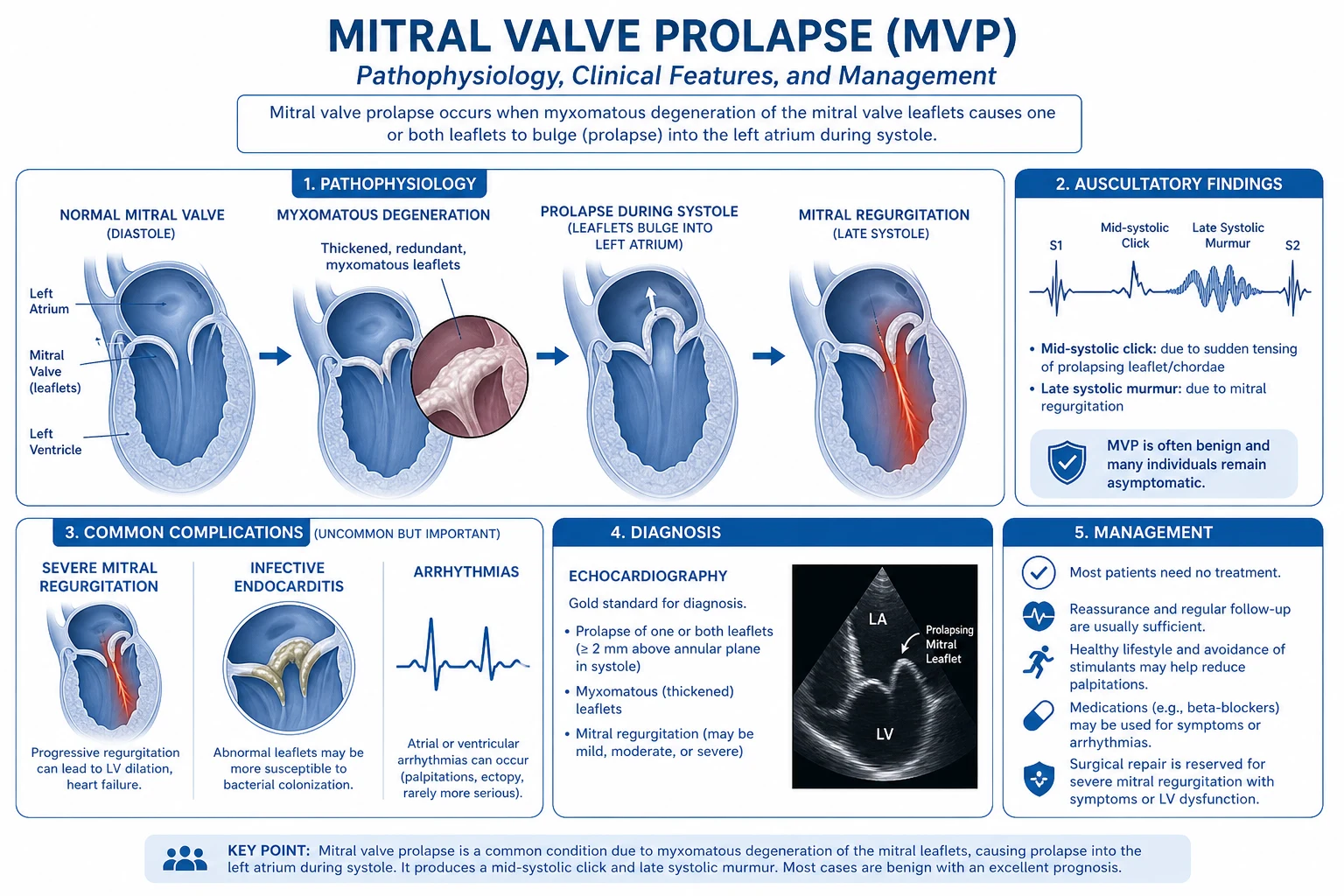
The valve abnormality is driven by myxomatous degeneration — a structural remodelling in which the leaflet spongiosa is infiltrated by proteoglycans, collagen is fragmented, and chordae tendineae are elongated (and prone to spontaneous rupture). When prolapse produces incomplete leaflet coaptation, mitral regurgitation (MR) follows; MR is the mechanical, symptomatic, prognostic and operative determinant of MVP. The mitral annulus may dilate (especially the posterior portion) and may separate from the ventricular myocardium — mitral annular disjunction (MAD) — providing the substrate for the rare arrhythmic MVP syndrome.[4][3]
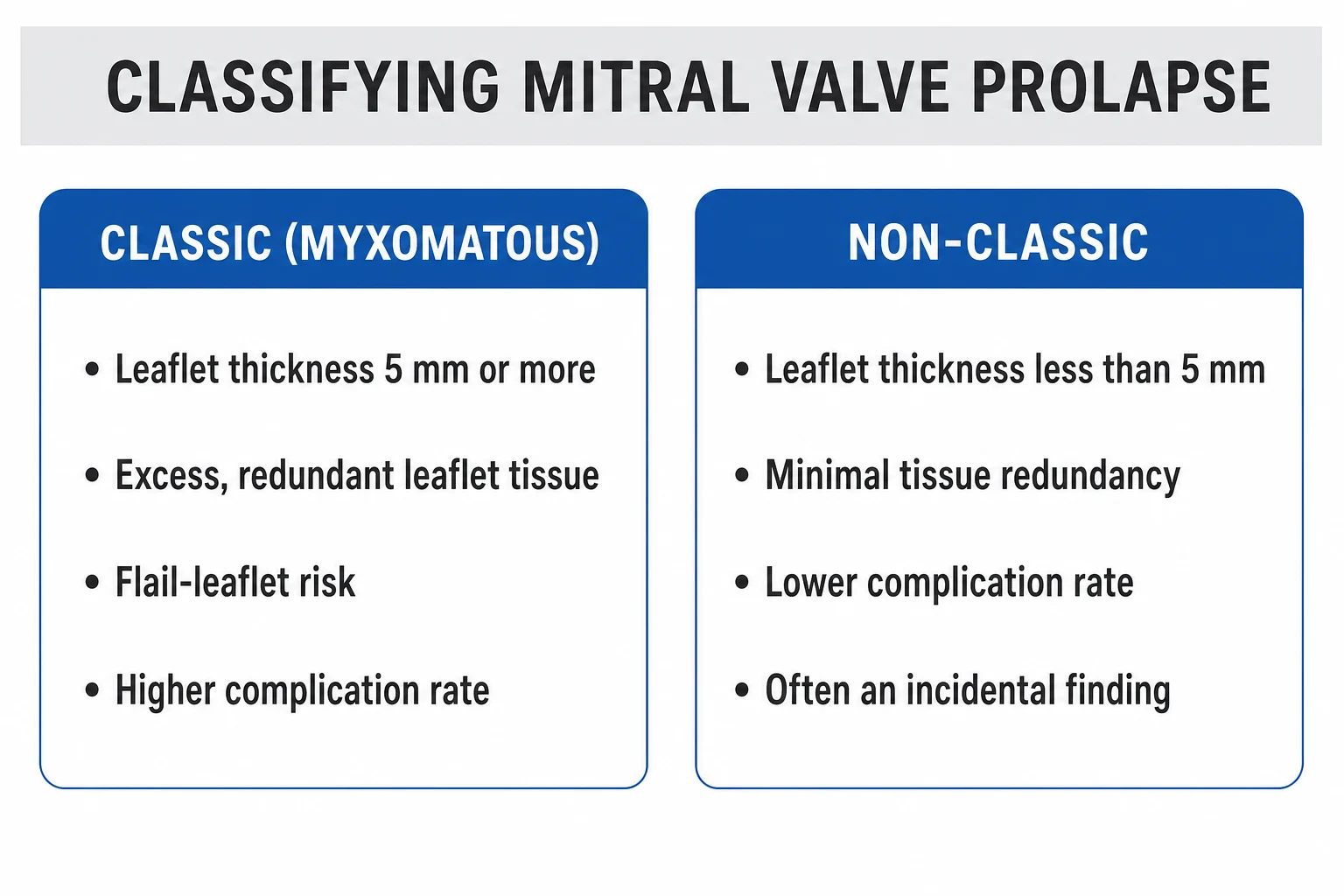
Two clinical patterns dominate. In the first, the prolapsing leaflet is thin, the patient is younger, and prolapse is focal (most often the P2 scallop of the posterior leaflet): this is fibroelastic deficiency (the German Feder-Elastische-Insuffizienz), characterised by abrupt chordal rupture and acute severe MR. In the second, the leaflets are diffusely thickened, redundant, multi-segmentally billowing (often bileaflet) and the annulus is markedly dilated: this is diffuse myxomatous Barlow's disease (Barlow-Pocock), typically presenting in middle life with chronic severe MR and arrhythmias.[5]
Why the definition matters for the examiner. Two misuses recur in the exam hall: (1) MVP is a structural/anatomical finding, not a clinical syndrome — the auscultatory click-murmur is one presentation of MVP, not its definition; the diagnosis is echocardiographic; (2) a prolapsing mitral leaflet is not automatically pathological — physiological prolapse (very low body-mass index, small LV volumes) falls above the modern threshold but is rarely clinically relevant.[1][2]
The clinical importance of MVP is its breadth: it is the commonest primary valvular abnormality in developed populations (2 to 3 percent prevalence), is usually benign, but accounts for most primary mitral surgery, much mitral endocarditis, and a recognisable proportion of sudden cardiac death in the young — and it is exactly these exceptions that the exam rewards.[1][2][3]
Classification
MVP is classified by aetiology (primary vs secondary), morphology (Barlow's diffuse vs fibroelastic deficiency vs MASS phenotype), leaflet involvement (anterior, posterior, bileaflet), MR severity, and, in a small subset, arrhythmic potential (arrhythmic MVP with MAD). The Carpentier classification of mitral repair is a surgical adjunct.[1][5]
Primary (degenerative) MVP
- The classic form — myxomatous degeneration of an otherwise structurally normal valve
- Fibroelastic deficiency (young, thin leaflet, focal P2 prolapse, chordal rupture, acute MR)
- Diffuse myxomatous Barlow's disease (multisegmental, bileaflet, redundant tissue, annular dilatation, chronic severe MR)
- MASS phenotype (Mitral valve, Aorta, Skeleton, Skin) — flail mitral, aortic root dilation, skeletal features, striae
- Familial autosomal-dominant myxomatous degeneration — rare single-gene forms (e.g. FLNA, DCHS1, DZIP1)
Secondary MVP
- Leaflet prolapse from another mechanism — not primary myxomatous degeneration
- Chordal elongation or rupture in rheumatic disease, endocarditis (post-inflammatory)
- Ruptured chord from fibrocalcific disease of the elderly
- Functional prolapse from LV geometry change (dilated cardiomyopathy, post-MI)
- Traumatic chordal rupture (rare)
Arrhythmic MVP with MAD
- A distinct high-risk phenotype — bileaflet prolapse + mitral annular disjunction + myocardial fibrosis of the inferolateral LV base + ventricular arrhythmia (PVC burden over 10 percent / 24 h, polymorphic VT, VF)
- Encompassing of MAD (Carmo 2010) and codified by the EHRA 2022 consensus
- Strong female preponderance, often young adult (30 to 50 years), preserved LV, mild-moderate MR
- Annual sudden-death event rate roughly 0.4 to 1.9 percent in those with sustained arrhythmia on Holter
- Risk stratification: Holter PVC burden, NSVT, complex morphology, late gadolinium enhancement on MRI, family history of SCD
The anatomic classification by leaflet involvement drives surgical planning: [1]
- Posterior-leaflet prolapse (most common) — usually P2; classic form, easiest to repair (quadrangular resection, neochordoplasty, ring annuloplasty).
- Anterior-leaflet prolapse — A2 prolapse; less common, technically harder to repair, more recurrence.
- Bileaflet prolapse — diffuse Barlow's disease; technically challenging; dedicated mitral centres report durable repair in over 90 percent.[1][2]
The MASS phenotype (Mitral valve, Aorta, Skeleton, Skin) is the connective-tissue-associated MVP variant — historically grouped with Marfan; in modern nosology it is an autosomal-dominant myxomatous degeneration of the mitral valve, aorta and skeleton, distinct from FBN1 Marfan but with phenotypic overlap. Marfan (FBN1), Ehlers-Danlos (vascular, classical), and Loeys-Dietz (TGFBR1/2) are connective-tissue disorders to screen for in young MVP patients with systemic features.[1][2]
Epidemiology & Risk Factors
Population prevalence of MVP is 2 to 3 percent when diagnosed by the modern echocardiographic (2 mm) criterion; older estimates of 5 to 15 percent used auscultation or looser echo thresholds and over-diagnosed thin-leaflet billowing.[1]
Age and sex: the classic form has a 2:1 female predominance, particularly under age 40. The prevalence in men rises with age as fibroelastic deficiency (often men, mid-60s, focal P2) becomes the leading mechanism of primary MR. The Framingham offspring cohort (Delling et al.) established the modern 2 to 3 percent figure and showed the highest valve-event rates in those with moderate-severe MR or leaflet thickening above 5 mm.[1]
Risk factors: [1]
- Female sex — the dominant risk for the classic form.
- Younger age for the classic Barlow's presentation; older age (60 to 80) for fibroelastic deficiency and chordal rupture.
- Connective-tissue disorders — Marfan syndrome (FBN1), Ehlers-Danlos (COL3A1, COL5A1/2), Loeys-Dietz (TGFBR1/2), osteogenesis imperfecta.
- Familial non-syndromic MVP — autosomal-dominant with variable penetrance; rare monogenic loci include FLNA, DCHS1, DZIP1.
- Low body-mass index and small LV volumes — slight increases in physiological prolapse (rarely pathological).
- Prior rheumatic fever, endocarditis, or chordal trauma — secondary MVP.
- Aortic root disease, scoliosis, pectus excavatum, joint hypermobility, skin striae — MASS phenotype overlap.[1][2]
Race and ethnicity: in Framingham the prevalence is roughly similar across white and Black Americans; some Asian populations show a lower prevalence of primary MR from myxomatous disease relative to rheumatic disease. [1]
The clinically important event rates in primary MVP: mitral surgery roughly 0.5 to 1 percent per year in the overall MVP population, endocarditis roughly 0.05 percent per year in normal MVP, rising above the population baseline in patients with MR, and sudden cardiac death roughly 0.2 to 0.4 percent per year overall with a higher rate in the arrhythmic MVP with MAD subtype.[3][1]
Pathophysiology
The pathophysiology of MVP is best understood as a structural and biomechanical failure of the leaflets and subvalvular apparatus, with downstream effects on left atrial (LA) and left ventricular (LV) remodelling and, in a minority, arrhythmogenic remodelling of the inferolateral basal myocardium. [1]
1. Leaflet and chordal pathology — myxomatous degeneration. The leaflet spongiosa layer is expanded by proteoglycan accumulation, collagen bundles are fragmented, and the fibrosa thins. The leaflets become redundant, thickened (above 5 mm) and prone to prolapse. Chordae tendineae elongate (allowing prolapse) and may rupture acutely (fibroelastic deficiency). The mitral annulus dilates, particularly at the posterior portion, accentuating the prolapse and non-coaptation. Strain distribution on the leaflet is altered, accelerating the degeneration — a positive-feedback biomechanical loop.[1][2][5]
2. Mitral annular disjunction (MAD). MAD is the systolic separation of the mitral annulus from the ventricular myocardium, visible on echocardiography as a wide-gap separation of the posterior annulus from the LV wall in the parasternal long-axis or on cardiac CT/MRI. First described by Carmo et al. (2010) in myxomatous MVP, MAD is associated with abnormal annular motion, hypermobility of the mitral apparatus and mechanical traction on the inferolateral basal myocardium, which is implicated in the arrhythmic MVP phenotype. MAD may be congenital or acquired and is highly prevalent in bileaflet prolapse.[4][3]
3. The inferolateral basal myocardium and ventricular arrhythmia. Cardiac MRI with late gadolinium enhancement (LGE) in arrhythmic MVP demonstrates focal fibrosis of the inferolateral LV base and sometimes the papillary muscles, with T-wave inversion in inferior leads on ECG at rest and frequent / complex PVCs (typically right-ventricular-outflow or fascicular or papillary-muscle-origin morphology) on Holter. The combination of mechanical traction (MAD) + myxomatous MV + basal myocardial fibrosis creates the substrate for polymorphic VT/VF.[3][1]
4. MR — the haemodynamic axis. Mild MR is late systolic and eccentric (posterior leaflet prolapse directs the jet anteriorly, hugging the LA wall — easy to underestimate on colour Doppler). Severe MR drives chronic volume overload: LA enlargement, AF onset, pulmonary venous hypertension and secondary right-heart strain, and LV eccentric hypertrophy eventually with falling LV EF below 60 percent (the surgical trigger). [1]
5. The neuro-endocrine / autonomic axis. A subset of patients has a dysautonomia-like phenotype — exaggerated sympathetic response, orthostatic intolerance and panic-like symptoms — that may account for palpitations and atypical chest pain independent of MR severity. Beta-blockade (with metoprolol or bisoprolol) is mechanistically rational in these patients.[1][2]
Clinical Presentation
Most patients with MVP are asymptomatic — the diagnosis is incidental on echo done for any reason (investigation of murmur, AF, syncope, or screening). The symptomatic presentations cluster into three groups: mechanical (related to MR), arrhythmic (related to MAD and basal fibrosis), and autonomic / constitutional. [1]
Symptoms (the high-yield list): [1]
- Palpitations — the most common symptom; usually premature ventricular contractions (PVCs) or non-sustained ventricular tachycardia (NSVT) of right- or left-ventricular origin, sometimes paroxysmal supraventricular tachycardia (PSVT) or atrial fibrillation in older patients with mitral regurgitation.
- Atypical chest pain — sharp, brief, variable location, often non-exertional; resembles pericarditis or panic more than angina. The mechanism is unclear (mechanical traction, papillary-muscle strain, autonomic).
- Dyspnoea on exertion and reduced exercise capacity — indicates haemodynamically significant MR; should prompt quantification.
- Fatigue, exercise intolerance, light-headedness — usually multifactorial.
- Syncope or pre-syncope — think of arrhythmia (NSVT, bradyarrhythmia from vagal excess), or autonomic dysfunction.
- Anxiety, panic-like episodes, hyperventilation — overlap with dysautonomia; panic disorder may be wrongly diagnosed.
- Neurological events — TIA or ischaemic stroke from AF-related thromboembolism in patients with MR and LA dilatation.[1][2]
Physical examination — the bedside essentials: [1]
- Mid-systolic click — high-pitched, heard best at the apex in left lateral decubitus, produced by the prolapsing leaflet snapping against the LA wall.
- Late systolic murmur — starts after the click, crescendo to S2, eccentric (anterior) jet if posterior leaflet, sometimes harsh or musical.
- Dynamic manoeuvres (the diagnostic test for the click-murmur):
- Valsalva (phase II, strain) and standing: venous return falls → LV volume falls → prolapse occurs earlier → click moves toward S1, murmur lengthens.
- Squatting or handgrip: venous return and afterload both rise → LV volume and pressure rise → prolapse occurs later → click moves toward S2, murmur shortens.
- Mid-systolic click that responds to Valsalva is the bedside signature of MVP.
- MR signs when MR is severe — apical thrill, pan-systolic radiation to axilla, displaced apex beat (LV dilatation), S3 with severe MR, signs of pulmonary hypertension (loud P2, raised JVP, hepatic congestion). [1]
Differential Diagnosis
The clinical skill is separating the click-murmur of MVP from its mimics, the prolapsing leaflet from physiological displacement, and the complications of MVP (MR, arrhythmia, endocarditis, embolic stroke) from primary conditions. [1]
Of mid-systolic click and late systolic murmur: [1]
- Hypertrophic cardiomyopathy (HOCM) — produces a mid-systolic murmur of LVOT obstruction that increases with Valsalva, but without the click of MVP; the carotid pulse is brisk and bisferiens, and there is usually a family history or asymmetric septal hypertrophy on echo.
- Tricuspid valve prolapse / tricuspid annular redundancy — usually a separate mid-systolic click, heard at the left lower sternal edge; often co-exists with MVP.
- Papillary-muscle dysfunction — from ischaemia or post-MI; the murmur is usually late systolic but lacks the earlier click of MVP and the response to Valsalva is opposite to that of MR from LV dilatation.
- Aortic stenosis (mild) — a crescendo-decrescendo ejection murmur starting after S1 and ending before S2, with a click from bicuspid aortic valve occasionally confused for MVP. Echo clarifies.
- Physiological mitral prolapse and innocent murmurs — benign, no MR on Doppler, no symptoms; do not over-diagnose.[1][2]
Of atypical chest pain in a young patient: [1]
- Panic disorder / anxiety with hyperventilation — frequently mistaken for or coexists with MVP; treat the panic.
- Acute coronary syndrome in the young — SCAD, anomalous coronary, premature CAD; pain is typically exertional and pressure-like.
- Pericarditis / myopericarditis — pleuritic, position-dependent pain with diffuse ST elevation on ECG.
- Pulmonary embolism — pleuritic, with dyspnoea, sinus tachycardia, low SpO2, computed-tomography pulmonary angiogram diagnostic.
- Costochondritis / chest-wall pain — reproducible on palpation, no echo correlates. [1]
Of dyspnoea and reduced exercise tolerance in MVP: [1]
- Primary MR from another cause — rheumatic, post-endocarditis, papillary muscle rupture.
- Heart failure with preserved EF (HFpEF) — older patients, HTN, LA enlargement, raised E/e'; may co-exist with MVP.
- Anaemia, thyroid disease — explain fatigue and palpitations; check TSH before attributing to MVP.
- Autonomic dysfunction / POTS — orthostatic tachycardia may accompany MVP.[1]
Of palpitation and arrhythmia: [1]
- Primary atrial fibrillation with a normal valve — MVP may be incidental.
- SVT (AVNRT, AVRT) in young patients — with structurally normal heart; treat with ablation.
- Long QT / Brugada / CPVT — risk-stratify young patients with MVP and syncope by 12-lead ECG at rest, Holter, and signal-averaged ECG.
- PVCs from RVOT — usually idiopathic in young patients without MVP. [1]
Of arrhythmic MVP and sudden cardiac death: [1]
- Arrhythmogenic right ventricular cardiomyopathy (ARVC) — RV dilatation, fat replacement, Epsilon wave on ECG, similar age profile.
- Long QT syndrome (LQTS) and Brugada syndrome — consider in family-history-positive syncope/SCD.
- Hypertrophic cardiomyopathy — syncope and SCD in young athletes, with asymmetric hypertrophy on echo.
- Primary coronary anomaly — present with sudden cardiac death in the young.[3][4]
The single best discriminator is the bedside echo + Holter + signal-averaged ECG — bileaflet prolapse with MAD, inferior T-wave inversion, complex PVCs on Holter, and LGE of the inferolateral basal myocardium identifies arrhythmic MVP. [1]
Clinical & Bedside Assessment
Step-by-step bedside assessment of suspected MVP (the examiner's algorithm): [1]
- History — symptoms (palpitations, atypical chest pain, dyspnoea, syncope, panic); systemic features (Marfanoid habitus, hypermobility); family history of MVP, SCD, connective-tissue disease.
- Inspection — pectus, scoliosis, joint hyperextension, skin hyperextensibility/striae, low body-mass index.
- Auscultation in the left lateral decubitus position with the bell and then the diaphragm, at the apex.
- Dynamic manoeuvres, each held 15 to 20 seconds:
- Valsalva (phase II) and standing → click moves earlier, murmur longer.
- Squatting (best performed on the floor or with a chair for safety) and handgrip → click moves later, murmur shorter.
- Position changes — the click in MVP is usually earlier while standing and delayed in squatting; HOCM murmur does the opposite (louder with Valsalva, softer with squatting) — opposite response is the bedside rule for distinguishing HOCM from MVP.[1][2]
- Estimate MR severity clinically — apical thrill, S3, axillary radiation, displaced apex, signs of pulmonary hypertension.
- Pulse and rhythm — regular or AF (AF is a marker of severity and a surgical trigger).
Risk stratification at presentation — the questions that decide "benign follow-up" vs "high-risk work-up": [1]
- Symptoms? — palpitations, atypical pain, dyspnoea, syncope.
- Severity of MR? — clinical and echo.
- LV size and function? — end-systolic diameter above 40 mm and EF below 60 percent are triggers for surgery.
- Pulmonary pressures? — raised pulmonary artery systolic pressure (>50 mmHg) is a surgical trigger.
- Rhythm? — AF is a surgical trigger.
- Connective-tissue features? — aortic root dilatation, skeletal features.
- Family history of SCD? — consider Holter and MRI for arrhythmic MVP.[1][3]
Investigations
Investigation is stepped: clinical suspicion → transthoracic echo (TTE) as the diagnostic workhorse → Holter and exercise ECG for arrhythmia assessment → TOE and cardiac MRI for the surgical / arrhythmic subset → bloods and ECG to complete the picture. [1]
First-line — for any patient with suspected MVP: [1]
- 12-lead ECG — usually normal; inferior T-wave inversion (II, III, aVF) and biphasic lateral T-waves are markers of arrhythmic MVP with basal fibrosis. Q waves are absent. ST elevation as in pericarditis should not be attributed to MVP.
- Transthoracic echocardiography (TTE) — the diagnostic cornerstone:
- Parasternal long-axis — leaflet displacement at least 2 mm above the annulus, usually P2; leaflet thickness (above or below 5 mm); mitral annular diameter; MAD (annular-ventricular separation in systole).
- Apical 4-chamber — leaflet coaptation line, leaflet billowing, prolapse extent, MR severity.
- MR severity — vena contracta (mm), EROA (mm^2), regurgitant volume (mL/beat) by PISA; an eccentric, anterior, wall-hugging jet is characteristic of posterior leaflet prolapse and is easy to underestimate.
- LV size and EF — end-systolic diameter, end-diastolic diameter, EF (Simpson's biplane).
- Left atrial volume index (LAVI) — >40 mL/m^2 is a marker of chronic MR and a surgical trigger.
- Pulmonary pressures — PASP from tricuspid regurgitation velocity; >50 mmHg is a surgical trigger.
- Right ventricular size and function.
- Chest X-ray — usually normal; LA and LV enlargement with severe chronic MR; pulmonary venous congestion when decompensated.[1][2]
Second-line — for symptoms or risk stratification: [1]
- 24 to 48 hour Holter (or 14-day patch) — PVC burden (%), NSVT, PSVT, paroxysmal AF. PVC burden above 10 percent and NSVT are markers of arrhythmic MVP.
- Exercise ECG / cardiopulmonary exercise test — assess functional capacity, exercise-induced arrhythmia, BP response.
- Cardiac MRI with LGE — indicated in arrhythmic MVP, syncope or family history of SCD to detect fibrosis of the inferolateral LV base and papillary muscles (a major risk marker); cine imaging also quantifies MR regurgitant fraction and LV volumes when echo is suboptimal.
- Signal-averaged ECG — late potentials may be positive in arrhythmic MVP.
- TOE — for preoperative planning (anatomy of prolapse, annular geometry, presence of clefts); not a first-line diagnostic test for MVP itself.
- 3D TTE / 3D TOE — at tertiary valve centres to plan repair (leaflet segmentation: A1-3 / P1-3). [1]
Specific situations: [1]
- Family history of SCD in MVP — TTE + Holter + cardiac MRI; consider ICD if criteria met.
- Marfan / connective-tissue features — aortic-root diameter (M-mode/sweep at sinuses of Valsalva), aortic-root Z-score; consider CT of the aorta if any dilatation. Genetic testing.
- Discordance between clinical MR severity and echo — exercise or dobutamine stress echo with PASP to detect dynamic MR.
- Acute severe MR from chordal rupture — urgent TTE ± TOE, urgent surgery. Vasodilator bridge (sodium nitroprusside 0.5 to 10 mcg/kg/min) plus IABP if needed.[1][3][1]
Drug-dose literal list (for treatment sections): [1]
- metoprolol 25 to 100 mg twice daily (symptomatic palpitations; start low, titrate).
- bisoprolol 2.5 to 10 mg daily (alternative; once daily preferred).
- verapamil 80 to 240 mg daily (when beta-blocker is not tolerated).
- amiodarone 200 mg three times daily loading then 200 mg daily (for ventricular arrhythmia in arrhythmic MVP).
- amoxicillin 2 g orally 30 to 60 minutes before the procedure (endocarditis prophylaxis only in highest-risk).[1][2][3]
Management — Resuscitation
MVP rarely requires acute resuscitation — but two emergency presentations do. [1]
1. Acute severe MR from chordal rupture (Sudden-Pugh syndrome). Acute posterior or anterior leaflet flail produces sudden severe MR, flash pulmonary oedema, cardiogenic shock in the unprepared ventricle, and acute refractory pulmonary hypertension. The bed-side triad is sudden dyspnoea, a new loud pan-systolic murmur (often with thrill) and acute decompensated heart failure in a previously well patient. [1]
- Resuscitation: ABCDE and oxygen, large-bore IV access, intravenous diuretics (furosemide 40 to 80 mg IV bolus, repeated), vasodilator (sodium nitroprusside 0.5 to 10 mcg/kg/min titrated to SBP 100 to 110) to reduce afterload and forward flow into the aorta, inotropic support if hypotensive (dobutamine 2 to 20 mcg/kg/min), intra-aortic balloon pump (IABP) for afterload reduction and forward flow, non-invasive or invasive ventilation.
- Urgent TTE ± TOE to confirm diagnosis and anatomy.
- Emergency mitral valve surgery — repair (preferred) or replacement within 24 hours, especially if the patient stabilises on medical therapy; mortality climbs steeply every day of delay.
- Percutaneous edge-to-edge repair (MitraClip) is a bridge or palliative option when surgical risk is prohibitive, but durable in selected centres.[1][2][4]
2. Out-of-hospital cardiac arrest / polymorphic VT in arrhythmic MVP with MAD. Standard Advanced Cardiac Life Support (ACLS): high-quality CPR, defibrillation for VF, amiodarone 300 mg IV bolus (then 150 mg) for refractory VT/VF, lidocaine 1 to 1.5 mg/kg IV as second line. After ROSC, manage in an arrhythmia / cardiac ICU with targeted temperature management, antiarrhythmic therapy (amiodarone 200 mg three times daily loading then 200 mg daily, or sotalol), and ICD evaluation for secondary prevention — the EHRA consensus is that secondary prevention with ICD is indicated after cardiac arrest or sustained VT in arrhythmic MVP regardless of LV EF.[3]
Most MVP patients do NOT require emergency care, but ABCD diagnosis-then-disposition is required for any MVP patient presenting acutely with syncope, dyspnoea or neurological symptoms. [1]
Management — Definitive & Stepwise
The definitive management of MVP is risk-stratified and stepped by severity of MR, LV / LA / pulmonary-pressure consequences, rhythm, and connective-tissue / arrhythmic phenotype.[1][2][3]
Stepwise Protocol
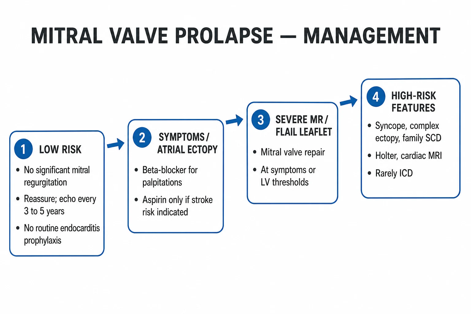
Step 1 — Asymptomatic, no MR or only mild MR: reassure, advise lifestyle (avoid extreme dehydration, maintain hydration for symptoms, encourage exercise), good dental hygiene to reduce the small risk of endocarditis, no antibiotics for routine procedures (the modern ESC and AHA position is no endocarditis prophylaxis except for the highest-risk subset — prosthetic valves, prior endocarditis, congenital cyanotic heart disease, transplanted valvulopathy). Repeat echo in 3 to 5 years unless symptoms change.[1][2]
Step 2 — Symptomatic palpitations, atypical chest pain, mild-moderate MR: beta-blocker first line — metoprolol 25 to 100 mg twice daily (start 12.5 to 25 mg twice daily, titrate) for palpitation and chest pain; bisoprolol 2.5 to 10 mg daily is an option with once-daily dosing; verapamil 80 to 240 mg daily in divided doses is an alternative when beta-blocker is not tolerated (avoid in severe LV dysfunction or significant MR). Treat coexisting anxiety / panic; reassure about the overall benign prognosis.[1]
Step 3 — Arrhythmic MVP with complex PVCs / NSVT on Holter: optimise beta-blockade (metoprolol 25 to 100 mg twice daily); antiarrhythmic therapy (amiodarone 200 mg three times daily loading for 5 to 7 days then 200 mg daily) for sustained or highly symptomatic VT; catheter ablation of PVCs (papillary muscle origin or fascicular origin) in tertiary centres for drug-refractory arrhythmia; ICD for secondary prevention after cardiac arrest or sustained VT. The EHRA consensus advocates individualised risk stratification for ICD in primary prevention (family history of SCD, syncope, NSVT, LGE on MRI).[3]
Step 4 — Severe MR — operative decision: mitral valve surgery. The 2020 ACC/AHA and 2014 AHA/ACC guidelines give Class I triggers for surgery: (1) severe primary MR with symptoms (NYHA II-IV), OR (2) asymptomatic severe MR with LV EF below 60 percent (falling EF in MR is a surgical trigger), OR (3) asymptomatic severe MR with LVESD above 40 mm. Additional Class IIa triggers: pulmonary hypertension (PASP above 50 mmHg), new-onset AF, severe MR with progressive LA enlargement (LAVI above 60 mL/m^2), connective-tissue disease with aortic-root involvement.[1][2]
Type of operation: [1]
- Mitral valve repair is preferred over replacement — lower operative mortality, better long-term survival, freedom from reoperation, better LV preservation and avoidance of anticoagulation in patients without AF. Repair rates exceed 90 percent at dedicated centres. Techniques: triangular or quadrangular resection of the prolapsing segment, neochordoplasty (artificial PTFE chordae), ring or band annuloplasty.
- Mitral valve replacement (MVR) with bioprosthesis for patients with repair failure, extensive disease, or calcification, particularly the elderly. Mechanical MVR for chronic AF requiring long-term anticoagulation and the young.
- Concomitant procedures — left atrial appendage ligation, MAZE / radiofrequency pulmonary-vein isolation for AF, aortic-root replacement for connective-tissue disease with aortic-root above 50 mm.[1][2]
Step 5 — Endocarditis prophylaxis (only for the highest-risk subset). The 2007 AHA guidelines, retained in the 2020 update, restrict antibiotic prophylaxis to: prosthetic valves, previous infective endocarditis, uncorrected cyanotic congenital heart disease, completely repaired congenital heart disease with prosthesis in the first 6 months, and repaired congenital lesions with residual defect at the device or adjacent site. MVP without MR is NOT in this list. MVP with severe MR undergoing high-risk dental work may warrant periprocedural amoxicillin 2 g orally 30 to 60 minutes before the procedure (alternatives: ampicillin, clindamycin 600 mg, azithromycin 500 mg, according to allergy status).[1][2]
Step 6 — Pregnancy and connective-tissue disease: cover in Special Populations. [1]
Step 7 — Anticoagulation for AF: standard CHA2DS2-VASc-driven oral anticoagulation — NOAC preferred over warfarin if no mechanical valve or moderate mitral stenosis; warfarin acceptable. Rate control with beta-blocker; rhythm control if symptomatic and feasible; LA appendage closure if long-standing AF and high bleeding risk.[1]
Step 8 — Follow-up: TTE annually for moderate MR or asymptomatic severe MR; 3 to 5 yearly for mild MR; immediate for any new symptom. [1]
Specific Subtypes & Scenarios
The exam rewards the recognition of specific scenarios that drive management. [1]
- Fibroelastic deficiency with acute chordal rupture — sudden severe MR; urgent repair with neochordoplasty + annuloplasty; acute presentation, repair within days. Operative mortality well under 5 percent at high-volume centres.
- Diffuse myxomatous Barlow's disease with chronic severe MR — bileaflet, multi-segmental prolapse, redundant tissue, severe annular dilatation; repair by experienced surgeons (complex resection, multiple neochordae, large annuloplasty ring). Durable repair but technically demanding; recurrence rate is higher than in fibroelastic deficiency.
- MASS phenotype (Mitral valve, Aorta, Skeleton, Skin) — Flail leaflet + aortic-root dilatation + skeletal features + skin hyperextensibility/striae; monitor aortic root, screen family.
- Arrhythmic MVP with MAD and cardiac arrest / sustained VT — ICD for secondary prevention, beta-blockade (metoprolol 25 to 100 mg twice daily, titrated), amiodarone 200 mg three times daily loading then 200 mg daily; catheter ablation for drug-refractory arrhythmia; mitral repair for severe MR may reduce arrhythmia burden in selected patients.
- MVP with new-onset AF — anticoagulation per CHA2DS2-VASc; rate/rhythm control; surgery for severe MR + MAZE / PVI at the time of repair.
- MVP with endocarditis — Duke criteria apply; blood cultures × 3, TTE then TOE, intravenous antibiotics (per local protocol, e.g. ampicillin + gentamicin, vancomycin + ceftriaxone, daptomycin); early surgery for heart failure, uncontrolled infection, embolic phenomena, large vegetations above 10 mm, or fungal infection.[3][4]
Complications
Cardiac: [1]
- Progressive mitral regurgitation — most important; severe MR within 10 years in roughly 5 to 10 percent of MVP overall, rising in those with leaflet thickness above 5 mm or MAD.
- Atrial fibrillation — from LA enlargement in chronic MR; embolism risk.
- Heart failure, pulmonary hypertension, right-heart failure.
- Endocarditis — highest risk in MVP with MR; cumulative lifetime risk roughly 0.05 to 0.1 percent per year, much higher in patients with prior endocarditis.
- Chordal rupture — acute severe MR.
- Ventricular arrhythmia and sudden cardiac death — arrhythmic MVP with MAD; NSVT in roughly 15 to 30 percent of MVP, sustained VT/VF in a small fraction (roughly 0.2 to 0.4 percent per year overall, higher in arrhythmic MVP).[3][1]
Embolic: [1]
- Ischaemic stroke from AF-related thromboembolism in MVP with chronic MR and LA dilatation; cryptogenic stroke in young patients with MAD is uncommon but described. [1]
Connective-tissue-related: [1]
- Aortic root dilatation, aortic dissection in Marfan (especially with mitral involvement).
- Progressive skeletal / skin features in systemic connective-tissue disorders. [1]
Surgical complications: [1]
- Repair failure / recurrence — 5 to 10 percent over 10 years at high-volume centres.
- Repair-related complications — systolic anterior motion (SAM), residual MR, haemolysis, ring dehiscence.
- Prosthetic-valve complications (replacement) — bleeding from anticoagulation, thrombosis, endocarditis, structural degeneration (bioprosthesis). [1]
Prognosis & Disposition
The prognosis of MVP is overwhelmingly benign. Most patients have mild MR, normal life expectancy and never require surgery. The clinical task is to identify the high-risk subsets and act: [1]
- Mild MR, asymptomatic — normal life expectancy, follow-up every 3 to 5 years.
- Moderate MR, asymptomatic — close follow-up and re-imaging 1 to 2 yearly; risk of progression to surgery roughly 10 to 15 percent at 10 years.
- Severe MR, treated conservatively — roughly 10 percent annual mortality in older data; modern cohort shows worse outcomes without surgery. Surgery restores life expectancy to normal in those operated before LV dysfunction sets in.
- Severe MR after repair — operative mortality under 2 percent at high-volume centres; 10-year survival 85 to 90 percent; freedom from reoperation 90 to 95 percent.
- Arrhythmic MVP with MAD, after cardiac arrest — high recurrence; ICD improves survival. [1]
Disposition: [1]
- Asymptomatic MVP — outpatient follow-up, with echo intervals as above.
- Symptomatic or severe MR — valve-clinic follow-up with cardiology, cardiac surgeon, and imaging.
- Acute severe MR / syncope / pulmonary oedema — admit for medical stabilisation and surgical decision.
- Cardiac arrest or sustained VT — admit to cardiac ICU / arrhythmia service for ICD evaluation.[1][2][3]
Special Populations
- Pregnancy — most MVP is well tolerated in pregnancy; severe MR with symptoms is managed medically with diuretics, beta-blocker (metoprolol preferred — long safety record), and inotropes for decompensated heart failure; surgery in pregnancy carries high foetal risk and is performed only for refractory cases (preferably postpartum). Peripartum decompensation in previously asymptomatic MVP may be the first presentation. Avoid angiotensin-converting-enzyme inhibitors and angiotensin-receptor blockers throughout pregnancy.
- Athletes — asymptomatic MVP without MR is not a bar to competitive sport; MVP with severe MR, LV dilatation, previous arrhythmia or systemic connective-tissue disease may require sports restriction — guidelines distinguish recommendations by severity. Systolic anterior motion (SAM) with MVP raises the differential of HOCM.
- Marfan syndrome and connective-tissue disease — regular aortic-root measurement (TTE annually if above 40 mm or rising), MRI/CT for monitoring, beta-blocker (metoprolol 25 to 100 mg twice daily, titrated) ± losartan for aortic protection; beta-blocker reduces aortic complications; MVP surgery may be combined with aortic-root replacement when aortic-root diameter approaches 50 mm.
- Elderly — fibroelastic deficiency predominates; chordal rupture is a common presentation; repair still preferred over replacement when feasible; transcatheter edge-to-edge repair (TEER, MitraClip) an option for high-risk elderly.
- Children — MVP is rare, usually associated with Marfan or connective-tissue disease; MASS phenotype in younger siblings of an index case.[1][2]
Evidence & Guidelines
Anchoring guidelines: [1]
- 2020 ACC/AHA Guideline for the Management of Patients With Valvular Heart Disease (Otto CM et al.) — the comprehensive primary reference for MVP diagnosis, follow-up, surgical indications and endocarditis prophylaxis. Establishes the Class I triggers for severe primary MR surgery.[1]
- 2014 AHA/ACC Guideline for the Management of Patients With Valvular Heart Disease (Nishimura RA et al.) — the precursor guideline; many of its staging recommendations and rheumatic-disease references remain in clinical use, especially the Class I surgical trigger for primary severe MR.[2]
- 2022 EHRA Expert Consensus Statement on Arrhythmic Mitral Valve Prolapse and Mitral Annular Disjunction (Sabbag A et al.) — the modern authority on MAD, arrhythmic-MVP risk stratification, antiarrhythmic therapy, ICD decision-making and ablation.[3]
Key primary references: [1]
- Barlow JB, Pocock WA (1979) — the original "specific billowing mitral leaflet syndrome" paper anchoring the Barlow phenotype.[5]
- Carmo P et al. (2010) — first systematic characterisation of mitral annular disjunction in myxomatous MVP.[4]
- Delling FN et al. (Framingham Heart Study) — modern prevalence data (2 to 3 percent) and outcome data in MVP.[1]
Controversies and ongoing trials: [1]
- Optimal ICD threshold for primary prevention in arrhythmic MVP (the consensus calls for individualised risk scores; a formal scoring system is awaited).
- Role of catheter ablation of papillary-muscle-origin PVCs in arrhythmia suppression (increasingly reported).
- Whether MAD repair during mitral surgery reduces arrhythmia burden (data emerging).
- Genetics — the role of cascade screening in familial non-syndromic MVP.
- Percutaneous edge-to-edge repair (MitraClip) versus surgery in high-risk patients (COAPT and MITRA-FR in mitral regurgitation are adjacent trials). [1]
Exam Pearls
- MVP = systolic billowing of one or both mitral leaflets at least 2 mm above the annulus (echo definition). Most patients are asymptomatic.
- Mid-systolic click + late systolic murmur at the apex; Valsalva shifts the click earlier (the bedside signature), squatting shifts it later — opposite to HOCM.
- Two main morphologies: fibroelastic deficiency (thin leaflet, focal P2, chordal rupture, acute MR) vs diffuse myxomatous Barlow's (multisegmental, bileaflet, annular dilatation, chronic severe MR).
- Prevalence 2 to 3 percent; 2:1 female predominance for the classic form; Marfan / Ehlers-Danlos / Loeys-Dietz association.
- Arrhythmic MVP with mitral annular disjunction (MAD) is a recognised cause of polymorphic VT/VF and sudden cardiac death (EHRA 2022 consensus).
- Management is risk-stratified: reassurance for asymptomatic, beta-blocker — metoprolol 25 to 100 mg twice daily (or bisoprolol 2.5 to 10 mg daily, verapamil 80 to 240 mg daily as alternative) for symptomatic palpitations or atypical chest pain; amiodarone 200 mg three times daily loading then 200 mg daily for ventricular arrhythmia; amoxicillin 2 g orally 30 to 60 minutes before the procedure for endocarditis prophylaxis in highest-risk patients only.
- Surgery (repair preferred over replacement) for severe primary MR, LV EF below 60 percent, LVESD above 40 mm, PASP above 50 mmHg, new AF.
- No endocarditis prophylaxis for routine MVP — only for prosthetic valves, prior endocarditis, cyanotic CHD, CHD prosthesis in first 6 months, or repaired CHD with residual defects.
- Anticoagulation for AF per CHA2DS2-VASc — NOAC preferred. [1]
Mitral valve prolapse — mnemonic
CLICK
Mid-systolic click + late systolic murmur at apex; moves EARLIER with Valsalva/standing, LATER with squatting
Echo criterion: ≥2 mm displacement above annulus in parasternal long-axis; thickness ≥5 mm for classic
Inferior T-wave inversion (II/III/aVF) marks arrhythmic MVP with MAD; PVCs and NSVT on Holter
Chordal elongation allows prolapse; acute rupture causes acute severe MR (Sudden-Pugh)
Repair (preferred over MVR) for severe MR with symptoms or LV EF below 60% or LVESD above 40 mm
Exam application bank (NEET-PG / INICET)
One-line answer
Mitral valve prolapse (MVP) is the systolic billowing of one or both mitral leaflets above the mitral annulus into the left atrium, defined echocardiographically as leaflet displacement of at least 2 mm beyond the mitral annular plane in the parasternal long-axis view during systole. The underlying pathology is myxomatous degeneration of the leaflet (proteoglycan-rich matrix, fragmented collagen) with chordal elongation and often annular dilatation or mitral annular disjunction (MAD). MVP is the commonest primary valve abnormality in developed countries, with a population prevalence of 2 to 3 percent and a 2:1 female predominance in classic (younger) forms. Clinically, two morphological phenotypes are recognised: fibroelastic deficiency (younger patients, thinner leaflets, focal prolapse, often P2 scallop, sudden chordal rupture, acute MR) and diffuse myxomatous Barlow's disease (multise
Worked stems (answer without another resource)
Stem 1 — Classic presentation. Map symptoms to mechanism; name the first investigation and first treatment step with dose/route if drug therapy is standard. [1]
Stem 2 — Unstable / complicated. List red flags that force immediate resuscitation, theatre, ICU, antidote, or reperfusion — and what you do in the first 15 minutes. [1]
Stem 3 — Atypical group. Elderly, pregnancy, child, or immunocompromised: how presentation and thresholds change. [1]
Stem 4 — Differential trap. Name the three closest mimics and one discriminator for each. [1]
Stem 5 — Disposition. Who goes home with safety-netting, who is admitted, who needs HDU/ICU/theatre, and what follow-up is mandatory. [1]
Rapid viva checklist
- Definition + classification
- Pathophysiology chain
- Bedside signs / criteria
- Score with exact components (if any)
- Emergency bundle
- Definitive therapy with doses
- Complications of disease and of treatment
- Special populations
- Guideline/trial name if classic
- Three exam traps
Coverage self-check
If you cannot answer any stem above from this page alone, re-read the matching section — the page is intended to be self-sufficient for final-prof and NEET-PG/INICET questions on Mitral Valve Prolapse.
[1]References
- [1]Otto CM, Nishimura RA, Bonow RO, et al. 2020 ACC/AHA Guideline for the Management of Patients With Valvular Heart Disease: A Report of the American College of Cardiology/American Heart Association Joint Committee on Clinical Practice Guidelines Circulation, 2021.PMID 33332150
- [2]Nishimura RA, Otto CM, Bonow RO, et al. 2014 AHA/ACC Guideline for the Management of Patients With Valvular Heart Disease: a report of the American College of Cardiology/American Heart Association Task Force on Practice Guidelines Circulation, 2014.PMID 24589853
- [3]Sabbag A, Essayagh B, Barrera JA, et al. EHRA expert consensus statement on arrhythmic mitral valve prolapse and mitral annular disjunction complex in collaboration with the ESC Council on valvular heart disease and the European Association of Cardiovascular Imaging endorsed cby the Heart Rhythm Society, by the Asia Pacific Heart Rhythm Society, and by the Latin American Heart Rhythm Society Europace, 2022.PMID 35951656
- [4]Carmo P, Andrade MJ, Aguiar C, et al. Mitral annular disjunction in myxomatous mitral valve disease: a relevant abnormality recognizable by transthoracic echocardiography Cardiovasc Ultrasound, 2010.PMID 21143934
- [5]Barlow JB, Pocock WA. Mitral valve prolapse, the specific billowing mitral leaflet syndrome, or an insignificant non-ejection systolic click Am Heart J, 1979.PMID 420065