cardiology · cardiology
Myocarditis
Also known as Myocarditis · Viral myocarditis · Lymphocytic myocarditis · Fulminant myocarditis · Giant cell myocarditis · Inflammatory cardiomyopathy
Myocarditis is an inflammatory disease of the myocardium confirmed by histological, immunological and immunohistochemical criteria, most commonly caused by viruses (coxsackievirus B3, adenovirus, parvovirus B19, enteroviruses, SARS-CoV-2). It presents with a clinical triad of chest pain, heart-failure symptoms and arrhythmia, frequently after a viral prodrome 1 to 2 weeks earlier. Diagnosis is suspected on chest pain plus raised high-sensitivity troponin with normal coronary angiography, and confirmed by cardiac MRI using the Lake Louise Criteria; endomyocardial biopsy remains the gold standard. Management is predominantly supportive (heart-failure therapy, arrhythmia control, activity restriction) — NSAIDs are avoided; giant cell, eosinophilic and sarcoid myocarditis respond to immunosuppression. Most patients recover fully; a minority progress to dilated cardiomyopathy or suffer sudden cardiac death, especially athletes.
On this page & tools
Your progress
Saved locally on this device.
Exam tags
Red flags
Overview & Definition
Myocarditis is an inflammatory disease of the myocardium confirmed by established histological (Dallas criteria), immunological and immunohistochemical criteria. The current ESC definition additionally requires a clinical picture compatible with myocarditis — symptoms, raised troponin, ECG/echo/MRI abnormalities — together with either biopsy-proven inflammation or characteristic cardiac MRI features (Lake Louise Criteria).[2][1]
Inflammatory cardiomyopathy is myocarditis in which ventricular dysfunction (usually dilated phenotype) develops as a consequence of the inflammation. The two terms sit on a continuum and are linked by the WHO/ISFC definition of cardiomyopathies (1995), which classifies inflammatory cardiomyopathy as a specific entity.[2]
The clinical skill is not the textbook triad (which is uncommon in full) but recognising the three overlapping phenotypes myocarditis can present as — (a) acute coronary syndrome mimic, (b) new heart failure / cardiomyopathy, (c) arrhythmia / sudden death — and excluding obstructive coronary disease, then risk-stratifying for fulminant, giant cell or refractory disease that changes management.[3][4]
Classification
Several classification axes run in parallel. The examinable ones are aetiology, histology, clinical course and clinical phenotype.[2][3]
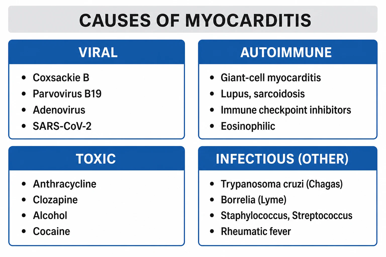
By aetiology (the axis that drives therapy):[3]
- Idiopathic — no cause found (declining as work-up intensifies).
- Viral (commonest in the developed world) — coxsackievirus B3 (the classical exam answer), adenovirus, parvovirus B19 (PVB19, now the commonest PCR-positive virus in adult biopsies), enteroviruses, human herpesvirus 6 (HHV-6), Epstein-Barr virus, cytomegalovirus, influenza A/B, SARS-CoV-2, HIV, hepatitis C, echovirus, mumps, rubella.
- Bacterial — Corynebacterium diphtheriae (toxin-mediated, classical), Borrelia burgdorferi (Lyme — with AV block), Mycoplasma, Streptococcus, Staphylococcus.
- Protozoal / parasitic — Trypanosoma cruzi (Chagas disease — commonest worldwide cause of myocarditis-related cardiomyopathy in endemic regions of Latin America), Toxoplasma gondii (immunocompromised), Trichinella.
- Fungal — Aspergillus, Candida (immunocompromised).
- Immune-mediated — systemic lupus erythematosus, rheumatoid arthritis, inflammatory bowel disease, giant cell myocarditis (idiopathic autoimmune), sarcoidosis.
- Hypersensitivity / drug-induced (eosinophilic) — clozapine, penicillins, sulfonamides, cephalosporins, diuretics (thiazides, furosemide), dobutamine, methyldopa, phenytoin, carbamazepine, tetanus toxoid, smallpox vaccine.
- Toxic — anthracyclines (doxorubicin, daunorubicin), cocaine, alcohol, radiation, chemotherapeutics (trastuzumab, cyclophosphamide, 5-fluorouracil), carbon monoxide, lead, snake/scorpion venom.
- Vaccine-associated — smallpox vaccine historically; mRNA COVID-19 vaccines (notably in young males after the second dose). [1]
By histology / Dallas classification (biopsy-defined, Aretz 1987, still the histological language):[2]
- Active myocarditis — characteristic inflammatory infiltrate with myocyte necrosis/degeneration not attributable to ischaemic damage.
- Borderline myocarditis — inflammatory infiltrate without myocyte damage.
- No myocarditis. [1]
By cellular infiltrate: lymphocytic (commonest, viral), eosinophilic (hypersensitivity, parasitic, Churg-Strauss), giant-cell (idiopathic giant cell myocarditis — aggressive autoimmune), granulomatous (sarcoid), neutrophilic (early bacterial, sepsis). [1]
By clinical course (McCarthy / Mayo, 2000) — fulminant vs acute (non-fulminant):[7]
- Fulminant myocarditis — distinct viral prodrome, rapid onset of severe heart failure within days, fever, frequently cardiogenic shock; on echo, severe global LV dysfunction with a non-dilated, thick-walled ventricle. Paradoxical good long-term prognosis if the patient is supported through the acute phase (mechanical circulatory support, VA-ECMO).
- Acute (non-fulminant) myocarditis — less distinct prodrome, more insidious onset of heart failure, dilated thin-walled ventricle; higher risk of progression to dilated cardiomyopathy and worse long-term survival than fulminant. [1]
By clinical phenotype (practical, guides work-up): ACS-mimic; new-onset heart failure / cardiomyopathy; arrhythmia or sudden cardiac death; incidental (subclinical troponin rise).[3]
Epidemiology & Risk Factors
True population incidence is unknown because most cases are subclinical and self-limiting. Autopsy series place myocarditis as the cause in approximately 5 to 10% of sudden cardiac deaths in young adults and up to 20% in athletes under 35.[3]
Demographic and host risk factors:[2][3]
- Young adults (under 40) — peak incidence 20 to 40 years; male predominance (2:1).
- Male sex — males develop more severe disease; oestrogen is cardioprotective in mouse models.
- Athletes — disproportionate sudden cardiac death risk; exertion during viraemia worsens injury.
- Pregnancy and peripartum period — peripartum cardiomyopathy overlaps.
- Immunocompromised — HIV, transplant, chemotherapy — atypical organisms (Toxoplasma, CMV, fungal).
- Genetic predisposition — HLA-DR haplotypes; rare familial giant cell myocarditis.
- Endemic exposures — Chagas disease in Latin America; tick bites (Lyme); young military recruits (smallpox vaccine).
- Recent viral illness, vaccine dose, or new drug (clozapine, sulfonamide, anthracycline). [1]
Pathophysiology
The human disease mirrors the classical three-phase mouse coxsackievirus B3 model (Woodruff; Liu & Mason):[3]
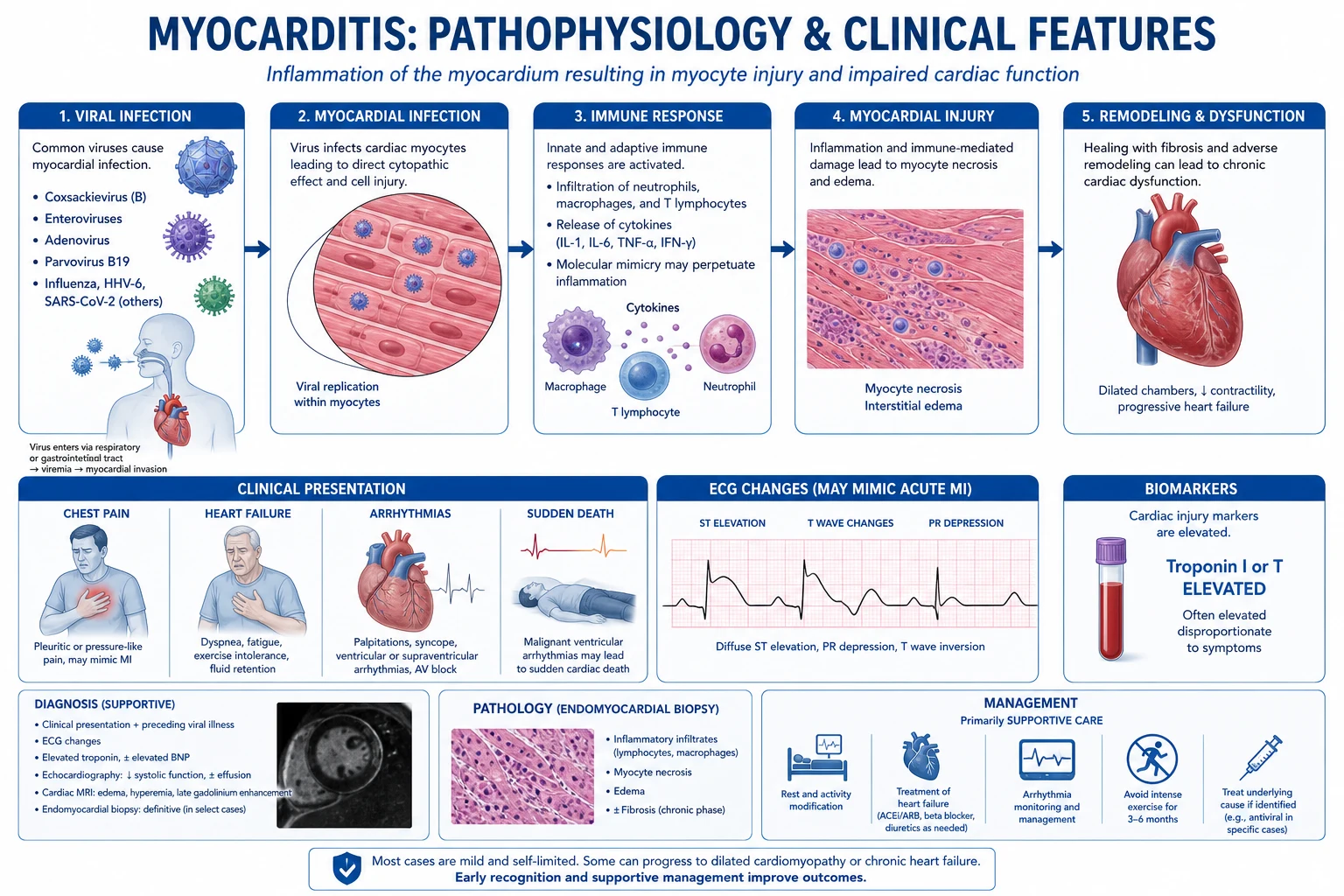
Phase 1 — acute injury (first ~7 days). The virus binds the coxsackievirus-adenovirus receptor (CAR) on the intercalated disc — a tight-junction protein that coxsackie B and adenoviruses share — with decay accelerating factor (DAF, CD55) as a co-receptor. Internalisation, replication and release of viral proteases 2A and 3C cleave host proteins including dystrophin, rupturing the dystrophin-glycoprotein complex and the sarcolemma, which allows intracellular calcium influx and myocyte necrosis with troponin leak.[3]
Phase 2 — immune-mediated injury (days 7 to 14). Necrotic myocytes release damage-associated molecular patterns (DAMPs) — cardiac myosin, heat-shock proteins, ATP — sensed by TLR3 and TLR4 and the NLRP3 inflammasome. Innate immunity (NK cells, M1 macrophages, type I interferons) is followed by adaptive immunity: CD8+ cytotoxic T cells kill infected cells via MHC class I, CD4+ Th1/Th17 cells recruit more inflammation, and B cells produce anti-cardiac myosin autoantibodies through molecular mimicry (anti-myosin, anti-β1-adrenergic receptor). It is this immune phase — not the virus — that dominates pathology by week two, which is why immunosuppression can be harmful in straightforward lymphocytic myocarditis but helpful in autoimmune forms.[2][3]
Phase 3 — chronic phase (weeks to months). A subset progress through viral persistence (PCR-positive biopsy) and/or ongoing autoimmunity to adverse remodelling — dilated cardiomyopathy with fibrosis. Predictors include certain viruses (adenovirus worse than PVB19 in some series), HLA-DR4, sustained LV dysfunction, and recurrent inflammation.[2]
Why the ECG and troponin mimic MI: patchy myocyte necrosis and adjacent pericardial inflammation produce regional ST elevation, T-wave inversion, PR depression (perimyocarditis) and troponin release indistinguishable from plaque rupture — hence the need for coronary angiography when the clinical setting (young, prodrome, atypical pain) does not fit obstructive CAD.[3]
Clinical Presentation
Myocarditis is a chameleon. The classical triad — chest pain, heart-failure symptoms, arrhythmia — is present in full only in a minority. The four clinical phenotypes below are the examinable patterns:[2][3]
Phenotype 1 — acute coronary syndrome mimic (commonest in young adults). Pleuritic or atypical chest pain, often preceded by a viral prodrome (fever, myalgia, sore throat, diarrhoea) 1 to 2 weeks earlier. ECG shows ST elevation, T-wave inversion or non-specific ST-T changes; troponin is markedly raised. Diffuse ST elevation with PR depression suggests perimyocarditis.[3]
Phenotype 2 — new heart failure / cardiomyopathy. Dyspnoea, orthopnoea, fatigue, fluid retention, sometimes rapidly progressive heart failure. Echo shows LV dysfunction, often global, sometimes regional. Fulminant myocarditis sits at the severe end of this spectrum: cardiogenic shock within days of a viral illness, hypotension, pulmonary oedema, multisystem hypoperfusion.[7]
Phenotype 3 — arrhythmia / sudden cardiac death. Palpitations, syncope, ventricular tachycardia/fibrillation, atrioventricular block (high-grade block in giant cell myocarditis, Lyme disease, sarcoid), or sudden death — particularly in athletes. An unexplained troponin rise after resuscitated cardiac arrest is a clue.[3]
Phenotype 4 — subclinical / incidental. Troponin rise in the setting of systemic viral illness (e.g., influenza, COVID-19) without cardiac symptoms; recognised increasingly with widespread troponin testing. [1]
Signs at the bedside: fever, tachycardia (often out of proportion to fever — relative bradycardia may occur in viral prodrome), S3 gallop, bibasal crackles, elevated JVP, pericardial friction rub (perimyocarditis), murmurs of functional mitral or tricuspid regurgitation, signs of low cardiac output (cool peripheries, oliguria, confusion) in fulminant disease. A patient who is systemically well with normal haemodynamics, normal echo and a small troponin rise is most likely to have mild myocarditis with a good prognosis.[2]
Atypical presentations (the examiner's favourites):[4]
- Elderly — presentation is dominated by heart failure rather than chest pain; prodrome may be absent; easily mistaken for ischaemic cardiomyopathy.
- Diabetic — pain may be muted; silently present as new HF or arrhythmia.
- Pregnant / peripartum — overlaps with peripartum cardiomyopathy; high index of suspicion.
- Immunocompromised — atypical organisms (CMV, Toxoplasma, fungal); can present as fulminant with a paucity of viral prodrome.
- Athlete — may present only as syncope or sudden death during exertion. [1]
Differential Diagnosis
Myocarditis is a diagnosis of pattern + exclusion of mimics. The differentials that must be actively considered and excluded:[3][4]
Acute MI / ACS
- Older, classic risk factors, exertional crescendo pain, reciprocal ST depression, regional wall-motion abnormality in a coronary distribution
- Culprit lesion on coronary angiography — the defining exclusion
- Troponin pattern: rise-and-fall with a peak; myocarditis troponin can stay elevated for days
Acute pericarditis
- Diffuse concave ST elevation, PR depression, PR-segment reciprocal changes in aVR; sharp, positional, pleuritic pain relieved by sitting forward
- Pericardial friction rub (three components)
- Troponin normal or mildly raised; concomitant myocarditis = perimyocarditis (the overlap is common)
Takotsubo (stress) cardiomyopathy
- Emotional/physical stressor, post-menopausal female, apical ballooning on echo/ventriculography
- Recovery of LV function over weeks
- Brain natriuretic peptide high; biopsy negative for inflammation
Sepsis-induced cardiomyopathy
- Circulating inflammatory cytokines (IL-1, IL-6, TNF) depress contractility in septic shock
- Reversible with sepsis resolution
- No myocardial necrosis pattern on biopsy
Pulmonary embolism
- Dyspnoea disproportionate to ECG changes, S1Q3T3 (uncommon), right-heart strain on echo, D-dimer raised, risk factors
- CT pulmonary angiography defines it
Arrhythmogenic RV cardiomyopathy (ARVC)
- Inherited desmosomal disease (plakoglobin, desmoplakin), epsilon waves, VT with LBBB morphology
- Fibro-fatty replacement on MRI/CMR, family history of sudden death
Cardiac sarcoidosis
- Granulomatous infiltrate on biopsy, AV block, PET-avid uptake, extra-cardiac sarcoid (hilar lymphadenopathy, uveitis)
- Confluent basal/intraseptal LGE on MRI
Giant cell myocarditis
- Rapidly progressive HF, sustained VT, high-grade AV block in middle age
- Multinucleated giant cells on biopsy; urgent biopsy in any fulminant or arrhythmic case
- Immunosuppression with cyclosporine + corticosteroid dramatically improves survival
Hypertrophic / dilated cardiomyopathy
- Long-standing, family history, characteristic echo (asymmetric HCM, dilated DCM)
- Biopsy shows non-specific changes, no florid inflammation
The single highest-yield differential question is myocarditis vs ACS in a young patient: if the coronary angiogram is normal in a patient with chest pain, raised troponin and ST changes, myocarditis (or Takotsubo) jumps to the top of the list — confirm with cardiac MRI.[3]
Clinical & Bedside Assessment
There is no bedside sign pathognomonic of myocarditis, so assessment focuses on (a) excluding alternative diagnoses and (b) recognising severity / the fulminant-giant-cell-spectrum.[3][4]
Vital signs drive triage: temperature (fever favours myocarditis or pericarditis over ACS), heart rate, respiratory rate, oxygen saturation, blood pressure, lactate (rising lactate signals poor perfusion / cardiogenic shock), urine output, conscious level. [1]
Cardiovascular examination: S3 gallop (LV dysfunction), bibasal crackles, elevated JVP, pericardial friction rub (perimyocarditis — best heard at left sternal border, leaning forward, in full expiration), functional mitral regurgitation murmur from LV dilatation, signs of low output (cool peripheries, weak pulse, prolonged capillary refill, mottled skin) in fulminant disease. Look for extra-cardiac clues: rash, lymphadenopathy (viral, sarcoid), arthritis (SLE, rheumatoid), uveitis/hilar fullness (sarcoid), erythema chronicum migrans (Lyme), recent vaccination site. [1]
Severity red flags at the bedside (warrant ICU and biopsy): sustained ventricular arrhythmia, high-grade or complete AV block, haemodynamic instability / cardiogenic shock, rapidly progressive heart failure, multisystem organ dysfunction — these together with a fulminant presentation raise giant cell myocarditis, which is biopsy-defined and treatable.[4]
Investigations
No single test diagnoses myocarditis; the diagnosis is syndromic — compatible clinical picture + raised troponin + ECG/echo abnormalities + characteristic CMR ± biopsy, after exclusion of coronary artery disease.[2][9]
First-line blood tests:[3]
- High-sensitivity troponin (hs-cTnT or hs-cTnI) — the hallmark; typically markedly and persistently raised, often higher than in ACS or with a different kinetic profile (sustained elevation rather than rise-and-fall). A normal troponin does not exclude myocarditis but makes florid disease unlikely.
- BNP / NT-proBNP — raised with myocardial strain / heart failure; prognostic.
- ESR / CRP — non-specific inflammation; raised in active disease, normal in chronic phase.
- Full blood count — eosinophilia (Churg-Strauss, hypersensitivity), lymphopenia (viral, HIV), atypical lymphocytes (EBV).
- U&E, LFTs, TFTs — renal/liver function (multisystem involvement in fulminant disease; exclude thyroid storm).
- Creatine kinase (CK) and CK-MB — can be elevated, especially if there is concurrent myositis.
- Blood cultures if bacteraemia/sepsis suspected; lactate for perfusion. [1]
Viral serology and PCR (selected, not routine):[2]
- SARS-CoV-2 PCR / antigen, influenza/RSV PCR during respiratory seasons.
- Enterovirus / coxsackievirus, parvovirus B19, adenovirus, HHV-6, EBV, CMV PCR (blood and/or nasopharyngeal) when clinically indicated.
- HIV serology; Borrelia antibodies if tick exposure; Trypanosoma cruzi serology if travel/exposure risk (Chagas).
- Autoimmune screen (ANA, anti-dsDNA, ANCA, rheumatoid factor) when immune-mediated disease is suspected. [1]
12-lead ECG — abnormal in most symptomatic patients but non-specific:[2]
- Sinus tachycardia (commonest), non-specific ST-T changes, T-wave inversion.
- ST elevation (regional — mimics MI; or diffuse concave with PR depression — perimyocarditis).
- Pathological Q waves, low voltages, AV block (any degree — high-grade or complete heart block suggests giant cell, sarcoid, Lyme, diphtheria).
- Atrial and ventricular arrhythmias — atrial fibrillation, ventricular tachycardia, ventricular fibrillation, sudden death.
- Paced rhythm / defibrillator — not relevant in the de novo case. [1]
Chest X-ray — usually normal early; may show cardiomegaly, pulmonary venous congestion, pulmonary oedema, pleural effusion in heart failure. Look for hilar lymphadenopathy (sarcoid). [1]
Echocardiography (first-line imaging):[3]
- Findings may be normal in early or mild disease — a normal echo does not exclude myocarditis.
- LV systolic dysfunction — global (favours myocarditis/DCM) or regional (favours ACS but can occur in myocarditis).
- Increased wall thickness / increased echo brightness — oedema in fulminant myocarditis.
- Wall-motion abnormalities, pericardial effusion (perimyocarditis), intracardiac thrombus (a complication to seek).
- Fulminant pattern: severe global dysfunction with a thick-walled, non-dilated ventricle (vs the dilated thin ventricle of acute non-fulminant myocarditis) — McCarthy's key distinguishing features.[7]
Coronary angiography — mandatory in any patient presenting as ACS-mimic to exclude obstructive epicardial coronary disease; normal coronaries + syndrome = myocarditis (or Takotsubo) climbs the differential.[2]
Cardiac MRI — the non-invasive diagnostic standard, Lake Louise Criteria.[5][6]
Lake Louise Criteria — original (Friedrich 2009). In combination with a compatible clinical presentation, the diagnosis is supported if 2 or more of 3 are present:[6]
- Regional LV wall-motion abnormality (hypo-, a-, dyskinesia) with or without global LV dysfunction (RV can also be involved).
- Myocardial oedema — T2-weighted ratio of signal intensity of myocardium to skeletal muscle ≥ 1.8, OR absolute T2 relaxation time ≥ 59 ms.
- Late gadolinium enhancement (LGE) — non-ischaemic pattern: mid-wall, subepicardial, or patchy; typically inferolateral or anteroseptal; sparing the subendocardium (which distinguishes it from ischaemic LGE). [1]
Lake Louise Criteria — updated (Ferreira 2018). Adds parametric mapping. The diagnosis is supported if at least one T2-based AND one T1-based criterion is positive:[5]
- T2-based (oedema): regional or global increase in T2 signal — T2 mapping ≥ 2 standard deviations above normal OR T2 ratio ≥ 1.8.
- T1-based (injury / fibrosis / increased extracellular volume): native T1 mapping increased (≥ 2 SD above normal), or extracellular volume fraction (ECV) increased, or non-ischaemic LGE present.
- (Mapping is now preferred over T2 ratio because it is more reproducible across centres.) [1]
CMR also detects pericardial involvement (perimyocarditis), RV involvement, intracardiac thrombus, and quantifies LV/RV function and oedema extent (prognostic — larger oedema burden predicts worse outcome). [1]
Endomyocardial biopsy (EMB) — the gold standard, but selective.[8]
- Dallas criteria (histology): active myocarditis (inflammatory infiltrate with myocyte necrosis/degeneration), borderline myocarditis (infiltrate without myocyte damage), or no myocarditis.
- Immunohistochemistry: raises sensitivity — CD3+ T lymphocytes, CD68+ macrophages (≥ 14 cells/mm² for active), HLA class II upregulation.
- PCR for viral genome on biopsy — PVB19, enterovirus, adenovirus, HHV-6, EBV, CMV; viral persistence influences prognosis and some management decisions. [1]
EMB is recommended (ESC/AHA consensus) when it will change management:[2][8]
- Fulminant or rapidly progressive heart failure (suspect giant cell myocarditis — needs immunosuppression).
- New, unexplained sustained ventricular arrhythmia or high-grade AV block (suspect giant cell, sarcoid).
- Refractory cardiogenic shock being considered for mechanical circulatory support or transplant.
- Suspected eosinophilic / hypersensitivity / drug-induced myocarditis (confirm and stop drug + steroids).
- Deteriorating inflammatory cardiomyopathy where immunosuppression is being considered. [1]
Routine EMB is not required for typical, mild lymphocytic myocarditis where the diagnosis is comfortably established by CMR and the patient is haemodynamically stable. [1]
Lake Louise Criteria — what the numbers are
Management — Resuscitation
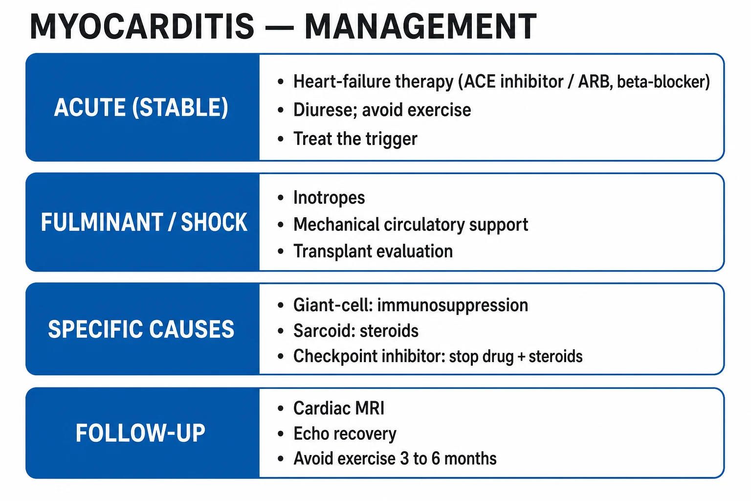
ABCDE first. Oxygen only if hypoxic — target SpO₂ 94 to 98% (or 88 to 92% in COPD/at risk of CO₂ retention).[2]
Cardiogenic shock or fulminant myocarditis — this is the resuscitation problem:[4][7]
- Two large-bore cannulae, monitoring (arterial line, central line, urinary catheter), continuous ECG, pulse oximetry, lactate trend.
- Inotropes / vasopressors — dobutamine 2 to 20 microgram/kg/min IV (β1 agonist, supports the failing LV) and/or milrinone 0.125 to 0.75 microgram/kg/min (PDE3 inhibitor — useful if tachyarrhythmia limits β-agonist use); add noradrenaline 0.05 to 1 microgram/kg/min for vasoplegia.
- Mechanical circulatory support (MCS) — escalate early in fulminant disease: intra-aortic balloon pump (IABP), Impella (percutaneous axial-flow LV assist), veno-arterial extracorporeal membrane oxygenation (VA-ECMO) — buys time for the inflamed myocardium to recover.
- Diurese the overloaded patient — IV furosemide 20 to 40 mg bolus (or infusion) to relieve pulmonary congestion; non-invasive ventilation (CPAP/BiPAP) for pulmonary oedema.
- Treat arrhythmias per ACLS — DC cardioversion for unstable VT/VF; correct electrolytes (K⁺ 4.0 to 5.0 mmol/L, Mg²⁺ ≥ 2.0); temporary transvenous pacing for symptomatic high-grade AV block.
- Anticoagulate if LV thrombus or severe LV dysfunction with a large apical akinetic region (prophylactic LMWH/heparin).
- Admit to ICU/CCU, urgent cardiology and (if fulminant) heart-failure/transplant team referral. [1]
Activity restriction starts now — strict bed rest in the acute phase; no exercise — exertion during viraemia/inflammation markedly increases sudden-death risk and extension of injury.[2]
Key drug-class caveats during resuscitation:[2]
- Avoid NSAIDs — they worsen myocardial inflammation, raise blood pressure, retain sodium, and are associated with worse outcomes in myocarditis (in contrast to their central role in pericarditis).
- Avoid high-dose digoxin (pro-arrhythmic in inflamed myocardium) and class Ic anti-arrhythmics in structurally abnormal hearts.
- Use beta-blockers and ACE inhibitors cautiously in the acute, haemodynamically unstable phase — they are started once the patient is stabilising. [1]
Management — Definitive & Stepwise
There is no specific antiviral or anti-inflammatory therapy proven for typical lymphocytic myocarditis. Definitive management is aetiology-driven and supportive:[2][4]
Step 1 — supportive heart-failure therapy (every phenotype):[2][4]
- ACE inhibitor / ARNI (e.g., ramipril 1.25 to 10 mg PO once daily, titrated; or sacubitril/valsartan 24/26 to 97/103 mg PO twice daily) — once haemodynamically stable; reduces afterload, opposes adverse remodelling.
- Beta-blocker (e.g., bisoprolol 1.25 to 10 mg PO once daily, or carvedilol 3.125 to 25 mg PO twice daily) — introduced only after decongestion and haemodynamic stability; reduces arrhythmia and sudden death.
- Mineralocorticoid receptor antagonist — spironolactone 12.5 to 50 mg PO once daily (or eplerenone 25 to 50 mg) if LVEF under 35% or symptomatic HF.
- Loop diuretic — furosemide 20 to 80 mg PO/IV for congestion; de-escalate as patient dries out.
- SGLT2 inhibitor (e.g., dapagliflozin 10 mg PO once daily or empagliflozin 10 mg PO once daily) — per recent heart-failure guidelines for reduced EF, once stable.
- Treat arrhythmia — rate control for AF (beta-blocker); anticoagulation (DOAC/warfarin) if AF, LV thrombus, or severe LV dysfunction with apical akinesia.
- Avoid NSAIDs. Use paracetamol for fever/pain. [1]
Step 2 — phenotype-specific therapy:[4]
- Typical lymphocytic viral myocarditis (the commonest form) — no immunosuppression. The landmark Myocarditis Treatment Trial (Mason et al., 1995) randomised biopsy-proven lymphocytic myocarditis to immunosuppression (prednisone + cyclosporine or azathioprine) vs placebo and showed no improvement in survival or LVEF; immunosuppression is therefore not routinely used. Treatment is supportive.[8]
- Giant cell myocarditis — urgent, high-dose immunosuppression: cyclosporine (target trough 200 to 300 nanogram/mL initially) combined with corticosteroids (e.g., methylprednisolone 0.5 to 1 g IV daily for 3 days, then prednisolone 1 mg/kg/day tapering) ± azathioprine or mycophenolate; transforms median survival from approximately 11 months (untreated) to over 12 months to several years; without therapy, death is nearly universal within months.[10]
- Eosinophilic / hypersensitivity myocarditis — withdraw the offending drug immediately (clozapine, sulfonamide, penicillin, cephalosporin, diuretic, etc.) and give systemic corticosteroids (prednisolone 1 mg/kg/day, taper over weeks); outcome is usually good if recognised early.
- Cardiac sarcoidosis — corticosteroids (prednisolone 30 to 60 mg/day, tapering over months) ± steroid-sparing agents (methotrexate, azathioprine, mycophenolate) for refractory disease; pacing/ICD for conduction disease and arrhythmia.
- Lyme carditis with high-grade AV block — IV ceftriaxone 2 g once daily (or oral doxycycline 100 mg twice daily for milder disease) for 14 to 21 days, plus temporary pacing for symptomatic block; usually fully reversible.
- Chagas myocarditis — benznidazole 5 to 8 mg/kg/day orally for 60 days (or nifurtimox) for acute/recent infection; manage chronic Chagas cardiomyopathy with standard HF therapy.
- Diphtheritic myocarditis — equine diphtheria antitoxin IV/IM urgently + penicillin G or erythromycin; supportive HF care; preventable by vaccination.
Step 3 — mechanical circulatory support and transplant. Refractory cardiogenic shock in fulminant myocarditis (especially giant cell) is an indication for VA-ECMO, Impella, or surgical LVAD as a bridge to decision, bridge to recovery, or bridge to transplant. Cardiac transplantation is reserved for irreversible end-stage inflammatory cardiomyopathy (note: giant cell myocarditis can recur in the transplanted heart, but survival is still good).[4]
Step 4 — long-term follow-up and return-to-play.[2][4]
- Serial echocardiography and CMR to monitor recovery of LV function, resolution of oedema, evolution of fibrosis.
- Activity restriction: no competitive sport, no moderate-to-vigorous exercise, for 3 to 6 months after diagnosis (ESC and AHA recommendation).
- Return-to-play criteria (after 3 to 6 months): (1) clinical recovery (asymptomatic), (2) normal serum biomarkers (troponin, BNP), (3) normal resting ECG (or stable, no new arrhythmia on Holter), (4) normal biventricular systolic function on echo/CMR, (5) no significant arrhythmia on exercise testing and ambulatory monitoring. [1]
Myocarditis causes — MYOCARD
MYOCARD
respiratory and systemic RNA viruses
diphtheria, borrelia, mycoplasma, staph, strep
clozapine, anthracyclines, cocaine, alcohol, radiation
the exam-favourite viral cause; uses CAR receptor
SLE, RA, IBD, sarcoid, idiopathic giant cell
drug-induced eosinophilic myocarditis — withdraw drug + steroids
penicillins, sulfonamides, cephalosporins
Specific Subtypes & Scenarios
Fulminant myocarditis. Defined by rapid onset of severe heart failure or cardiogenic shock within days of a viral illness, fever, and a severely depressed but non-dilated, thick-walled LV on echo. The paradoxical point — McCarthy NEJM 2000 — is that survival is BETTER than non-fulminant myocarditis if the patient is supported through the acute phase (often with VA-ECMO): 11-year transplant-free survival 93% vs 45%.[7]
Giant cell myocarditis. Idiopathic autoimmune disease of middle age (median 40s), often with other autoimmune disease (thyroiditis, myasthenia, ulcerative colitis). Presents with rapidly progressive heart failure, sustained ventricular arrhythmias, and high-grade AV block — the triad that should trigger urgent biopsy. Histology shows multinucleated giant cells. Treatment with cyclosporine + corticosteroids markedly improves survival; untreated, median survival is around 11 months.[10][4]
Eosinophilic / hypersensitivity myocarditis. Peripheral eosinophilia, rash, fever, new cardiac symptoms 2 to 10 weeks after starting a new drug. Classical offenders: clozapine (clozapine-induced myocarditis, ~1% incidence, monitor troponin and CRP during titration), penicillins, sulfonamides, cephalosporins, diuretics, dobutamine, phenytoin, carbamazepine. Also seen in eosinophilic granulomatosis with polyangiitis (Churg-Strauss) and parasitic infestation. Stop the drug + corticosteroids; outcome usually good.[4]
Cardiac sarcoidosis. Granulomatous inflammation, often basal/intraseptal, with AV block, ventricular arrhythmia, and cardiomyopathy. Look for extra-cardiac sarcoid (bilateral hilar lymphadenopathy, uveitis, erythema nodosum). Diagnosis: PET-CT uptake, characteristic CMR LGE, biopsy. Treat with corticosteroids ± steroid-sparing agents; ICD for sustained VT.[10]
Perimyocarditis. Simultaneous myocarditis and pericarditis — diffuse ST elevation, PR depression, pericardial rub, raised troponin, pericardial effusion. Treat pericardial inflammation with colchicine (acute pericarditis: colchicine 0.5 mg once/twice daily for 3 months ± aspirin 750 to 1000 mg every 8 hours for 1 to 2 weeks, tapering); avoid NSAIDs in significant LV dysfunction.[1]
COVID-19-related myocarditis and MIS-C. SARS-CoV-2 can cause myocarditis by direct viral injury, systemic hyperinflammation, and immune-mediated mechanisms. Multisystem inflammatory syndrome in children (MIS-C) — usually 2 to 6 weeks after COVID — presents with fever, gastrointestinal symptoms, rash, and cardiac dysfunction; treat with IV immunoglobulin (2 g/kg) and corticosteroids. Adult COVID-19 myocarditis ranges from incidental troponin rise to fulminant disease.[4]
mRNA COVID-19 vaccine-associated myocarditis. Characterised by chest pain within ~1 week of vaccination, raised troponin, more common after the second dose, in young males (peak 16 to 24 years), at a population rate of approximately 10 to 100 per million doses depending on age/sex. Usually mild and self-limiting; managed supportatively. Risk from COVID-19 infection itself is substantially higher, supporting continued vaccination.[4]
Sepsis-induced cardiomyopathy. Reversible biventricular dysfunction driven by circulating inflammatory cytokines (IL-1, IL-6, TNF-α, nitric oxide) in septic shock. No myocardial necrosis on biopsy. Resolves with sepsis resolution; treat the underlying sepsis.[3]
Toxic myocarditis. Anthracyclines (doxorubicin, daunorubicin) cause dose-dependent cardiotoxicity — both acute (myopericarditis) and chronic (DCM); cumulative lifetime doxorubicin dose is kept below 400 to 450 mg/m²; monitor with serial echo and troponin; dexrazoxane (iron chelator) is cardioprotective. Trastuzumab (HER2 therapy) causes a usually reversible cardiomyopathy. Cocaine causes coronary spasm, ischaemia and direct myocyte toxicity. Alcohol is a chronic cause of DCM.[3]
Complications & Pitfalls
Cardiac: heart failure (acute and chronic), progression to dilated cardiomyopathy (approximately 5 to 10% of biopsy-proven myocarditis), arrhythmia (atrial fibrillation, ventricular tachycardia/fibrillation, AV block), sudden cardiac death (especially athletes), cardiogenic shock in fulminant disease, intracardiac thrombus and systemic embolism, pericardial effusion and tamponade (perimyocarditis), recurrence.[2]
Systemic: multi-organ failure in cardiogenic shock; thromboembolism; complications of immunosuppression (infection, diabetes, osteoporosis) when used; transplant-related complications. [1]
- Treating myocarditis with NSAIDs (they are first-line in pericarditis — but harmful in myocarditis with LV dysfunction).
- Missing giant cell myocarditis by not performing EMB in a patient with fulminant HF, sustained VT or high-grade AV block (it is treatable with immunosuppression).
- Mislabelling myocarditis as "mild ACS with normal coronaries" and discharging the patient to sport — risking sudden death.
- Failing to restrict activity for 3 to 6 months — exercise during recovery is the single modifiable sudden-death risk.
- Stopping an ACE inhibitor or beta-blocker because of acute hypotension rather than recognising fulminant disease and escalating to inotropes/MCS.
- Giving immunosuppression to typical lymphocytic myocarditis — the Myocarditis Treatment Trial showed no benefit.
- Not stopping clozapine in a patient with rising troponin during clozapine titration (eosinophilic myocarditis).
- Forgetting Lyme in AV block with tick exposure and erythema chronicum migrans. [1]
Prognosis & Disposition
Most patients with mild, lymphocytic myocarditis recover fully within weeks to months. The fulminant paradox: despite appearing the sickest, fulminant myocarditis has better long-term survival than non-fulminant when supported through the acute phase (McCarthy 2000: 93% transplant-free survival at 11 years vs 45% for non-fulminant).[7]
Predictors of poor outcome:[2][4]
- Reduced LVEF (under 40%), persistent LV dysfunction, LV dilatation.
- Heart failure at presentation.
- Sustained ventricular arrhythmia, syncope, sudden-death history.
- Giant cell histology (aggressive course, but treatable).
- Persistent viral genome on biopsy (some viruses; adenovirus worse than PVB19 in several series).
- Advanced NYHA class, BNP elevation, renal dysfunction.
- Late gadolinium enhancement extent on CMR (larger = worse).
- Older age, comorbidity. [1]
Disposition:[2]
- Mild, haemodynamically stable, normal echo: ward-level monitoring, telemetry, supportive therapy; discharge when stable with strict activity restriction and early cardiology follow-up with repeat echo/CMR.
- LV dysfunction, arrhythmia, or significant symptoms: CCU/high-dependency; consider transfer to a heart-failure centre.
- Fulminant disease, cardiogenic shock, sustained VT, high-grade AV block: ICU, mechanical circulatory support, urgent cardiology and heart-failure/transplant referral, endomyocardial biopsy. [1]
Follow-up with serial echocardiography ± CMR at 1 to 3 months and again at 6 months; troponin and BNP trends; Holter monitoring for arrhythmia; assess return-to-play criteria in athletes.[4]
Special Populations
- Athletes — disproportionate sudden cardiac death risk. No competitive sport or moderate-vigorous exercise for 3 to 6 months. Return-to-play only when all five criteria are met (clinical recovery, normal biomarkers, normal ECG/Holter, normal LV function on imaging, no arrhythmia on exercise testing).[2]
- Children — myocarditis is an important cause of paediatric heart failure and sudden death; more often PCR-positive; can present as nonspecific respiratory/gastrointestinal illness with tachypnoea, hepatomegaly, poor feeding. High index of suspicion; aggressive supportive care; paediatric cardiology and transplant referral for refractory cases.
- Pregnancy and peripartum — overlaps with peripartum cardiomyopathy (last month of pregnancy to 5 months postpartum); both can produce LV dysfunction and thromboembolism; treat with pregnancy-safe heart-failure therapy (avoid ACEi/ARNI/MRA in pregnancy; use hydralazine + nitrates + beta-blocker; bromocriptine is emerging for peripartum cardiomyopathy).
- Elderly — presentation dominated by heart failure rather than chest pain; comorbid ischaemic heart disease complicates interpretation; lower threshold for coronary angiography.
- Immunocompromised (HIV, transplant, chemotherapy) — atypical organisms (CMV, Toxoplasma, fungal, bacterial); broader PCR/microbiology; biopsy often required; aggressive opportunistic-infection treatment.
- Anticoagulated patients — therapeutic anticoagulation should continue; intracardiac thrombus and embolic events warrant intensification.
Evidence, Guidelines & Regional Differences
The 2025 ESC Guidelines for the management of myocarditis and pericarditis (Schulz-Menger et al.) are the most current comprehensive European guidance, consolidating CMR (2018 Lake Louise Criteria), the role of EMB, and the phenotype-based management approach.[1]
The 2013 ESC position statement (Caforio et al.) was the prior benchmark, defining the syndromic diagnosis and the role of CMR and biopsy.[2]
The 2020 Expert Consensus on acute myocarditis and chronic inflammatory cardiomyopathy (Ammirati et al.) gives a practical phenotype-based algorithm and defines when immunosuppression is justified.[4]
The 2009 Lake Louise Criteria (Friedrich et al., JACC White Paper) standardised CMR diagnosis; the 2018 update (Ferreira et al.) added parametric T1/T2 mapping.[6][5]
The 2007 AHA/ACC/ESC Scientific Statement (Cooper et al.) defines the role and technique of endomyocardial biopsy.[8]
The 1995 Myocarditis Treatment Trial (Mason et al.) is the landmark negative trial — immunosuppression did not improve typical lymphocytic myocarditis — establishing supportive care as the default.[8]
- Developed world — viral causes predominate (PVB19 commonest PCR-positive, then enteroviruses, adenovirus, HHV-6).
- Latin America — Chagas disease (Trypanosoma cruzi) is the commonest cause of myocarditis-related cardiomyopathy; treat with benznidazole/nifurtimox.
- Africa and South Asia — rheumatic carditis (post-streptococcal, predominantly in children and young adults) overlaps in differential diagnosis; diphtheria persists in under-immunised populations; HIV-related myocarditis where prevalence is high.
- mRNA COVID-19 vaccine programmes — vaccine-associated myocarditis must be balanced against the higher myocarditis risk from COVID-19 itself. [1]
Exam Pearls
- Classical cause: coxsackievirus B3. Uses the coxsackievirus-adenovirus receptor (CAR); viral proteases 2A and 3C cleave dystrophin.
- Triad: chest pain, heart failure, arrhythmia — often after a viral prodrome 1 to 2 weeks earlier.
- ACS-mimic with NORMAL coronary angiogram = think myocarditis (or Takotsubo); confirm with cardiac MRI.
- Lake Louise Criteria (CMR): original 2009 = 2 of 3 (wall motion, T2 oedema, LGE); updated 2018 = 1 T2-based + 1 T1-based (T1/T2 mapping). LGE is subepicardial / mid-wall, inferolateral — distinguishing it from ischaemic subendocardial LGE.
- Dallas criteria (biopsy): active (infiltrate + necrosis) vs borderline (infiltrate only).
- Fulminant myocarditis: paradoxical BETTER prognosis than non-fulminant — but needs ICU/MCS. McCarthy NEJM 2000.
- Avoid NSAIDs (in contrast to pericarditis). Treat HF with ACEi/ARNI, beta-blocker, MRA, diuretic.
- Immunosuppression is ONLY for giant cell, eosinophilic/hypersensitivity, and sarcoid myocarditis — NOT typical lymphocytic (Myocarditis Treatment Trial).
- Giant cell myocarditis triad: rapidly progressive HF + sustained VT + high-grade AV block → urgent biopsy + cyclosporine + corticosteroid.
- Activity restriction 3 to 6 months. Return-to-play: 5 criteria (asymptomatic, normal biomarkers, normal ECG, normal LV function, no arrhythmia on Holter/exercise test).
- EMB when it changes management: fulminant/rapidly progressive HF, unexplained sustained VT/high-grade AV block, refractory shock being considered for MCS/transplant, suspected eosinophilic/drug.
- Diphtheria → antitoxin + penicillin; Lyme → ceftriaxone (AV block, often reversible); Chagas → benznidazole.
- mRNA vaccine myocarditis — young male, after 2nd dose, mild, self-limiting; risk from COVID-19 itself is higher. [1]
Exam application bank (NEET-PG / INICET)
One-line answer
Myocarditis is an inflammatory disease of the myocardium confirmed by histological, immunological and immunohistochemical criteria, most commonly caused by viruses (coxsackievirus B3, adenovirus, parvovirus B19, enteroviruses, SARS-CoV-2). It presents with a clinical triad of chest pain, heart-failure symptoms and arrhythmia, frequently after a viral prodrome 1 to 2 weeks earlier. Diagnosis is suspected on chest pain plus raised high-sensitivity troponin with normal coronary angiography, and confirmed by cardiac MRI using the Lake Louise Criteria; endomyocardial biopsy remains the gold standard. Management is predominantly supportive (heart-failure therapy, arrhythmia control, activity restriction) — NSAIDs are avoided; giant cell, eosinophilic and sarcoid myocarditis respond to immunosuppression. Most patients recover fully; a minority progress to dilated cardiomyopathy or suffer sudden
Worked stems (answer without another resource)
Stem 1 — Classic presentation. Map symptoms to mechanism; name the first investigation and first treatment step with dose/route if drug therapy is standard. [1]
Stem 2 — Unstable / complicated. List red flags that force immediate resuscitation, theatre, ICU, antidote, or reperfusion — and what you do in the first 15 minutes. [1]
Stem 3 — Atypical group. Elderly, pregnancy, child, or immunocompromised: how presentation and thresholds change. [1]
Stem 4 — Differential trap. Name the three closest mimics and one discriminator for each. [1]
Stem 5 — Disposition. Who goes home with safety-netting, who is admitted, who needs HDU/ICU/theatre, and what follow-up is mandatory. [1]
Rapid viva checklist
- Definition + classification
- Pathophysiology chain
- Bedside signs / criteria
- Score with exact components (if any)
- Emergency bundle
- Definitive therapy with doses
- Complications of disease and of treatment
- Special populations
- Guideline/trial name if classic
- Three exam traps
Coverage self-check
If you cannot answer any stem above from this page alone, re-read the matching section — the page is intended to be self-sufficient for final-prof and NEET-PG/INICET questions on Myocarditis.
References
- [1]Schulz-Menger J, Tschöpe C, Cooper LT Jr, et al. 2025 ESC Guidelines for the management of myocarditis and pericarditis Eur Heart J, 2025.PMID 40878297
- [2]Caforio AL, Pankuweit S, Arbustini E, et al. Current state of knowledge on aetiology, diagnosis, management, and therapy of myocarditis: a position statement of the European Society of Cardiology Working Group on Myocardial and Pericardial Diseases Eur Heart J, 2013.PMID 23824828
- [3]Sagar S, Liu PP, Cooper LT Jr. Myocarditis Lancet, 2012.PMID 22185868
- [4]Ammirati E, Veronese G, Bottiroli M, et al. Management of Acute Myocarditis and Chronic Inflammatory Cardiomyopathy: An Expert Consensus Document Circ Heart Fail, 2020.PMID 33176455
- [5]Ferreira VM, Schulz-Menger J, Holmvang G, et al. Cardiovascular Magnetic Resonance in Nonischemic Myocardial Inflammation: Expert Recommendations J Am Coll Cardiol, 2018.PMID 30545455
- [6]Friedrich MG, Sechtem U, Schulz-Menger J, et al. Cardiovascular magnetic resonance in myocarditis: A JACC White Paper J Am Coll Cardiol, 2009.PMID 19389557
- [7]McCarthy RE 3rd, Boehmer JP, Hruban RH, et al. Long-term outcome of fulminant myocarditis as compared with acute (nonfulminant) myocarditis N Engl J Med, 2000.PMID 10706898
- [8]Cooper LT, Baughman KL, Feldman AM, et al. The role of endomyocardial biopsy in the management of cardiovascular disease: a scientific statement from the American Heart Association, the American College of Cardiology, and the European Society of Cardiology. Endorsed by the Heart Failure Society of America and the Heart Failure Association of the European Society of Cardiology J Am Coll Cardiol, 2007.PMID 17980265
- [9]Biesbroek PS, Hirsch A, Zweerink A, et al. Diagnosis of myocarditis: Current state and future perspectives Int J Cardiol, 2015.PMID 25974197
- [10]Okura Y, Dec GW, Hare JM, et al. A clinical and histopathologic comparison of cardiac sarcoidosis and idiopathic giant cell myocarditis J Am Coll Cardiol, 2003.PMID 12535829