Cardiology · Cardiology
Non-ST-Elevation Acute Coronary Syndrome (NSTE-ACS)
Also known as NSTE-ACS · NSTEMI · Non-ST-elevation myocardial infarction · Unstable angina · Non-STEMI · Acute coronary syndrome without ST elevation
Non-ST-elevation acute coronary syndrome (NSTE-ACS) is myocardial ischaemia at rest or on minimal exertion caused by a partially or intermittently occlusive intra-coronary thrombus over a disrupted atherosclerotic plaque, but without the ST elevation that mandates immediate reperfusion. It comprises NSTEMI (myocardial necrosis demonstrated by a rise and/or fall of high-sensitivity cardiac troponin with at least one value above the 99th centile) and unstable angina (ischaemia without troponin release). Diagnosis rests on the 12-lead ECG within 10 minutes plus serial high-sensitivity troponin (0-hour/1-hour or 0-hour/3-hour algorithms). Risk-stratify with the GRACE (death) and TIMI (composite endpoint) scores to triage the timing of invasive coronary angiography: immediate (within 2 hours) for very high-risk (haemodynamic instability, life-threatening arrhythmia, mechanical complication, acute heart failure, recurrent dynamic ST-T change), early (within 24 hours) for high risk (GRACE above 140, dynamic ST-T change, troponin rise-fall), and within 72 hours for intermediate risk. Every patient receives immediate dual antiplatelet therapy (aspirin 300 mg load then 75 mg daily plus ticagrelor 180 mg load then 90 mg twice daily, or prasugrel/clopidogrel) and a parenteral anticoagulant (fondaparinux 2.5 mg subcutaneous daily, enoxaparin, unfractionated heparin, or bivalirudin), a high-intensity statin (atorvastatin 80 mg) within 24 hours, and beta-blockade and an ACE inhibitor where not contraindicated. NSTE-ACS is more common than STEMI in contemporary registries and carries a long-term mortality that equals or exceeds STEMI because of the larger burden of multivessel disease and recurrent events.
On this page & tools
Your progress
Saved locally on this device.
Exam tags
Red flags
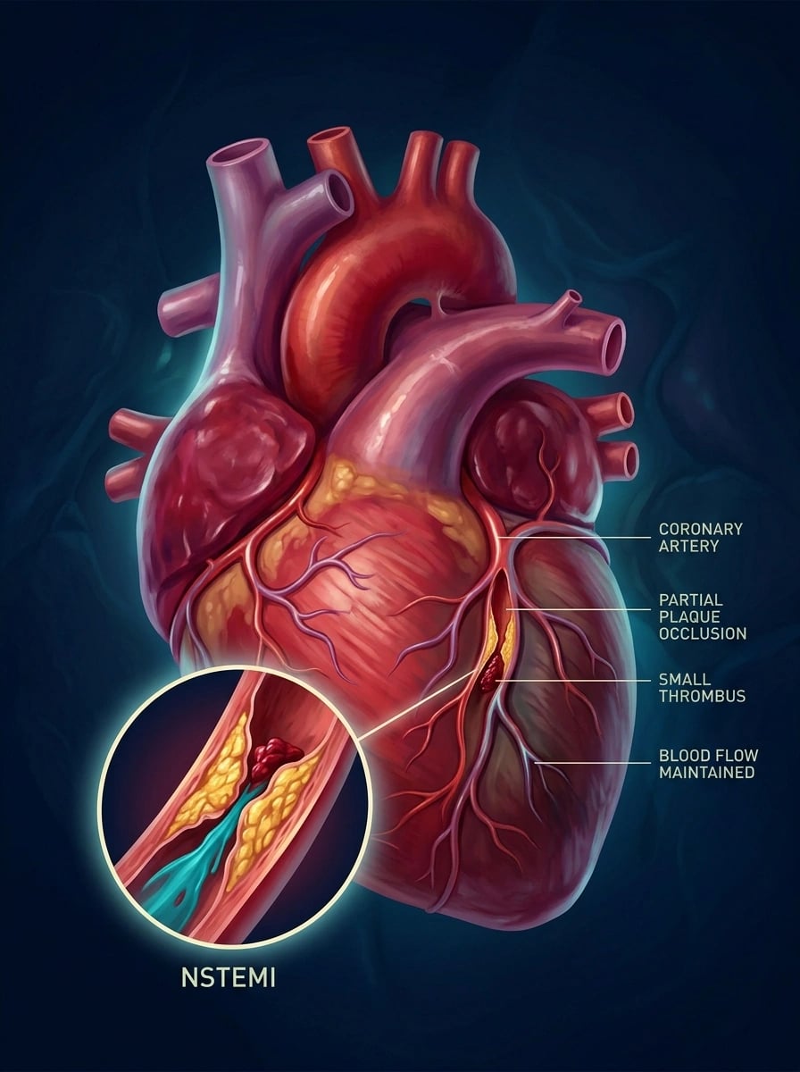
Overview & Definition
Acute coronary syndrome (ACS) is the spectrum of acute myocardial ischaemia caused by an abrupt reduction in coronary blood flow. It is divided, by the 12-lead ECG, into ST-elevation ACS (STE-ACS = STEMI) — complete coronary occlusion requiring immediate reperfusion — and non-ST-elevation ACS (NSTE-ACS), in which the culprit vessel is partially or only intermittently occluded and the ECG does not show ST elevation.[1]
NSTE-ACS is then split, by the presence of myocardial necrosis, into two entities with identical clinical presentation and management but different prognosis: [1]
- Non-ST-elevation myocardial infarction (NSTEMI) — myocardial necrosis is present, shown by a rise and/or fall of cardiac troponin with at least one value above the 99th centile upper reference limit of a healthy population, plus at least one of: symptoms of ischaemia, new ischaemic ECG changes, imaging evidence of new loss of viable myocardium or regional wall motion abnormality, or identification of an intracoronary thrombus at angiography or autopsy.[1]
- Unstable angina (UA) — ischaemia at rest/minimal exertion without biochemical evidence of necrosis; troponins do not rise (or rise without a dynamic pattern). With the near-universal adoption of high-sensitivity troponin assays, many former "unstable angina" patients now reclassify as NSTEMI, so UA has become a shrinking diagnosis reserved for ischaemic events with truly negative troponins.[1]
The Fourth Universal Definition of Myocardial Infarction classifies MI into five types; the vast majority of NSTE-ACS events are Type 1 MI (spontaneous MI from atherothrombotic plaque disruption — rupture, erosion, or dissection). Distinguishing Type 2 MI (an imbalance of myocardial oxygen supply and demand without plaque disruption — tachyarrhythmia, hypotension/shock, severe anaemia, hypoxia, hypertensive emergency, sepsis) is a recurring exam point: Type 2 has no plaque thrombus and is managed by treating the precipitant, not by routine catheterisation and intensive antithrombotic therapy.[1]
The clinical skill in NSTE-ACS is not recognising that the patient has ischaemia (the history and ECG usually do that) but three decisions that change outcome: (1) excluding STEMI and its mimics (aortic dissection, pericarditis) within the first 10 minutes; (2) separating NSTE-ACS from non-cardiac chest pain with high-sensitivity troponin; and (3) risk-stratifying to time the invasive strategy — because, unlike STEMI, the right answer is not "everyone to the lab immediately" but a graded, score-driven escalation.[1][2]
Classification
NSTE-ACS is classified along three axes that each drive a management decision. [1]
By the ECG versus the troponin (the two axes that define the ACS spectrum): [1]
STEMI (STE-ACS)
- ST elevation in at least 2 contiguous leads, or true posterior (ST depression V1 to V3) / new LBBB with ischaemic symptoms
- Complete coronary occlusion, transmural necrosis
- Immediate reperfusion: primary PCI within 120 min or fibrinolysis
- Troponin rises, but reperfusion is decided by the ECG, not by waiting for troponin
NSTEMI
- No persistent ST elevation; ST depression, T-wave inversion, transient ST elevation, or normal ECG
- Partial/intermittent occlusion; subendocardial (non-transmural) necrosis
- No immediate reperfusion mandate; risk-stratified invasive strategy
- High-sensitivity troponin positive with a rise and/or fall above the 99th centile
Unstable angina
- No ST elevation; may have dynamic ST depression or T inversion
- Ischaemia without necrosis
- Risk-stratified invasive strategy
- Troponin negative (no rise/fall above 99th centile)
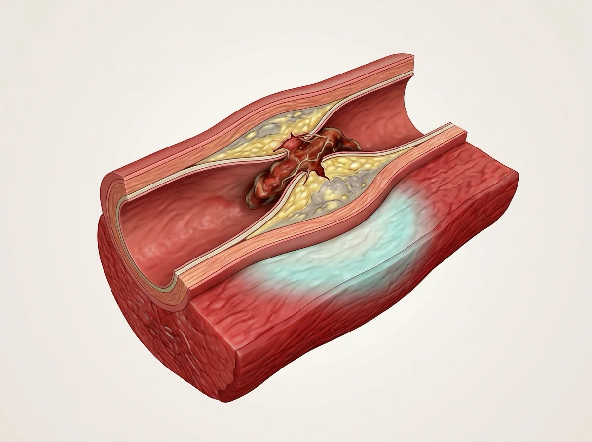
By Braunwald clinical presentation (classic classification, still examined): [1]
| Class | Definition |
|---|---|
| I B | New-onset severe angina or crescendo angina, not at rest (secondary if precipitated by an extracardiac condition) |
| II B | Angina at rest within the past month but not in the preceding 48 hours (sub-acute) |
| III B | Angina at rest within the preceding 48 hours (acute) — the commonest NSTE-ACS presentation |
| C | Post-myocardial infarction angina (within 2 weeks of an MI) |
(The suffixes A/B/C: A = secondary to an extracardiac oxygen-demand stress; B = primary/unstable; C = post-infarction. The higher the class, the higher the risk.) [1]
By the Universal Definition of MI type (the classification that drives mechanism): [1]
- Type 1 — spontaneous MI from atherothrombotic plaque disruption (the NSTE-ACS mechanism; managed as below).
- Type 2 — supply–demand mismatch without plaque disruption (treat the cause; troponin rises but there is no thrombotic substrate).
- Type 3 — MI causing sudden cardiac death before biomarkers can be obtained.
- Type 4a — MI related to PCI; 4b — stent/scaffold thrombosis; 4c — restenosis.
- Type 5 — MI related to CABG. [1]
Epidemiology & Risk Factors
NSTE-ACS is the commonest form of ACS in contemporary Western registries, accounting for roughly 70 per cent of all ACS admissions; STEMI has fallen with declines in smoking and improvements in prevention, while NSTE-ACS — driven by an ageing, multi-morbid population — has not. Incidence rises steeply with age and is higher in men until the female menopause, after which the sex gap narrows. South Asians (relevant to the Indian NEET-PG population) have among the highest rates of premature coronary disease worldwide, with NSTE-ACS frequently presenting a decade earlier than in Europeans.[1]
Crucially, although the in-hospital mortality of NSTEMI is lower than STEMI, the long-term (1-year and 5-year) mortality of NSTE-ACS equals or exceeds that of STEMI, because NSTE-ACS patients are older, carry more comorbidity (diabetes, chronic kidney disease, prior MI), and more often have multivessel disease and recurrent events. This is the central reason the disease is "critical" weight in any exam.[1]
Risk factors for the underlying atherosclerosis are the conventional, modifiable Framingham/INTERHEART cluster: tobacco smoking (the single largest population-attributable risk, especially in young South-Asian men), diabetes mellitus (doubles risk and confers a worse prognosis after NSTE-ACS), hypertension, dyslipidaemia (raised LDL, low HDL, raised apolipoprotein B), family history of premature coronary disease (first-degree male under 55, female under 65), age, male sex/post-menopausal, obesity and physical inactivity, psychosocial stress, and diet. The INTERHEART nine modifiable risk factors account for over 90 per cent of the population-attributable risk of MI.[1]
Precipitants of an acute event on a background of established (often subclinical) plaque (examiners love these): non-cardiac surgery (post-operative NSTE-ACS — the commonest peri-operative cardiac emergency), severe infection/sepsis, acute blood loss/anaemia, tachyarrhythmia (AF with rapid ventricular response), hypoxia (pneumonia, exacerbation of COPD), cocaine or amphetamine use (spasm plus thrombosis), thyrotoxicosis, and withdrawal of antiplatelet or statin therapy (a "statin holiday" or stopping aspirin precipitates rebound thrombosis). Recognising a Type 2 component (demand ischaemia) coexisting with a Type 1 event is part of the assessment. [1]
Pathophysiology
The unifying event in NSTE-ACS is myocardial ischaemia caused by an acute, dynamic reduction in coronary blood flow from an atherosclerotic plaque that has become unstable. The "five vicious Ps" — plaque, platelets, pressure (vasoconstriction), pumping (demand), and passage of emboli — are a useful memory frame. [1]
1. Plaque disruption. Most NSTE-ACS events begin with disruption of an atherosclerotic plaque. Two mechanisms: [1]
- Plaque rupture — a thin fibrous cap (under 65 micrometres, heavily loaded with macrophage-derived foam cells and scarce smooth muscle) tears, exposing the highly thrombogenic lipid/necrotic core (tissue factor, collagen, von Willebrand factor) to flowing blood. Rupture is the dominant mechanism in men and in STEMI.
- Plaque erosion — an intact but denuded endothelial surface over a plaque rich in proteoglycans recruits platelets directly, without a tear and without an exposed lipid core. Erosion is commoner in women and in sudden cardiac death, and tends to produce NSTE-ACS rather than STEMI. [1]
Plaques that precipitate ACS are typically only mildly to moderately stenotic (under 70 per cent) on prior angiography — they are vulnerable by virtue of a large lipid core, thin cap, and active inflammation (matrix metalloproteinases digesting the cap), not by their luminal narrowing. This is why a "normal" stress test or mild stenosis on a prior angiogram does not exclude future ACS.[1]
2. Platelet adhesion, activation, and aggregation. Exposure of subendothelial collagen and von Willebrand factor binds glycoprotein Ib-IX-V on platelets (especially under high shear in eroded plaques). Platelets activate, release ADP, thromboxane A2 (TXA2), and serotonin, and upregulate glycoprotein IIb/IIIa receptors, which bind fibrinogen to cross-link platelets into a growing aggregate. This is why the antiplatelet armamentarium targets three nodes: cyclo-oxygenase-1 (aspirin — blocks TXA2), the P2Y12 ADP receptor (clopidogrel, prasugrel, ticagrelor), and GPIIb/IIIa (abciximab, eptifibatide, tirofiban — the final common pathway).[5][6]
3. Coagulation cascade and thrombin generation. Tissue factor from the plaque core activates the extrinsic (VII) pathway, generating thrombin (Factor IIa). Thrombin both converts fibrinogen to fibrin (stabilising the clot as a mixed platelet-fibrin thrombus) and is the most powerful platelet activator known (via protease-activated receptors). Anticoagulants in NSTE-ACS therefore target thrombin or Factor Xa: unfractionated heparin and low-molecular-weight heparin (enoxaparin) potentiate antithrombin; fondaparinux is a selective indirect Factor Xa inhibitor; bivalirudin is a direct thrombin inhibitor.[8][9]
4. Dynamic obstruction and vasoconstriction. Serotonin and TXA2 released from platelets cause local coronary vasoconstriction, worsening stenosis. Superimposed epicardial spasm (variant/Prinzmetal component) or microvascular dysfunction may produce transient ST elevation. The obstruction in NSTE-ACS is therefore dynamic — waxing and waning — which is the substrate for crescendo/rest angina and for dynamic ST-T changes on serial ECGs.[1]
5. Distal embolisation and the troponin leak. Micro-fragments of the platelet-fibrin thrombus embolise downstream, plugging the microcirculation and producing patchy myocardial necrosis (subendocardial, non-transmural). Necrotic cardiomyocytes release cardiac troponin I and T into the circulation — detectable by high-sensitivity assays at 1 to 3 hours, peaking around 18 to 24 hours, and persisting for 7 to 14 days. The troponin rise/fall is the biochemical signature that separates NSTEMI from unstable angina. Because the necrosis is patchy and subendocardial (the inner, highest-demand layer perfused in systole and diastole), the ECG usually shows ST depression or T-wave inversion rather than ST elevation, and there is no pathological Q wave.[1]
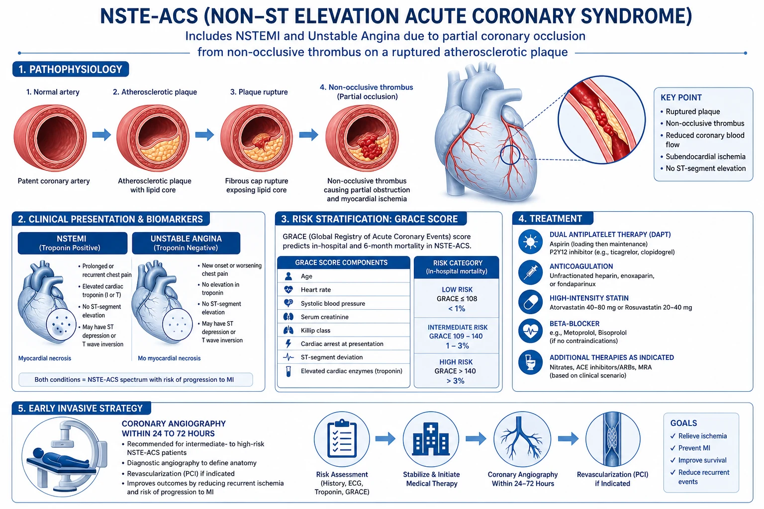
Clinical Presentation
Typical ischaemic chest pain of NSTE-ACS shares the same character as stable angina but breaks the "stable" rules — it is new, crescendo, or occurs at rest: [1]
- Quality — central, retrosternal pressure/tightness/band-like/heavy/crushing ("like an elephant on the chest"), rarely sharp or stabbing.
- Duration — typically 10 to 30 minutes; longer than stable angina, not the seconds of musculoskeletal pain or the hours-to-days of muscular/oesophageal pain.
- Radiation — to the left arm, both arms, neck, jaw, shoulders, epigastrium, or back.
- Provocation/relief — the defining feature of unstable angina: occurs at rest, or with progressively less exertion, or continues after exertion stops; not reliably relieved by sublingual GTN; may wake the patient from sleep.
- Associations — dyspnoea (the commonest anginal equivalent), diaphoresis, nausea, vomiting (especially inferior territory), fatigue, palpitations, sense of doom. [1]
The three classical unstable-angina presentations (Braunwald III B is the textbook presentation): [1]
- Rest angina — angina at rest, usually lasting over 20 minutes (the commonest).
- New-onset severe angina — new angina of at least Canadian Cardiovascular Society Class III (marked limitation of ordinary activity), within 2 months.
- Crescendo angina — previously diagnosed angina that has become distinctly more frequent, longer lasting, or lower in threshold, within 2 months. [1]
Anginal equivalents (pain-free ischaemia) — the high-risk atypical presentations: [1]
- Women, the elderly, and people with diabetes often present without the classic chest pressure. Instead: isolated dyspnoea (the single most common anginal equivalent, especially in heart failure or post-operatively), epigastric pain or "indigestion", fatigue, syncope, or acute delirium/confusion in the very old.
- Diabetic autonomic neuropathy produces silent ischaemia — the patient may have extensive troponin-positive NSTE-ACS with no pain at all, presenting with heart failure or an arrhythmia.
- Post-operative patients (after non-cardiac surgery) — pain masked by analgesia and surgical wounds; NSTE-ACS presents as hypotension, new AF, pulmonary oedema, or unexplained tachycardia.
- Cocaine-induced NSTE-ACS — young patient, chest pain after use, often with sympathetic toxicity (hypertension, tachycardia, mydriasis); mechanism is a mixture of spasm, thrombosis, and demand ischaemia. [1]
Differential Diagnosis
"Ischaemic-sounding chest pain" is a vast basket; the immediate job is to exclude the immediately life-threatening mimics of ACS before committing the patient to dual antiplatelet and anticoagulant therapy. The five immediately life-threatening causes of chest pain (the exam staple) are: ACS, aortic dissection, pulmonary embolism, tension pneumothorax, and oesophageal rupture (Boerhaave) — all but pneumothorax can masquerade as NSTE-ACS. [1]
Acute pericarditis
- **Positional** pain — worse on lying flat, **relieved by sitting forward**; pleuritic
- **Diffuse** concave ST elevation in many leads, **PR depression**, no reciprocal change, no mirror-image
- **Three-phase pericardial friction rub**
- Troponin may rise (myopericarditis) but ECG changes are **persistent**, not dynamic; fever/viral prodrome common
Aortic dissection
- **Tearing** pain radiating to the **back**, **sudden onset maximal at onset** (vs crescendo)
- **Pulse or blood-pressure differential between arms**, widened mediastinum on CXR, new aortic regurgitation murmur
- **Must exclude before antiplatelet/anticoagulation** — CT angiography is diagnostic
- Troponin may rise if dissection extends into coronary ostium (usually right coronary — inferior STEMI pattern)
Pulmonary embolism
- **Pleuritic** pain, **dyspnoea out of proportion**, risk factors (immobility, malignancy, pregnancy)
- **Sinus tachycardia, S1Q3T3, right-axis deviation, T inversion V1 to V4**, echocardiographic right-heart strain
- **D-dimer elevated**, CT pulmonary angiogram diagnostic
- Right-heart strain can cause a **small troponin rise** — do not be misled into NSTEMI
Gastro-oesophageal (reflux, spasm)
- **Burning**, related to food, **relieved by antacids**, postural (worse lying/bending)
- Oesophageal spasm can **mimic angina exactly** — even respond to GTN (smooth-muscle relaxant)
- Normal ECG and troponin; diagnosis of exclusion after ACS ruled out
Musculoskeletal / chest wall
- **Reproducible on palpation/position**, lasts seconds or days, **pleuritic**
- Costochondritis (Tietze): tender costochondral joints with swelling
- Normal ECG and troponin; beware — reproducible tenderness does NOT exclude ACS (can coexist)
Pneumonia / pneumothorax
- Pneumonia: fever, purulent sputum, consolidation on CXR, septic picture
- Pneumothorax: sudden dyspnoea + pleuritic pain, reduced breath sounds, **tracheal deviation if tension**
- CXR or CT resolves both; troponin usually normal
Pitfalls in the differential. (a) Right ventricular infarction in inferior STEMI can present with clear lung fields and hypotension — the ECG may show only subtle ST elevation in V4R. (b) Takotsubo (stress) cardiomyopathy mimics STEMI/NSTEMI with troponin rise and regional wall-motion abnormality beyond a single coronary territory; triggered by emotional/physical stress; angiography shows no obstructive culprit. (c) Myocarditis — troponin-positive with diffuse ST-T change and a viral prodrome; no culprit at angiography. (d) Boerhaave syndrome — vomiting then excruciating chest pain; subcutaneous emphysema; CT diagnostic. Always explicitly ask, document, and exclude aortic dissection before the first dose of aspirin.[1][2]
Clinical & Bedside Assessment
The first 10 minutes — airway, breathing, circulation; 12-lead ECG within 10 minutes of arrival; establish IV access; attach a cardiac monitor. The ECG is the first branch point: ST elevation or new LBBB → STEMI pathway; no ST elevation → NSTE-ACS pathway (but record serial ECGs for dynamic change).[1]
History — characterise the pain (SOCRATES), establish the unstable pattern (rest/crescendo/new-onset severe), identify risk factors and precipitants, and ask about prior aspirin use (relevant to TIMI score), recent bleeding, and anticoagulant/antiplatelet drugs. [1]
Examination — look for the haemodynamic and ischaemic phenotype that determines risk:
- Vital signs — heart rate, blood pressure, respiratory rate, oxygen saturation, temperature. Shock (hypotension, cold peripheries) and acute heart failure are very-high-risk features mandating immediate invasive strategy.
- Killip class (originally for STEMI but applied to all ACS; reproduced in the registry) — I no crackles/S3; II basilar crackles or S3; III pulmonary oedema; IV cardiogenic shock. Higher Killip = higher GRACE and higher mortality.
- New murmur — a new pansystolic murmur with sudden haemodynamic collapse days after an ACS is a mechanical complication (papillary muscle rupture causing acute mitral regurgitation, or ventricular septal rupture) — a surgical emergency.
- Signs of an alternative diagnosis — pericardial rub, unequal pulses (dissection), pleural rub/calf tenderness (PE), tender chest wall (musculoskeletal). [1]
Bedside tests during assessment — finger-prick glucose (diabetic ketoacidosis can mimic chest pain; hyperglycaemia worsens prognosis), haemoglobin (anaemia may be the Type 2 trigger and modifies the antithrombotic/bleeding risk), and a venous blood gas/lactate if shock is suspected. [1]
Investigations
First-line (in the first hour)
- 12-lead ECG within 10 minutes — the single most important and time-critical test. Look for ST depression (horizontal or downsloping, at least 0.5 mm in two contiguous leads), deep symmetrical T-wave inversion, transient ST elevation, pseudo-normalisation of previously inverted T waves, and posterior MI patterns (ST depression V1 to V3 with tall R waves and upright T waves — record posterior leads V7 to V9). Dynamic change on serial ECGs (every 15 to 30 minutes if pain persists) is a high-risk feature. Up to 20 per cent of NSTEMI patients have a normal ECG — a normal ECG never excludes ACS. ECG patterns with high-yield names to reproduce: De Winter T waves (up-sloping ST depression with tall, symmetrical T waves in precordial leads — a STEMI-equivalent), and Wellens syndrome (deep biphasic T waves in V2 to V3, pain-free at the time, critical proximal LAD stenosis).[1]
- High-sensitivity cardiac troponin (hs-cTn) — the biomarker of choice; never use CK-MB or myoglobin as the primary biomarker. Sample at presentation (0-hour) and use a validated accelerated protocol: ESC 0-hour/1-hour algorithm (three-band: rule-out, observe, rule-in based on absolute level and 1-hour delta) or 0-hour/3-hour if a 1-hour assay is unavailable. The diagnostic criterion is a rise and/or fall of troponin with at least one value above the 99th centile of a healthy reference population, in the clinical context of ischaemia. Serial sampling is essential — a single normal troponin at presentation does not exclude NSTEMI. Beware chronically elevated troponin (renal failure, severe heart failure, sepsis, pulmonary embolism, myocarditis) — the dynamic change (rise/fall) is what distinguishes an acute Type 1 event from a chronically elevated baseline.[1][3]
- Full blood count, urea and electrolytes, creatinine/eGFR (renal function governs anticoagulant/fondaparinux dosing and contrast risk), glucose, lipid profile within 24 to 48 hours (LDL falls after 24 to 48 h so an early sample is the accurate baseline), liver function, thyroid function (if AF), coagulation screen, and D-dimer if PE is in the differential. Group and save in case of bleeding or need for transfusion/angiography.
- Chest X-ray — to exclude pneumothorax, widened mediastinum (dissection), pulmonary oedema (Killip class), pneumonia. Not diagnostic of ACS but essential to the differential.
- Transthoracic echocardiogram — regional wall-motion abnormality supports ischaemia; assesses left ventricular function (drives ACE-inhibitor/beta-blocker benefit and prognosis); identifies mechanical complications; excludes aortic root dissection when coupled with the clinical picture.
Risk stratification — reproduce the named scores verbatim
TIMI risk score for NSTE-ACS (1 point each, maximum 7) — reproduced from the registry components:[2]
- Age 65 years or older.
- At least 3 risk factors for CAD (family history, hypertension, hypercholesterolaemia, diabetes, current smoker).
- Known CAD (coronary stenosis 50 per cent or more).
- Aspirin use in the past 7 days.
- Severe angina (at least 2 anginal events in the past 24 hours).
- ST-segment deviation 0.5 mm or more on ECG.
- Elevated cardiac markers (troponin/CK-MB). [1]
TIMI risk bands (14-day rate of all-cause mortality, new/recurrent MI, or severe recurrent ischaemia requiring urgent revascularisation): score 0 to 1 (about 4.7 per cent, low); 2 (about 8.3 per cent); 3 (about 13.2 per cent); 4 (about 19.9 per cent); 5 (about 26.2 per cent); 6 to 7 (about 40.9 per cent, high). A TIMI score of 3 or more generally supports an invasive strategy. [1]
GRACE (Global Registry of Acute Coronary Events) score — the ESC-preferred risk model for predicting in-hospital and 6-month mortality. Reproduced from the registry components, it combines eight weighted variables:[1]
- Age
- Heart rate at admission
- Systolic blood pressure at admission
- Creatinine (renal function)
- Killip class at presentation
- Cardiac arrest at admission
- ST-segment deviation on the ECG
- Abnormal cardiac enzymes (troponin) [1]
Because the points are non-linear and tabulated, GRACE is computed with an app/calculator. Risk bands (in-hospital mortality): low (GRACE below 109, mortality about 1 per cent or less); intermediate (109 to 140, mortality about 1 to 3 per cent); high (above 140, mortality about 3 per cent and rising sharply with the score). GRACE is the score the ESC uses to set the timing of invasive angiography.[1]
HEART score (used in the emergency department for undifferentiated chest pain to predict 6-week major adverse cardiac events) — reproduced in the registry: History, ECG, Age, Risk factors, Troponin (0 to 2 points each, maximum 10): 0 to 3 low risk (about 1.7 per cent MACE, consider discharge); 4 to 6 moderate; 7 to 10 high (about 50 per cent MACE).[3]
CRUSADE bleeding score — predicts in-hospital major bleeding to balance ischaemic against bleeding risk before intensifying antithrombotic therapy; components include baseline haematocrit, estimated glomerular filtration rate, heart rate, sex, congestive heart failure, prior vascular disease, diabetes, and systolic blood pressure. [1]
Confirmatory / in-hospital
- Coronary angiography — the definitive investigation; identifies the culprit lesion (ulceration, filling defect, thrombus, collateralisation) and defines anatomy for PCI/CABG. Performed on the invasive-strategy timeline dictated by risk (below).
- Multidetector CT coronary angiography (CTCA) — an alternative to rule out coronary disease in low-risk patients (after ACS excluded by troponin/ECG), per NICE NG185 and the 2021 AHA/ACC chest pain guideline. Not for the confirmed NSTE-ACS patient. [1]
Management — Resuscitation
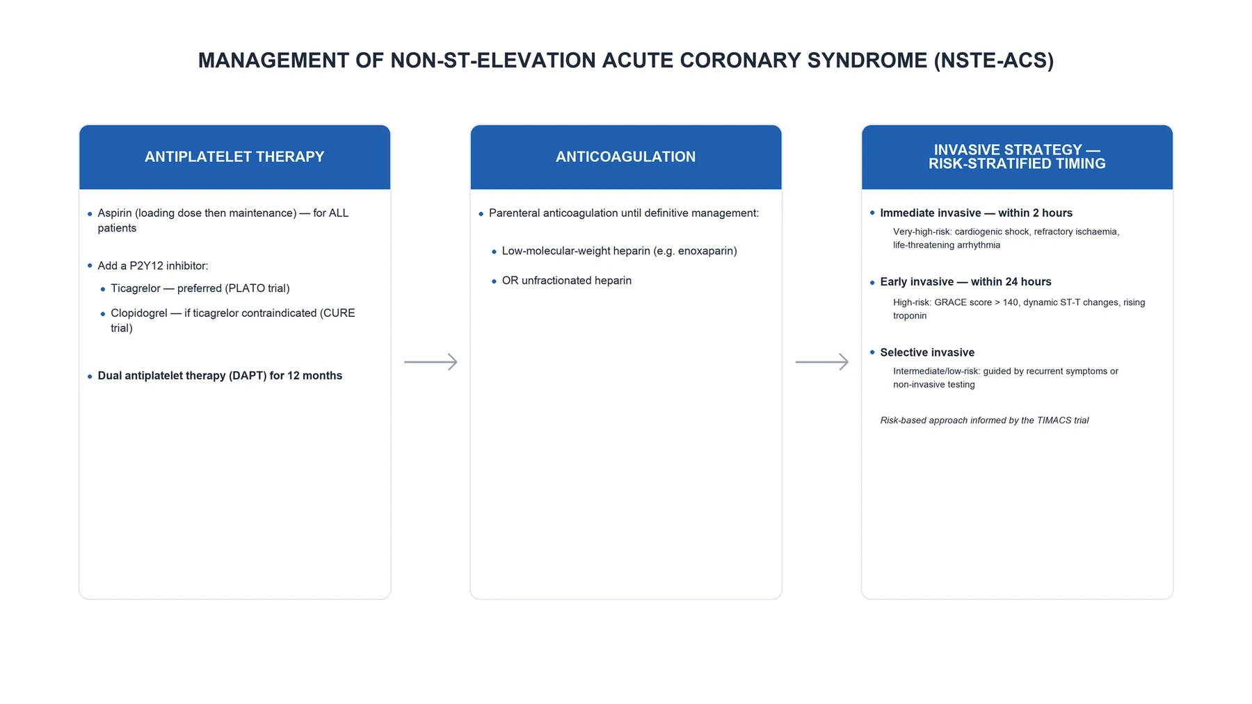
The immediate management of suspected NSTE-ACS is a standardised bundle delivered in parallel with the work-up; none of it should wait for the troponin if the clinical and ECG picture is consistent. [1]
ABCDE first; continuous ECG monitoring; IV access; defibrillator at the bedside. [1]
- Oxygen — give only if hypoxic (SpO2 under 90 per cent, or under 88 per cent in chronic CO2-retaining COPD). Routine oxygen in normoxic patients is harmful (the AVOID trial in STEMI showed larger infarcts with supplemental oxygen); target SpO2 94 to 98 per cent (88 to 92 per cent in COPD).[1]
- Aspirin — 300 mg (162 to 325 mg) oral, chewed immediately (non-enteric-coated, for fastest absorption), then 75 to 100 mg once daily indefinitely. Aspirin irreversibly acetylates cyclo-oxygenase-1, blocking thromboxane A2 for the lifespan of the platelet (8 to 10 days). The mortality benefit was established in ISIS-2 and the Antithrombotic Trialists' Collaboration.[4]
- Second antiplatelet (P2Y12 inhibitor) — added immediately on making the NSTE-ACS diagnosis, before or at angiography depending on the agent and strategy (see below). Ticagrelor preferred; clopidogrel if ticagrelor/prasugrel contraindicated or if on oral anticoagulation.
- Nitrates — sublingual glyceryl trinitrate (GTN) 0.4 mg (400 micrograms) every 5 minutes up to 3 doses for ongoing ischaemic pain; if pain persists or there is hypertension/acute left ventricular failure, start intravenous GTN starting at 10 micrograms/min, titrated up to 200 micrograms/min to pain/blood pressure. Nitrates reduce preload and coronary vasospasm.
- Parenteral analgesia — morphine 2 to 4 mg IV every 5 to 15 minutes for refractory ischaemic pain; also reduces sympathetic drive. Caution: morphine slows gastric emptying and delays absorption of oral P2Y12 inhibitors (ticagrelor, clopidogrel), with a contested signal of worse outcomes — give the oral antiplatelet first, use morphine judiciously, and consider crushing ticagrelor. Fentanyl has the same problem.
- Anticoagulation — a parenteral anticoagulant is started with the antiplatelets: fondaparinux 2.5 mg subcutaneous once daily (preferred for a conservative strategy — lowest bleeding, OASIS-5), enoxaparin 1 mg/kg subcutaneous twice daily, unfractionated heparin (UFH) 60 to 70 IU/kg bolus then 12 to 15 IU/kg/hour infusion titrated to APTT 1.5 to 2.5 times control, or bivalirudin for an invasive strategy.[8]
NSTE-ACS — the numbers an examiner wants
Management — Definitive & Stepwise
Definitive management proceeds in three streams: (A) antithrombotic therapy (antiplatelet + anticoagulant), (B) the ischaemic-strategy decision (timing of angiography), and (C) disease-modifying secondary-prevention drugs and revascularisation. [1]
A. Antiplatelet therapy — dual antiplatelet therapy (DAPT)
Aspirin
- **300 mg load PO, then 75 to 100 mg OD**, indefinitely
- Irreversible COX-1 blockade of thromboxane A2
- With ticagrelor, reduce to 75 mg OD (PLATO used 75 to 100 mg)
- Anchor of DAPT in every NSTE-ACS patient
Ticagrelor
- **180 mg load, then 90 mg twice daily** — preferred P2Y12 for all NSTE-ACS (invasive or conservative), PLATO mortality benefit
- Reversible, direct-acting; faster onset than clopidogrel
- **Contraindicated** in prior intracranial haemorrhage, active bleeding; caution in gout/hyperuricaemia and asthma (dyspnoea, bradycardia)
- No dose cut for age, weight, or renal function
Prasugrel
- **60 mg load, then 10 mg OD** — **only after coronary anatomy is known** (PCI patients)
- Most potent, fastest onset; **contraindicated in prior stroke/TIA**
- **Caution in age 75 or over and weight under 60 kg** — use 5 mg; avoid if high bleeding risk
- Not for the conservative-strategy patient (no known anatomy)
Clopidogrel
- **300 to 600 mg load, then 75 mg OD** (600 mg if PCI planned)
- Prodrug — genetically variable CYP2C19 activation (slower in about 30 per cent)
- Use when ticagrelor/prasugrel contraindicated, **in patients on oral anticoagulation** (with a DOAC/VKA), or if cost/unavailable
- CURE established its ACS benefit over aspirin alone
B. Parenteral anticoagulation (continued from resuscitation)
- Fondaparinux 2.5 mg subcutaneous once daily — preferred for a conservative (non-invasive) strategy: in OASIS-5 it matched enoxaparin for ischaemic events with significantly less major bleeding and lower mortality at 30 days and 6 months. If the patient proceeds to PCI on fondaparinux, add a single bolus of UFH because fondaparinux alone is associated with catheter thrombosis.[8]
- Enoxaparin 1 mg/kg subcutaneous twice daily (reduce to 1 mg/kg once daily if eGFR under 30 mL/min); a 0.5 mg/kg IV bolus is given at the time of PCI.
- Unfractionated heparin 60 to 70 IU/kg IV bolus (max 4000 to 5000 IU) then 12 to 15 IU/kg/hour infusion to APTT 1.5 to 2.5 times control — preferred when rapid reversibility is needed (renal failure, high bleeding risk, catheter-lab), and universally available.
- Bivalirudin 0.75 mg/kg IV bolus then 1.75 mg/kg/hour — a direct thrombin inhibitor; in ACUITY and MATRIX it gave comparable ischaemic protection to heparin plus a GPIIb/IIIa inhibitor with less bleeding, and is an option for an invasive strategy.[9][10]
Glycoprotein IIb/IIIa inhibitors (abciximab, eptifibatide, tirofiban) are no longer used routinely upstream. The EARLY-ACS trial showed that routine early, upstream eptifibatide was not superior and bled more than provisional (bail-out) use during PCI. Reserve them for thrombotic complications during PCI (large thrombus burden, slow/no reflow, bail-out).[14]
C. Disease-modifying and secondary-prevention drugs (the "ABCDE" of long-term therapy)
- High-intensity statin — atorvastatin 80 mg once daily (or rosuvastatin 20 to 40 mg) within 24 hours of admission, regardless of baseline LDL; PROVE-IT-TIMI 22 showed intensive lipid lowering (LDL target below 1.4 mmol/L, 55 mg/dL) reduced mortality and recurrent ischaemia after ACS. Add ezetimibe 10 mg if LDL target not met (IMPROVE-IT).[15][16]
- Beta-blocker — bisoprolol 5 mg OD, metoprolol 25 to 50 mg twice daily, or carvedilol within 24 hours if no contraindication (acute heart failure with low output, cardiogenic shock, advanced AV block, severe asthma, marked bradycardia). Reduces reinfarction, ventricular arrhythmia, and mortality; essential after anterior MI/LV dysfunction.
- ACE inhibitor (or ARB) — ramipril 2.5 mg twice daily titrated to 10 mg OD (or lisinopartan, perindopril) within 24 hours, especially with LV dysfunction, diabetes, hypertension, anterior MI, or heart failure (HOPE/ISIS-4 class benefit).[17]
- Aldosterone antagonist (eplerenone 25 to 50 mg OD) for LV ejection fraction 40 per cent or less with heart failure or diabetes (post-MI), with monitoring of potassium and creatinine.
- Nitrates for ongoing ischaemia; calcium-channel blocker (diltiazem or verapamil, non-dihydropyridine) if beta-blocker contraindicated and no LV dysfunction.
D. The ischaemic strategy — timing of invasive coronary angiography
The pivotal, evidence-based, exam-decisive decision is when to take the patient to the catheter laboratory. The ESC 2023 risk-stratified timing (reproduced verbatim):[1]
Early invasive strategy — within 24 hours (high risk): at least one of — rise/fall of troponin compatible with MI; dynamic ST/T changes (symptomatic or silent); GRACE score above 140. [1]
Invasive strategy — within 72 hours (intermediate risk): at least one of — diabetes mellitus; eGFR under 60 mL/min/1.73 m2; LVEF under 40 per cent or congestive heart failure; early post-infarction angina; prior PCI; prior CABG; GRACE 109 to 140. [1]
Selective (non-invasive) strategy (low risk): none of the above; the risk is low; pursue non-invasive ischaemia testing (stress imaging or CTCA) and manage accordingly — routine early angiography is not beneficial.[1]
The evidence base for an invasive strategy in medium- to high-risk NSTE-ACS comes from the FRISC II, RITA-3, and ICTUS trials and their meta-analyses: an early invasive approach reduces death, MI, and refractory ischaemia over the medium term (especially in high-risk, troponin-positive, ST-change-positive patients), with the benefit durable at 5 years. The FRISC II dose of "early" was within 7 days; modern practice has compressed the window to within 24 hours for high-risk patients based on the TIMAC, OPTIMA, and VERDICT trials showing non-inferiority (and bleeding advantages) of very-early angiography.[11][12][13]
Revascularisation — PCI (drug-eluting stent) for the culprit and, increasingly, complete revascularisation of significant non-culprit lesions (especially in multivessel disease) to reduce recurrent events. CABG for left main stem disease, three-vessel disease (especially with LV dysfunction or diabetes), and complex anatomy (SYNTAX score). The heart team decides. [1]
E. Duration of therapy and step-down
- DAPT for a minimum of 12 months after NSTE-ACS (with stenting, regardless of stent type — even with newer-generation drug-eluting stents) unless bleeding risk is prohibitive. Ticagrelor 60 mg twice daily plus low-dose aspirin is an option to prolong DAPT beyond 12 months in patients with high ischaemic and low bleeding risk (PEGASUS-TIMI 54).
- De-escalation (e.g. ticagrelor or clopidogrel monotherapy after 1 to 3 months) is considered in high bleeding risk, especially after PCI.
- Aspirin, high-intensity statin, beta-blocker, and ACE inhibitor are continued long-term; the statin is lifelong at the maximum tolerated intensity. [1]
Specific Subtypes & Scenarios
NSTE-ACS — the five subtypes examiners escalate on
SCOPE
Very high risk: haemodynamic instability, life-threatening arrhythmia, mechanical complication, acute HF — immediate invasive within 2 h
Rest/crescendo angina, troponin-negative = unstable angina — invasive strategy timed by GRACE/TIMI
Patient on warfarin/DOAC for AF — minimise antithrombotics: aspirin + clopidogrel, shortest triple therapy, prefer radial access
Type 2 component: sepsis, anaemia, tachyarrhythmia, hypoxia — treat the precipitant, weigh the antithrombotic risk
Painless ischaemia, dyspnoea, delirium — low threshold for troponin, modified antithrombotic dosing
- Unstable angina (troponin-negative NSTE-ACS) — managed identically to NSTEMI but prognosis is better; risk-stratify and time angiography on the same GRACE/TIMI criteria. The key exam point is the biochemical distinction (no necrosis) and the recognition that high-sensitivity troponin has shrunk the diagnosis.
- NSTE-ACS in patients requiring oral anticoagulation (AF, mechanical valve, VTE) — the highest bleeding-risk subgroup. Aspirin + clopidogrel (not ticagrelor/prasugrel) plus the OAC; minimise triple therapy to the peri-PCI window (up to 1 week); prefer radial access; consider left atrial appendage occlusion at the time of PCI. Triple therapy beyond a month substantially raises major bleeding.[1]
- Cocaine-induced NSTE-ACS — benzodiazepines first (relieve sympathetic drive and spasm), aspirin, nitrates, and a calcium-channel blocker; avoid beta-blockers acutely (unopposed alpha-mediated vasoconstriction — though modern evidence is less absolute, the exam answer remains beta-blocker caution). Anticoagulate and risk-stratify as usual.
- Type 2 MI (demand ischaemia) — treat the precipitant (tachyarrhythmia, sepsis, anaemia, hypoxia, hypertensive emergency); troponin rises but there is no thrombotic substrate, so routine intensive antithrombotic therapy and catheterisation are not indicated unless a concomitant Type 1 event is suspected. This is a frequent SAQ/viva discriminator.
- NSTE-ACS with cardiogenic shock — immediate invasive strategy within 2 hours; consider short-term mechanical circulatory support (IABP counter-pulsation, Impella, VA-ECMO) as a bridge; revascularise the culprit and significant lesions. Mortality is high.
Complications & Pitfalls
Ischaemic/electrical (first days): recurrent ischaemia and re-infarction; ventricular arrhythmias (VT/VF — monitor, defibrillator at bedside); atrial fibrillation (especially with atrial infarction and heart failure, and in the elderly); bradyarrhythmias and conduction block (especially inferior MI — AV nodal block; anterior MI — wide-complex infranodal block, ominous). [1]
Mechanical (classically days 2 to 7, after the necrosis softens):
- Acute mitral regurgitation — papillary muscle rupture (posteromedial papillary muscle, single blood supply from PDA, is the commonest) — sudden pulmonary oedema with a new pansystolic murmur at the apex radiating to the axilla.
- Ventricular septal rupture — a new loud pansystolic murmur at the lower left sternal border with a thrill, biventricular failure.
- Free-wall rupture — sudden cardiac tamponade and electromechanical dissociation arrest; usually fatal without emergency surgery.
- All three present with sudden haemodynamic collapse and a new murmur; urgent echocardiography is diagnostic, and they are surgical emergencies (mortality climbs hourly). [1]
Heart failure and cardiogenic shock — by extension of infarct size; managed by the shock pathway (inotropes, mechanical support, revascularisation). [1]
Bleeding — the complication of our own therapy, and the major determinant of outcome after NSTE-ACS (a major bleed doubles 30-day mortality). Commonest sites are gastrointestinal and access-site (groin). Prevent by radial access (60 to 70 per cent reduction in access bleeding), careful dosing (renal dose-adjust enoxaparin/fondaparinux), avoiding upstream GPIIb/IIIa, and using the CRUSADE score prospectively. Protamine reverses UFH (partially enoxaparin); fondaparinux is not reversed by protamine (consider recombinant factor VIIa in life-threatening bleed); and reversal of the antiplatelet effect is supportive (platelet transfusion for life-threatening bleeding on ticagrelor/prasugrel).[1]
Contrast-induced acute kidney injury — risk-stratify by eGFR; periprocedural intravenous isotonic saline hydration, minimise contrast volume, and hold nephrotoxins. [1]
Stent thrombosis — acute (under 24 h), subacute (1 to 30 days), late (1 to 12 months), very late (over 12 months); presents as recurrent ACS — often catastrophic. Adherence to DAPT is the single most important preventable cause. [1]
Classic pitfalls:
- Treating NSTE-ACS like STEMI — there is no immediate-reperfusion mandate; over-rapid intervention in low-risk patients adds bleeding without benefit.
- Missing the very-high-risk patient — failing to recognise haemodynamic instability or dynamic ST-T change as the trigger for immediate invasive strategy within 2 hours.
- Giving aspirin before excluding aortic dissection — a fatal, irreversible error.
- Over-interpreting a single troponin in renal failure or chronic heart failure — use the rise/fall pattern, not an isolated value.
- Routine supplemental oxygen in the normoxic patient.
- Stopping DAPT early (e.g. for a non-cardiac procedure) without bridging — precipitates stent thrombosis.
- Forgetting atypical presentations in the elderly, diabetic, and post-operative patient. [1]
Prognosis & Disposition
In-hospital mortality of NSTE-ACS is around 3 to 5 per cent — lower than STEMI — but 1-year mortality equals or exceeds STEMI (around 10 to 15 per cent) because NSTE-ACS patients are older, carry more comorbidity, and more often have multivessel and recurrent disease. The GRACE score is the validated predictor of in-hospital and 6-month mortality; TIMI predicts the composite ischaemic endpoint. Poor-outcome predictors include older age, diabetes, chronic kidney disease, heart failure/LV dysfunction, troponin elevation, dynamic ST changes, anaemia, and elevated inflammatory markers.[1]
Disposition. Admit to a monitored coronary care unit (CCU) or cardiac monitored bed with continuous ECG and a defibrillator at the bedside. Once stable and revascularised, transfer to a step-down ward, then home with a comprehensive secondary-prevention bundle: aspirin, P2Y12 inhibitor (to complete 12 months), high-intensity statin, beta-blocker, ACE inhibitor; smoking cessation, cardiac rehabilitation, diabetes and hypertension control, diet and exercise, influenza and pneumococcal vaccination, and a clear follow-up plan. Early outpatient review (within 2 to 4 weeks) to titrate medications, check adherence to DAPT, and address residual ischaemia. [1]
Special Populations
- Elderly (75 and over) — atypical presentations (dyspnoea, delirium, syncope) are the norm; higher bleeding risk so dose-adjust antithrombotics (prasugrel 5 mg if used, caution with enoxaparin), prefer radial access, and a lower threshold to admit and troponin-test. Frailty weighs into the invasive decision.
- Diabetes mellitus — more extensive disease, worse prognosis, often painless; benefit greatly from an invasive strategy and complete revascularisation; metformin is continued peri-procedurally with renal monitoring (contrast); SGLT2 inhibitors and GLP-1 receptor agonists are continued for long-term cardiovascular protection.
- Chronic kidney disease (eGFR under 60) — high-risk by definition (intermediate-risk invasive strategy within 72 h); troponin is chronically elevated so the delta (rise/fall) is decisive; dose-adjust enoxaparin/fondaparinux/bivalirudin; contrast-sparing angiography.
- Pregnancy — NSTE-ACS is rare (incidence rising with advanced maternal age and pre-eclampsia); spontaneous coronary artery dissection is an important mimic/cause in the peripartum period. Manage with aspirin, beta-blocker, and unfractionated heparin (does not cross the placenta; enoxaparin preferred if established); avoid statins (teratogenic, though data softening); prefer bare-metal stenting if PCI needed (shorter DAPT) and avoid GPIIb/IIIa unless essential. Multidisciplinary with obstetrics and cardiology.
- Patients already anticoagulated (warfarin, DOAC) — minimise antithrombotic load as above; bridge periprocedurally with heparin if warfarin is interrupted; the rhythm of choice for AF after ACS is a DOAC over a VKA when OAC is needed.
- Prior CABG — intermediate-risk invasive strategy; graft angiography is essential (vein graft disease is a common culprit); consider completeness of revascularisation.
- Anaemia/bleeding diathesis — balance ischaemic against bleeding risk with CRUSADE; avoid upstream GPIIb/IIIa; consider a conservative strategy and the lowest-bleeding anticoagulant (fondaparinux, bivalirudin). [1]
Evidence, Guidelines & Regional Differences
Landmark trials (know the question each answered): [1]
| Trial | Year | Question | Result |
|---|---|---|---|
| CURE | 2001 | Clopidogrel + aspirin vs aspirin alone in NSTE-ACS | Clopidogrel reduced CV death/MI/stroke; established DAPT in NSTE-ACS. Bleeding rose.[5] |
| PLATO | 2009 | Ticagrelor vs clopidogrel in ACS | Ticagrelor reduced all-cause mortality and MI without increased major bleeding (except non-CABG); became preferred P2Y12.[6] |
| TRITON-TIMI 38 | 2007 | Prasugrel vs clopidogrel in ACS (PCI) | Prasugrel reduced ischaemic events but increased fatal bleeding; contraindicated in prior stroke.[7] |
| OASIS-5 | 2006 | Fondaparinux vs enoxaparin in NSTE-ACS | Equal ischaemic efficacy, less bleeding, lower 30-day and 6-month mortality — fondaparinux preferred for conservative strategy.[8] |
| ACUITY | 2006 | Bivalirudin vs heparin + GPI in moderate-high-risk NSTE-ACS (invasive) | Bivalirudin non-inferior for ischaemia, less bleeding.[9] |
| MATRIX | 2015 | Bivalirudin vs UFH in ACS (invasive) | Less bleeding; numerically lower mortality (not significant for co-primary).[10] |
| FRISC II | 1999 | Early invasive vs conservative in unstable CAD | Invasive reduced death/MI at 6 months — established the invasive strategy.[11] |
| RITA-3 | 2002 | Interventional vs conservative in NSTE-ACS | Invasive reduced death/MI/refractory ischaemia; benefit at 5 years, concentrated in high-risk.[12] |
| ICTUS | 2005 | Early invasive vs selective invasive in troponin-positive NSTE-ACS | No overall advantage for routine early invasive; tempered the enthusiasm for an invasive strategy in lower-risk troponin-positive patients.[13] |
| EARLY-ACS | 2009 | Routine upstream vs provisional eptifibatide (invasive NSTE-ACS) | No benefit, more bleeding — moved GPIIb/IIIa to bail-out.[14] |
| PROVE IT-TIMI 22 | 2004 | Atorvastatin 80 vs pravastatin 40 after ACS | High-intensity statin reduced events — established early high-dose statin after ACS.[15] |
| IMPROVE-IT | 2015 | Ezetimibe + simvastatin vs simvastatin after ACS | Adding ezetimibe to lower LDL further reduced events — supports LDL target below 1.4 mmol/L.[16] |
| HOPE | 2000 | Ramipril in high-risk vascular patients (no HF) | Ramipril reduced mortality/MI/stroke — broadened ACE-inhibitor use after ACS.[17] |
| ISIS-2 | 1988 | Aspirin and streptokinase in suspected MI | Aspirin alone cut 5-week vascular mortality by 23 per cent — the foundation of aspirin in ACS.[4] |
Guidelines and regional deltas. The ESC 2023 ACS guideline is the current European standard for the risk-stratified invasive timing above.[1] The 2014 AHA/ACC NSTE-ACS guideline (Amsterdam) is the North American counterpart, with broadly concordant recommendations (a very early invasive strategy within 24 hours for high risk, high-intensity statin, and DAPT), and the 2021 AHA/ACC Chest Pain guideline (Gulati) reinforces tropanin as the preferred biomarker and a low threshold to test in the undifferentiated chest-pain patient.[2][3] NICE NG185 (chest pain of recent onset) emphasises CT coronary angiography to rule out disease in low-risk patients and a risk-stratified invasive pathway mirroring ESC timing for confirmed NSTE-ACS. Differences are minor and mostly in emphasis; the principles — early ECG, serial troponin, GRACE/TIMI risk stratification, DAPT + anticoagulant + statin, and a graded invasive strategy — are universal.
Controversies: (i) Optimal timing of the invasive strategy in high-risk NSTE-ACS has compressed from within 24 to within 12 hours (VERDICT) with non-inferior outcomes and a bleeding signal favouring earlier intervention; the 2-hour window is reserved for very-high-risk. (ii) Routine complete vs culprit-only revascularisation in NSTE-ACS has shifted toward complete in multivessel disease (COMPLETE in STEMI; observational NSTE-ACS). (iii) P2Y12 monotherapy (dropping aspirin after 1 to 3 months) is expanding in high bleeding-risk patients after PCI. [1]
Exam Pearls
- NSTE-ACS = NSTEMI (troponin-positive) + unstable angina (troponin-negative); no ST elevation; partial/intermittent occlusion; no immediate-reperfusion mandate.
- The single most important and time-critical test is the ECG within 10 minutes — to exclude STEMI (and posterior STEMI, De Winter, Wellens) before committing to the NSTE-ACS pathway.
- Troponin defines NSTEMI vs unstable angina; use high-sensitivity troponin with a 0/1-hour or 0/3-hour algorithm; the rise/fall distinguishes acute Type 1 from chronically elevated troponin (renal failure, HF, PE).
- Type 1 vs Type 2 MI is a favourite discriminator: Type 1 = plaque thrombus (treat as NSTE-ACS); Type 2 = demand ischaemia (treat the precipitant).
- Risk-stratify with GRACE (mortality) and TIMI (composite); they set the timing of angiography.
- Immediate invasive within 2 hours for very high risk (haemodynamic instability, life-threatening arrhythmia, mechanical complication, acute HF, recurrent dynamic ST-T change); within 24 hours for high risk; within 72 hours for intermediate risk; selective for low risk.
- DAPT = aspirin 300 mg load then 75 mg OD plus ticagrelor 180 mg then 90 mg BD (preferred) — or prasugrel (post-PCI, no prior stroke) — or clopidogrel (if on an OAC or intolerant).
- Anticoagulant = fondaparinux 2.5 mg SC OD (conservative), enoxaparin 1 mg/kg BD, UFH infusion, or bivalirudin (invasive).
- High-intensity statin within 24 h (atorvastatin 80 mg) regardless of baseline LDL — PROVE-IT.
- Routine oxygen is harmful in the normoxic patient — give only if SpO2 under 90 per cent.
- Exclude aortic dissection before the first aspirin (tearing back pain, pulse/BP differential, widened mediastinum).
- On an oral anticoagulant — aspirin + clopidogrel, minimise triple therapy, radial access, DOAC over VKA.
- Long-term mortality of NSTE-ACS equals or exceeds STEMI — because of older, sicker patients and multivessel disease.
- Mechanical complications (days 2 to 7) present with sudden collapse and a new murmur — urgent echocardiogram and surgery.
- CRUSADE for bleeding, GRACE for death, TIMI/HEART for ischaemia/risk — know which score answers which question.
- Plaques that rupture are often only mildly stenotic — a prior "normal" angiogram or stress test does not exclude ACS. [1]
Exam application bank (NEET-PG / INICET)
One-line answer
Non-ST-elevation acute coronary syndrome (NSTE-ACS) is myocardial ischaemia at rest or on minimal exertion caused by a partially or intermittently occlusive intra-coronary thrombus over a disrupted atherosclerotic plaque, but without the ST elevation that mandates immediate reperfusion. It comprises NSTEMI (myocardial necrosis demonstrated by a rise and/or fall of high-sensitivity cardiac troponin with at least one value above the 99th centile) and unstable angina (ischaemia without troponin release). Diagnosis rests on the 12-lead ECG within 10 minutes plus serial high-sensitivity troponin (0-hour/1-hour or 0-hour/3-hour algorithms). Risk-stratify with the GRACE (death) and TIMI (composite endpoint) scores to triage the timing of invasive coronary angiography: immediate (within 2 hours) for very high-risk (haemodynamic instability, life-threatening arrhythmia, mechanical complication, a
Worked stems (answer without another resource)
Stem 1 — Classic presentation. Map symptoms to mechanism; name the first investigation and first treatment step with dose/route if drug therapy is standard. [1]
Stem 2 — Unstable / complicated. List red flags that force immediate resuscitation, theatre, ICU, antidote, or reperfusion — and what you do in the first 15 minutes. [1]
Stem 3 — Atypical group. Elderly, pregnancy, child, or immunocompromised: how presentation and thresholds change. [1]
Stem 4 — Differential trap. Name the three closest mimics and one discriminator for each. [1]
Stem 5 — Disposition. Who goes home with safety-netting, who is admitted, who needs HDU/ICU/theatre, and what follow-up is mandatory. [1]
Rapid viva checklist
- Definition + classification
- Pathophysiology chain
- Bedside signs / criteria
- Score with exact components (if any)
- Emergency bundle
- Definitive therapy with doses
- Complications of disease and of treatment
- Special populations
- Guideline/trial name if classic
- Three exam traps
Coverage self-check
If you cannot answer any stem above from this page alone, re-read the matching section — the page is intended to be self-sufficient for final-prof and NEET-PG/INICET questions on Non-ST-Elevation Acute Coronary Syndrome (NSTE-ACS).
References
- [1]Byrne RA, Rossello X, Coughlan JJ, et al. 2023 ESC Guidelines for the management of acute coronary syndromes Eur Heart J, 2023.PMID 37622654
- [2]Amsterdam EA, Wenger NK, Brindis RG, et al. 2014 AHA/ACC Guideline for the Management of Patients with Non-ST-Elevation Acute Coronary Syndromes: a report of the American College of Cardiology/American Heart Association Task Force on Practice Guidelines J Am Coll Cardiol, 2014.PMID 25260718
- [3]Writing Committee Members; Gulati M, Levy PD, et al. 2021 AHA/ACC/ASE/CHEST/SAEM/SCCT/SCMR Guideline for the Evaluation and Diagnosis of Chest Pain: A Report of the American College of Cardiology/American Heart Association Joint Committee on Clinical Practice Guidelines J Am Coll Cardiol, 2021.PMID 34756653
- [4]ISIS-2 (Second International Study of Infarct Survival) Collaborative Group. Randomised trial of intravenous streptokinase, oral aspirin, both, or neither among 17,187 cases of suspected acute myocardial infarction: ISIS-2. ISIS-2 (Second International Study of Infarct Survival) Collaborative Group Lancet, 1988.PMID 2899772
- [5]Yusuf S, Zhao F, Mehta SR, et al. (CURE Investigators). Effects of clopidogrel in addition to aspirin in patients with acute coronary syndromes without ST-segment elevation N Engl J Med, 2001.PMID 11519503
- [6]Wallentin L, Becker RC, Budaj A, et al. (PLATO Investigators). Ticagrelor versus clopidogrel in patients with acute coronary syndromes N Engl J Med, 2009.PMID 19717846
- [7]Wiviott SD, Braunwald E, McCabe CH, et al. (TRITON-TIMI 38 Investigators). Prasugrel versus clopidogrel in patients with acute coronary syndromes N Engl J Med, 2007.PMID 17982182
- [8]Yusuf S, Mehta SR, Chrolavicius S, et al. (Fifth Organization to Assess Strategies in Acute Ischemic Syndromes Investigators; OASIS-5). Comparison of fondaparinux and enoxaparin in acute coronary syndromes N Engl J Med, 2006.PMID 16537663
- [9]Stone GW, McLaurin BT, Cox DA, et al. (ACUITY Investigators). Bivalirudin for patients with acute coronary syndromes N Engl J Med, 2006.PMID 17124018
- [10]Valgimigli M, Frigoli E, Leonardi S, et al. (MATRIX Investigators). Bivalirudin or Unfractionated Heparin in Acute Coronary Syndromes N Engl J Med, 2015.PMID 26324049
- [11]FRISC II Investigators. Invasive compared with non-invasive treatment in unstable coronary-artery disease: FRISC II prospective randomised multicentre study. FRagmin and Fast Revascularisation during InStability in Coronary artery disease Investigators Lancet, 1999.PMID 10475181
- [12]Fox KA, Poole-Wilson PA, Henderson RA, et al. (RITA-3 Investigators). Interventional versus conservative treatment for patients with unstable angina or non-ST-elevation myocardial infarction: the British Heart Foundation RITA 3 randomised trial. Randomized Intervention Trial of unstable Angina Lancet, 2002.PMID 12241831
- [13]de Winter RJ, Windhausen F, Cornel JH, et al. (Invasive versus Conservative Treatment in Unstable coronary Syndromes Investigators; ICTUS). Early invasive versus selectively invasive management for acute coronary syndromes N Engl J Med, 2005.PMID 16162880
- [14]Giugliano RP, White JA, Bode C, et al. (EARLY-ACS Investigators). Early versus delayed, provisional eptifibatide in acute coronary syndromes N Engl J Med, 2009.PMID 19332455
- [15]Cannon CP, Braunwald E, McCabe CH, et al. (Pravastatin or Atorvastatin Evaluation and Infection Therapy-Thrombolysis in Myocardial Infarction 22 Investigators; PROVE IT-TIMI 22). Intensive versus moderate lipid lowering with statins after acute coronary syndromes N Engl J Med, 2004.PMID 15007110
- [16]Cannon CP, Blazing MA, Giugliano RP, et al. (IMPROVE-IT Investigators). Ezetimibe Added to Statin Therapy after Acute Coronary Syndromes N Engl J Med, 2015.PMID 26039521
- [17]Heart Outcomes Prevention Evaluation Study Investigators. Effects of an angiotensin-converting-enzyme inhibitor, ramipril, on cardiovascular events in high-risk patients N Engl J Med, 2000.PMID 10639539