cardiology · cardiology
Pericardial Disease
Also known as Pericarditis · Acute pericarditis · Pericardial effusion · Cardiac tamponade · Constrictive pericarditis · Dressler syndrome · Post-cardiac injury syndrome
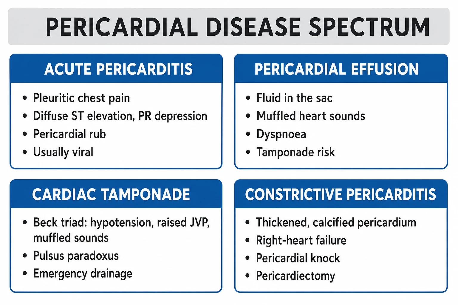
Pericardial disease spans three overlapping syndromes: acute pericarditis (inflammation of the pericardium with pleuritic chest pain, pericardial rub and diffuse ST elevation with PR depression), pericardial effusion with cardiac tamponade (fluid under pressure impairing diastolic filling; Beck triad, pulsus paradoxus), and constrictive pericarditis (chronic thickening producing Kussmaul sign and a pericardial knock). Commonest cause is idiopathic/viral; in India TB pericarditis is a major differential. Diagnose clinically plus ECG; echocardiography is the key imaging. Treat acute pericarditis with NSAIDs (ibuprofen 600 to 800 mg TID) plus colchicine 0.5 mg BD for 3 months; corticosteroids are NOT first-line (raise recurrence). Tamponade is drained by echo-guided pericardiocentesis; constriction needs pericardiectomy.
On this page & tools
Your progress
Saved locally on this device.
Practise this topic
Exam tags
Red flags
Overview & Definition
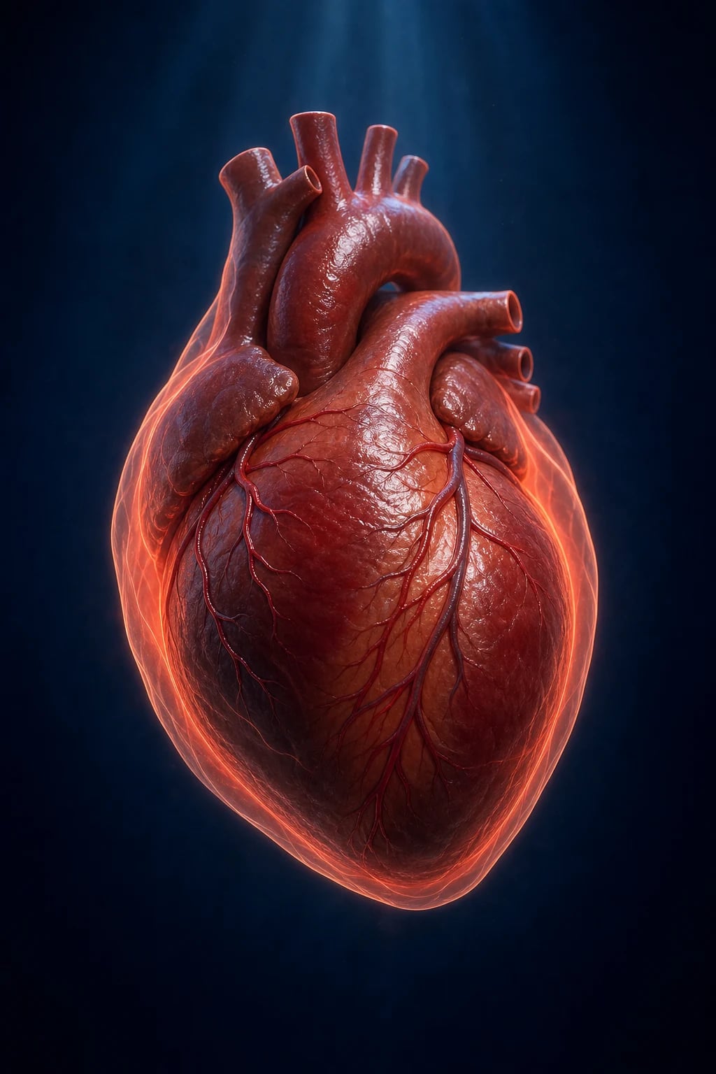
Pericardial disease is a family of disorders of the pericardial sac and its contents, with three overlapping but clinically distinct syndromes:[1]
- Acute pericarditis — acute inflammation of the pericardium lasting under 3 months, defined by the ESC diagnostic criteria (see Investigations). Includes first episode, incessant (over 4 to 6 weeks but under 3 months), recurrent (new episode after a symptom-free interval of at least 4 to 6 weeks) and chronic (over 3 months) pericarditis.
- Pericardial effusion — abnormal accumulation of fluid in the pericardial space; clinical importance depends on rate of accumulation more than absolute volume. Cardiac tamponade is the decompensated end-state where fluid under pressure impairs ventricular filling and cardiac output.
- Constrictive pericarditis — chronic thickening, scarring and sometimes calcification of the pericardium that limits diastolic filling, producing a clinical picture of right heart failure. [1]
The clinical skill is threefold: distinguish pericarditis from acute MI (both can cause chest pain and ST elevation), recognise tamponade early before haemodynamic collapse, and differentiate constriction from restrictive cardiomyopathy (both have raised JVP and oedema but radically different treatments).[1][6]
Classification
Acute pericarditis
- Inflammation of pericardium, under 3 months
- ESC criteria: 2 of 4 (chest pain, rub, ECG changes, effusion)
- Commonest cause idiopathic/viral
- NSAID + colchicine first-line; steroids NOT first-line
Pericardial effusion
- Fluid in pericardial space (transudate, exudate, haemorrhagic, purulent)
- Haemodynamic impact depends on RATE of accumulation
- Tamponade when pressure exceeds filling pressure
- Echo: chamber collapse, IVC plethora, respiratory variation
Cardiac tamponade
- Decompensated effusion impairing cardiac filling
- Beck triad: hypotension, raised JVP, muffled heart sounds
- Pulsus paradoxus over 10 mmHg
- Emergency echo-guided pericardiocentesis
Constrictive pericarditis
- Chronic thick, scarred (often calcified) pericardium
- Kussmaul sign: JVP rises on inspiration
- Pericardial knock (early diastolic sound)
- Definitive: pericardiectomy
Recurrent pericarditis
- Recurrence after symptom-free interval of at least 4 to 6 weeks
- Recurrence rate 15 to 30 per cent without colchicine
- Colchicine halves recurrence (COPE, ICAP)
- Refractory cases: low-dose colchicine, IL-1 blocker (anakinra)
Post-cardiac injury syndrome
- Inflammatory pericarditis 1 to 8 weeks after MI, surgery, trauma, catheter
- Dressler syndrome: post-MI autoimmune pericarditis
- Pleuritic pain, fever, raised inflammatory markers
- Aspirin first-line (post-MI); avoid steroids and NSAIDs early after MI
Epidemiology & Risk Factors
Acute pericarditis accounts for about 5 per cent of all non-ischaemic chest pain presentations to the emergency department; it is the commonest form of pericardial disease. Peak incidence 20 to 50 years, male predominance, often follows a viral upper respiratory infection.[1]
Aetiology and the causes that map to it (high-yield): [1]
- Idiopathic / viral (commonest in developed countries; 80 to 90 per cent) — coxsackievirus, echovirus, adenovirus, influenza, EBV, HIV.
- Bacterial (purulent) — Staphylococcus, Streptococcus, Pneumococcus; TB pericarditis is a leading cause in India, sub-Saharan Africa, and other TB-endemic regions (5 to 70 per cent of cases depending on region).
- Uraemic — renal failure (pericarditis is an indication for urgent dialysis).
- Post-cardiac injury — acute MI (first 1 to 3 days, fibrinous pericarditis), Dressler syndrome (1 to 8 weeks post-MI), post-cardiac surgery (post-pericardiotomy syndrome), post-trauma, post-catheter/ablation.
- Autoimmune / connective tissue disease — SLE, rheumatoid arthritis, scleroderma, vasculitis, sarcoidosis, inflammatory bowel disease.
- Neoplastic — lung cancer, breast cancer, lymphoma, melanoma (metastatic much commoner than primary).
- Radiation — mediastinal radiotherapy (months to years later).
- Drugs — procainamide, hydralazine, isoniazid (drug-induced lupus), phenytoin, doxorubicin.
- Endocrine — hypothyroidism (myxoedema), rarely pregnancy. [1]
Risk factors for the major causes: immunocompromise (HIV, transplant, chemotherapy) for viral/bacterial/mycobacterial; chronic dialysis for uraemic; recent open-heart surgery for post-pericardiotomy syndrome; ** mediastinal radiotherapy or known cancer** for neoplastic; untreated active TB exposure for tuberculous. Constriction most often follows tuberculous or viral pericarditis, after cardiac surgery, or mediastinal radiation.[1][5]
Pathophysiology
The pericardium is normally non-distensible in the acute setting. This single physical fact drives the pathophysiology of all three syndromes.[6]
Acute pericarditis. Insult (virus, autoimmune, uraemia, MI) triggers a localised inflammatory response in the pericardial layers with fibrinous exudate, neutrophil and lymphocyte infiltrate, and sometimes a sympathetic pericardial effusion. The inflamed pericardial surfaces rub against each other producing the pericardial friction rub; subepicardial inflammation produces the characteristic diffuse ST elevation (epicardial injury current) and PR segment depression (atrial injury current).[1]
Cardiac tamponade. Because the fibrous pericardium cannot stretch acutely, fluid accumulating rapidly (200 to 300 mL over minutes to hours) raises intra-pericardial pressure sharply. Pressure-volume relationship is non-linear: a slow effusion can be tolerated (over 1 to 2 L) but a fast one cannot. When intra-pericardial pressure exceeds ventricular filling pressure, the chambers collapse in sequence: right atrium in late systole/early diastole first, then right ventricle in early diastole. The interventricular septum shifts leftward during inspiration (the right heart fills more on inspiration due to negative intrathoracic pressure, but in tamponade total cardiac volume is fixed, so the left heart is compressed) → exaggerated drop in left-sided stroke volume and systolic BP on inspiration = pulsus paradoxus (over 10 mmHg fall in SBP).[3]
Constrictive pericarditis. Chronic inflammation heals by fibrosis, scarring and often calcification, fusing the pericardial layers into a rigid shell. The heart is held in a non-compliant casing that abruptly halts ventricular filling in mid-to-late diastole (rapid early filling, then sudden stop) — producing the pericardial knock and a prominent y descent on the JVP. Because the pericardium isolates the heart from intrathoracic pressure changes, inspiration cannot augment systemic venous return normally; instead venous pressure rises on inspiration = Kussmaul sign.[7]
Ventricular interdependence and the pressure-volume curve. The pericardial pressure-volume relationship is exponential, not linear: once the flat portion of the curve is reached, tiny increments in volume produce large rises in pressure. This explains why 200 mL accumulating over minutes (aortic dissection, catheter perforation, trauma) causes tamponade while 2 L accumulating over months (neoplastic, myxoedema) may be tolerated. In tamponade, all four chambers share a fixed total volume within a non-distensible sac, so filling of one chamber steals from the others — this is ventricular interdependence, exaggerated to the point of haemodynamic collapse. On inspiration, negative intrathoracic pressure augments systemic venous return and right-heart filling; because total cardiac volume is fixed, the interventricular septum shifts leftward, reducing left ventricular filling, stroke volume and systolic blood pressure. A fall under 10 mmHg is normal physiology; a fall over 10 mmHg defines pulsus paradoxus.[3]
Why constriction differs from tamponade. Both impair filling and raise venous pressure, but the mechanism differs fundamentally. In tamponade the fluid compresses the heart throughout diastole, so there is no rapid early filling (blunted y descent). In constriction the pericardium is rigid but does not oppose early diastolic filling — the ventricle fills rapidly until it hits the rigid shell and stops abruptly in mid-diastole (deep y descent, pericardial knock). Because the pericardium in constriction insulates the heart from intrathoracic pressure swings, inspiration cannot lower intrapericardial pressure, so systemic venous pressure does not fall (and may rise — Kussmaul sign), while pulmonary venous pressure still falls normally on inspiration; the gradient between pulmonary veins and left atrium narrows, reducing left-sided filling. The absence of pulsus paradoxus in most constrictive cases is an important bedside discriminator.[7]
Clinical Presentation
Acute pericarditis — the classic triad is chest pain, pericardial friction rub and ECG changes (any two of the ESC four suffice for diagnosis).[1]
- Pain is sharp, pleuritic, worse on deep inspiration and lying flat, relieved by sitting forward. Often radiates to the left trapezial ridge (phrenic nerve C3 to C5, pericardium shares innervation with shoulder). Distinguish from MI (pressure, radiates arm/jaw, not pleuritic, not positional).
- Pericardial friction rub — best heard at the left sternal border, with the patient leaning forward in full expiration. Three components (atrial systole, ventricular systole, rapid ventricular filling) but often only one or two heard — a high-pitched, scratching, leather-like, superficial sound. Highly specific but evanescent (re-examine).
- Low-grade fever, myalgia, recent viral prodrome. [1]
Cardiac tamponade — the clinical picture ranges from incidental effusion to Beck triad in the decompensated state:[3]
- Beck triad (acute): hypotension, raised JVP, muffled heart sounds.
- Pulsus paradoxus — drop in SBP over 10 mmHg on normal inspiration (normal is under 10). Severe: over 25 mmHg.
- Dyspnoea, tachycardia, tachypnoea; the patient may be anxious, sitting upright. Shock with clear lungs (unlike pulmonary oedema) and raised JVP is the key discriminator.
- A pulsatile effusion in a patient with a known aortic dissection or recent cardiac procedure suggests haemopericardium — emergency. [1]
Constrictive pericarditis — presents as right heart failure with no primary lung disease:[7]
- Raised JVP with prominent x and y descents (M-shape); the y descent is deep and rapid. Kussmaul sign — JVP rises on inspiration (paradoxical).
- Pericardial knock — high-pitched early diastolic sound (0.06 to 0.12 s after S2),** louder and earlier** than the S3 of heart failure.
- Hepatomegaly, ascites, peripheral oedema — often disproportionate to pulmonary congestion. May present with cachexia, atrial fibrillation in advanced disease.
- Pulsus paradoxus is usually absent (constriction = fixed volume but pressures transmitted — distinguishes from tamponade). [1]
Atypical presentations: elderly/immunocompromised — pain and rub absent; presents as confusion, dyspnoea, falling BP, raised JVP. Post-MI early fibrinous pericarditis may have only a transient rub 1 to 3 days after STEMI. Uraemic pericarditis may be painless with only a rub and effusion. TB pericarditis is insidious with constitutional features (fever, night sweats, weight loss). Pregnant patient — physiological S3 and mild JVP elevation make tamponade harder to spot; rely on echo.[5]
Differential Diagnosis
Distinguish pericarditis from the other causes of acute chest pain and from the other causes of raised JVP/right heart failure.[6]
1. Acute MI (STEMI) — both produce chest pain and ST elevation; both may coexist (early post-MI pericarditis). [1]
| Feature | Acute pericarditis | STEMI |
|---|---|---|
| Pain character | Sharp, pleuritic, positional | Pressure, crushing |
| Radiation | Trapezial ridge | Arm, jaw |
| ST morphology | Diffuse, concave | Leads reflecting culprit artery, convex (tombstone) |
| PR segment | Depression (common) | Elevation (aVR) in some |
| Reciprocal changes | Absent | Present |
| Evolution | Stages I–IV over days to weeks | Hours (Q waves, T inversion) |
| Troponin | Mildly raised or normal | Markedly raised |
2. Pulmonary embolism — pleuritic pain and dyspnoea but no diffuse ST elevation, no pericardial rub; risk factors, tachycardia, sometimes right heart strain on ECG (S1Q3T3, right axis). D-dimer, CTPA. [1]
3. Aortic dissection — tearing chest/back pain, pulse/BP differential; if complicated by haemopericardium, presents as tamponade — NEVER anticoagulate until excluded. [1]
4. Restrictive cardiomyopathy vs constrictive pericarditis (the classic discriminator, see Investigations): [1]
| Feature | Constriction | Restriction |
|---|---|---|
| Pericardial knock | Present | Absent (S3 may be present) |
| Pericardial calcification (CXR/CT) | Present in up to 50 per cent | Absent |
| Kussmaul sign | Present | Often absent |
| BNP | Normal/mildly raised | Markedly raised |
| Echo septal bounce | Present | Absent |
| Cardiac MRI LGE | Pericardial LGE, thickening | Myocardial LGE |
| Treatment | Pericardiectomy | Treat underlying cause (amyloid, etc.) |
5. Other causes of raised JVP/right heart failure — decompensated cirrhosis, cor pulmonale, tricuspid regurgitation, RV infarction (Kussmaul sign in RV MI). Distinguish by history, JVP waveform, echo. [1]
Clinical & Bedside Assessment
Vital signs drive urgency: BP, pulsus paradoxus, JVP, SpO2, RR, temperature. Measure pulsus paradoxus by sphygmomanometer — deflate slowly; the first Korotkoff sound heard only on expiration is the upper limit, the point where it is heard throughout the respiratory cycle the lower; difference over 10 mmHg is abnormal.[3]
JVP waveform in constriction shows deep y descent and the Kussmaul sign (paradoxical rise on inspiration). In tamponade the x descent is preserved, y descent is blunted or absent (no rapid early filling). [1]
Auscultation of the rub — patient leaning forward, in full expiration, diaphragm at left lower sternal border. Re-examine — the rub is evanescent. [1]
Pericardial knock — best heard at the left sternal border or apex, high-pitched, early diastolic. [1]
Look for the underlying cause: stigmata of SLE (malar rash, oral ulcers), RA (hand deformity), uraemia (fistula, dialysis signs), TB (cachexia, lymphadenopathy), malignancy (cachexia, clubbing), recent surgery/scars (post-pericardiotomy). [1]
Investigations
ESC diagnostic criteria for acute pericarditis — at least 2 of 4 must be present:[1]
- Pericarditic chest pain (sharp, pleuritic, positional).
- Pericardial rub.
- ECG with new widespread ST elevation or PR depression.
- New or worsening pericardial effusion on imaging. [1]
Additional supportive markers: raised inflammatory markers (CRP, ESR), mild troponin elevation (myopericarditis if troponin markedly raised with focal wall motion abnormality). [1]
First-line investigations:[1]
- 12-lead ECG — the cardinal test.
- Echocardiography — assess effusion size, chamber collapse, IVC plethora, respiratory variation; exclude concomitant myocarditis/regional wall motion.
- Bloods — FBC, CRP, ESR, troponin, urea and electrolytes (uraemia), LFTs, TSH; blood cultures if septic; viral serology generally unhelpful; autoimmune screen (ANA, dsDNA, RF) only if clinical suspicion; HIV serology in all.
- Chest X-ray — may be normal; large effusion produces a water-bottle heart; pericardial calcification suggests constriction; lung fields clear in tamponade/constriction.
- If TB suspected (endemic region) — sputum AFB, GeneXpert/MTB/RIF, Mantoux/IGRA, adenosine deaminase in pericardial fluid (over 40 U/L supports TB). [1]
ECG stages of acute pericarditis (reproduced):[1]
- Stage I (hours to days) — diffuse concave ST elevation in most leads (I, II, aVL, aVF, V3 to V6), PR depression in the same leads (PR elevation in aVR/V1), no reciprocal changes.
- Stage II (days to weeks) — ST segments return to baseline; PR depression persists; T waves begin to flatten.
- Stage III — T wave inversion (diffuse).
- Stage IV — T waves normalise; recovery. [1]
Echocardiographic findings in tamponade (reproduced, the high-yield list):[3]
- Right atrial collapse in late systole/early diastole (sensitive if over one-third of cardiac cycle).
- Right ventricular diastolic collapse (more specific).
- IVC plethora (over 2.1 cm, under 50 per cent collapse on sniff — highly sensitive).
- Respiratory variation in ventricular inflow — over 25 per cent change in mitral E, over 40 per cent in tricuspid E on inspiration.
- Swinging heart (electrical alternans) in large effusions. [1]
Cardiac MRI/CT — when echocardiography non-diagnostic, to assess pericardial thickness (over 2 mm abnormal; constriction typically over 3 to 4 mm), pericardial LGE (active inflammation), calcification (CT more sensitive), and to differentiate constriction from restriction.[7]
Pericardial fluid analysis (after drainage) — cell count, protein, LDH, glucose, Gram stain, AFB and GeneXpert, cytology, adenosine deaminase (TB). [1]
Management — Resuscitation
ABCDE first. The patient with tamponade is the resuscitation emergency.[3]
Effusion size on echocardiography (the ESC semiquantitative grading):[1]
| Size | Echo appearance | Approximate volume |
|---|---|---|
| Trivial | Echo-free space in systole only, under 0.5 cm | under 50 mL |
| Small | Under 1 cm, posterior only | 50 to 100 mL |
| Moderate | 1 to 2 cm, circumferential | 100 to 500 mL |
| Large | Over 2 cm, circumferential | over 500 mL |
Echo in constriction — the key signs beyond tamponade: septal bounce (abrupt early diastolic septal motion towards the LV as the RV hits the constraint), pericardial thickening (over 3 mm, often with a bright echo-dense band), respirophasic variation in mitral and tricuspid inflow (over 25 per cent mitral E-wave reduction on inspiration), a plethoric IVC, and preserved or exaggerated tissue Doppler e-prime (e') at the medial annulus — the so-called annulus paradoxus (mitral annulus moves normally despite restrictive transmitral filling), which distinguishes constriction (normal myocardium) from restriction (reduced e').[7]
Cardiac MRI and CT — when echo is non-diagnostic or constriction is suspected: CT is the modality of choice for pericardial calcification (seen in up to 50 per cent of constrictive cases, best along the AV grooves and right heart border) and for pericardial thickness (over 2 mm abnormal; over 3 to 4 mm typical of constriction). MRI adds tissue characterisation: pericardial late gadolinium enhancement (LGE) indicates active inflammation (may predict response to medical therapy in transient constriction), real-time cine imaging confirms the septal bounce and ventricular interdependence, and tagged sequences show pericardial-myocardial adherence (failure of normal sliding) — a specific sign of constriction. MRI also excludes myocardial disease (LGE, T1 mapping for amyloid) that would point to restrictive cardiomyopathy instead.[7]
- Oxygen to maintain SpO2 94 to 98 per cent.
- IV access, bloods, group and save; avoid delays for tests.
- Volume expansion — 250 to 500 mL bolus crystalloid may transiently improve preload in the volume-depleted tamponade patient; avoid in established volume overload.
- Avoid vasodilators, diuretics and positive-pressure ventilation if possible (reduce venous return, worsen collapse) — intubate only if essential, with full volume loading first.
- Pericardiocentesis — echo-guided subxiphoid approach, ideally in a cardiac catheterisation/theatre setting; send fluid for analysis. Surgical drainage (subxiphoid pericardiostomy) preferred for loculated, recurrent, or malignant effusions, or where tissue biopsy is needed (TB).
- In extremis with no immediate echo — emergency blind pericardiocentesis (subxiphoid, aim for left shoulder) is justified if death is imminent; aspiration of even 50 mL can be lifesaving. [1]
Acute pericarditis is rarely a resuscitation emergency unless large effusion or tamponade — but always assess for haemodynamic compromise. [1]
Management — Definitive & Stepwise
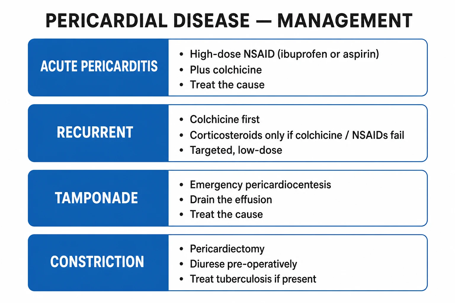
Acute pericarditis — first-line (ESC 2015, reproduced with doses):[1][2]
- Aspirin 750 to 1000 mg PO every 8 hours OR ibuprofen 600 to 800 mg PO every 8 hours (the two first-line NSAIDs). Continue 1 to 2 weeks (until pain and CRP settle), then taper over weeks.
- Plus colchicine 0.5 mg PO once daily if body weight under 70 kg, OR 0.5 mg twice daily if over 70 kg — for 3 months (reduces recurrence from 30 to 15 per cent; ICAP, COPE trials).[2][4]
- PPI gastroprotection (e.g. omeprazole 20 mg OD).
- Restricted physical activity until symptom-free and CRP normal; athletes at least 3 months.
- Treat the underlying cause (haemodialysis for uraemic, antibiotics for purulent, anti-TB, immunosuppression for autoimmune).
Corticosteroids — NOT first-line (high recurrence, chronicity, drug-related; clear increase in recurrence rate in observational studies and randomised trials). Reserve for:[1][6]
- Indications: pericarditis refractory to NSAID + colchicine, autoimmune aetiology (SLE, RA), uraemic (after dialysis), pregnancy (NSAIDs contraindicated), specific contraindication to NSAID/colchicine.
- Dose: prednisolone 0.2 to 0.5 mg/kg/day (low dose preferred; the ESC recommends against high-dose in the first episode), taper slowly over weeks to months after symptom resolution and CRP normalisation.
- Avoid corticosteroids in suspected TB without cover; avoid in early post-MI pericarditis (impairs scar, raises risk of rupture). [1]
Post-MI pericarditis / Dressler syndrome: [1]
- Early fibrinous pericarditis (1 to 3 days post-STEMI) — usually benign; aspirin 650 mg every 4 to 6 hours preferred (NSAIDs and steroids impair healing, raise rupture risk in first weeks).
- Dressler syndrome (1 to 8 weeks post-MI) — autoimmune; high-dose aspirin first-line; corticosteroids reserved for refractory cases after the infarct has healed. [1]
Recurrent pericarditis — defined as recurrence after a symptom-free interval of at least 4 to 6 weeks. Recurrence rate without colchicine is 15 to 30 per cent after the first episode and rises with each subsequent episode (up to 50 per cent after three). Colchicine is the cornerstone (COPE-2, CORP trials — halve recurrence); treat each recurrence with the standard NSAID + colchicine regimen, extending colchicine to 6 months or longer for multiple recurrences. Target CRP-guided tapering: continue NSAIDs until pain and CRP normalise, then taper over 2 to 4 weeks. Refractory recurrent pericarditis (three or more recurrences or steroid-dependent) is transformed by IL-1 blockade: anakinra (recombinant IL-1 receptor antagonist) 1 to 2 mg/kg/day subcutaneously (up to 100 mg/day) for several months then tapered, or rilonacept (IL-1 trap) 320 mg loading then 160 mg weekly subcutaneously — both targeting the NLRP3 inflammasome-driven cytokine cascade that drives recurrent inflammation. Corticosteroids (prednisolone 0.2 to 0.5 mg/kg/day, tapered over months) remain an option but carry the highest recurrence on withdrawal. Azathioprine or IV immunoglobulin are reserved for colchicine- and IL-1-blocker-resistant disease.[6]
Tuberculous pericarditis — the Indian context:[5][1]
- Standard anti-tubercular therapy (RIPE) — rifampicin 10 mg/kg, isoniazid 5 mg/kg, pyrazinamide 25 mg/kg, ethambutol 15 mg/kg daily for 2 months intensive, then rifampicin + isoniazid 4 months (total 6 months).
- Adjunctive corticosteroids — prednisolone (often 60 mg/day tapering over 6 to 12 weeks) reduces constrictive sequelae and mortality in proven TB pericarditis; recommended by WHO and ICMR; benefit greatest in HIV-positive patients. Do not give without concomitant anti-TB therapy.
- Pericardiocentesis for tamponade; pericardiectomy for established constriction. [1]
Purulent (bacterial) pericarditis — IV antibiotics (vancomycin 1 g BD + third-generation cephalosporin e.g. ceftriaxone 2 g OD, then tailored) plus urgent surgical/percutaneous drainage; mortality high without drainage. [1]
Neoplastic effusion — pericardiocentesis, often indwelling pericardial catheter, intrapericardial sclerosants (e.g. bleomycin, doxycycline), and treatment of the underlying tumour. [1]
Constrictive pericarditis — definitive treatment is pericardiectomy: surgical resection of the pericardium (phrenic-to-phrenic). Best results in early/established constriction without end-stage myocardial disease. Operative mortality 5 to 15 per cent. Transient constrictive physiology (post-viral, post-surgery) may resolve with 2 to 3 months of NSAID + colchicine + low-dose steroid — re-image before committing to surgery.[7]
Pericardiocentesis — technique the examiner expects
Indications: cardiac tamponade with haemodynamic compromise; large symptomatic effusion (over 2 cm circumferential with dyspnoea); diagnostic fluid sampling (suspected TB, bacterial, neoplastic); and effusive-constrictive physiology where drainage both relieves and diagnoses.[3]
Approach: the subxiphoid (subcostal) route is standard. With the patient supine at 30 to 45 degrees, the needle enters 1 to 2 cm below the left xiphocostal angle, aimed at the left shoulder at 30 degrees to the skin, with continuous ECG monitoring (ST elevation or ectopy if the myocardium is touched) and ideally continuous echocardiographic guidance (apical or subcostal window to watch the needle tip enter the effusion). An apical approach (5th to 7th intercostal space at the cardiac apex, 1 cm lateral to the point of maximum impulse) is the modern echo-guided alternative and avoids the liver. Blind pericardiocentesis is reserved for imminent arrest — aspirate just enough (50 to 100 mL) to relieve the haemodynamic emergency and gain time for definitive drainage. [1]
Technique: aseptic preparation and local anaesthesia (1 per cent lidocaine); advance an 18-gauge needle on a syringe with slight negative pressure; once fluid is aspirated, pass a guidewire (Seldinger) into the pericardial space, dilate, and place a multi-side-hole pigtail catheter (6 to 8 Fr). Aspirate until drainage slows; leave the catheter on closed drainage for 24 to 72 hours, draining recurrent accumulation (common in malignant effusion). [1]
Send fluid for: cell count and differential, protein, LDH, glucose, Gram stain and culture, AFB smear and culture plus GeneXpert MTB/RIF, adenosine deaminase (over 40 U/L supports TB), cytology (malignancy), and pH (under 7.1 suggests purulent or bacterial). Transudate versus exudate (using adapted Light's criteria) helps narrow the differential. [1]
Complications: cardiac chamber laceration (right ventricle most common — the anterior structure), pneumothorax, liver injury, arrhythmia (atrial ectopy as the needle nears the atrium), coronary or internal mammary artery laceration, and re-expansion pulmonary oedema. The risk is halved by echocardiographic guidance; never attempt blind pericardiocentesis when echo and a trained operator are available.[3]
Surgical options — when the needle is not enough
Subxiphoid pericardiotomy (pericardial window): surgical creation of a communication between pericardial and pleural or subcutaneous space under general or local anaesthesia; preferred for recurrent malignant effusion, loculated effusion, purulent pericarditis (needs debridement), and when tissue for histology (TB, malignancy) is required. Video-assisted thoracoscopic (VATS) pericardiectomy and balloon pericardiotomy are alternatives for recurrent drainage. Pericardiectomy (complete, phrenic-to-phrenic stripping of the pericardium) is the definitive operation for constriction — performed via median sternotomy or left thoracotomy on cardiopulmonary bypass when dense calcification is present; operative mortality 5 to 15 per cent, higher in radiation-induced disease where coexisting myocardial damage limits recovery.[7]
Specific Subtypes & Scenarios
- Viral/idiopathic pericarditis — the commonest; excellent prognosis; colchicine halves recurrence.
- Tuberculous pericarditis — insidious onset over weeks with constitutional features (fever, night sweats, weight loss); the effusion is often large and haemorrhagic or serosanguineous; an effusive-constrictive phenotype may evolve during treatment. Diagnosis is challenging — combine pericardial fluid adenosine deaminase over 40 U/L, GeneXpert MTB/RIF positive, AFB culture, and pericardial biopsy with caseating granulomas. High risk of constriction (20 to 30 per cent even with treatment); treat with RIPE for 6 months plus adjunctive corticosteroids; pericardiectomy if established constriction. HIV co-infection is common in endemic regions and worsens prognosis.[5]
- Purulent pericarditis — abrupt, septic, medical emergency; rapid tamponade; surgical drainage mandatory.
- Uraemic pericarditis — painless, often with rub and effusion; urgent intensified dialysis is the treatment; avoid NSAIDs (renal risk).[1]
- Dressler syndrome — autoimmune, 1 to 8 weeks post-MI, fever, pleuritic pain, raised ESR/CRP; aspirin high-dose first-line; rare in the modern primary-PCI era.
- Post-pericardiotomy syndrome — weeks after cardiac surgery; colchicine prophylaxis (COPPS trial) reduces incidence.[4]
- Neoplastic effusion — lung/breast/lymphoma; often large, haemorrhagic; palliative drainage; consider pericardial window.
- Radiation pericarditis — months to years after mediastinal radiotherapy; can present as acute, effusive, or constrictive.
- Effusive-constrictive pericarditis — tamponade physiology persists after pericardiocentesis because the visceral pericardium (epicardium) is the constrictor, not just the parietal layer. Suspect it when JVP fails to fall after drainage and the right atrial pressure remains elevated. Definitive treatment is pericardiectomy with epicardiectomy (stripping the visceral layer as well).[7]
- Recurrent pericarditis — recurrence after a symptom-free interval of at least 4 to 6 weeks; cumulative recurrence rises with each episode. Colchicine is the cornerstone; IL-1 blockade (anakinra, rilonacept) is transformative for steroid-dependent or multi-recurrent disease. A minority develop a chronic autoreactive/idiopathic pattern requiring long-term low-dose colchicine with periodic CRP-guided drug holidays.[6]
Complications & Pitfalls
Complications of acute pericarditis: recurrence (15 to 30 per cent), cardiac tamponade (more in neoplastic, bacterial, TB), progression to constrictive pericarditis (TB, purulent, radiation), myopericarditis (raised troponin, focal wall motion abnormality; treat as myocarditis). [1]
Pitfalls: [1]
- Misdiagnosing STEMI as pericarditis (or vice versa) — check for reciprocal changes, PR depression, diffuse concave ST elevation; if in doubt, urgent coronary angiography.
- Missing tamponade because BP looks normal — pulsus paradoxus and JVP examination are essential; rely on echo for any large effusion.
- Giving steroids for first-episode idiopathic pericarditis — raises recurrence 2- to 3-fold.[1]
- Using NSAIDs/steroids in early post-MI pericarditis — aspirin only; risk of free-wall rupture.
- Anticoagulating haemorrhagic effusion (e.g. post-catheter) — worsens tamponade.
- Forgetting TB in the Indian patient with effusion — a missed TB pericarditis will progress to constriction.
- Mislabelling constrictive pericarditis as cirrhosis (ascites with raised JVP = constriction; cirrhosis has low JVP).
Prognosis & Disposition
Acute idiopathic pericarditis — excellent; 80 per cent recover fully with one course of NSAID + colchicine. Recurrence risk 15 to 30 per cent (halved by colchicine).[2][6]
Cardiac tamponade — prognosis depends on aetiology and speed of drainage; untreated is fatal; post-pericardiocentesis depends on the underlying disease. [1]
Constrictive pericarditis — progressive right heart failure without surgery; pericardiectomy 5-year survival 80 per cent in idiopathic/viral, lower in post-radiation (myocardial damage coexists). Radiation-induced and renal-failure-associated carry the worst prognosis.[7]
TB pericarditis — even with treatment, 20 to 30 per cent develop constriction; mortality 8 to 17 per cent; HIV co-infection worsens outcomes.[5]
Disposition: acute pericarditis with small/no effusion, no fever, no high-risk feature — outpatient with NSAID + colchicine + safety net. Admit if fever over 38 deg C, large effusion, tamponade, immunocompromise, trauma, oral anticoagulation, failure of outpatient therapy, myopericarditis — the ESC high-risk triage markers.[1]
Special Populations
- Pregnant — NSAIDs (especially ibuprofen, indometacin) contraindicated after 20 weeks (fetal renal effects, premature closure of ductus); colchicine safe; corticosteroids are first-line for symptomatic pericarditis in pregnancy.[1]
- Renal failure / dialysis — uraemic pericarditis is an indication for intensified dialysis; NSAIDs and colchicine (renal excretion) must be dose-adjusted or avoided; aspirin at dialysis-adjusted dose preferred.
- Elderly / immunocompromised — atypical presentation, painless, higher rate of TB/neoplastic; lower threshold to investigate and admit.
- Children — colchicine weight-based (0.5 mg daily under 5 years; up to 1.5 mg/day in divided doses older); purulent pericarditis more common in young children.
- Anticoagulated patient — high risk of haemorrhagic effusion/tamponade; reverse anticoagulation if effusion enlarges or tamponade; do not start anticoagulation in pericarditis unless essential.
Evidence, Guidelines & Regional Differences
Key ESC 2015 guideline changes:[1]
- Diagnostic criteria restated (2 of 4).
- Triage by risk markers — admit if any of: fever over 38 deg C, subacute onset, large effusion, tamponade, immunocompromise, trauma, oral anticoagulation, myopericarditis, failure of NSAID.
- Colchicine is now first-line for the index episode and recurrence.
- Corticosteroids downgraded — only for specific indications; low-dose prednisolone preferred. [1]
Landmark trials: [1]
- COPE (2010) — colchicine plus conventional therapy halved recurrence at 18 months in first episode.[4]
- ICAP (2013, NEJM) — colchicine 0.5 mg for 3 months halved recurrent pericarditis (16.7 vs 37.5 per cent).[2]
- COPPS (2011) — colchicine prophylaxis halved post-pericardiotomy syndrome after cardiac surgery.
- Imazio JAMA 2015 systematic review — colchicine effective in acute, recurrent and post-pericardiotomy pericarditis.[6]
Regional delta — India: TB pericarditis is endemic and the leading cause of constriction; any large or recurrent effusion in the Indian patient warrants evaluation for TB (sputum, GeneXpert, IGRA, pericardial ADA). Anti-TB therapy (6 months) plus adjunctive corticosteroids is recommended by ICMR/NTEP for proven or strongly suspected TB pericarditis. Purulent pericarditis and rheumatic pericarditis are more common than in Western series.[5][1]
Exam Pearls
- Pericarditis triad: chest pain + rub + ECG changes (any 2 of ESC 4 = diagnostic).
- Pain: sharp, pleuritic, worse on lying flat, relieved by sitting forward; radiates to trapezial ridge.
- Rub: three-component, scratching, superficial, left sternal border, leaning forward in expiration, evanescent.
- ECG stage I: diffuse concave ST elevation + PR depression, no reciprocal changes — the discriminator from STEMI.
- Troponin may be mildly raised (myopericarditis) — does not equal MI.
- Tamponade — Beck triad: hypotension + raised JVP + muffled heart sounds; pulsus paradoxus over 10 mmHg; echo: RA/RV collapse, IVC plethora.
- Pulsus paradoxus is also seen in severe asthma/COPD, tension pneumothorax, PE — distinguish by context.
- Constriction — Kussmaul sign (JVP rises on inspiration); pericardial knock; pericardial calcification on CXR/CT.
- Kussmaul sign also seen in: RV MI, severe TR, restrictive cardiomyopathy, severe pulmonary hypertension.
- Constriction vs restriction: calcification + knock + septal bounce + normal BNP = constriction; myocardial LGE + high BNP = restriction.
- Treatment acute pericarditis: aspirin 750 to 1000 mg TID or ibuprofen 600 to 800 mg TID + colchicine 0.5 mg BD for 3 months.
- Steroids are NOT first-line — raise recurrence; reserve for autoimmune, uraemic, pregnancy, refractory.
- Post-MI pericarditis — aspirin only, NOT NSAIDs/steroids (rupture risk).
- Dressler syndrome: 1 to 8 weeks post-MI, autoimmune, raised ESR/CRP.
- TB pericarditis in India: anti-TB (RIPE 6 months) + adjunctive corticosteroids; high risk of constriction.
- Effusive-constrictive: tamponade physiology persists after drainage — visceral pericardium is the constrictor.
- Pericardiocentesis = echo-guided, subxiphoid; surgical window for recurrent/loculated/malignant.
- Constriction — definitive treatment: pericardiectomy. [1]
Exam application bank (NEET-PG / INICET)
One-line answer
Pericardial disease spans three overlapping syndromes: acute pericarditis (inflammation of the pericardium with pleuritic chest pain, pericardial rub and diffuse ST elevation with PR depression), pericardial effusion with cardiac tamponade (fluid under pressure impairing diastolic filling; Beck triad, pulsus paradoxus), and constrictive pericarditis (chronic thickening producing Kussmaul sign and a pericardial knock). Commonest cause is idiopathic/viral; in India TB pericarditis is a major differential. Diagnose clinically plus ECG; echocardiography is the key imaging. Treat acute pericarditis with NSAIDs (ibuprofen 600 to 800 mg TID) plus colchicine 0.5 mg BD for 3 months; corticosteroids are NOT first-line (raise recurrence). Tamponade is drained by echo-guided pericardiocentesis; constriction needs pericardiectomy. [1]
Worked stems (answer without another resource)
Stem 1 — Classic presentation. Map symptoms to mechanism; name the first investigation and first treatment step with dose/route if drug therapy is standard. [1]
Stem 2 — Unstable / complicated. List red flags that force immediate resuscitation, theatre, ICU, antidote, or reperfusion — and what you do in the first 15 minutes. [1]
Stem 3 — Atypical group. Elderly, pregnancy, child, or immunocompromised: how presentation and thresholds change. [1]
Stem 4 — Differential trap. Name the three closest mimics and one discriminator for each. [1]
Stem 5 — Disposition. Who goes home with safety-netting, who is admitted, who needs HDU/ICU/theatre, and what follow-up is mandatory. [1]
Rapid viva checklist
- Definition + classification
- Pathophysiology chain
- Bedside signs / criteria
- Score with exact components (if any)
- Emergency bundle
- Definitive therapy with doses
- Complications of disease and of treatment
- Special populations
- Guideline/trial name if classic
- Three exam traps
Coverage self-check
If you cannot answer any stem above from this page alone, re-read the matching section — the page is intended to be self-sufficient for final-prof and NEET-PG/INICET questions on Pericardial Disease.
Pericardial disease — the numbers examiners ask
CAUSES of pericarditis — mnemonic
PERICARD
early fibrinous, Dressler syndrome, post-pericardiotomy
uraemia, hypothyroidism
mediastinal radiotherapy (months to years later)
coxsackie, echovirus, EBV, HIV — commonest
SLE, RA, scleroderma, vasculitis
procainamide, hydralazine, isoniazid
rheumatic fever, IBD
doxorubicin, aortic dissection causing haemopericardium, blunt chest trauma
TAMPONADE bedside — mnemonic
TAMPON
compensatory, then bradycardia in agonal phase
no rapid early filling; x descent preserved
fluid around heart dampens sound
SBP drops over 10 mmHg on inspiration
raised systemic venous pressure
SBP falls but DBP preserved early
References
- [1]Adler Y, Charron P, Imazio M, et al. 2015 ESC Guidelines for the diagnosis and management of pericardial diseases: The Task Force for the Diagnosis and Management of Pericardial Diseases of the European Society of Cardiology (ESC)Endorsed by: The European Association for Cardio-Thoracic Surgery (EACTS) Eur Heart J, 2015.PMID 26320112
- [2]Imazio M, Brucato A, Cemin R, et al. Reduced cortisol metabolism during critical illness N Engl J Med, 2013.PMID 23902495
- [3]Roy CL, Minor MA, Brookhart MA, Choudhry NK. Residency review committee for surgery: an update Arch Surg, 2007.PMID 17576896
- [4]Imazio M, Bobbio M, Cecchi E, et al. MicroRNA-494 targeting both proapoptotic and antiapoptotic proteins protects against ischemia/reperfusion-induced cardiac injury Circulation, 2010.PMID 20837890
- [5]Mayosi BM, Ntsekhe M, Volmink JA, Commerford PJ. Synergy between sulforaphane and selenium in the induction of thioredoxin reductase 1 requires both transcriptional and translational modulation Carcinogenesis, 2003.PMID 12663510
- [6]Imazio M, Galletta F, LeWinter MM. Directly targeting transcriptional dysregulation in cancer Nat Rev Cancer, 2015.PMID 26493648
- [7]Bertog SC, Thambidorai SK, Parakh K, et al. Lysophosphatidic acid up-regulates expression of interleukin-8 and -6 in granulosa-lutein cells through its receptors and nuclear factor-kappaB dependent pathways: implications for angiogenesis of corpus luteum and ovarian hyperstimulation syndrome J Clin Endocrinol Metab, 2008.PMID 18171700