Cardiology · General Medicine
Peripheral Arterial Disease
Also known as Peripheral arterial disease · PAD · Peripheral vascular disease · Intermittent claudication · Critical limb ischaemia · Acute limb ischaemia · Leriche syndrome
Peripheral arterial disease (PAD) is atherosclerotic narrowing of the lower-limb arteries — a clinical expression of systemic atherosclerosis and a powerful marker of coronary and cerebrovascular risk. The hallmark symptom is intermittent claudication, reproducible calf (or buttock/thigh) pain on walking that is relieved within minutes by rest, progressing in advanced disease to rest pain and chronic limb-threatening ischaemia with ulceration or gangrene. The bedside test is the ankle–brachial index (ABI): a value below 0.9 confirms PAD, below 0.4 indicates critical ischaemia, and over 1.3 signals non-compressible calcified vessels. Management has two aims — reduce cardiovascular risk (smoking cessation, high-intensity statin, antiplatelet, tight blood-pressure and glycaemic control, supervised exercise) and improve the leg (cilostazol or naftidrofuryl for claudication; endovascular or surgical bypass revascularisation for disabling symptoms or limb threat). Acute limb ischaemia, the sudden 6-P occlusion from embolism or thrombosis, is a vascular emergency requiring immediate heparin and revascularisation. Smoking and diabetes dominate the risk profile.
On this page & tools
Your progress
Saved locally on this device.
Exam tags
Red flags
Overview & Definition
Peripheral arterial disease (PAD) is the atherosclerotic narrowing or occlusion of the arteries supplying the lower limb (and, far less commonly, the upper limb, mesenteric or renal beds). It is not a disease of the leg alone — it is systemic atherosclerosis made visible in the leg, and a powerful marker of concomitant coronary and cerebrovascular disease. A patient newly diagnosed with PAD is, statistically, far more likely to die of a myocardial infarction or stroke than to lose a leg, which is why cardiovascular risk reduction sits at the centre of management.[1][3]
Three clinical syndromes share the same underlying pathology and require distinct responses: asymptomatic PAD (detected by an abnormal ABI but no limb symptoms), intermittent claudication (the symptomatic-but-stable end), and chronic limb-threatening ischaemia (CLTI) — rest pain, ulceration or gangrene — that threatens the limb. Layered on top of these chronic states is acute limb ischaemia (ALI), a sudden occlusion from embolism or thrombosis that is a time-critical vascular emergency.[1]
Classification
PAD is classified by symptom severity (Fontaine, Rutherford), by lesion anatomy and extent (TASC II), and — for the threatened limb — by a combined Wound, Ischaemia and foot Infection (WIfI) stage that predicts amputation risk and guides the urgency of revascularisation.
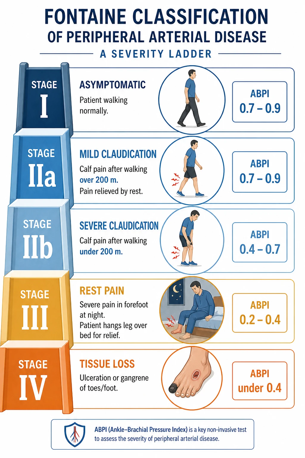
[3]The Fontaine system, used widely in Europe and the classical exam answer, stages PAD by its clinical evolution. Stage I is asymptomatic disease — an abnormal ABI without symptoms. Stage II is intermittent claudication, subdivided into IIa (claudication beyond 200 metres) and IIb (claudication within 200 metres). Stage III is ischaemic rest pain, typically nocturnal forefoot pain relieved by dependency. Stage IV is chronic limb-threatening ischaemia with ulceration or gangrene.[3]
The Rutherford categories, favoured in North America, align closely with Fontaine but incorporate objective treadmill walking data: 0 asymptomatic, 1 mild claudication, 2 moderate claudication, 3 severe claudication, 4 ischaemic rest pain, 5 minor tissue loss (non-healing ulcer or focal gangrene), and 6 major tissue loss (extending above the transmetatarsal level). Rutherford categories 4 to 6 correspond to chronic limb-threatening ischaemia.[2]
Fontaine I
Fontaine II
Fontaine III
Fontaine IV
Rutherford 4–6
The TASC II lesion classification governs the choice of revascularisation strategy. Lesions in each anatomical bed (aortoiliac, femoropopliteal, infrapopliteal) are graded A (single focal stenosis — endovascular first) through D (long, multilevel occlusion — surgical bypass first), with B and C occupying the intermediate zone where operator experience, conduit availability and patient comorbidity decide. The 2015 TASC supplement extended the classification to the below-the-knee arteries.[6]
For the threatened limb, the WIfI system grades Wound, Ischaemia (ABI, toe pressure, TcPO₂) and foot Infection each from 0 to 3, yielding a composite stage (1 to 4) that predicts one-year amputation risk and informs whether revascularisation is urgent. WIfI is increasingly used alongside the Global Vascular Guidelines' GLASS (Global Limb Anatomic Staging System) to plan revascularisation.[1]
Epidemiology & Risk Factors
PAD is a disease of enormous and growing global burden. The landmark systematic review by Fowkes and colleagues estimated that approximately 236 million adults aged 25 years and over were living with PAD worldwide in 2010, a rise of almost a quarter over the previous decade, driven principally by ageing populations and the spread of diabetes and smoking in low- and middle-income countries.[4]
Prevalence rises steeply with age — fewer than 3 percent of 40-year-olds are affected, rising to over 20 percent of those older than 70, with the highest burden in those who smoke, have diabetes, or both. Smoking is the strongest modifiable risk factor, with a dose-dependent two- to five-fold increase in PAD incidence and a marked effect on progression to amputation. Diabetes mellitus increases PAD risk two- to four-fold, preferentially affects the distal (infrapopliteal) arteries, and is the dominant driver of chronic limb-threatening ischaemia and major amputation. The remaining risk factors mirror those of all atherosclerotic disease: age, hypertension, dyslipidaemia (raised LDL and low HDL), chronic kidney disease, obesity, family history and the metabolic syndrome.[4][1]
Chronic kidney disease deserves special emphasis: it accelerates atherosclerosis, predisposes to medial arterial calcinosis (Monckeberg sclerosis) that renders vessels non-compressible, and is associated with poor outcomes after revascularisation. South Asian and Black ethnicities carry higher PAD risk at any given risk-factor burden.[1]
Pathophysiology
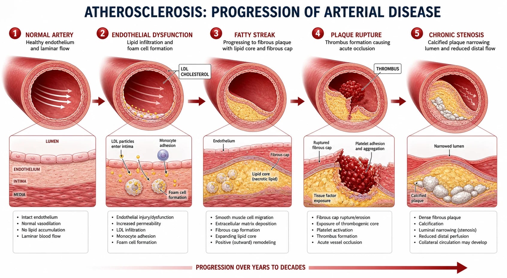
PAD shares the same mechanism as every other manifestation of atherosclerosis — a chronic inflammatory response to endothelial injury. The cascade begins with endothelial dysfunction (driven by tobacco smoke, hyperglycaemia, oxidised LDL, hypertension and the shear stress of turbulent flow). Low-density lipoproteins infiltrate the intima, become oxidised, and trigger monocyte adhesion and migration into the subendothelial space, where they transform into lipid-laden foam cells — the cellular hallmark of the early fatty streak.[1]
Over years, smooth-muscle cells migrate into the intima and lay down a fibrous cap over a necrotic, lipid-rich core, producing the fibrous plaque. Progressive plaque growth and intimal-medial thickening narrow the lumen; positive (outward) remodelling initially preserves the lumen, but a plaque encroaches on flow once it reduces the cross-sectional area by more than about 70 percent (corresponding to a diameter stenosis over 50 percent). Plaque complications — calcification, intra-plaque haemorrhage, cap rupture and superficial erosion — expose the thrombogenic core, triggering platelet aggregation and thrombus that can either cause stepwise chronic occlusion or, catastrophically, acute limb ischaemia.[1][3]
At the cellular and molecular level, atherosclerosis is a smouldering inflammatory disease. Endothelial dysfunction — from nitric-oxide depletion and adhesion-molecule upregulation — recruits monocytes that differentiate into pro-inflammatory M1 macrophages within the intima and scavenge oxidised LDL to become foam cells. T-lymphocytes and smooth-muscle cells follow; the balance between matrix metalloproteinases (which thin the fibrous cap) and their tissue inhibitors (which stabilise it) decides whether a plaque matures quietly or becomes a thin-cap fibroatheroma with a large lipid core and heavy macrophage infiltrate — the lesion prone to rupture. A ruptured or eroded cap exposes tissue factor, igniting the coagulation cascade and producing a platelet-rich thrombus. This biology is why antiplatelet and intensive lipid-lowering therapy target the plaque itself, not merely the haemodynamics of the stenosis.[1]
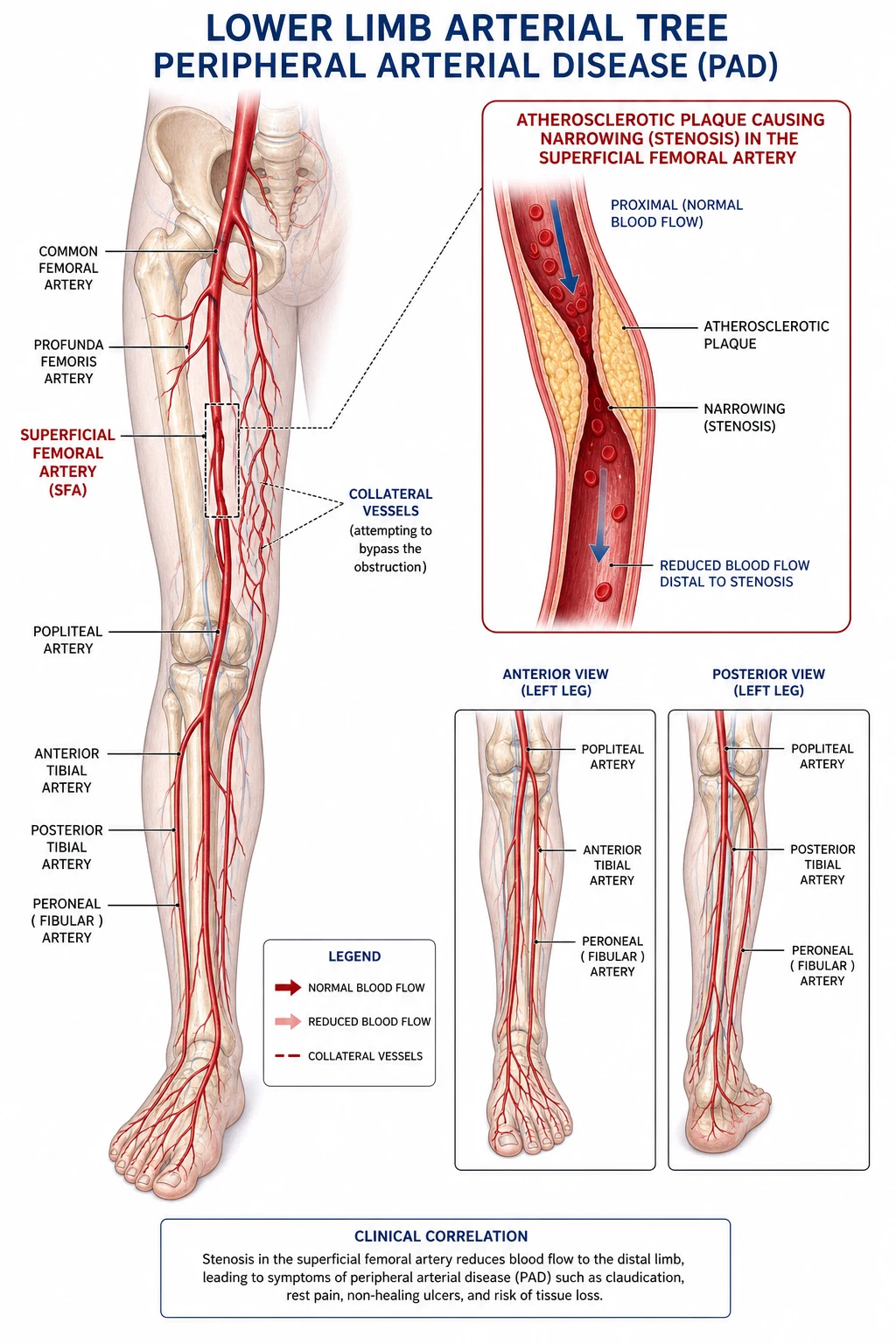
The haemodynamic consequence of a fixed stenosis is best understood through Poiseuille's law: flow is proportional to the fourth power of the radius, so a halving of radius reduces flow sixteen-fold. At rest, the limb demands little flow and a moderate stenosis is silent; on exercise, the calf muscle demands a many-fold increase in blood flow that the stenotic vessel cannot deliver, producing a supply–demand mismatch — claudication — that resolves within minutes of rest as demand falls. As stenosis worsens, perfusion fails even at rest, producing ischaemic rest pain, and ultimately tissue breakdown with ulceration and gangrene.[3]
In diabetes and chronic kidney disease, medial arterial calcinosis deposits calcium in the medial layer, stiffening the vessel wall and rendering it non-compressible to the cuff. This produces a falsely elevated ABI (often over 1.3) and is the reason toe pressures must be measured in these patients.[1]
Clinical Presentation
The clinical face of PAD spans the spectrum from a silent abnormal ABI through to a cold, pulseless, gangrenous limb. Recognising each presentation — and its atypical variants — is the core clinical skill examiners test.
[2]Intermittent claudication is the cardinal symptom: a reproducible, cramping calf (or buttock or thigh) pain that comes on after a predictable walking distance, is relieved within minutes by standing still, and recurs at the same distance. Crucially, the relief is with rest alone — not with sitting, leaning forward, or changing posture. The site of pain localises the level of disease: buttock and thigh claudication with erectile dysfunction points to aortoiliac disease (Leriche syndrome); calf claudication is typical of superficial femoral artery disease; foot claudication (rare) suggests infrapopliteal involvement.[2]
Ischaemic rest pain is severe, persistent pain in the forefoot, classically worse at night and relieved by hanging the leg out of bed or walking to another room (gravity aids perfusion). It indicates that resting blood flow is now inadequate — a limb-threatening state.
[3]Claudication
Rest pain
Chronic limb-threatening ischaemia
Acute limb ischaemia
Chronic limb-threatening ischaemia (CLTI) — formerly critical limb ischaemia — is defined as rest pain persisting for more than two weeks despite optimal medical management, or ischaemic ulceration or gangrene, in the absence of an alternative cause. Ischaemic ulcers are punched-out, painful, and located on the toes, the lateral malleolus or other pressure points, with a pale, dry base and surrounding callus.[3]
Leriche syndrome is the classic chronic aortoiliac occlusion triad of buttock and thigh claudication, absent femoral pulses, and erectile dysfunction, seen most often in middle-aged male smokers. It is a perennial favourite in vivas.[2]
Acute limb ischaemia presents as a sudden, dramatic drop in limb perfusion, captured in the 6 Ps: Pain, Pallor, Pulselessness, Paraesthesia, Paralysis and Perishing cold. The first three are early; the last three — sensory and motor loss with profound coldness — mark a threatened limb in which minutes count. The order matters: a viable limb can wait for imaging; a threatened limb cannot.[1]
6 Ps
Atypical presentations are the trap. In diabetes, peripheral neuropathy may mask both claudication and rest pain, so the patient first presents with a painless ulcer on a pressure point. In the elderly, claudication is misattributed to arthritis or spinal stenosis, or masked by immobility. Women report claudication less often and more often present with atypical leg pain or are diagnosed later.[1]
Differential Diagnosis
Not every leg pain on walking is claudication. The discriminating features are the pattern of relief, the ABI, and the neurological and musculoskeletal examination. A candidate who can distinguish claudication from its mimics in a sentence earns marks.
[2]Vascular claudication (PAD)
Neurogenic claudication (spinal stenosis)
Venous claudication
Baker cyst / calf strain
Osteoarthritis (hip/knee)
Chronic exertional compartment syndrome
DVT
The two highest-yield discriminating features examiners want named are: PAD claudication is relieved by rest alone within minutes, whereas neurogenic claudication is relieved by sitting down or leaning forward (the shopping-cart sign) and is not promptly relieved by standing still; and the ABI is normal in neurogenic, venous and musculoskeletal causes. A resting ABI can be normal in mild PAD — if the history is suggestive but the resting ABI is normal, perform a treadmill exercise test and recheck the ABI (a drop of over 0.15 confirms PAD).[2]
Clinical & Bedside Assessment
The vascular examination is structured: look, feel, listen — and always include the abdomen and the contralateral limb.
[2]Inspection reveals the chronic signs of ischaemia: hair loss on the lower leg, thin, shiny, dry skin, thickened and slow-growing nails, muscle wasting, and — in advanced disease — pallor on elevation, rubor on dependency, coolness, ulceration or dry gangrene of the toes. Buerger's test is performed by elevating the leg to 45 degrees: pallor of the sole within 60 seconds is abnormal, and the Buerger's angle (the angle at which pallor appears) is a rough index of severity — the lower the angle, the worse the perfusion. On dependency, the foot flushes a dusky red (reactive hyperaemia).[2]
Palpation assesses all the lower-limb pulses — femoral, popliteal, posterior tibial and dorsalis pedis — graded 0 (absent) through to 2 (normal), with 3 indicating a bounding, aneurysmal pulse. A prominent or expansile popliteal pulse suggests a popliteal aneurysm (and prompts screening for an abdominal aortic aneurysm). Skin temperature is compared side to side, and the level of temperature drop localises the occlusion. Capillary refill is prolonged.[2]
Auscultation listens for bruits over the abdomen (aortic or renal), the femoral triangle (aortoiliac or common femoral disease) and the carotids (concomitant cerebrovascular disease). An abdominal aortic aneurysm must be excluded in any PAD patient, especially a male smoker over 65.
[2]Ulcer morphology at the bedside distinguishes the three ulcer types: arterial ulcers are punched-out, sharply demarcated, painful, dry, and lie on the toes or lateral malleolus; venous ulcers are shallow, exudative, mildly painful, in the gaiter area (above the medial malleolus) and accompanied by varicose eczema and lipodermatosclerosis; neuropathic ulcers are painless, deep, at pressure points (under the metatarsal heads, heel), surrounded by callus, in a warm, insensate foot.[3]
For acute limb ischaemia, examination assesses the limb's viability category: viable (not immediately threatened, sensory and motor intact, Doppler audible) — investigate and treat urgently; threatened (sensorimotor deficit, inaudible arterial Doppler) — revascularise immediately; irreversible (fixed mottling, anaesthesia with rigidity, absent venous Doppler) — primary amputation. This single judgement decides whether to image first or to proceed straight to theatre.[1]
Investigations
Investigation answers three questions: is PAD present, where is the disease, and is the limb at threat? The first is answered at the bedside with the ankle–brachial index; the second with anatomical imaging; the third with severity measures (toe pressure, TcPO₂) and the WIfI stage.
[2]The ankle–brachial index (ABI) is the first-line test. With the patient supine, systolic pressures are measured at both brachial and both ankle arteries (posterior tibial and dorsalis pedis) using a handheld Doppler and a manual cuff. The ABI is the higher of the two ankle pressures divided by the higher brachial pressure for each leg.[2]
Over 1.3
0.9 to 1.3
0.7 to 0.9
0.4 to 0.7
0.4 and below
Under 0.3
When the ABI is over 1.3 (non-compressible), when it is normal despite a strongly suggestive history, or in diabetes and chronic kidney disease where medial calcinosis is common, measure the toe–brachial index (TBI): a TBI under 0.7 is abnormal and under 0.3 critical, as the digital vessels are spared by medial calcinosis. An exercise ABI (treadmill or active plantar-flexion until symptoms, then re-measure ABI) — a drop of over 0.15 confirms PAD in equivocal claudication.[1]
Segmental limb pressures and pulse-volume recordings (PVR), performed with cuffs at the thigh, calf and ankle, localise the level of disease by the step-down in pressure between segments. Duplex ultrasound is the first-line anatomical imaging: it locates stenoses and occlusions, grades their haemodynamic significance from the peak systolic velocity ratio, and identifies the conduit (great saphenous vein) for bypass. CT angiography (CTA) provides a road map for revascularisation, shows calcification, and is fast and widely available — at the cost of iodinated contrast and radiation. MR angiography (MRA) is an alternative that avoids iodinated contrast, but gadolinium carries the risk of nephrogenic systemic fibrosis in advanced kidney disease, and overestimates stenosis. Digital subtraction angiography (DSA) remains the gold standard for anatomical detail and is usually reserved for when an intervention is already planned — it is invasive, carries contrast and puncture risks, and is not a diagnostic first-line test.[2][3]
Duplex ultrasound
CT angiography (CTA)
MR angiography (MRA)
Digital subtraction angiography
TcPO2
Transcutaneous oxygen pressure (TcPO₂) measures tissue-level oxygenation and predicts wound healing: a value over 40 mmHg predicts healing, under 30 mmHg predicts failure, and the test guides the decision to revascularise or amputate in CLTI. Blood tests complete the work-up: lipid profile, HbA1c, fasting glucose, urea and electrolytes, eGFR, full blood count; an ECG (atrial fibrillation as a source of embolism in ALI, silent ischaemia); and, where renovascular disease or renal dysfunction is suspected, relevant imaging. Screening for abdominal aortic aneurysm (ultrasound) is warranted in male smokers with PAD.[1]
Management — Resuscitation
Two scenarios demand immediate action: acute limb ischaemia and the septic, threatened diabetic foot. Both are time-critical.
[1]In acute limb ischaemia, the moment the diagnosis is entertained, give analgesia (IV opioid, e.g. morphine 2.5 to 5 mg titrated) and immediate intravenous unfractionated heparin — an 80 units/kg bolus followed by an 18 units/kg/hour infusion (or a weight-based LMWH such as enoxaparin 1 mg/kg twice daily subcutaneously if heparin is unavailable) — to limit thrombus propagation and protect the microcirculation. Do not elevate the limb (it worsens ischaemia); keep it dependent. Mobilise the vascular team immediately — in a threatened limb (paraesthesia or paralysis), proceed to revascularisation without waiting for cross-sectional imaging: a Fogarty embolectomy under general anaesthesia is both diagnostic and therapeutic in an embolic occlusion. A viable limb allows time for angiography to localise the lesion and decide between thrombolysis and surgery.[1][3]
In the septic diabetic foot with spreading cellulitis or wet gangrene, admit for broad-spectrum intravenous antibiotics (e.g. amoxicillin–clavulanate 1.2 g IV three times daily plus metronidazole 500 mg IV three times daily in severe infection, adjusted to culture), urgent surgical debridement of necrotic tissue, glycaemic control with an insulin infusion, and urgent vascular imaging to plan revascularisation. The IWGDF/IDSA infection grading (mild, moderate, severe) guides antibiotic choice and depth.[9][10]
Management — Definitive & Stepwise
PAD management has two parallel goals that must be pursued together: reduce cardiovascular risk (save the patient) and relieve leg symptoms or save the limb. Best medical therapy (BMT) is the foundation for every PAD patient, including the asymptomatic patient with an abnormal ABI; revascularisation is added on top of, never instead of, BMT.
[1]Lifestyle and risk-factor modification. Smoking cessation is the single most effective intervention to halt claudication progression and reduce both cardiovascular events and amputation — pharmacotherapy (varenicline, nicotine replacement, bupropion) and behavioural support should be offered to every smoker. A Mediterranean or low-salt diet, weight reduction toward a body-mass index under 25, and meticulous foot care (especially in diabetes) are reinforced at every visit.[1][3]
Supervised exercise therapy (SET) is first-line for claudication and matches or exceeds the benefit of pharmacotherapy: a structured programme of 30 to 45 minutes of walking, three times a week, for at least 12 weeks, walking to near-maximal claudication each time. SET improves both pain-free and maximal walking distance and quality of life. Home-based exercise with a step counter and structured coaching is an alternative where SET is unavailable.[1]
Pharmacotherapy — cardiovascular protection. An antiplatelet agent is recommended for all symptomatic PAD and for asymptomatic PAD with a high-risk feature: aspirin 75 to 100 mg once daily, or clopidogrel 75 mg once daily (clopidogrel is preferred by many guidelines after the CAPRIE trial showed superiority over aspirin in PAD). A high-intensity statin is given to every PAD patient regardless of baseline LDL — atorvastatin 40 to 80 mg once daily or rosuvastatin 20 to 40 mg once daily, targeting an LDL cholesterol under 1.8 mmol/L (70 mg/dL) and ideally under 1.4 mmol/L in high-risk disease. Blood pressure is controlled to under 130/80 mmHg with an ACE inhibitor (e.g. ramipril 10 mg once daily) or angiotensin-receptor blocker as first-line; beta-blockers are not contraindicated in PAD. Diabetes is managed to a target HbA1c under 53 mmol/mol (7 percent), with SGLT2 inhibitors and GLP-1 receptor agonists preferred for their cardiovascular and renal benefit.[1][3]
Antiplatelet
Statin (high-intensity)
ACE inhibitor
Anticoagulant (selected)
Claudication drug
Diabetes
Low-dose rivaroxaban plus aspirin (rivaroxaban 2.5 mg twice daily plus aspirin 100 mg daily) reduced major adverse limb events and mortality in patients with stable PAD in the COMPASS trial, and is an option in patients without a high bleeding risk. It is not used in CLTI or after recent revascularisation where the evidence differs.[8]
Claudication-specific drugs. Cilostazol, a phosphodiesterase-3 inhibitor, improves both pain-free and maximal walking distance — the Cochrane review confirmed a modest benefit of roughly 25 to 30 percent over placebo. The dose is 100 mg twice daily (50 mg twice daily if intolerant), taken 30 minutes before or two hours after food. Cilostazol is absolutely contraindicated in heart failure (all PDE-3 inhibitors increase mortality in this group) and is used cautiously with CYP3A4 or P-gp inhibitors. Naftidrofuryl (a 5-HT₂ antagonist and vasodilator), 100 to 200 mg three times daily, is the first-line vasoactive agent in the UK (NICE) and is generally well tolerated; it is an alternative where cilostazol is contraindicated. Pentoxifylline 400 mg three times daily has weak evidence and is a third-line option.[12][3]
Revascularisation is offered for disabling claudication that has failed best medical therapy and supervised exercise for at least 3 months, and urgently for chronic limb-threatening ischaemia to salvage the limb. The choice of endovascular (percutaneous transluminal angioplasty with or without stenting) versus surgical bypass is guided by the TASC II lesion classification, the GLASS anatomic stage, the availability of a suitable autologous vein (great saphenous), and the patient's comorbidity and life expectancy.[1][6]
For aortoiliac disease, endovascular intervention (angioplasty ± stenting) is preferred for TASC A and B lesions (short, focal stenoses or short occlusions), with excellent patency. For extensive TASC D aortoiliac occlusions, an aortobifemoral bypass offers durable patency in fit patients. For femoropopliteal disease, angioplasty with selective stenting (drug-eluting stents and drug-coated balloons reduce restenosis) suits TASC A–C lesions; long SFA occlusions in a fit patient with a good vein are best served by a femoropopliteal vein bypass. Femorodistal (tibial or pedal) bypass is reserved for CLTI with infrapopliteal disease, where vein is overwhelmingly preferred over prosthetic conduit. The BASIL trial informed the strategy of vein-bypass-first versus best-endovascular-first in severe limb ischaemia, showing broadly similar short-term survival and amputation-free survival, with newer trials (BEST-CLI, BASIL-2) refining who benefits from which strategy.[11]
Amputation is reserved for irreversible ischaemia (non-viable limb), uncontrolled sepsis, intractable rest pain unsuitable for revascularisation, or a non-ambulatory patient in whom revascularisation offers no functional benefit. The aim is always the most distal amputation that will heal — transmetatarsal, below-knee or above-knee.[1]
Specific Subtypes & Scenarios
Acute limb ischaemia
Acute limb ischaemia is a sudden reduction in limb perfusion that threatens viability. The two mechanisms — embolic and thrombotic — are distinguished at the bedside and dictate management. An embolus classically arises from the heart (atrial fibrillation, mural thrombus after myocardial infarction, valvular vegetations) or an aneurysm (popliteal, abdominal aortic), lodges at a vessel bifurcation (common femoral, popliteal, aortic), and presents in a patient without prior collaterals — a profoundly ischaemic limb with a clear demarcation and no history of claudication. Thrombosis occurs on a ruptured pre-existing plaque or within an occluded bypass graft, in a patient with chronic collaterals, and so may present less catastrophically.[1]
Embolic
Thrombotic
The Rutherford classification of acute limb ischaemia grades the limb by its viability category and dictates urgency. Category I (viable): not immediately threatened, sensation and power intact, both arterial and venous Doppler audible — investigate and treat urgently, a window of hours exists. Category IIa (marginally threatened): salvageable if promptly revascularised, minimal sensory loss, no motor deficit, arterial Doppler inaudible but venous audible — image then revascularise. Category IIb (immediately threatened): salvageable only with immediate revascularisation, marked sensory and early motor loss — operate now, do not wait for imaging. Category III (irreversible): major permanent tissue loss, profound paralysis and anaesthesia with rigor, no venous Doppler — primary amputation or palliation. The moment paraesthesia or paralysis appears, ALI converts from "urgent" to "theatre now".[1]
Management follows the resuscitation principles above: immediate analgesia and IV heparin, then emergency revascularisation. For an embolic occlusion in a threatened limb, a Fogarty catheter embolectomy under general or regional anaesthesia — passed through a transverse arteriotomy, the balloon inflated and withdrawn to extract the clot — is both diagnostic and therapeutic, and is followed by an on-table angiogram and fasciotomies if the ischaemia time is prolonged. For a thrombotic occlusion on a known plaque or graft, catheter-directed thrombolysis (alteplase infused via an intra-arterial catheter over 12 to 24 hours, with heparin cover) lyses clot and exposes the underlying lesion for angioplasty or stenting, and is preferred when the limb is viable. The limb-salvage window is 4 to 6 hours before irreversible muscle necrosis; after successful revascularisation of a long ischaemia time, prophylactic fasciotomy prevents reperfusion compartment syndrome.[1][3]
Chronic limb-threatening ischaemia
CLTI is the end of the chronic PAD continuum — rest pain for over two weeks, or ulceration or gangrene — and demands urgent vascular referral and revascularisation planning. The WIfI stage stratifies amputation risk; the GLASS anatomic stage guides whether an endovascular or surgical strategy is likely to achieve an angiosome-directed or best-flow revascularisation. Post-revascularisation, the patient needs wound care, offloading, infection control and indefinite BMT. The IWGDF 2023 guidance on diabetic foot prevention applies in full.[9][1]
WIfI staging in practice. Each of Wound, Ischaemia and foot Infection is graded 0 to 3, and the combination yields a clinical stage 1 to 4 that predicts one-year amputation risk. A stage 1 limb (minor wound, mild ischaemia, no infection) carries an amputation risk under 10 percent and may heal with wound care alone, whereas a stage 4 limb (extensive wound, severe ischaemia, severe infection) carries an amputation risk over 25 percent and demands urgent combined revascularisation and debridement. WIfI thus converts the old binary "critical limb ischaemia" label into a graded limb-salvage prognosis and triages the urgency of revascularisation.[1]
The angiosome concept refines revascularisation planning in CLTI. The foot is divided into distinct arterial territories: the posterior tibial artery supplies the medial plantar surface, the anterior tibial artery (continuing as the dorsalis pedis) supplies the dorsum, and the peroneal artery supplies the heel and lateral border. Direct angiosome-targeted revascularisation — restoring flow to the specific artery feeding the ulcer — improves wound healing and limb salvage compared with indirect revascularisation, particularly in diabetics with distal disease. Where a single straight-line flow to the foot is the only option, the best-flow (rather than strictly angiosome) strategy is used, and the GLASS stage summarises the expected difficulty of achieving it.[1][6]
Diabetic foot
The diabetic foot sits at the intersection of neuropathy, ischaemia and infection, and is the leading cause of non-traumatic amputation. Three ulcer types are recognised: neuropathic (painless, on pressure points, in a warm, dry, callused, insensate foot with palpable pulses), ischaemic (painful, punched-out, in a cool, pulseless foot), and neuroischaemic (the commonest pattern, combining both). Management is multidisciplinary — podiatry, diabetes, vascular surgery, orthotics and microbiology — and follows the IWGDF/IDSA framework: debridement of necrotic tissue, offloading (total-contact cast for plantar ulcers), revascularisation for ischaemic/neuroischaemic ulcers, targeted antibiotics for infection graded by severity, and wound care. Prevention — daily foot inspection, correctly fitted footwear, regular podiatry, and glycaemic control — is paramount.[9][10]
Infection grading (IWGDF/IDSA) sets the antibiotic depth. Grade 1 (mild) — local cellulitis under 2 cm: oral amoxicillin–clavulanate 500 mg three times daily for one to two weeks. Grade 2 (moderate) — cellulitis over 2 cm or deep tissue involvement: intravenous amoxicillin–clavulanate 1.2 g three times daily plus metronidazole 500 mg three times daily, with Gram-negative cover for chronic wounds. Grade 3 (severe) — systemic sepsis or critical ischaemia: broad-spectrum intravenous therapy such as piperacillin–tazobactam 4.5 g three times daily, source control and intensive-care support. A positive probe-to-bone test over an exposed, necrotic ulcer base raises osteomyelitis — confirm with plain radiograph, MRI, and bone biopsy for culture and histology.[9][10]
Buerger disease (thromboangiitis obliterans)
Thromboangiitis obliterans (Buerger disease) is a distinct, non-atherosclerotic, segmental inflammatory vasculitis of the small and medium arteries and veins of the distal extremities, strongly associated with heavy tobacco use in young men (onset before age 45, often under 35). The pathology is distinctive — an occlusive inflammatory thrombus with giant cells preserving the internal elastic lamina, and skip lesions — and the clinical picture is of distal ischaemia (claudication of the foot or calf, rest pain, digital ulceration or gangrene) accompanied by superficial migratory thrombophlebitis and Raynaud's phenomenon. Proximal pulses are preserved while distal pulses are absent, and arteriography shows segmental occlusions with corkscrew collaterals around the occluded segments. The only effective treatment is absolute and complete abstinence from all tobacco — including nicotine — which halts progression in most; revascularisation is rarely feasible because the disease is distal and diffuse. Amputation may be required for established gangrene.[7]
Leriche syndrome
Leriche syndrome is chronic aortoiliac occlusion, classically in a middle-aged male smoker, presenting with the triad of buttock and thigh claudication, absent femoral pulses, and erectile dysfunction. Investigation with CTA shows a distal aortic and bilateral common iliac occlusion; revascularisation is by endovascular recanalisation and stenting or, in fit patients with extensive occlusion, aortobifemoral bypass.[2]
Complications & Pitfalls
The feared limb complications are progression to chronic limb-threatening ischaemia, major amputation, and — in acute limb ischaemia — limb loss if revascularisation is delayed beyond the salvage window. Infection of an ischaemic ulcer can precipitate wet gangrene and sepsis; osteomyelitis complicates deep diabetic foot ulcers and is diagnosed by probe-to-bone, MRI and bone culture.
[1]The most common and most lethal complications are not in the leg. Patients with PAD die predominantly of myocardial infarction and stroke — the systemic atherosclerosis that the leg is signalling — which is why aggressive medical therapy is non-negotiable.[1]
Procedure-related complications include contrast-induced nephropathy (mitigate with isotonic saline hydration and the lowest contrast volume, hold metformin and nephrotoxins), reperfusion injury and compartment syndrome after revascularisation (prophylactic fasciotomy after prolonged ischaemia), graft or stent thrombosis and restenosis (the rationale for antiplatelets and, where appropriate, dual pathway inhibition), pseudoaneurysm and bleeding at the puncture site, and infection of prosthetic graft. Cholesterol embolisation (blue toes, livedo, renal failure, eosinophilia) is a rare but serious complication of catheter manipulation in a diseased aorta.[3]
The classic pitfalls are: failing to measure an ABI in a patient with leg pain (PAD is missed); trusting a normal ABI in diabetes without checking a toe pressure (medial calcinosis); delaying heparin and revascularisation in a threatened limb while awaiting imaging; prescribing cilostazol to a heart-failure patient; and treating the leg while neglecting the cardiovascular risk that will actually kill the patient.[1]
Prognosis & Disposition
Prognosis in PAD is best expressed in two parallel stories — the limb and the life.
[1]For the limb, intermittent claudication runs a relatively benign course: with risk-factor modification and supervised exercise, around 70 to 80 percent of claudicants remain stable or improve, only 20 to 30 percent progress, and fewer than 5 percent require major amputation over five years. Revascularisation for claudication is offered only when symptoms are disabling and refractory — most claudicants never need it. By contrast, chronic limb-threatening ischaemia carries a one-year major amputation rate of around 20 to 25 percent and a one-year mortality of around 20 to 25 percent without revascularisation — outcomes that revascularisation substantially improves but does not normalise. Acute limb ischaemia salvages over 90 percent of limbs if revascularised within 6 hours, falling steeply thereafter; mortality at 30 days is 10 to 15 percent, reflecting the underlying cardiovascular disease.[1][3]
For life, the dominant message is that five-year mortality in symptomatic PAD is around 25 to 30 percent, and the deaths are overwhelmingly cardiovascular. Prognosis tracks smoking status, diabetes control, statin adherence, renal function and revascularisation patency. Disposition is to a vascular surgeon for all patients with CLTI or ALI; patients with stable claudication are managed in primary care with BMT and SET, with vascular referral for disabling or worsening symptoms.[1]
Special Populations
Diabetes mellitus is the most important special population: PAD is two to four times more prevalent, preferentially affects the distal (infrapopliteal) arteries, is complicated by neuropathy that masks symptoms, and progresses more often to ulceration and amputation. Medial calcinosis renders the ABI falsely high — measure the toe pressure. Screen diabetics over 50 (or younger with risk factors) with an ABI, examine the feet at every visit, and refer any ulcer to a multidisciplinary diabetic foot service. SGLT2 inhibitors and GLP-1 receptor agonists are preferred agents for their cardiovascular-renal benefit.[9][10]
Chronic kidney disease and dialysis patients have aggressive, often calcific, multilevel disease; minimise iodinated contrast (prefer CO₂ angiography or MRA where feasible), optimise hydration, and anticipate non-compressible vessels. Elderly patients may attribute claudication to arthritis or immobility, present late, and tolerate revascularisation less well — the risk-benefit of intervention shifts toward BMT. Women are under-diagnosed, report claudication less often, and may present with atypical leg pain; ABI thresholds are the same. Pregnancy and hormone therapy modestly increase venous thromboembolism risk but do not typically worsen PAD. Anticoagulated patients need periprocedural bridging around revascularisation, balancing thrombosis against bleeding.[1][3]
Buerger disease (young male smokers) is discussed above; absolute tobacco abstinence is the only effective treatment, and the prognosis is limb-threatening without it.[7]
Evidence, Guidelines & Regional Differences
The contemporary global standards are the 2024 ACC/AHA PAD Guideline (Gornik and colleagues) and the 2017 ESC Guideline on Peripheral Arterial Diseases (Aboyans and colleagues), both of which rest on the foundational 2016 AHA/ACC Guideline (Gerhard-Herman). The TASC II document (Norgren, 2007) and its 2015 supplement (Jaff) codify the lesion classification that still governs revascularisation strategy. All are aligned on the central message — best medical therapy for every patient, supervised exercise for claudication, revascularisation for limb threat.[1][3][2][6]
Key recent trials shape contemporary practice. The COMPASS trial established low-dose rivaroxaban 2.5 mg twice daily plus aspirin as a strategy to reduce major adverse limb events and mortality in stable PAD.[8] The BASIL trial (and its successors BASIL-2 and the BEST-CLI trial) refined the choice of vein-bypass-first versus best-endovascular-first for severe limb ischaemia, generally favouring a good-quality vein bypass in fit patients with complex CLTI and an endovascular-first approach in those with limited life expectancy or poor conduit.[11] The Cochrane review of cilostazol (Brown, 2021) confirmed a modest but real improvement in walking distance, underpinning its role in claudication (with the heart-failure caveat).[12]
Controversies include the paclitaxel-coated balloon and stent mortality signal first raised in 2018 (Katsanos meta-analysis), which substantially altered practice before being largely resolved by larger, longer-term pooled analyses and the 2024 vascular society reviews showing no excess mortality — though device-level monitoring continues. The role of dual antithrombotic therapy (rivaroxaban plus aspirin) in CLTI and after revascularisation remains under active study.[1]
Exam Pearls
A handful of one-liners and discriminating facts decide most PAD questions. Memorise them verbatim.
[1]The eight pearls that decide a PAD answer:
[1]Exam application bank (NEET-PG / INICET)
One-line answer
Peripheral arterial disease (PAD) is atherosclerotic narrowing of the lower-limb arteries — a clinical expression of systemic atherosclerosis and a powerful marker of coronary and cerebrovascular risk. The hallmark symptom is intermittent claudication, reproducible calf (or buttock/thigh) pain on walking that is relieved within minutes by rest, progressing in advanced disease to rest pain and chronic limb-threatening ischaemia with ulceration or gangrene. The bedside test is the ankle–brachial index (ABI): a value below 0.9 confirms PAD, below 0.4 indicates critical ischaemia, and over 1.3 signals non-compressible calcified vessels. Management has two aims — reduce cardiovascular risk (smoking cessation, high-intensity statin, antiplatelet, tight blood-pressure and glycaemic control, supervised exercise) and improve the leg (cilostazol or naftidrofuryl for claudication; endovascular or s
Worked stems (answer without another resource)
Stem 1 — Classic presentation. Map symptoms to mechanism; name the first investigation and first treatment step with dose/route if drug therapy is standard. [1]
Stem 2 — Unstable / complicated. List red flags that force immediate resuscitation, theatre, ICU, antidote, or reperfusion — and what you do in the first 15 minutes. [1]
Stem 3 — Atypical group. Elderly, pregnancy, child, or immunocompromised: how presentation and thresholds change. [1]
Stem 4 — Differential trap. Name the three closest mimics and one discriminator for each. [1]
Stem 5 — Disposition. Who goes home with safety-netting, who is admitted, who needs HDU/ICU/theatre, and what follow-up is mandatory. [1]
Rapid viva checklist
- Definition + classification
- Pathophysiology chain
- Bedside signs / criteria
- Score with exact components (if any)
- Emergency bundle
- Definitive therapy with doses
- Complications of disease and of treatment
- Special populations
- Guideline/trial name if classic
- Three exam traps
Coverage self-check
If you cannot answer any stem above from this page alone, re-read the matching section — the page is intended to be self-sufficient for final-prof and NEET-PG/INICET questions on Peripheral Arterial Disease.
DABSI
The clinical bottom line: PAD is systemic atherosclerosis made visible in the leg. The leg tells you about the heart and brain; treat both, measure the ABI, recognise the threatened limb, and act on time.
[1]References
- [1]Gornik HL, Beckman JA, Bhatt DL, et al. 2024 ACC/AHA/AACVPR/APMA/ABC/SCAI/SVM/SVN/SVS/SIR/VESS Guideline for the Management of Lower Extremity Peripheral Artery Disease: A Report of the American College of Cardiology/American Heart Association Joint Committee on Clinical Practice Guidelines Circulation, 2024.PMID 38743805
- [2]Gerhard-Herman MD, Gornik HL, Barrett C, et al. 2016 AHA/ACC Guideline on the Management of Patients With Lower Extremity Peripheral Artery Disease: A Report of the American College of Cardiology/American Heart Association Task Force on Clinical Practice Guidelines J Am Coll Cardiol, 2017.PMID 27851992
- [3]Aboyans V, Ricco JB, Bartelink MEL, et al. 2017 ESC Guidelines on the Diagnosis and Treatment of Peripheral Arterial Diseases, in collaboration with the European Society for Vascular Surgery (ESVS): Document covering atherosclerotic disease of extracranial carotid and vertebral, mesenteric, renal, upper and lower extremity arteriesEndorsed by: the European Stroke Organization (ESO)The Task Force for the Diagnosis and Treatment of Peripheral Arterial Diseases of the European Society of Cardiology (ESC) and of the European Society for Vascular Surgery (ESVS) Eur Heart J, 2018.PMID 28886620
- [4]Fowkes FG, Rudan D, Rudan I, et al. Comparison of global estimates of prevalence and risk factors for peripheral artery disease in 2000 and 2010: a systematic review and analysis Lancet, 2013.PMID 23915883
- [5]Rooke TW, Hirsch AT, Misra S, et al. 2011 ACCF/AHA Focused Update of the Guideline for the Management of Patients With Peripheral Artery Disease (updating the 2005 guideline): a report of the American College of Cardiology Foundation/American Heart Association Task Force on Practice Guidelines J Am Coll Cardiol, 2011.PMID 21963765
- [6]TASC Steering Committee, Jaff MR, White CJ, et al. An Update on Methods for Revascularization and Expansion of the TASC Lesion Classification to Include Below-the-Knee Arteries: A Supplement to the Inter-Society Consensus for the Management of Peripheral Arterial Disease (TASC II) Vasc Med, 2015.PMID 26268268
- [7]Piazza G, Goldhaber SZ. Thromboangiitis obliterans Circulation, 2010.PMID 20421527
- [8]Anand SS, Bosch J, Eikelboom JW, et al. Major Adverse Limb Events and Mortality in Patients With Peripheral Artery Disease: The COMPASS Trial J Am Coll Cardiol, 2018.PMID 29540326
- [9]Bus SA, Lavery LA, Monteiro-Soares M, et al. Guidelines on the prevention of foot ulcers in persons with diabetes (IWGDF 2023 update) Diabetes Metab Res Rev, 2024.PMID 37302121
- [10]Fitridge R, Chuter VH, Mills JL, et al. Editor's Choice - The Intersocietal IWGDF, ESVS, SVS Guidelines on Peripheral Artery Disease in People With Diabetes Mellitus and a Foot Ulcer Eur J Vasc Endovasc Surg, 2023.PMID 37724984
- [11]Bradbury AW, Adam DJ, Bell J, et al. Editor's Choice - Bypass versus Angioplasty in Severe Ischaemia of the Leg (BASIL)-2 Trial: Analysis of the Timing and Causes of Death in Participants Randomised to an Infrapopliteal Vein Bypass or Best Endovascular Treatment First Revascularisation Strategy Eur J Vasc Endovasc Surg, 2025.PMID 39069068
- [12]Brown T, Smith A, Hinchliffe R, et al. Cilostazol for intermittent claudication Cochrane Database Syst Rev, 2021.PMID 34192807