Cardiology · Cardiology
Pulmonary Hypertension
Also known as Pulmonary arterial hypertension · PAH · Pulmonary hypertensive vascular disease · Pre-capillary pulmonary hypertension · Cor pulmonale (chronic)
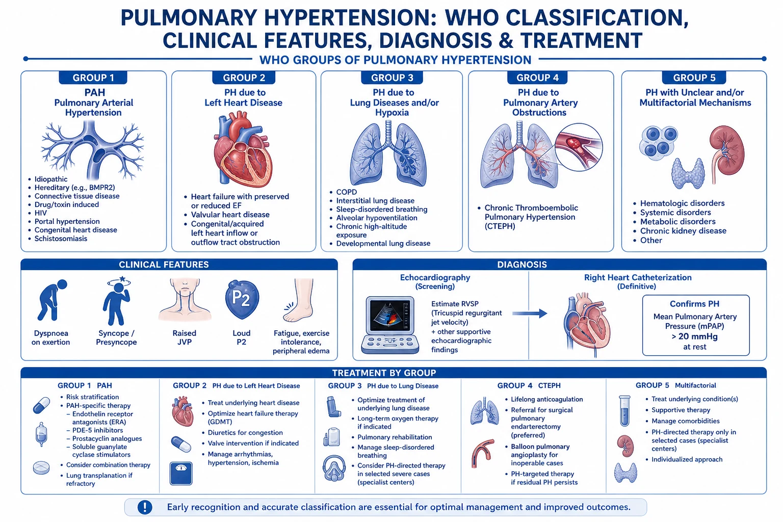
Pulmonary hypertension (PH) is a haemodynamic and pathophysiological syndrome defined as a mean pulmonary arterial pressure (mPAP) above 20 mmHg at rest measured by right heart catheterisation (RHC), confirmed by a pulmonary arterial wedge pressure (PAWP) of 15 mmHg or less in pre-capillary disease and pulmonary vascular resistance (PVR) above 2 Wood units (WU) in pulmonary arterial hypertension (PAH). The 2022 ESC/ERS Guidelines classify PH into five groups by aetiology: Group 1 — pulmonary arterial hypertension (PAH) (idiopathic, heritable, drug- and toxin-induced, and associated with connective tissue disease, HIV, portal hypertension, congenital heart disease); Group 2 — PH due to left heart disease (heart failure with preserved or reduced ejection fraction, valvular disease); Group 3 — PH due to lung disease or hypoxia (COPD, interstitial lung disease, sleep-disordered breathing); Group 4 — PH due to pulmonary artery obstruction (chronic thromboembolic pulmonary hypertension, CTEPH); Group 5 — PH with unclear or multifactorial mechanisms (haematological, systemic, metabolic disorders, sarcoidosis). Clinical presentation is dominated by progressive exertional dyspnoea, fatigue, pre-syncope or syncope on exertion, signs of right ventricular (RV) failure (raised JVP, peripheral oedema, hepatomegaly, ascites), and on examination a loud pulmonary component of the second heart sound (P2), a left parasternal (RV) heave, a pansystolic murmur of tricuspid regurgitation, and a right ventricular S4. The diagnostic algorithm is stepwise: transthoracic echocardiography with Doppler estimation of peak tricuspid regurgitant velocity (TR Vmax) — used to derive right ventricular systolic pressure (RVSP) — is the screening test of choice, followed by confirmation with RHC (the only definitive test, providing mPAP, PAWP, PVR, cardiac output, and vasoreactivity testing). Ventilation–perfusion (V/Q) scan is mandatory to exclude CTEPH (Group 4). The mainstay of management is risk-stratified PAH-specific therapy in Group 1 (endothelin receptor antagonists [ERAs — bosentan 62.5 mg BD for 4 weeks then 125 mg BD, ambrisentan 5 to 10 mg daily, macitentan 10 mg daily], phosphodiesterase-5 inhibitors [PDE5i — sildenafil 20 mg three times daily, tadalafil 40 mg daily], the soluble guanylate-cyclase stimulator riociguat 1 mg three times daily titrated to 2.5 mg three times daily, prostacyclin analogues [epoprostenol 1 to 2 ng/kg/min IV titrated to 10 to 20 ng/kg/min, iloprost 5 mcg inhaled 6 to 9 times daily, treprostinil subcutaneous or inhaled], and the selective IP-receptor agonist selexipag 200 mcg twice daily titrated up by 200 mcg twice daily every 2 weeks to a maximum of 1600 mcg twice daily), combined with supportive measures (oxygen, furosemide 40 mg IV or PO daily with metolazone if needed, supervised rehabilitation, iron replacement when deficient, cautious anticoagulation in selected idiopathic PAH). CTEPH (Group 4) is potentially curable by pulmonary endarterectomy, with riociguat as the only licensed medical therapy in inoperable or residual disease. Acute RV failure in PH is resuscitated with parenteral prostacyclin (epoprostenol or iloprost), cautious diuresis with furosemide 40 mg IV, inotropic support (dobutamine or milrinone) to maintain systemic blood pressure and RV perfusion, and — for refractory cases — intubation with avoidance of positive-pressure ventilation, mechanical RV support (VA-ECMO), and urgent transfer to a PH centre. Pulmonary hypertension remains a progressive disease with mortality dominated by RV failure, with five-year survival in untreated idiopathic PAH historically around 30 to 40 percent but improving to more than 60 percent at five years on modern combination therapy.
On this page & tools
Your progress
Saved locally on this device.
Practise this topic
Exam tags
Red flags
Overview & Definition
Pulmonary hypertension (PH) is a clinical and haemodynamic syndrome characterised by a mean pulmonary arterial pressure (mPAP) above 20 mmHg at rest, measured directly by right heart catheterisation (RHC), revised downwards from the historical threshold of 25 mmHg in the 2022 ESC/ERS Guidelines to improve early detection.[1]
The definition is paired with two further haemodynamic specifications that separate pre-capillary from post-capillary PH and drive treatment: [1]
- Pre-capillary PH (Groups 1, 3, 4 and most of 5) — mPAP above 20 mmHg, with pulmonary arterial wedge pressure (PAWP) of 15 mmHg or less (a left-heart cause excluded) and pulmonary vascular resistance (PVR) above 2 Wood units (the accepted threshold for pulmonary vascular disease that warrants therapy).
- Isolated post-capillary PH (Group 2, left-heart-driven) — mPAP above 20 mmHg, PAWP above 15 mmHg, with PVR at or below 2 WU; treated by managing the left-heart cause (diuretics, valve surgery, GDMT for HF).
- Combined pre- and post-capillary PH — PAWP above 15 mmHg and PVR above 2 WU; a hybrid that may need PAH therapy on top of left-heart treatment in selected cases.[1][2]
The distinct subgroup of pulmonary arterial hypertension (PAH, WHO Group 1) has a stricter haemodynamic definition: mPAP above 20 mmHg, PAWP at or below 15 mmHg, and PVR above 2 WU — this is the population in whom PAH-specific therapy has trial evidence.[1][2]
The clinical importance is both diagnostic and prognostic: PH is common (PH of any cause is the third most common cardiovascular syndrome after ischaemic heart disease and stroke, with Group 2 PH dominating prevalence), and pulmonary arterial hypertension (Group 1) is rare but lethal — untreated idiopathic PAH has a median survival of about 2.8 years from diagnosis in historical series, while modern combination therapy has improved five-year survival to over 60 percent.[1][3] The exam-relevant skills are (1) suspecting PH early (exertional dyspnoea disproportionate to examination is the clue, not wheeze); (2) classifying it correctly (Groups 1–5; the group dictates therapy); (3) confirming it by RHC, not echo; (4) screening for CTEPH in every patient with a V/Q scan; and (5) applying risk-stratified combination therapy with attention to the patient's functional class, BNP, six-minute walk distance and imaging.[1][3]
Classification
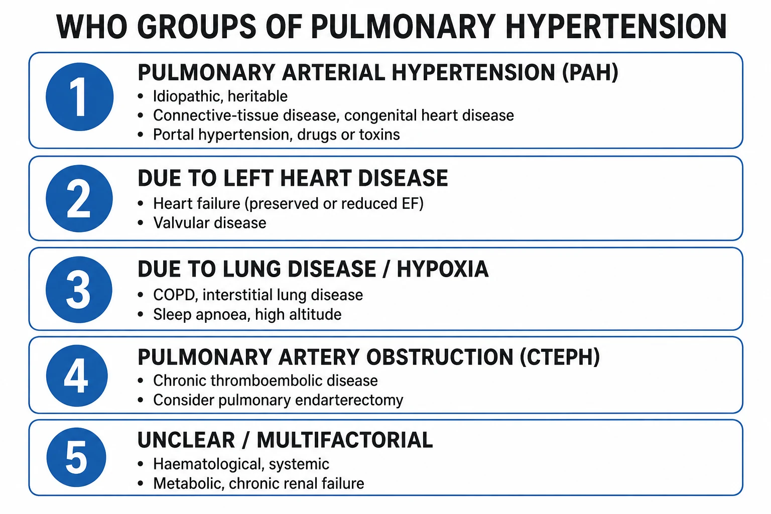
The WHO clinical classification of PH (Geneva, 2022 update of the ESC/ERS classification) organises PH into five groups by underlying pathophysiology and treatment rather than by severity.[1][2]
Group 1 — Pulmonary arterial hypertension (PAH)
- Idiopathic and heritable (BMPR2, ACVRL1, ENG, SMAD9, CAV1, KCNK3 mutations); 15 to 25 percent familial
- Drug- and toxin-induced (anorexigens like fenfluramine, dexfenfluramine, aminorex, benfluorex; SSRIs in newborns; dasatinib; interferons)
- Associated with connective tissue disease (systemic sclerosis — most important, SLE, mixed CTD, RA, MCTD); portalservices (portopulmonary); HIV infection; congenital heart disease (Eisenmenger)
- Pulmonary veno-occlusive disease (PVOD) and pulmonary capillary haemangiomatosis (PCH); haemodynamic response to calcium-channel blockers is rare in idiopathic disease
- Treatment by PAH-specific therapy: ERAs, PDE5i, riociguat, parenteral prostacyclin, selexipag; supportive oxygen and diuresis
Group 2 — PH due to left heart disease
- Most common cause of PH overall (65 to 80 percent)
- Includes heart failure with reduced ejection fraction (HFrEF), HFpEF, valvular disease (mitral stenosis, aortic stenosis, mitral regurgitation), and congenital/acquired left-heart inflow-outflow obstruction
- Haemodynamics: PAWP above 15 mmHg; PVR below or above 2 WU depending on whether there is a reactive pre-capillary component (isolated vs combined post- and pre-capillary PH)
- Treatment: optimise GDMT, diuretics, valve surgery; PAH-specific therapy has not been shown to help (negative trials in HFrEF and HFpEF) and may worsen gas exchange
Group 3 — PH due to lung disease or hypoxia
- COPD (the commonest), interstitial lung disease (especially combined pulmonary fibrosis and emphysema), sleep-disordered breathing (obstructive sleep apnoea), high-altitude exposure, developmental lung disease
- Severity is usually modest (mPAP 25 to 35 mmHg), with severe PH in a minority (about 1 to 5 percent of COPD, more in ILD)
- Treatment: optimise underlying lung disease (bronchodilators, oxygen 15 h/day for chronic hypoxaemia, CPAP in OSA, smoking cessation); PAH-specific therapy unproven and discouraged in severe lung disease; trial of inhaled treprostinil an option in ILD-PH in the US
Group 4 — PH due to pulmonary artery obstruction
- Chronic thromboembolic pulmonary hypertension (CTEPH) after acute pulmonary embolism (about 4 percent of survivors)
- Other obstructions: pulmonary artery sarcoma, tumour embolism, hydatid embolism, fibrosing mediastinitis, congenital pulmonary artery stenosis
- V/Q scan is the gold-standard screening tool (mismatched segmental perfusion defects) — every newly diagnosed PH patient needs one
- Treatment: pulmonary endarterectomy (PEA, potentially curative — first-line); riociguat (1 mg TDS titrated to 2.5 mg TDS) for inoperable or residual disease; balloon pulmonary angioplasty (BPA) for distal lesions in experienced centres
Group 5 — PH with unclear or multifactorial mechanisms
- Haematological disorders (chronic haemolytic anaemia including sickle cell and thalassaemia, myeloproliferative disorders, splenectomy)
- Systemic disorders (sarcoidosis with pulmonary vascular involvement, pulmonary Langerhans cell histiocytosis, neurofibromatosis, vasculitis)
- Metabolic disorders (glycogen storage disease, Gaucher disease, thyroid disease)
- Other: chronic kidney disease with dialysis, fibrosing mediastinitis, tumour compression
- Treatment: support underlying disease; no PAH-specific therapy generally indicated; referral to a PH centre
WHO functional class (I–IV) is the simple, bedside severity scale carried over from the NYHA system:[1]
- Class I — asymptomatic with ordinary physical activity; no breathlessness, fatigue, chest pain or pre-syncope.
- Class II — slight limitation; symptoms with ordinary activity (climbing two flights of stairs, walking uphill), comfortable at rest.
- Class III — marked limitation; comfortable only at rest, symptoms with less-than-ordinary activity (dressing, walking a few metres on the flat).
- Class IV — symptoms at rest; unable to carry out any physical activity without symptoms; signs of right-heart failure (peripheral oedema, syncope). [1]
The other two indispensable classification tools are risk stratification (see Management — Definitive & Stepwise: low / intermediate / high risk based on WHO class, six-minute walk distance, BNP/NT-proBNP, haemodynamics, RV imaging) and vasoreactivity testing (see Investigations — a positive test, defined as a fall in mPAP of at least 10 mmHg to a value of 40 mmHg or less with maintained or increased cardiac output on inhaled nitric oxide or intravenous adenosine or epoprostenol, identifies the small subset (about 5 percent) of idiopathic, heritable or drug-induced PAH who benefit from calcium-channel blockers — amlodipine 5 to 20 mg daily or nifedipine 30 to 120 mg daily or diltiazem 120 to 360 mg daily — titrated to BP tolerance).[1][3]
Epidemiology & Risk Factors
Pulmonary hypertension is heterogeneous in incidence and prevalence by group. The most useful numbers for examination: [1]
- PAH (Group 1) — estimated prevalence 15 to 50 per million adults in Europe (a so-called rare disease at the orphan-disease threshold); incidence about 2 to 5 per million per year in Western registries; female:male ratio roughly 3 to 4:1, median age at diagnosis 50 to 65 years (children and young adults in heritable forms); heritable PAH about 15 to 25 percent of idiopathic-like cases (BMPR2 the commonest pathogenic variant).[1][3]
- PH due to left heart disease (Group 2) — the commonest cause of PH overall, present in 40 to 75 percent of HFrEF, 50 to 80 percent of HFpEF, and almost all severe mitral stenosis at the time of valve-replacement referral.
- PH due to lung disease (Group 3) — present in roughly 30 to 70 percent of advanced COPD, 30 to 50 percent of interstitial lung disease, and most severe obesity-hypoventilation syndrome and OSA (Group 3 estimated to account for about 25 to 40 percent of PH in Western registries).
- CTEPH (Group 4) — cumulative incidence of about 4 percent after acute symptomatic pulmonary embolism; an under-recognised cause that explains roughly 30 to 50 per million adult prevalence in registries with structured follow-up.
- Group 5 PH — variable; usually secondary to the systemic or haematological disease.[1]
Established and emerging risk factors for PAH (Group 1): [1]
- Connective tissue disease — systemic sclerosis (scleroderma) is the most important, with PAH in 8 to 12 percent by echocardiographic screening and annual echo with TR Vmax plus annual DLCO is the recommended surveillance (mortality in SSc-PAH is higher than in idiopathic PAH). SLE, mixed CTD, RA, MCTD, dermatomyositis, Sjogren syndrome also carry risk.
- Drugs and toxins — anorexigens (fenfluramine, dexfenfluramine, aminorex, benfluorex) carry the strongest causal signal (clusters emerged in the 1960s and 1990s); dasatinib (tyrosine-kinase inhibitor used in chronic myeloid leukaemia); interferon alfa and beta; SSRI exposure in newborn period (persistent PH of the newborn); methamphetamine; possible association with hormonal therapy and l-tryptophan in supplements.
- HIV — PAH develops in roughly 0.5 percent of people with HIV; HIV is a definite but uncommon cause (mechanism unclear — likely HIV-protein and chronic inflammation–driven endothelial remodelling); modern antiretroviral therapy has not abolished the risk.
- Portal hypertension — portopulmonary hypertension in roughly 2 to 6 percent of patients with cirrhosis and portal hypertension (raised PVR on RHC with normal PAWP); recognised before liver transplantation candidacy because severe PH increases perioperative mortality.
- Congenital heart disease (Eisenmenger syndrome) — uncorrected or unrepaired large left-to-right shunts (VSD, PDA, ASD, aortopulmonary window) that over decades raise PVR to systemic levels, reverse the shunt to right-to-left, and produce cyanosis, polycythaemia and exercise intolerance; roughly half of Eisenmenger patients survive into the fifth decade.
- Heritable PAH — autosomal-dominant BMPR2 mutations in 70 to 80 percent of familial and 10 to 25 percent of idiopathic PAH; ACVRL1 / ALK1, ENG, SMAD9, CAV1, KCNK3 are rarer causes. Genetic counselling and family screening are recommended in heritable PAH.[1][3]
Risk factors for CTEPH (Group 4) — large perfusion defect at the index PE; unprovoked PE; recurrent PE; delay from symptom onset to diagnosis of acute PE; antiphospholipid antibody syndrome; myeloproliferative disease; splenectomy; chronic indwelling central venous catheters; ventriculoatrial shunts; male sex.[1]
Risk factors for PH in general — increasing age (Groups 2 and 3 rise sharply with age), female sex (PAH), genetic susceptibility (PAH), comorbid heart, lung, liver or connective-tissue disease (raises the specific group), and modifiable contributors (tobacco smoking, intravenous drug use, untreated OSA, high altitude).[1]
Pathophysiology
The final common pathway of all PH is an imbalance between vasoconstriction, vasoproliferation and thrombosis versus vasodilatation, apoptosis and antithrombosis in the small pulmonary arteries, producing vascular remodelling, raised PVR, raised mPAP, RV pressure overload, RV hypertrophy, eventual RV dilation and failure, and death.[1][3]
1. Pulmonary vascular remodelling — the histological hallmark. All three vessel-wall layers are affected: intima (endothelial proliferation, intimal thickening and fibrosis, concentric or eccentric; plexiform lesions at the advanced end of the spectrum in PAH); media (smooth-muscle hypertrophy, smooth-muscle recruitment in previously non-muscularised distal vessels); adventitia (fibroblast proliferation, collagen deposition). The combined effect narrows the lumen and stiffens the wall, raising PVR.[1]
2. The three classical vasomotor pathways. Disease activity is centred on reduced nitric oxide (NO) and prostacyclin (PGI2) signalling and increased endothelin-1 (ET-1) signalling; this triad is the pharmacological substrate for every approved PAH therapy.[1]
- Nitric oxide / cGMP axis. Endothelial NO synthase (eNOS) generates NO, which activates soluble guanylate cyclase to produce cGMP, causing smooth-muscle relaxation; cGMP is degraded by phosphodiesterase-5 (PDE5). In PAH, eNOS expression falls and PDE5 expression rises, lowering cGMP. PDE5 inhibitors (sildenafil, tadalafil) restore cGMP; riociguat directly stimulates soluble guanylate cyclase, raising cGMP by a different route.
- Prostacyclin / cAMP axis. Endothelial prostacyclin synthase produces PGI2, which raises cAMP in smooth-muscle cells, causing vasodilation and inhibiting smooth-muscle proliferation. Prostacyclin also inhibits platelet aggregation. In PAH, PGI2 synthase expression falls and thromboxane rises. Prostacyclin analogues (epoprostenol, iloprost, treprostinil, beraprost) and the selective IP-receptor agonist selexipag substitute for the deficiency.
- Endothelin-1 axis. Endothelin-1 is the most potent endogenous vasoconstrictor, also drives smooth-muscle proliferation and fibrosis; ET-1 binds ETA receptors on smooth muscle (vasoconstriction) and ETB receptors on endothelium (clearance, NO and PGI2 release, but in PAH these effects are lost). ET-1 levels are markedly raised in PAH. Endothelin-receptor antagonists (ERAs) (bosentan, ambrisentan, macitentan) block the receptor; each is disease-modifying in PAH.[1][3]
3. Vasoconstriction, thrombosis and inflammation in situ. Remodelled pulmonary arteries exhibit exaggerated hypoxic vasoconstriction (Euler-Liljestrand mechanism), endothelial dysfunction with a pro-thrombotic state (fibrin deposition in situ, platelet activation) and inflammatory infiltrate (B and T lymphocytes, macrophages, mast cells). The plasma biomarkers BNP, NT-proBNP, uric acid, troponin, growth-differentiation factor 15 (GDF-15) and inflammatory cytokines are elevated and are used as risk-stratification tools.[1][3]
4. Right ventricular response. Sustained pressure overload (PVR above about 2.5 to 3 WU) initially produces adaptive RV hypertrophy (preserved contractility, increased stroke work via Anrep and Frank-Starling mechanisms); with time the RV transitions to maladaptive dilation, eccentric remodelling, tricuspid annular dilatation with functional tricuspid regurgitation, RV–LV interaction (septal shift towards the LV in systole), and reduced forward cardiac output. Right coronary perfusion falls because aortic pressure falls and RV wall tension rises, producing RV ischaemia that compounds failure. Acute RV failure is the commonest mode of death in PH.[4]
5. Leftward bias of the septum and ventriculoarterial coupling. When the failing RV is pressure-overloaded, the interventricular septum shifts leftward in diastole (the ECG hallmark of right atrial enlargement, P pulmonale, right axis deviation, right bundle branch block, RV hypertrophy, and on echo D-shaped LV, paradoxical septal motion). Forward LV preload falls despite normal LV function; systemic pressure drops and organ perfusion suffers.[4]
6. Group-specific pathophysiology. Group 1 (PAH) — primary vascular disease outlined above. Group 2 (PH-LHD) — passive backward transmission of raised left-atrial pressure. Group 3 (PH-lung disease) — hypoxic vasoconstriction plus obliterative vascular remodelling in fibrosis (especially combined pulmonary fibrosis and emphysema). Group 4 (CTEPH) — organised fibrotic thrombus in major pulmonary arteries, web and band lesions, distal microvasculopathy. Group 5 PH — variable — hyperviscosity, high-output cardiac states, vasculitis, sarcoidosis-related granulomas in vessel walls.[1]
Clinical Presentation
The clinical presentation is dominated by the combination of exertional symptoms and signs of RV pressure overload and failure, with a signature mismatch between the dramatic severity of the disease and the normal-appearing lung examination.[1]
History (the pattern depends on the subgroup): [1]
- Exertional dyspnoea — gradually progressive, often out of proportion to examination; the commonest presenting symptom in PAH and in all PH groups (FEV1 and chest examination in Group 1 PAH are typically near-normal).
- Fatigue — loss of cardiac output reserve on exertion, deconditioning.
- Pre-syncope or syncope on exertion — fixed PVR is high, cardiac output cannot increase on exertion (a sinister sign in PAH — suggests low output on exertion, raised risk in WHO functional class III–IV).
- Chest pain — RV ischaemia (raised wall tension and low aortic pressure, drop in right coronary flow).
- Palpitations — supraventricular and ventricular arrhythmias (atrial flutter and AF in advanced disease from dilated right atrium).
- Cough — non-specific; minor haemoptysis from rupture of hypertrophied bronchial arteries.
- Right-heart failure symptoms — abdominal distension, right upper-quadrant pain (hepatic congestion), early satiety (bowel oedema), ankle swelling, weight gain.[1][3]
Vital signs: [1]
- Tachycardia — compensatory (HR above 100 suggests hypotension and low output).
- Hypotension — narrow pulse pressure with low SBP suggests low output in advanced disease.
- Cyanosis — central cyanosis in advanced PH, Eisenmenger syndrome, and severe hypoxaemia.
- Low oxygen saturation — in Group 1 PAH with a patent foramen ovale and right-to-left shunting.
- Clubbing — not specific but suggests chronic hypoxaemia, Eisenmenger, ILD. [1]
Cardiovascular examination: [1]
- JVP — raised with prominent a-wave (and v-wave with tricuspid regurgitation); Kussmaul sign (raised JVP on inspiration) indicates a constrictive element.
- Left parasternal (RV) heave — RV hypertrophy or dilation.
- Loud pulmonary component of the second heart sound (P2) — raised pulmonary artery diastolic pressure; a single P2 or palpable P2 is a classic sign.
- Right ventricular S4 — a low-frequency presystolic sound over the lower left sternal edge.
- Tricuspid regurgitation murmur — pansystolic at the lower left sternal edge, louder on inspiration.
- Pulmonary regurgitation (Graham Steell murmur) — early diastolic decrescendo at upper left sternal edge in advanced disease.
- Hepatojugular reflux, tender pulsatile hepatomegaly, ascites, peripheral oedema — signs of right-heart failure. [1]
Atypical presentations: [1]
- Older patients — commoner presentation with HFpEF (Group 2 with combined post- and pre-capillary component) rather than PAH; look for coronary disease, valve disease, hypertension.
- Pregnancy — symptoms dismissed as breathlessness of pregnancy; deteriorating dyspnoea with new peripheral oedema is suspect.[1]
Differential Diagnosis
The first task in suspected PH is distinguishing PAH (Group 1) from the four other groups because treatment is fundamentally different. Every patient with newly suspected PH needs comprehensive evaluation to assign group before PAH-specific therapy is considered.[1]
- Group 2 — PH due to left heart disease — the most common cause; history of ischaemic heart disease, hypertension, diabetes, atrial fibrillation; exertional dyspnoea with paroxysmal nocturnal dyspnoea or orthopnoea (PAH rarely has PND/orthopnoea); examination with loud P2 but with S3 of LV failure, bibasal crackles, mitral murmur, lateral displacement of the apex; echo with left atrial enlargement, LV hypertrophy, LV diastolic dysfunction, valve lesion; PAWP above 15 mmHg on RHC. Treat the left-heart cause; PAH therapy does not help.
- Group 3 — PH due to lung disease or hypoxia — long history of smoking, occupational exposure, chronic cough, wheezing, snoring, or connective-tissue-disease interstitial lung disease; smoke-stained fingers, hyperinflated chest, wheeze, fine bibasal crackles, finger-clubbing in IPF; echo with right ventricular changes in context of elevated right atrial pressure plus abnormal lung parenchyma on CT and obstructive or restrictive pattern on PFTs. Treat the lung (oxygen, bronchodilator, CPAP, iloprost/tadalafil may have a trial role in selected severe PH–lung combinations).
- Group 4 — CTEPH — history of pulmonary embolism (in about 75 percent, often unrecognised); a vein of discordant features — severe dyspnoea with clear lung fields; ECG with right-heart strain; V/Q scan with multiple segmental or lobar mismatched perfusion defects; confirming computed-tomography pulmonary angiography (CTPA) or digital-subtraction pulmonary angiogram shows organised thrombus, webs, bands and intimal irregularities; RHC shows pre-capillary PH (PAWP at or below 15 mmHg) with elevated PVR; treat by pulmonary endarterectomy (potentially curative) — riociguat 1 mg TDS titrated to 2.5 mg TDS for inoperable or residual disease, balloon pulmonary angioplasty for distal disease.[1][5]
- Group 5 — multifactorial PH — underlying sickle cell disease, sarcoidosis, myeloproliferative disorder, chronic kidney disease on dialysis, thyroid disease, HIV, splenectomy; treat the underlying condition; PAH-specific therapy has limited or no trial evidence.
- Eisenmenger syndrome (Group 1.4.1) — central cyanosis, finger-clubbing, polycythaemia, signs of right-to-left shunt through a previously left-to-right defect (VSD, PDA, ASD), fixed splitting of S2, central cyanosis with mild-to-moderate dyspnoea despite very low oxygen saturation; echo demonstrates right-to-left shunting and severe RV hypertrophy; treat with PAH-specific therapy, oral anticoagulation cautiously (bleeding risk high), haematocrit control with venesection if symptomatic hyperviscosity.[1]
- Pulmonary veno-occlusive disease (PVOD) — a rare Group 1 entity with haemodynamic features of PAH but pulmonary venous and capillary involvement, presenting with severe dyspnoea, hypoxaemia, crackles and ground-glass opacities on high-resolution CT, septal lines and mediastinal lymphadenopathy; vasodilator therapy may precipitate life-threatening pulmonary oedema, and lung transplantation is the definitive treatment.
- Chronic severe anaemia (Group 5) — high-output cardiac state; treat anaemia.
- Severe aortic or mitral stenosis (Group 2) — exertional syncope (aortic stenosis), presyncope with LV failure signs; valve replacement corrects the PH.[1]
Clinical & Bedside Assessment
The bedside assessment of suspected PH has four aims: screen, identify the group, gauge severity, and trigger the right investigations.[1]
ABCE (omitting D, which is dilators, not a bedside assessment step in PH unless about to deliver emergency prostacyclin): [1]
- A — Airway is preserved; central cyanosis, stridor absent.
- B — Breathing — tachypnoea, hyperpnoea, low SpO2 (SpO2 below 92 percent alerts to severe hypoxaemia, common in Eisenmenger, late PAH, severe lung-disease PH); auscultation — lungs are typically clear in PAH despite dyspnoea (a clinical clue — wheeze and crackles shift attention to Groups 2 and 3).
- C — Circulation — tachycardia, narrow-pulse-pressure hypotension, raised JVP with prominent a-wave (and v-wave with severe TR), left parasternal RV heave, single loud P2 with palpable P2, right S4, TR murmur (pansystolic at lower left sternal edge, louder on inspiration), PR murmur (Graham Steell), hepatojugular reflux, pulsatile hepatomegaly, ascites, ankle oedema.
- E — Exposure — finger-clubbing, peripheral cyanosis, surgical sternotomy/lateral-thoracotomy scars (correction of congenital shunt), signs of systemic disease (sclerodactyly, telangiectasia, calcinosis — CREST), central-line catheters (chronic catheter-related PE, dialysis access), hepatosplenomegaly. [1]
Bedside severity cues: [1]
- Resting tachycardia above 100 — hypoperfused or exhausted RV.
- Syncope on standing or walking a few steps — class IV.
- Peripheral oedema up to thighs with ascites — overt right-heart failure.
- Cool peripheries, prolonged capillary refill — low output.
- Single loud P2 with audible PR (Graham Steell) — severe PH.
- JVP above the ear when sitting upright — high right atrial pressure.[1][4]
Look for clues to specific groups: [1]
- Sclerodactyly, sclerodermatous face, telangiectasia — systemic sclerosis (scleroderma CREST) → Group 1.4.1 CTD-PAH; lupus facies, photosensitive rash, oral ulcers — SLE PAH; Raynaud phenomenon — connective-tissue-disease PAH.
- Varicose veins, venous congestion of lower limbs, palpable spleen, jaundice, palmar erythema — portal hypertension (cirrhosis) → portopulmonary hypertension (Group 1.4.2).
- Central cyanosis, finger clubbing, polycythaemia, surgical scars from prior shunt repair — Eisenmenger (Group 1.4.3).
- Cachexia, digital clubbing, oxygen requirement — COPD/ILD → Group 3; spirometry and CT chest will confirm.
- History of PE, calf swelling, JVP raised but no peripheral oedema on examination, no syndromic features — CTEPH → Group 4.[1]
Investigations
The investigation pathway in suspected PH is structured, with the screening tool first, the confirmatory test second, and the aetiology work-up third:[1]
Screening (anyone with unexplained dyspnoea): [1]
- Transthoracic echocardiography (TTE) with Doppler — the screening test of choice; reports peak tricuspid regurgitant velocity (TR Vmax), used in the Bernoulli equation (RVSP ≈ 4 × [TR Vmax]² + right atrial pressure) as a surrogate for systolic PAP; the probability bands are:
- Low — TR Vmax at or below 2.8 m/s (RVSP at or below ~36 mmHg + RAP) — PH unlikely (but coexistent with no other features);
- Intermediate — TR Vmax 2.9 to 3.4 m/s (RVSP 37 to ~50 mmHg + RAP) with or without other features — needs second-line screening;
- High — TR Vmax above 3.4 m/s (RVSP above ~50 mmHg + RAP) plus other signs (RV hypertrophy, dilation, septal flattening, pulmonary artery dilation, RA enlargement, IVS shift) — PH likely, expedite RHC.[1]
- Other echo features — RV free-wall hypertrophy, RV dilation (RV basal diameter above 4.2 cm), TAPSE (tricuspid annular plane systolic excursion) for RV systolic function — low TAPSE (below 1.8 cm) suggests RV failure; RA area above 18 cm²; RV fractional-area change; pericardial effusion (a poor prognostic sign); IVC plethora (raised RAP).
Confirmatory — right heart catheterisation (RHC): the definitive test; every patient in whom PH is suspected and who is being considered for PH-specific therapy requires RHC unless clearly inoperable or terminally ill. RHC gives: [1]
- mPAP (mean of several cardiac cycles of PA tracing) — must exceed 20 mmHg for PH.
- PAWP — at or below 15 mmHg for pre-capillary (Groups 1, 3, 4, 5); above 15 mmHg for post-capillary (Group 2).
- PVR — mPAP − PAWP divided by cardiac output; above 2 WU (1 WU = 1 mmHg/L/min).
- Cardiac output and cardiac index — by thermodilution or Fick.
- Vasoreactivity testing — inhaled nitric oxide (10 to 80 ppm) or intravenous epoprostenol; a positive vasoreactivity test is defined as a fall in mPAP of at least 10 mmHg to a value of 40 mmHg or less, with maintained or increased cardiac output — identifies the small subset (~5 percent) of idiopathic, heritable or drug-induced PAH who benefit from high-dose calcium-channel blockers (amlodipine 5 to 20 mg daily or nifedipine 30 to 120 mg daily or diltiazem 120 to 360 mg daily); non-vasoreactive patients get upfront combination PAH therapy.
- Pulmonary angiography — performed during RHC if CTEPH is in the differential.[1][2]
Aetiology work-up — what to do once the haemodynamic definition is established: [1]
- Ventilation–perfusion (V/Q) scan — mandatory in every newly diagnosed PH patient to exclude CTEPH (Group 4); the V/Q scan is more sensitive than CTPA for chronic organised thrombus; one or more mismatched segmental or larger perfusion defects — CTEPH until proven otherwise.
- CT pulmonary angiography (CTPA) — defines the anatomy of thrombus (surgical accessibility for CTEPH), tumour embolism, mediastinal fibrosis.
- High-resolution CT chest — interstitial lung disease, emphysema.
- Pulmonary function tests with DLCO — DLCO is reduced in PAH (often below 60 percent of predicted); reduced DLCO with preserved FVC suggests pulmonary vascular disease rather than ILD; DLCO below 40 percent is a poor prognostic marker.
- Arterial blood gas — PaO2 normal-to-low in PAH; PaCO2 low from hyperventilation (a clue); high PaCO2 suggests chronic hypoventilation.
- Cardiopulmonary exercise testing (CPET) — reduced peak VO2 (< 15 mL/kg/min in PAH), reduced peak oxygen pulse, raised VE/VCO2 slope, low oxygen reserve; used for risk stratification and disability assessment.
- Six-minute walk distance (6MWD) — a core functional outcome; below 165 m is high risk, above 440 m is low risk.
- Biomarkers — NT-proBNP and BNP track right-heart strain (rising with decompensation, falling with successful therapy); troponin elevated in acute decompensation.
- Connective-tissue-disease screening — ANA, anti-centromere, anti-Scl-70, anti-RNA polymerase III, anti-RNP, anti-dsDNA, complement — driven by clinical features.
- HIV serology, hepatitis serology and liver function, thyroid panel, haemoglobin electrophoresis (sickle cell, thalassaemia).
- Thrombophilia screen for CTEPH work-up — antiphospholipid antibodies, factor V Leiden, prothrombin G20210A, protein C and S, antithrombin, JAK2.
- Abdominal ultrasound with portal venous Doppler — signs of cirrhosis / portal vein flow reversal.
- Cardiac MRI — gold standard for RV volume, ejection fraction, mass, fibrosis (late gadolinium enhancement at the RV–LV insertion points is characteristic of PAH).[1]
Risk stratification labs and thresholds: [1]
| Tool | Low risk | Intermediate | High risk |
|---|---|---|---|
| WHO functional class | I–II | III | IV |
| 6MWD | above 440 m | 165 to 440 m | below 165 m |
| BNP / NT-proBNP | below 50 ng/L / below 300 ng/L | 50 to 300 / 300 to 1400 | above 300 / above 1400 |
| Cardiac index | above 2.5 L/min/m² | 2.0 to 2.5 | below 2.0 |
| RAP | below 8 mmHg | 8 to 14 | above 14 |
| TAPSE | above 1.8 cm | 1.0 to 1.8 | below 1.0 |
Pulmonary hypertension — key numbers
Management — Resuscitation (Acute RV Failure)
Acute right-heart failure (cor pulmonale or acute PAH crisis) is the commonest mode of death in advanced PH and the most feared perioperative complication. It is precipitated by intercurrent infection, anaesthesia and positive-pressure ventilation, pulmonary embolism, withdrawal of PAH therapy, arrhythmia (AF or atrial flutter), pregnancy, volume overload or inappropriate cessation of prostacyclin.[1][4]
Airway and breathing — avoid intubation and positive pressure where possible. Positive-pressure ventilation raises RV afterload, lowers RV preload and worsens RV ischaemia; the patient with pulmonary hypertension who deteriorates after intubation has a mortality above 50 percent in most series. Pre-oxygenate, sit the patient up (right ventricle likes a low afterload and a high preload), deliver non-invasive ventilation if needed (CPAP 5 to 10 cm H2O), and only intubate as a last resort — with a vasopressor infusion already running to maintain systemic pressure, induction agent that does not depress the RV (ketamine 1 to 2 mg/kg IV, etomidate 0.3 mg/kg IV, fentanyl), low tidal volume (6 mL/kg predicted body weight), low plateau pressure (under 30 cm H2O), low PEEP (below 5 cm H2O), short inspiratory time (I:E ratio 1:3 or longer expiratory) and immediate inhaled nitric oxide or iloprost via the ventilator circuit.[4]
Circulation: [1]
- Volume — RV filling is critical but excess volume worsens RV dilation and TR. If the JVP is not raised, cautious 250 mL crystalloid challenge with haemodynamic check; if the JVP is raised or visibly distended, immediate diuresis with furosemide 40 mg IV (may repeat or escalate to 80 mg IV, then continuous infusion 5 to 20 mg/hour in diuretic-resistant cases), aiming for RAP at or below 8 mmHg.
- Vasopressor — noradrenaline 0.05 to 1.0 mcg/kg/min titrated to MAP above 65 mmHg to maintain right coronary perfusion; vasopressin 0.5 to 4 mU/kg/min if noradrenaline is failing; the rationale is to defend systemic pressure for RV perfusion and to maintain LV afterload for systemic output.
- Inotrope — dobutamine 2 to 10 mcg/kg/min or milrinone 0.125 to 0.75 mcg/kg/min (or inhaled for selective pulmonary vasodilation in some centres) to improve RV contractility; milrinone lowers PVR and SVR (cautious with hypotension — combine with a vasopressor).
- Pulmonary vasodilators — inhaled nitric oxide 10 to 40 ppm (rapid selective pulmonary vasodilator, no systemic effects), inhaled iloprost 5 mcg via nebuliser 6 to 9 times daily or IV epoprostenol 1 to 2 ng/kg/min titrated to 10 to 20 ng/kg/min (the rescue agent of choice for acute PAH crisis; started low, titrated every 15 minutes; vasodilator of choice in Eisenmenger crisis); selexipag or oral ERAs continued but slow uptitration.
- Mechanical support — VA-ECMO (veno-arterial) as a bridge to recovery or to transplant for refractory acute RV failure; right ventricular assist device (RVAD, surgical) for persistent isolated RV failure; oxygenation must be supported if there is lung disease. Periprocedural decisions (extubation timing, RV biopsy, transplant listing) should be led by a PH centre.[1][4]
Management — Definitive & Stepwise
The management of PH is risk-stratified combination therapy in Group 1 PAH, group-specific therapy in Groups 2 to 5.[1][3]
Step 1 — Risk stratification at diagnosis and at every follow-up visit (a low, intermediate or high-risk category drives drug choice and follow-up intensity): [1]
- Low risk — WHO class I–II, 6MWD above 440 m, BNP below 50 ng/L, cardiac index above 2.5 L/min/m², RAP below 8 mmHg.
- Intermediate risk — most patients at diagnosis (class III, 6MWD 165 to 440 m, intermediate BNP/CI/RAP).
- High risk — class IV, 6MWD below 165 m, BNP above 300 ng/L, CI below 2.0 L/min/m², RAP above 14 mmHg, signs of RV failure (pericardial effusion on echo, progressive renal dysfunction). [1]
Step 2 — Initial oral monotherapy or upfront oral combination therapy for low / intermediate-risk patients (2022 ESC/ERS endorse upfront oral combination therapy for most patients without significant contraindications, supported by the AMBITION trial of ambrisentan plus tadalafil at initial diagnosis):[1][3][6]
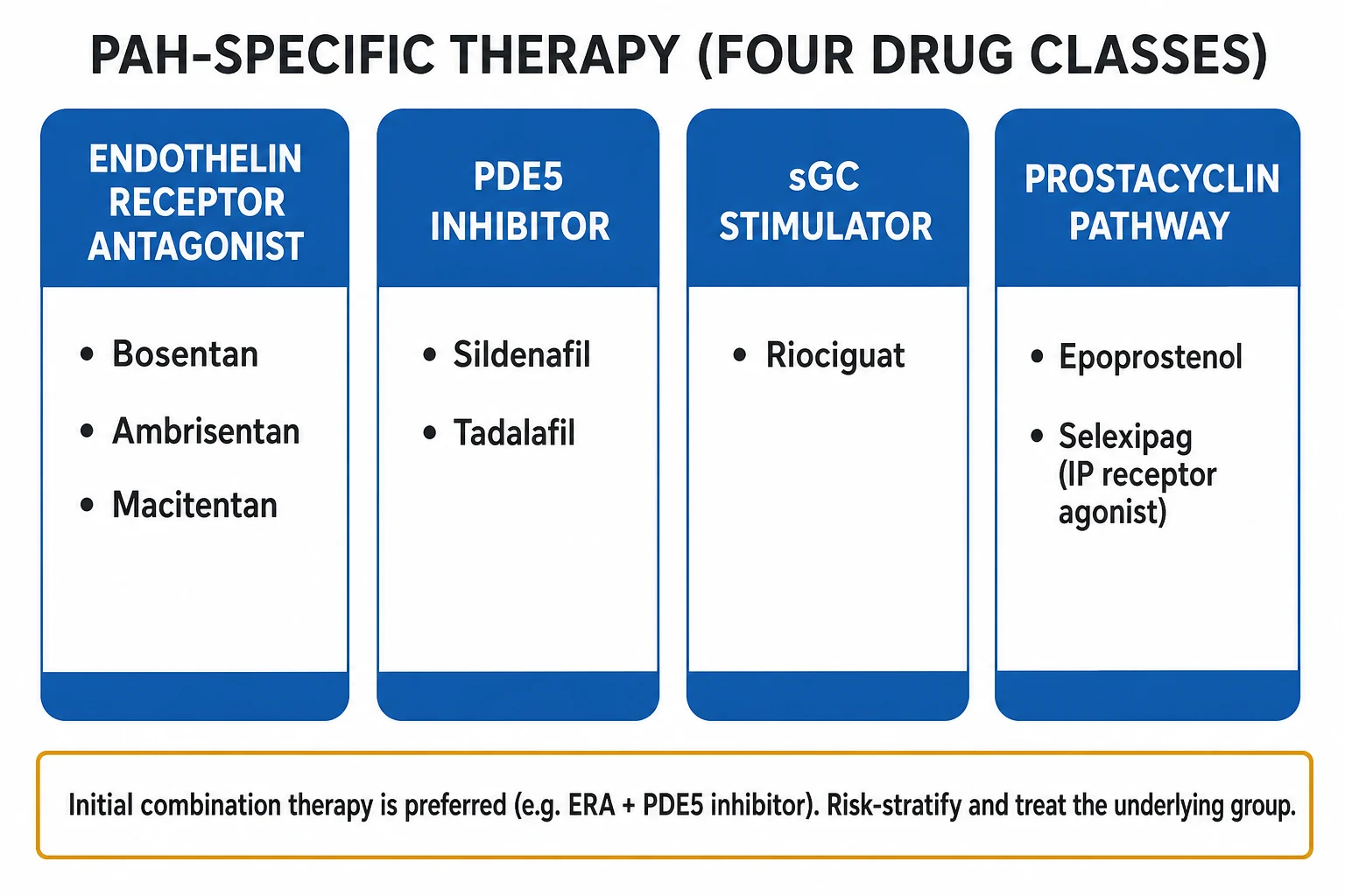
- Endothelin-receptor antagonist (ERA) — start ambrisentan 5 mg daily, titrated to 10 mg daily if tolerated (hepatic-function monitoring minimal), or bosentan 62.5 mg twice daily for 4 weeks, then 125 mg twice daily, with monthly LFTs (avoid if transaminases above 3× upper limit), or macitentan 10 mg daily (no LFT monitoring needed).
- Phosphodiesterase-5 inhibitor (PDE5i) — sildenafil 20 mg three times daily (avoid co-administration with nitrates and strong CYP3A4 inhibitors; rare optic neuropathy); or tadalafil 40 mg daily (longer half-life).
- Soluble guanylate-cyclase stimulator — riociguat 1 mg three times daily, titrated by 0.5 mg every 2 weeks to a maximum of 2.5 mg three times daily — only licensed agent that is disease-modifying in CTEPH in addition to PAH; avoid in pregnancy; monitor blood pressure.[1][5]
Step 3 — Prostacyclin pathway — reserve for high-risk patients, rapid progressors, and as add-on when oral combination does not achieve low-risk profile: [1]
- Epoprostenol (synthetic prostacyclin) — given as continuous intravenous infusion starting at 1 to 2 ng/kg/min and titrated to 10 to 20 ng/kg/min over days to weeks (typical target in class III is 5 to 10 ng/kg/min, in class IV is 10 to 20 ng/kg/min) — improves survival (the only PAH therapy proven to do so in a randomised trial, Barst et al. NEJM 1996); requires central venous catheter and continuous infusion pump; side effects: jaw pain, flushing, diarrhoea, headache, thrombocytopenia, line sepsis.
- Iloprost — inhaled 5 mcg per dose, 6 to 9 inhalations per day; or IV continuous infusion; selective pulmonary vasodilator with brief duration; convenient for ambulatory patients but adherence is demanding.
- Treprostinil — subcutaneous, IV, inhaled or oral; SC infusion is the original formulation but causes infusion-site pain.
- Beraprost — oral, less potent; available in some Asian markets only.
- Selexipag — oral selective IP-receptor agonist starting at 200 mcg twice daily and titrated in 200 mcg twice-daily increments every 2 weeks to the maximum tolerated dose up to 1600 mcg twice daily (median dose at 1 year in trials 1000 mcg twice daily); headache, jaw pain, flushing, diarrhoea; can be combined with ERAs and PDE5i.[1][3]
Step 4 — Reassess and escalate at every follow-up (3 to 6 months): [1]
- Low risk achieved — continue current regimen, monitor.
- Intermediate risk — add parenteral prostacyclin, consider balloon atrial septostomy as bridge (creates a right-to-left shunt that unloads the RV at the cost of hypoxaemia), or refer for transplant.
- High risk — urgent parenteral prostacyclin (IV epoprostenol), VA-ECMO, urgent referral for lung transplantation (single or bilateral; combined heart-lung for Eisenmenger). [1]
Step 5 — Supportive measures (for all groups): [1]
- Oxygen — correct hypoxaemia to keep SpO2 above 92 percent in PAH, above 90 percent in CTEPH and lung-disease PH (15 hours/day oxygen trial historically in COPD; symptomatic use in Eisenmenger to suppress hypoxic drive to shunt).
- Diuretics — furosemide 40 mg IV or orally once or twice daily with metolazone 2.5 to 5 mg pre-dose if diuretic resistance; combine with spironolactone 25 to 50 mg daily for RAAS and potassium conservation.
- Supervised rehabilitation — improves 6MWD and quality of life; avoid unsupervised high-intensity exercise.
- Iron replacement if ferritin below 100 mcg/L or transferrin saturation below 20 percent.
- Vaccination — influenza annually, pneumococcal, COVID-19 — to avoid intercurrent infection triggering RV failure.
- Anticoagulation — controversial; warfarin INR 2.0 to 3.0 often used in idiopathic, heritable and drug-induced PAH; not in CTD-PAH or portopulmonary; DOACs have not shown the protective effect of warfarin in registries in idiopathic PAH.
- Pregnancy is contraindicated in PAH (Group 1); pre-conception counselling and offered termination in established pregnancy, except in mild disease where specialist PH and obstetric teams may support continuation with parenteral prostacyclin, planned C-section and ECMO on standby. [1]
Step 6 — Group-specific therapy: [1]
- Group 2 — Left-heart disease — optimise GDMT; treat valvular lesions surgically; cautious diuresis; PAH-specific therapy not indicated; enrolment in clinical trials (for combined post- and pre-capillary PH) in specialist centres.
- Group 3 — Lung disease — treat the lung; oxygen 15 h/day for chronic hypoxia in COPD; no PAH-specific therapy unless severe PH (case-by-case trial of inhaled treprostinil in ILD-PH).
- Group 4 — CTEPH — pulmonary endarterectomy (PEA, surgically curative) first line if surgical; riociguat 1 mg TDS titrated to 2.5 mg TDS for inoperable or residual disease after PEA (CHEST-1 trial); balloon pulmonary angioplasty (BPA) for inoperable distal disease in experienced centres.[1][5]
- Group 5 — treat the underlying cause.[1]
Subtypes & Scenarios
Connective-tissue-disease PAH (Group 1.4.1) — systemic sclerosis is the most important; PAH in 8 to 12 percent; annual echo with TR Vmax plus annual DLCO is the screening standard (a fall in DLCO may precede the rise in TR Vmax). Initial therapy is upfront oral combination (e.g. ambrisentan 5 to 10 mg daily plus tadalafil 40 mg daily); uptitrate to parenteral prostacyclin if intermediate-high risk. Mortality is higher than in idiopathic PAH.[1]
HIV-associated PAH (Group 1.4.3) — occurs in about 0.5 percent of HIV-positive patients; same diagnostic and treatment pathway as idiopathic PAH (single PAH-specific agent plus antiretroviral therapy) — antiretroviral therapy may improve haemodynamics. Bosentan is contraindicated in patients on protease inhibitors and certain NNRTIs due to hepatotoxicity and drug interactions. [1]
Drug- and toxin-induced PAH (Group 1.3) — anorexigens (fenfluramine, dexfenfluramine, aminorex, benfluorex) are definite; dasatinib, methamphetamines, interferons probable. Stop the drug; treat as PAH; reverse-remodelling is possible. [1]
Portopulmonary hypertension (Group 1.4.2) — 2 to 6 percent of patients with portal hypertension; PVR above 3 WU and mPAP above 35 mmHg exclude liver transplantation candidacy in the international guidelines; sildenafil and tadalafil are first-choice (avoid ERAs due to hepatotoxicity in cirrhosis). Parenteral epoprostenol is reserved as bridge to transplant. [1]
Eisenmenger syndrome (Group 1.4.3.1) — uncorrected or unrepaired large left-to-right shunts that reverse to right-to-left after PVR exceeds systemic vascular resistance. Symptoms: central cyanosis, finger clubbing, polycythaemia, syncope, right-heart failure, secondary erythrocytosis, paradoxical embolism (stroke, brain abscess), gout, hyperviscosity. Treatment: PAH-specific therapy with bosentan or tadalafil improves symptoms and 6MWD; avoid systemic vasodilators that worsen shunt; supervised exercise; venesection reserved for severe hyperviscosity; pregnancy is contraindicated; transplant is heart-lung or bilateral lung with repair of the defect.[1]
CTEPH (Group 4) — defined as pre-capillary PH (mPAP above 20 mmHg, PAWP at or below 15 mmHg) with mismatched perfusion defects on V/Q scan and signs of chronic organised thrombus on CTPA / DSA more than 3 months after adequate anticoagulation; treatment is pulmonary endarterectomy (PEA) — potentially curative, with riociguat 1 mg TDS titrated to 2.5 mg TDS for inoperable or residual disease; BPA in expert centres for distal disease.[1][5]
Paediatric PH — predominantly idiopathic or heritable PAH in term infants, BPD in preterm, congenital diaphragmatic hernia repair, congenital heart disease with residual PH; treatment is by paediatric PH specialists; acute vasodilator testing performed with inhaled nitric oxide; many oral PAH therapies have paediatric extensions; transitional care to adult PH centre at age 16 to 18 years. [1]
Complications
Cardiac complications: right-heart failure — the principal life-limiting complication (oedema, ascites, anasarca, hepatic congestion, cardiorenal syndrome); tricuspid regurgitation from RV and annular dilation; atrial flutter / atrial fibrillation in the dilated right atrium (loss of atrial kick critically reduces RV preload — restore sinus rhythm); ventricular arrhythmias in advanced RV failure; pericardial effusion (sign of severe decompensation and a poor prognostic marker); sudden cardiac death from RV ischaemia, arrhythmia or massive PE.[1][4]
Thrombotic and embolic complications: in-situ pulmonary artery thrombosis (more than 50 percent of severe PAH on autopsy); paradoxical embolism through a patent foramen ovale or atrial septal defect (stroke, brain abscess); CTEPH underlying many Group 4 patients. [1]
Haemoptysis — rupture of dilated bronchial arteries (massive haemoptysis is rare but potentially fatal). [1]
Other complications: hypoxaemia from V/Q mismatch, right-to-left shunt through PFO, hypoventilation; pregnancy (decompensation and maternal mortality up to 30 to 50 percent); NSAID or anaesthesia-related deterioration (cyclo-oxygenase inhibition raises PVR; general anaesthesia depresses RV); sudden withdrawal of prostacyclin (rapid rebound PH crisis). [1]
Therapy-related: ERAs — hepatotoxicity (LFT elevations above 3× upper limit, monthly LFTs with bosentan, minimal with ambrisentan and macitentan), peripheral oedema, anaemia, teratogenicity (pregnancy must be avoided — barrier contraception + monthly pregnancy tests for women of child-bearing age), reduced spermatogenesis; PDE5i — headache, flushing, dyspepsia, blue-tinted vision (sildenafil — rare non-arteritic anterior ischaemic optic neuropathy), epistaxis, avoid with nitrates; riociguat — hypotension, dyspepsia, headache, teratogenicity, syncope, bleeding (avoid anticoagulants with bleeding source); parenteral prostacyclin — flushing, jaw pain, diarrhoea, headache, infusion-site pain (treprostinil SC), line infection and pump failure (epoprostenol); selexipag — headache, jaw pain, flushing, hyperthyroidism.[1][4]
Prognosis & Disposition
Survival — five-year survival in idiopathic PAH has improved from below 35 percent in the pre-treatment era to over 60 percent with modern combination therapy; the REVEAL 2.0 risk score and the 2022 ESC/ERS three-strata risk model (low, intermediate, high) predict one-year mortality of less than 5 percent for low risk, 5 to 20 percent for intermediate and above 20 percent for high risk based on WHO class, 6MWD, BNP, RAP, cardiac index and pericardial effusion. [1]
Predictors of poor outcome: [1]
- Low WHO functional class IV at presentation — strongest single marker.
- Six-minute walk distance below 165 m.
- NT-proBNP above 1400 ng/L or BNP above 300 ng/L.
- Cardiac index below 2.0 L/min/m².
- RAP above 14 mmHg.
- Pericardial effusion on echo.
- DLCO below 40 percent of predicted.
- Reduced TAPSE (below 1.5 cm).
- Long-standing intravascular volume overload and chronic diuretic dependence.
- Frequent hospitalisations for RV failure.
- Pregnancy in Group 1 PAH (maternal mortality up to 30 to 50 percent even in modern series). [1]
Disposition: [1]
- All newly suspected PH patients — specialist PH centre referral within 2 to 6 weeks.
- Established PAH — expert PH centre for initiation of IV prostacyclin and transplant listing.
- Acute RV failure — emergency admission to cardiac ICU with right-heart catheter and PA catheter available, ideally at a PH centre with ECMO.
- CTEPH — multidisciplinary team of PH physician, cardiothoracic surgeon, interventional radiologist and pharmacist; transfer to the nearest PEA centre.[1][3]
Special Populations
Pregnancy — Group 1 PAH is a contraindication to pregnancy because maternal mortality in published series is 30 to 50 percent, even with modern PAH therapy. Pre-conception counselling with a PH specialist and a high-risk obstetrician is mandatory; long-acting contraception (e.g. levonorgestrel IUS, sterilisation) is recommended for women of childbearing age. If pregnancy occurs: multidisciplinary counselling about termination; if continuation, planned admission to a PH centre in the second trimester, parenteral prostacyclin (epoprostenol) titrated upward, planned delivery by C-section under epidural with ECMO on standby, avoidance of ergometrine (vasoconstriction); vaginal delivery has lower volume shifts but is more stressful haemodynamically.[1]
Paediatrics — separate considerations: idiopathic/heritable PAH dominates; persistent pulmonary hypertension of the newborn (PPHN) is a separate Group 1 entity; BPD-associated PH in preterm infants; CDH repair-associated PH; congenital heart disease residual PH and Eisenmenger; paediatric-specific dosing and formulations; transition to adult PH centres planned at 16 to 18 years. [1]
Elderly — Group 2 PH and Group 3 PH dominate; less aggressive PAH-specific therapy because of comorbidities; Group 1 PAH is rare after age 70. [1]
Pre-existing left heart disease — PAH therapy may worsen gas exchange and fluid retention; trials are negative in HFrEF and HFpEF; do not add ERAs or PDE5i purely for raised PVR in Group 2. [1]
Liver disease — portopulmonary hypertension (Group 1.4.2); concerns of hepatotoxicity with ERAs; sildenafil and tadalafil preferred; transplant candidacy requires PVR less than 3 WU and mPAP less than 35 mmHg.[1]
Race and ethnicity — Black and Hispanic patients are over-represented in Group 2 (HFpEF and hypertensive heart disease) and Group 5 PH (sickle cell disease, sarcoidosis); sickle cell disease PH is a Group 5 entity (chronic haemolysis and NO depletion); treat the underlying disease and consider hydroxyurea and exchange transfusion. [1]
Evidence, Guidelines & Regional Differences
The single most important guideline: 2022 ESC/ERS Guidelines for the Diagnosis and Treatment of Pulmonary Hypertension (Humbert et al.) updates the 2015 (Galie et al.) classification, lowers the mPAP threshold to above 20 mmHg, refines the PAWP-PVR schema, and endorses upfront oral combination therapy and risk-stratified treatment. The 2021 ESC/EHA statement on acute RV failure (Harjola et al.) is the reference for resuscitation.[1][2][4]
Landmark trials the candidate must know: [1]
- AMBITION (Galie et al., NEJM 2015) — initial combination of ambrisentan plus tadalafil halved clinical failure events versus monotherapy in treatment-naive PAH.[6]
- CHEST-1 (Ghofrani et al., NEJM 2013) — riociguat improved 6MWD in inoperable or residual CTEPH — the only licensed medical therapy in CTEPH.[5]
- GRIPHON (Sitbon et al., NEJM 2015) — selexipag reduced morbidity/mortality composite in PAH when added to background ERA ± PDE5i.
- SERAPHIN (Pulido et al., NEJM 2013) — macitentan reduced morbidity/mortality in PAH.
- COMPASS-2 / REVEAL risk score validate risk-stratification tools.
- Barst et al. NEJM 1996 — epoprostenol improved survival in class III–IV idiopathic PAH — the only PAH drug so far with a survival benefit in a randomised trial.
Regional deltas: [1]
- UK (NHS / NICE) — national PH service network; riociguat, macitentan, selexipag, sildenafil and tadalafil commissioned; PH centres at Papworth, Royal Brompton, Hammersmith, Freeman, Sheffield; NICE NG145 (2024 update) endorses 2022 ESC/ERS risk-stratified therapy. Surgery-first model for CTEPH at Royal Brompton / Papworth.
- US (AHA / ACC) — 6th World Symposium on Pulmonary Hypertension updated classification; FDA-approved drugs include sildenafil, tadalafil, bosentan, ambrisentan, macitentan, riociguat, epoprostenol, treprostinil (multiple formulations), iloprost, selexipag; inhaled treprostinil for ILD-PH; CCB only for confirmed vasoreactive idiopathic PAH (amiloride, nifedipine, diltiazem).
- EU — 2022 ESC/ERS adopted; PAH centres certified by European Reference Network (ERN-LUNG); AMBITION-style upfront oral combination is standard.
- India / South Asia — anorexigen exposure (fenfluramine) clustering in the past; CTD-PAH is common; cost barriers to macitentan, selexipag and epoprostenol — generic bosentan and sildenafil are first-line by necessity; CTEPH surgery at a few centres; the NEET-PG / INICET exam emphasis is on classification, bedside signs, RHC and first-line therapy recognition.[1]
Exam Pearls
- PH = mPAP above 20 mmHg at rest on right heart catheterisation (revised down from 25 mmHg in 2022); pre-capillary needs PAWP at or below 15 mmHg and PVR above 2 WU; echo screening then RHC confirms.[1]
- WHO Groups 1 to 5 — the classification that drives treatment: Group 1 PAH (idiopathic, heritable, drug-induced, CTD, HIV, portal HTN, congenital), Group 2 left-heart, Group 3 lung disease, Group 4 CTEPH, Group 5 multifactorial.[1][2]
- V/Q scan — mandatory in every newly diagnosed PH (the patient with mismatched perfusion defects has CTEPH until proven otherwise and may be curable by surgery).[1]
- Bedside signs — loud P2 + RV heave + raised JVP = look hard for PH; TR murmur is functional in advanced disease; a single loud P2 alone in a young woman with dyspnoea = PAH until proven otherwise.[1]
- Upfront oral combination — ambrisentan + tadalafil (or any ERA + PDE5i) is the new default in low-/intermediate-risk treatment-naïve PAH (AMBITION).[6]
- Riociguat is the only drug licensed in CTEPH (CHEST-1); avoid in PAH-ERA combinations (no added benefit, added hypotension).[5]
- Acute RV failure — avoid intubation where possible (positive-pressure ventilation raises RV afterload and lowers preload, and worsens ischaemia); use inhaled nitric oxide, IV epoprostenol or inhaled iloprost, cautious diuresis with furosemide 40 mg IV, inotropy, and VA-ECMO for refractory cases.[1][4]
- Vasoreactivity testing — a positive test (fall in mPAP of at least 10 mmHg to a value at or below 40 mmHg with maintained CO) identifies the 5 percent of idiopathic/heritable PAH who respond to calcium-channel blockers (amlodipine 5 to 20 mg, nifedipine 30 to 120 mg, diltiazem 120 to 360 mg daily); non-responders need PAH-specific combination therapy.[1]
- CTEPH surgery (pulmonary endarterectomy, PEA) is the only potentially curative therapy in PH — refer every CTEPH patient to a PEA centre.
- Anorexigen exposure (fenfluramine family) is a specific drug-cause of PAH; ask about weight-loss medication history.
- Scleroderma — annual echo with TR Vmax + DLCO screening (PAH in 8 to 12 percent; mortality higher than idiopathic PAH).[1]
- Pregnancy is contraindicated in Group 1 PAH (maternal mortality up to 30 to 50 percent).[1]
- Moderate / high dose diuretics (furosemide 40 mg IV/PO daily, with metolazone or spironolactone as needed) for RV failure.[1]
Pulmonary hypertension aetiology by WHO group — mnemonic
PAH LUNG CLOT MISC
Idiopathic, heritable (BMPR2), drug- and toxin-induced (anorexigens, dasatinib), CTD (scleroderma, SLE, MCTD), HIV, portal HTN, congenital (Eisenmenger), PVOD/PCH
HFrEF, HFpEF, valvular disease (mitral stenosis, aortic stenosis, mitral regurgitation), constrictive pericarditis, restrictive cardiomyopathy
COPD (the commonest), interstitial lung disease (combined pulmonary fibrosis and emphysema), sleep-disordered breathing (OSA, OHS), high-altitude exposure, developmental lung disease
Chronic thromboembolic PH (CTEPH) after acute PE; pulmonary artery sarcoma, tumour embolism, fibrosing mediastinitis
Haematological (sickle cell, thalassaemia, myeloproliferative, splenectomy), systemic (sarcoidosis, histiocytosis, vasculitis), metabolic (Gaucher, glycogen storage, thyroid), CKD with dialysis
Group 4 is potentially curable by pulmonary endarterectomy — never miss CTEPH
Exam application bank (NEET-PG / INICET)
One-line answer
Pulmonary hypertension (PH) is a haemodynamic and pathophysiological syndrome defined as a mean pulmonary arterial pressure (mPAP) above 20 mmHg at rest measured by right heart catheterisation (RHC), confirmed by a pulmonary arterial wedge pressure (PAWP) of 15 mmHg or less in pre-capillary disease and pulmonary vascular resistance (PVR) above 2 Wood units (WU) in pulmonary arterial hypertension (PAH). The 2022 ESC/ERS Guidelines classify PH into five groups by aetiology: Group 1 — pulmonary arterial hypertension (PAH) (idiopathic, heritable, drug- and toxin-induced, and associated with connective tissue disease, HIV, portal hypertension, congenital heart disease); Group 2 — PH due to left heart disease (heart failure with preserved or reduced ejection fraction, valvular disease); Group 3 — PH due to lung disease or hypoxia (COPD, interstitial lung disease, sleep-disordered breathing); G
Worked stems (answer without another resource)
Stem 1 — Classic presentation. Map symptoms to mechanism; name the first investigation and first treatment step with dose/route if drug therapy is standard. [1]
Stem 2 — Unstable / complicated. List red flags that force immediate resuscitation, theatre, ICU, antidote, or reperfusion — and what you do in the first 15 minutes. [1]
Stem 3 — Atypical group. Elderly, pregnancy, child, or immunocompromised: how presentation and thresholds change. [1]
Stem 4 — Differential trap. Name the three closest mimics and one discriminator for each. [1]
Stem 5 — Disposition. Who goes home with safety-netting, who is admitted, who needs HDU/ICU/theatre, and what follow-up is mandatory. [1]
Rapid viva checklist
- Definition + classification
- Pathophysiology chain
- Bedside signs / criteria
- Score with exact components (if any)
- Emergency bundle
- Definitive therapy with doses
- Complications of disease and of treatment
- Special populations
- Guideline/trial name if classic
- Three exam traps
Coverage self-check
If you cannot answer any stem above from this page alone, re-read the matching section — the page is intended to be self-sufficient for final-prof and NEET-PG/INICET questions on Pulmonary Hypertension.
References
- [1]Humbert M, Kovacs G, Hoeper MM, et al. Temperature-Dependent Superhydrophobic Functionalized Coordination Polymers (SFCPs) for Selective Adsorption of C(2)H(4) over C(2)H(6) Inorg Chem, 2022.PMID 36027580
- [2]Galie N, Humbert M, Vachiery JL, et al. Can be tracing of surgeon's hand movement useful in laparoscopic and endoscopic learning curve? Bratisl Lek Listy, 2015.PMID 26084743
- [3]Galie N, Channick RN, Frantz RP, et al. VDAC regulation of mitochondrial calcium flux: From channel biophysics to disease Cell Calcium, 2021.PMID 33529977
- [4]Harjola VP, Mebazaa A, Celutkiene J, et al. Notum palmitoleoyl-protein carboxylesterase regulates Fas cell surface death receptor-mediated apoptosis via the Wnt signaling pathway in colon adenocarcinoma Bioengineered, 2021.PMID 34402722
- [5]Ghofrani HA, D'Armini AM, Grimminger F, et al. Blockade of electron transport before ischemia protects mitochondria and decreases myocardial injury during reperfusion in aged rat hearts Transl Res, 2012.PMID 22698829
- [6]Galie N, Barbera JA, Frost AE, et al. Endovascular Total Arch Repair Using In Situ Fenestration for Arch Aneurysm and Chronic Type A Dissection Ann Thorac Surg, 2016.PMID 26387722