Cardiology · Cardiology
Sudden Cardiac Death
Also known as SCD · Sudden cardiac arrest · Cardiac sudden death · Aborted sudden cardiac death
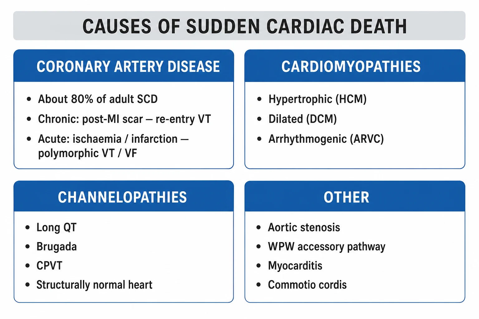
Sudden cardiac death (SCD) is the sudden, unexpected death from a cardiac cause occurring within one hour of symptom onset (witnessed), or within 24 hours of last being seen alive and well (unwitnessed). It is the leading natural cause of death in the industrialised world, accounting for roughly 300,000 to 400,000 events per year in the United States and 4 to 5 million globally. The final common pathway in 80 to 90 percent of cases is a ventricular tachyarrhythmia (rapid polymorphic VT degenerating to ventricular fibrillation), with bradyasystole and pulseless electrical activity accounting for the rest. Coronary artery disease underlies 75 to 80 percent of adult SCD; the remainder arise from cardiomyopathies (hypertrophic, dilated, arrhythmogenic right ventricular), inherited channelopathies (long QT, Brugada, CPVT), severe valvular disease, myocarditis, drug toxicity, electrolyte disturbance, anomalous coronary arteries, commotio cordis, and massive pulmonary embolism. Management is the chain of survival (early CPR, early defibrillation, early advanced life support), targeted temperature management at 32 to 36 degrees C for 24 hours, urgent coronary angiography when a cardiac cause is suspected, and ICD implantation for survivors (secondary prevention) and for high-risk primary-prevention subgroups defined by the landmark trials (MADIT, MADIT-II, MUSTT, SCD-HeFT, DINAMIT, DANISH). First-degree relatives of young SCD victims require cascade clinical and genetic screening.
On this page & tools
Your progress
Saved locally on this device.
Exam tags
Red flags
Overview & Definition
Sudden cardiac death is defined as natural death from a cardiac cause, heralded by abrupt loss of consciousness within one hour of the onset of acute symptoms (in the witnessed case); when unwitnessed, the definition extends to a person known to be alive and clinically stable within the preceding 24 hours. The mechanism is almost always an abrupt cessation of effective cardiac output — most commonly a malignant ventricular tachyarrhythmia — occurring against a substrate of structural or electrical heart disease that may have been previously unrecognised. Crucially, the time and mode of death are unexpected, which separates SCD from the terminal arrhythmia of advanced heart failure or septic shock.[1]
The distinction between sudden cardiac arrest (SCA) and sudden cardiac death (SCD) is operational. SCA is the event; if the patient survives to hospital discharge it is termed aborted SCD. Approximately 10 percent of out-of-hospital cardiac arrests survive to discharge, so SCA far outnumbers SCD. The reversible window is short: brain injury begins within 4 to 6 minutes of circulatory arrest, which is why bystander CPR and early defibrillation dominate survival more than any hospital intervention. [1]
SCD is the first manifestation of cardiac disease in roughly half of all victims, and the first manifestation of coronary disease in one quarter. This sobering fact drives the entire prevention strategy: risk stratification tools (left ventricular ejection fraction, Holter, signal-averaged ECG, heart rate variability, cardiac MRI scar) identify only a minority of those who will die, so population-level modification of coronary risk factors, family screening of young victims, and disease-specific therapy for high-risk cardiomyopathies and channelopathies are all required.[2][3]
Classification
SCD is usefully classified along two axes: the underlying aetiology (which dictates prevention) and the terminal rhythm (which dictates the resuscitation). Aetiologically, ischaemic heart disease dominates in adults over 35, while inherited cardiomyopathies, channelopathies, and congenital coronary anomalies dominate in the young. By rhythm, the broad split is shockable (VF, pulseless VT) versus non-shockable (asystole, PEA) — a distinction that determines whether the first action is a shock or chest compressions and adrenaline. [1]
Ischaemic (75 to 80 percent)
- Acute MI: plaque rupture, thrombosis, acute ischaemia-driven VF
- Chronic post-MI scar: re-entry VT, often monomorphic, years after infarct
- Ischaemic cardiomyopathy: combined scar and LV dysfunction
- Coronary anomaly: anomalous left coronary from right sinus, intramural course
Non-ischaemic cardiomyopathy
- Dilated cardiomyopathy (genetic, viral, alcoholic, tachycardia-induced, anthracycline)
- Hypertrophic cardiomyopathy: the commonest cause in the young athlete
- Arrhythmogenic right ventricular cardiomyopathy (ARVC): fibro-fatty replacement
- Infiltrative: cardiac sarcoidosis, amyloidosis, haemochromatosis, Chagas
Channelopathy (structurally normal heart)
- Long QT syndrome (LQT1/2/3): adrenergic or sleep-triggered torsades
- Brugada syndrome: type 1 coved ST in V1 to V3, resting VF risk
- Catecholaminergic polymorphic VT (CPVT): exertional bidirectional VT
- Early repolarisation and short QT syndromes
Valvular and other structural
- Severe aortic stenosis: LVH, subendocardial ischaemia
- Severe aortic or mitral regurgitation
- Aortic dissection with tamponade or coronary compromise
- Congenital aortic stenosis, Ebstein anomaly, post-repair tetralogy
Acquired and external
- Myocarditis (coxsackie, parvovirus B19, giant cell)
- Drug toxicity: cocaine, QT-prolonging agents, digoxin, sodium-channel blockers
- Severe electrolyte disturbance: hypokalaemia, hypomagnesaemia, hypocalcaemia
- Massive pulmonary embolism: PEA arrest
- Commotio cordis: blunt precordial blow in the vulnerable repolarisation window
By terminal rhythm, the data from monitored arrests are striking and examinable: [1]
Epidemiology & Risk Factors
SCD is the single largest natural killer in the industrialised world, responsible for roughly half of all cardiovascular deaths and 15 to 20 percent of all-cause mortality. Incidence in adults is 1 to 2 per 1,000 person-years, rising steeply with age: the peak decade is 65 to 74 in men, with a male-to-female ratio of 3 to 4:1 before the seventh decade. After 75, the sex gap narrows. The absolute number of SCD events is therefore highest in the elderly, but the proportion of deaths that are sudden is highest in the young — which is why youthful SCD carries such disproportionate societal weight.[1]
The established risk factors fall into three tiers. Population risk factors are the conventional coronary risk factors (age, male sex, hypertension, dyslipidaemia, smoking, diabetes, obesity, family history of premature coronary disease) — they predict SCD largely because they predict coronary disease. Cardiac substrate markers are far stronger individual predictors: a left ventricular ejection fraction of 35 percent or less raises SCD risk five- to tenfold and is the single most important variable used in guidelines to select patients for a primary-prevention ICD. Arrhythmic markers (prior cardiac arrest, sustained VT, unexplained syncope with structural disease, non-sustained VT on Holter, frequent premature ventricular complexes) further stratify risk in those with a substrate.[2][3]
Major risk factors (high yield)
- Prior SCA or sustained VT — strongest predictor
- LVEF 35 percent or less after MI or in non-ischaemic DCM on optimal therapy
- NYHA class II to III heart failure with reduced EF
- First 30 days after MI (highest risk window of life)
- Family history of SCD in a first-degree relative under 40
Moderate risk factors
- Prior MI with residual scar (especially anterior)
- LV hypertrophy on ECG or echo
- Frequent PVCs (over 10 percent burden) or non-sustained VT on monitoring
- Unexplained syncope, especially with structural heart disease
- Severe sleep apnoea, chronic kidney disease
Specific high-risk diseases
- Hypertrophic cardiomyopathy (1 percent per year overall; higher with risk factors)
- Arrhythmogenic RV cardiomyopathy
- Long QT (0.1 to 0.9 percent per year, gene-specific)
- Brugada syndrome (variable; highest in symptomatic)
- Cardiac sarcoidosis, amyloidosis, Chagas disease
A note on athletes: the overall risk of SCD in athletes is lower than in the age-matched general population, but among young athletes (under 35) SCD is the leading cause of death, with hypertrophic cardiomyopathy the commonest substrate in the US (followed by anomalous coronary artery and commotio cordis) and arrhythmogenic RV cardiomyopathy the commonest in Italy (where ECG screening has long been mandatory). Above 35, coronary disease dominates in athletes as in the general population. [1]
Pathophysiology
The final common pathway in roughly 80 to 90 percent of SCD is a rapid ventricular tachyarrhythmia. Three factors — the substrate, the trigger, and the modulating influence — must interact for the arrhythmia to ignite and sustain. Understanding this triplet explains why a person can carry a scarred myocardium for decades without event, then arrest during an adrenergic surge, an electrolyte disturbance, or an episode of acute ischaemia.[1][2]
The substrate. In ischaemic disease the substrate is a post-infarction scar with surviving myocyte channels embedded in fibrous tissue, creating zones of slow conduction and unidirectional block that permit re-entry. In hypertrophic and infiltrative cardiomyopathy the substrate is disorganised architecture, interstitial fibrosis, and myocyte disarray. In channelopathies there is no structural scar, but the myocyte membranes carry a mutation (potassium loss-of-function in LQT1/2, sodium gain-of-function in LQT3, sodium loss-of-function in Brugada, ryanodine-receptor leak in CPVT) that prolongs repolarisation or allows calcium-driven after-depolarisations. [1]
The trigger. The initiating event is usually a premature ventricular complex that lands on the vulnerable repolarisation window — the downslope of the T wave, roughly 10 to 30 ms before its peak, when dispersion of repolarisation is maximal. The short-coupled PVC captures a tissue that is partly refractory, partly excitable, and sets up a re-entrant or spiral-wave circuit. In acute ischaemia the trigger is more chaotic: heterogeneous depolarisation across the ischaemic border zone generates automatic and triggered activity that rapidly organises into polymorphic VT and then VF. [1]
The modulator. Autonomic tone, electrolytes, drugs, and ischaemia shift the threshold. A sympathetic surge (exercise, emotion, the morning cortisol surge) lowers the VF threshold; hypokalaemia and hypomagnesaemia prolong repolarisation and promote early after-depolarisations; QT-prolonging drugs (macrolides, fluoroquinolones, antipsychotics, methadone) do the same in susceptible individuals; acute ischaemia superimposed on a chronic scar is the classical double-hit that produces monomorphic VT. [1]
The degeneration cascade is then mechanical as much as electrical: VF produces no effective cardiac output, coronary perfusion collapses, the myocardium becomes progressively more ischaemic and acidotic, and the rhythm becomes harder to terminate with each passing minute. After 8 to 10 minutes the myocardium enters an unresponsive phase; VF often degrades to asystole. Bradyasystolic arrest (asystole and PEA) represents a different mechanism — usually the terminal manifestation of severe myocardial failure, hypoxia, or a massive mechanical catastrophe (tamponade, massive PE, tension pneumothorax, hypovolaemia) — and carries a worse prognosis because it usually signals end-stage disease rather than a treatable electrical event.[1]
Clinical Presentation
The cardinal presentation is sudden collapse with loss of consciousness, absence of pulse, and apnoea or agonal gasping. The event is abrupt — there is no progressive build-up — which distinguishes it from cardiogenic shock or sepsis. In 25 to 50 percent of cases there is a prodrome in the preceding hours to weeks: chest pain, dyspnoea, palpitations, fatigue, or syncope. These symptoms are non-specific and frequently overlooked in retrospect, but a careful witness history (was the patient exertional, emotional, asleep, post-prandial?) can point to aetiology — exertional arrest suggests HCM, ARVC, anomalous coronary, or CPVT; arrest in sleep or at rest suggests long QT 3 or Brugada; arrest in water suggests long QT 1.[1]
Two under-recognised presentations are worth flagging. First, agonal breathing is not normal breathing — bystanders and dispatchers frequently misinterpret it as a sign of life and withhold CPR. Second, seizure-like activity (myoclonic jerks from cerebral hypoperfusion) is misdiagnosed as epilepsy; a small but real fraction of "first seizure in an adult" presentations are aborted cardiac arrests, particularly in long QT syndrome. [1]
Survivors of cardiac arrest who achieve return of spontaneous circulation (ROSC) present in the post-cardiac arrest syndrome: comatose or confused, often intubated and ventilated, with variable haemodynamic instability from myocardial stunning and the underlying cause. There may be signs of end-organ ischaemia — acute kidney injury, transaminitis, ileus, lactic acidosis — and the neurological exam may range from normal to brainstem areflexia. The first hours are critical for both neuroprotection (targeted temperature, normoxia, normocapnia, normoglycaemia, seizure control) and cause identification (ECG, echo, angiography).[3]
Differential Diagnosis
By definition, the SCD victim is dead, so the differential applies to aborted SCD (the survivor) and to the sudden-collapse mimic. The broad categories are cardiac and non-cardiac, and within cardiac they subdivide into arrhythmic versus mechanical catastrophe. [1]
Cardiac causes (resuscitated)
- Acute MI / ischaemia-driven VF (most common)
- Scar-based re-entry VT (post-MI, DCM, ARVC)
- Channelopathy: long QT torsades, Brugada, CPVT
- Severe structural: HCM, severe AS, aortic dissection with tamponade
- Myocarditis (especially giant cell), infiltrative disease
Non-cardiac arrest causes
- Massive pulmonary embolism (PEA, hypoxia, distended neck veins)
- Aortic dissection (tearing pain, BP differential, widened mediastinum)
- Tension pneumothorax (asymmetrical breath sounds, tracheal deviation)
- Cardiac tamponade (Beck triad, electrical alternans)
- Anaphylaxis (urticaria, angioedema, exposure history)
Collapse mimics
- Vasovagal syncope: long prodrome, situational, rapid full recovery
- Seizure: aura, tonic-clonic phase, post-ictal confusion, tongue biting
- Drug overdose: opioid (miotic, slow), TCA (wide QRS), beta-blocker (bradycardic)
- Subarachnoid haemorrhage: thunderclap headache, focal deficit
- Hypoglycaemia: sweating, tremor, rapid response to glucose
The discriminator that examiners love is the rhythm at arrest. VF or pulseless VT in a middle-aged adult is ischaemic until proven otherwise and mandates urgent coronary angiography. PEA with a narrow complex and a history of immobility, recent surgery, or malignity points to pulmonary embolism — for which thrombolysis during CPR is indicated. PEA with electrical alternans and a history of malignity or trauma suggests tamponade and pericardiocentesis or resuscitative thoracotomy. Asystole in the unwitnessed, prolonged down-time arrest is usually medical futility unless a reversible cause is found.[2]
Clinical & Bedside Assessment
At the scene the priority is the chain of survival, not diagnosis. The assessment is brief and action-oriented: confirm unresponsiveness, check for normal breathing (not agonal) and a central pulse for no more than 10 seconds, call for help and a defibrillator, and begin CPR. Once an AED or monitor is attached, the rhythm decides the next move — shockable (coarse VF, pulseless VT) or non-shockable (asystole, PEA).[2]
The focused history (taken from witnesses and relatives while CPR continues) targets reversible causes and pre-existing disease: did the patient have chest pain (ischaemia), shortness of breath (PE, heart failure), a recent fall or fracture (fat embolism, hypovolaemia), a drug ingestion or overdose, a known cardiomyopathy or channelopathy, a family history of sudden death? The drug history specifically hunts for QT-prolonging agents, digoxin, beta-blockers, calcium-channel blockers, and cocaine. The examination during CPR looks for signs of the 4 Hs and 4 Ts — distended neck veins and muffled heart sounds (tamponade), unequal breath sounds (tension pneumothorax), a hemothorax or external bleeding (hypovolaemia), needle marks or pill bottles (toxins), a swollen leg (PE). [1]
After ROSC, the secondary survey is systematic: 12-lead ECG within minutes to look for STEMI (which mandates immediate angiography), focused echo at the bedside to assess LV function, RV strain (PE), tamponade, and unsuspected structural disease, arterial blood gas for acidosis and hypoxia, bedside ultrasound to exclude free fluid, and a focused neurological examination (GCS, pupillary response, brainstem reflexes, myoclonus) which anchors prognostication. [1]
Investigations
The investigation strategy in a survivor of cardiac arrest is a hunt for the cause, because the cause determines whether the patient receives an ICD, a revascularisation, an ablation, medical therapy for a cardiomyopathy, or simply reassurance. The work-up is layered. [1]
Immediate (within the first hour). A 12-lead ECG is the single most important test. Look for ST elevation (STEMI — go straight to angiography), ST depression, T-wave inversion, pathological Q waves (old infarct), LVH with strain (HCM, AS), Brugada type 1 pattern (coved ST in V1 to V3), prolonged corrected QT (over 470 ms in men, 480 ms in women), epsilon waves and T-wave inversion in V1 to V3 (ARVC), a delta wave (WPW), or low voltage (infiltrative disease, tamponade). Repeat the ECG serially, as ischaemic changes may evolve. Arterial blood gas reveals acidaemia (a marker of downtime and resuscitation quality), hypoxia, hyperkalaemia, and lactate. Bloods include troponin (rise occurs with infarction and is also non-specifically elevated after prolonged CPR), electrolytes (K, Mg, Ca, phosphate), renal and liver function, glucose, lactate, full blood count, coagulation, and a toxicology screen (cocaine, opioids, methadone, TCA). Beta-HCG in women of reproductive age (peripartum cardiomyopathy).[2][3]
Within the first day. Coronary angiography is recommended in all survivors in whom a cardiac cause is suspected and in whom STEMI or haemodynamic instability is present — and is reasonable in all survivors without an obvious non-cardiac cause, because occult plaque rupture is common. Transthoracic echocardiography assesses LV systolic function (the most important variable for ICD decisions), regional wall motion, valvular disease, hypertrophic cardiomyopathy, ARVC, infiltrative disease, tamponade, RV dysfunction (PE), and congenital anomalies. High-sensitivity troponin trended over 3 to 6 hours distinguishes infarction from resuscitation-related injury. [1]
Within the first days (once stable). Cardiac MRI is the gold standard for tissue characterisation — late gadolinium enhancement identifies subendocardial scar (ischaemic), mid-wall enhancement (DCM, myocarditis, sarcoid), and patchy involvement (amyloid); it is essential in the work-up of suspected myocarditis, ARVC, sarcoidosis, and infiltrative disease. Holter monitoring (24 to 48 hours) or an implantable loop recorder captures paroxysmal arrhythmia. An electrophysiology study (programmed ventricular stimulation) has a declining role in the primary-prevention era but is still useful in suspected scar-based VT (inducibility guides ablation) and in selected inherited syndromes.[2]
Genetic testing and family screening. In survivors under 40, those with a phenotype suggesting an inherited syndrome (HCM, ARVC, long QT, Brugada, CPVT), and in all young SCD victims (post-mortem), a cardiomyopathy and channelopathy gene panel is indicated. Identifying a pathogenic variant in the proband allows cascade genetic testing of first-degree relatives — the cornerstone of family screening. A molecular autopsy (post-mortem genetic testing on blood or tissue) is recommended when a young person dies suddenly and autopsy is negative, as up to 25 percent of such cases carry a pathogenic channelopathy variant.[2][3]
Selective imaging. CT pulmonary angiogram if PE is suspected, CT aorta if dissection is in the differential, CT head if there is head trauma, focal neurology, or a suspicion of intracranial haemorrhage as the precipitant. Drug challenge with ajmaline or flecainide is used to unmask concealed Brugada syndrome in survivors with a borderline ECG and a family history. [1]
Management — Resuscitation
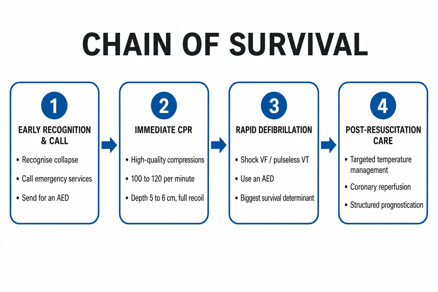
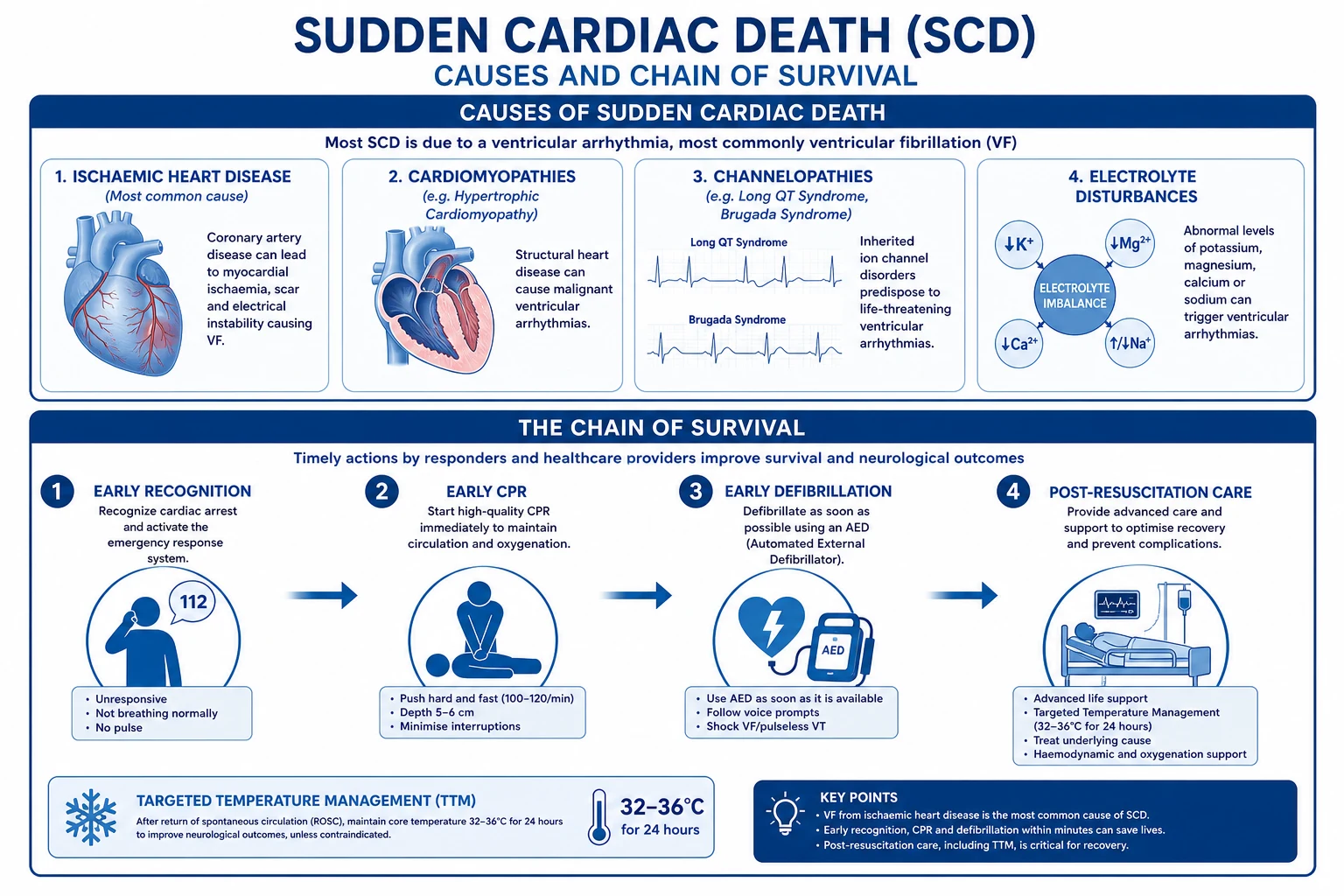
Resuscitation is the chain of survival: early recognition and call for help, early CPR with emphasis on high-quality chest compressions, early defibrillation, and early advanced life support with integrated post-resuscitation care. Survival falls by roughly 10 percent for every minute without defibrillation in VF, but good bystander CPR roughly halves that slope.[1][2]
High-quality CPR is the foundation: compress the chest at a rate of 100 to 120 per minute, to a depth of 5 to 6 cm, allowing full chest recoil between compressions, with minimal interruption (chest-compression fraction above 0.6, ideally above 0.8). Ventilate at a ratio of 30 compressions to 2 breaths with a bag-valve-mask or advanced airway, avoiding excessive ventilation (which raises intrathoracic pressure and reduces coronary perfusion). Once an advanced airway is in place, compressions become continuous at 100 to 120 per minute with one breath every 6 seconds. [1]
Confirm arrest, call for help
Unresponsive, no normal breathing, no pulse (under 10 s check). Activate emergency response, summon a defibrillator.
Begin CPR
30 compressions to 2 breaths, rate 100 to 120/min, depth 5 to 6 cm, full recoil, minimise interruptions.
Attach defibrillator, assess rhythm
Shockable (VF, pulseless VT) vs non-shockable (asystole, PEA).
Shockable loop
Shock (biphasic 150 to 200 J), resume CPR 2 min, reassess rhythm, repeat. Adrenaline 1 mg IV after the 2nd shock, then every 3 to 5 min. Amiodarone 300 mg IV after the 3rd shock, 150 mg after the 5th.
Non-shockable loop
Adrenaline 1 mg IV immediately, then every 3 to 5 min. CPR 2 min, reassess. Identify and treat reversible causes (4 Hs and 4 Ts).
Reversible causes
Hypovolaemia, Hypoxia, Hypo/hyperkalaemia, Hypothermia, Hydrogen ion (acidosis); Tension pneumothorax, Tamponade, Toxins, Thrombosis (PE, coronary).
ROSC
Begin post-resuscitation care: targeted temperature, normoxia, normocapnia, MAP above 65, 12-lead ECG, coronary angiography if indicated.
Shockable rhythms (VF, pulseless VT). The single most important intervention is immediate defibrillation. Deliver a biphasic shock at 150 to 200 J (or the manufacturer-recommended energy), and escalate to maximum (typically 360 J biphasic) for subsequent shocks. Immediately resume CPR for 2 minutes before reassessing rhythm — pausing to check a pulse immediately post-shock wastes the window of recovering perfusion. Give adrenaline 1 mg intravenously every 3 to 5 minutes (started after the second shock) to raise coronary and cerebral perfusion pressure. Give amiodarone 300 mg intravenous bolus after the third shock, then 150 mg after the fifth shock if VF/VT persists; lidocaine 1 to 1.5 mg/kg IV is an alternative. Identify and treat the reversible causes throughout.[2]
Non-shockable rhythms (asystole, PEA). There is no role for defibrillation. The algorithm centres on adrenaline 1 mg IV immediately, then every 3 to 5 minutes, 2-minute cycles of CPR with rhythm and pulse checks, and an aggressive hunt for the reversible cause — because in PEA the rhythm is a symptom of a mechanical catastrophe, and the patient will not survive unless that catastrophe is reversed. Atropine is no longer recommended for asystole or PEA. [1]
4 Hs and 4 Ts
Targeted temperature management (TTM). In comatose survivors of cardiac arrest (any rhythm), targeted temperature management at 32 to 36 degrees C for at least 24 hours is a class I intervention that improves both survival and neurological outcome. The TTM trial (Nielsen 2013) showed no difference between 33 and 36 degrees C, shifting practice toward 36 degrees C with strict fever avoidance; both are acceptable. Maintain normoxia (SpO2 94 to 98 percent, avoiding hyperoxia), normocapnia (PaCO2 35 to 45 mmHg), mean arterial pressure above 65 mmHg, glucose 8 to 10 mmol/L, and treat seizures. Continuous EEG is used to detect non-convulsive status. Neurological prognostication is delayed to at least 72 hours after ROSC and is multimodal (clinical exam, somatosensory evoked potentials, EEG, neuron-specific enolase, brain MRI).[11][12]
Urgent coronary angiography. Survivors with ST elevation on the post-ROSC ECG, or with haemodynamic instability, go to the catheter lab immediately (within 2 hours) for PCI. In those without ST elevation but with a suspected cardiac cause, early angiography (within 2 to 24 hours) is reasonable and frequently identifies an culprit lesion. Out-of-hospital cardiac arrest with no obvious non-cardiac cause should be presumed ischaemic until proven otherwise. [1]
Management — Definitive & Stepwise
Once ROSC is achieved and the cause is identified, definitive management centres on three pillars: disease-specific therapy (revascularisation, heart-failure therapy, ablation, valve surgery), antiarrhythmic and autonomic modulation, and the implantable cardioverter-defibrillator (ICD) for secondary or primary prevention. [1]
Secondary prevention ICD. Any survivor of cardiac arrest due to VF or haemodynamically unstable VT, in whom the cause is not fully reversible, should receive an ICD before discharge. This is a class I indication supported by the three classic RCTs — AVID (NEJM 1997), CIDS, and CASH — which together showed roughly a 25 to 30 percent relative reduction in mortality with ICD versus antiarrhythmic drugs, predominantly amiodarone. Reversible causes that do not mandate an ICD include VF within the first 48 hours of an acute STEMI (ischaemia-driven, treated by reperfusion), VF caused by severe electrolyte disturbance (corrected), and proarrhythmia from a drug (withdrawn).[4]
Primary prevention ICD. The decision to implant an ICD in a patient who has not yet had a cardiac arrest is built on a scaffold of landmark RCTs, each of which defines a specific high-risk subgroup. The candidate must be on optimal medical therapy for at least 3 months (for non-ischaemic DCM) or at least 40 days after MI and 3 months after revascularisation (for ischaemic), with an expected survival with meaningful functional status of more than 1 year. [1]
MADIT (1996)
PMID 8960472
Population: Post-MI, LVEF under 35 percent, non-sustained VT on Holter, inducible sustained VT at EP study not suppressed by procainamide
Key finding
ICD reduced all-cause mortality by 54 percent versus conventional therapy
MUSTT (1999)
PMID 0
Population: Coronary disease, LVEF 40 percent or less, NSVT, inducible VT
Key finding
EP-guided therapy (predominantly ICD) reduced arrhythmic death/cardiac arrest by 27 percent; benefit entirely from ICD
MADIT-II (2002)
PMID 11907286
Population: Prior MI (over 1 month), LVEF 30 percent or less
Key finding
ICD reduced all-cause mortality by 31 percent versus conventional therapy
SCD-HeFT (2005)
PMID 15659722
Population: NYHA II to III heart failure, LVEF 35 percent or less (ischaemic and non-ischaemic)
Key finding
ICD reduced all-cause mortality by 23 percent; amiodarone had no benefit and possible harm in NYHA III
DINAMIT (2004)
PMID 15590950
Population: 6 to 40 days post-MI, LVEF 35 percent or less, impaired heart rate variability or high resting heart rate
Key finding
ICD reduced arrhythmic death but increased non-arrhythmic death; no overall mortality benefit — basis for the 40-day waiting rule
COMPANION (2004)
PMID 15152059
Population: NYHA III to IV, QRS over 120 ms, LVEF 35 percent or less
Key finding
CRT-D reduced all-cause mortality by 36 percent versus medical therapy; CRT-P also beneficial
DANISH (2016)
PMID 27571011
Population: Non-ischaemic systolic heart failure, LVEF 36 percent or less, NYHA II to IV (or III to IV if EF above 30)
Key finding
No significant reduction in all-cause mortality with primary-prevention ICD — has tempered enthusiasm for ICD in non-ischaemic DCM, especially in younger patients with CRT indication
The synthesis, encoded in the 2015 ESC and 2017 AHA/ACC/HRS guidelines, is: [1]
Class I primary-prevention ICD
- Symptomatic HF (NYHA II to III), LVEF 35 percent or less after at least 3 months of optimal therapy, expected survival over 1 year — ischaemic (post-MI, over 40 days) or non-ischaemic
- LVEF 30 percent or less post-MI (over 40 days), NYHA I — ischaemic (MADIT-II)
- Survivors of haemodynamically unstable sustained VT not from a reversible cause
Class IIa (HCM, ARVC, channelopathy)
- HCM with estimated 5-year SCD risk at least 4 percent (HCM Risk-SCD calculator)
- ARVC with sustained VT or VF; selected with multiple risk factors
- Long QT with recurrent syncope on beta-blocker, or sustained VT
- Brugada with syncope and spontaneous type 1 pattern; VF survivors
Not recommended / no benefit
- Within 40 days of MI (DINAMIT) — wait, reassess LVEF; wearable CV (LifeVest) as a bridge
- Within 3 months of CABG/PCI — reassess
- NYHA IV symptoms refractory to therapy (unless CRT-D candidate or transplant/bridge)
- Non-cardiac life expectancy under 1 year, or cause fully reversible
Wearable cardioverter-defibrillator (WCD). A vest-based external defibrillator, the WCD bridges the 40-day post-MI or 3-month post-DCM-diagnosis waiting period in patients with severely impaired LVEF and recent sustained VT/VF or high-risk features. The VEST trial (2018) showed a non-significant trend toward reduced sudden death but significant reduction in arrhythmic mortality; it is an option, not a mandate.[3]
Catheter ablation of VT. In patients with recurrent scar-based VT (post-MI, ARVC, DCM) — particularly those with electrical storm (three or more sustained VT/VF episodes in 24 hours) or recurrent ICD shocks — catheter ablation reduces recurrence and improves quality of life. The VTACH and SMASH-VT trials support ablation as an adjunct to the ICD in scar-based VT. In PVC-induced cardiomyopathy or idiopathic VT, ablation may be curative. [1]
Pharmacological adjuncts. Beta-blockers are the single most effective drug class for reducing SCD across heart failure and post-MI populations (metoprolol succinate, bisoprolol, carvedilol). Amiodarone is the antiarrhythmic of choice for recurrent VT/VF, but it does not improve overall mortality in primary prevention (SCD-HeFT showed no benefit and possible harm in NYHA III) and carries cumulative toxicity (pulmonary, thyroid, hepatic, cutaneous). Sotalol is an alternative in selected patients. Statin therapy reduces coronary events and SCD indirectly. In long QT syndrome, beta-blockade (nadolol, propranolol) is first-line; in CPVT, beta-blockade plus flecainide in refractory cases; in Brugada, quinidine or isoproterenol for electrical storm, with ICD for high-risk patients. [1]
Specific Subtypes & Scenarios
Each high-risk substrate has its own prevention logic, and examiners test the distinctions. [1]
Post-MI SCD. The risk is highest in the first 30 days after MI, declining over the first year. Early VF (within 48 hours of infarction) is usually ischaemia-driven and treated by reperfusion — it does not by itself mandate an ICD. Late VF or sustained VT (over 48 hours) reflects a scar substrate and is a secondary-prevention ICD indication. The 40-day and 3-month waiting periods (DINAMIT, IRIS) prevent futile ICD implantation in patients whose LVEF may recover with modern therapy — roughly one third of patients with LVEF under 35 percent at the time of MI will recover above 35 percent at 40 days.[8]
Hypertrophic cardiomyopathy. SCD risk is roughly 1 percent per year overall but rises sharply with major risk factors: family history of SCD, massive LVH (wall over 30 mm), unexplained syncope, non-sustained VT on Holter, abnormal blood pressure response to exercise, and apical aneurysm. The HCM Risk-SCD calculator (O'Mahony 2018) integrates age, wall thickness, LA size, gradient, family history, NSVT, and syncope to estimate 5-year risk; an estimate of 4 percent or higher justifies an ICD (class IIa), and 6 percent or higher is class I in most guidelines.[2]
Arrhythmogenic RV cardiomyopathy. Fibro-fatty replacement of the RV myocardium produces scar-based re-entry VT, often with left bundle branch block morphology. The Padua task force criteria (revised 2010) combine imaging, biopsy, ECG (epsilon wave, T-wave inversion V1 to V3), arrhythmia, family history, and genetics. ICD is indicated for sustained VT, VF, or significant risk factors; competitive sport is contraindicated; and cascade screening of first-degree relatives is mandatory because the disease is autosomal dominant (desmoplakin, plakophilin-2).[2]
Long QT syndrome. Congenital LQTS affects roughly 1 in 2,000. LQT1 (KCNQ1) is exercise- and emotion-triggered, LQT2 (KCNH2) is post-partum and auditory-triggered, LQT3 (SCN5A) is rest- and sleep-triggered. The cardinal ECG finding is a corrected QT interval over 470 ms (men) or 480 ms (women), with gene-specific T-wave morphology. Beta-blockade (nadolol preferred) is first-line; an ICD is indicated for recurrent syncope on beta-blocker, sustained VT, or a QTc over 500 ms with high-risk genotype. Acquired long QT (drugs, hypokalaemia, hypomagnesaemia) is far more common than congenital and is treated by withdrawing the offending agent and correcting electrolytes.[2]
Brugada syndrome. An autosomal-dominent sodium-channelopathy (SCN5A in 20 to 30 percent) producing a type 1 coved ST-segment elevation in V1 to V3 and a propensity to polymorphic VT and VF, classically at rest or during fever. The diagnosis requires a spontaneous or drug-provoked (ajmaline, flecainide) type 1 pattern. Risk stratification is by symptoms (syncope, prior cardiac arrest), the spontaneous (versus drug-induced) pattern, QRS fragmentation, and inducibility at EP study. Symptomatic or spontaneous-pattern patients receive an ICD; quinidine or isoproterenol are adjuncts for electrical storm.[2]
Catecholaminergic polymorphic VT (CPVT). A ryanodine-receptor (RYR2) calcium-handling disorder in which structurally normal hearts develop bidirectional or polymorphic VT during adrenergic stress (exercise, emotion). The resting ECG is normal; the diagnosis is made on exercise testing or Holter. Beta-blockade (nadolol) is first-line, with flecainide added for breakthrough; an ICD is indicated for syncope or sustained VT on therapy. Competitive sport is contraindicated. [1]
Commotio cordis. A blunt, non-penetrating blow to the precordium (baseball, hockey puck, karate kick) delivered during a 10 to 30 ms vulnerable window before the T-wave peak triggers VF in a structurally normal heart — a uniquely mechanical-electrical catastrophe. Survival depends on immediate CPR and defibrillation; outcomes are poor without rapid response. Survivors undergo a full work-up to exclude underlying disease; if none is found, an ICD is generally recommended for protection.[3]
Anomalous coronary artery. An anomalous origin of the left coronary from the right (or, less often, the right from the left) with an interarterial or intramural course is a recognised cause of SCD in the young, often during exercise from compression and ischaemia. Surgical repair (reimplantation or unroofing) is indicated for symptomatic patients and for those with high-risk anatomy. [1]
Massive pulmonary embolism. PE may present as cardiac arrest with PEA, hypoxia, and distended neck veins. Thrombolysis during CPR (alteplase 50 mg IV bolus) is indicated when PE is the suspected cause; surgical or catheter-directed embolectomy is an alternative in centres with capability. [1]
Complications & Pitfalls
Even after successful resuscitation, the post-cardiac arrest syndrome is a multi-organ process. The major complications and the classic pitfalls are listed below. [1]
Neurological
- Anoxic brain injury — the dominant cause of death in comatose survivors
- Post-anoxic myoclonus (often a poor prognostic sign)
- Seizures, including non-convulsive status (monitor with continuous EEG)
Cardiovascular
- Post-arrest myocardial stunning (reversible systolic dysfunction)
- Recurrent VT/VF — electrical storm (3 or more episodes in 24 hours)
- Recurrent ischaemia or stent thrombosis if PCI performed
ICD-related
- Inappropriate shocks (sinus tachycardia, AF, T-wave oversensing) — major quality-of-life issue
- Lead fracture, displacement, or infection
- Device infection (pocket or endovascular) — requires extraction
Systemic
- Acute kidney injury from hypoperfusion and contrast
- Hepatic dysfunction (transaminitis, coagulopathy)
- Ileus, stress ulceration, hyperglycaemia, sepsis
The classic clinical pitfalls are several. Agonal breathing mistaken for normal breathing leads bystanders and dispatchers to withhold CPR — a top priority of public education. Pausing compressions to check a pulse or rhythm lowers coronary perfusion and survival — compressions should resume immediately after each shock and continue for 2 minutes before a rhythm check. Premature prognostication — declaring futility within 72 hours, before TTM has been completed and rewarming is finished — leads to inappropriate withdrawal of life-sustaining therapy; prognostication must be delayed, multimodal, and ideally performed after normothermia for at least 24 hours. Failing to screen the family of a young SCD victim misses the opportunity to prevent the next event. Implanting an ICD within 40 days of MI violates the DINAMIT evidence and exposes the patient to a procedure that has no mortality benefit in that window.[3][8]
Prognosis & Disposition
Overall survival to hospital discharge after out-of-hospital cardiac arrest is 8 to 10 percent, but this single number hides enormous variation. Witnessed VF arrest with bystander CPR and early defibrillation can achieve survival above 50 percent; unwitnessed asystolic arrest has survival under 2 percent. The Utstein template stratifies outcomes by rhythm and circumstance and is the benchmark for comparing systems. Of those who survive to discharge, 50 to 80 percent have a good neurological outcome (Cerebral Performance Category 1 or 2); good outcome is more likely with witnessed arrest, bystander CPR, shockable rhythm, short downtime, and effective TTM.[2]
Long-term survival after a secondary-prevention ICD is 80 to 90 percent at 5 years and 70 to 80 percent at 10 years, with most late deaths from non-arrhythmic causes (heart failure, recurrent MI). The risk of recurrent VT/VF is highest in the first year. Disposition after the index admission is to cardiology / electrophysiology follow-up with ICD checks every 3 to 12 months, optimisation of heart-failure therapy, control of coronary risk factors, and — for inherited syndromes — coordination of family screening through a clinical genetics service. Driving restrictions apply after ICD implantation and after an arrhythmic event: in the UK, no driving for 6 months after a secondary-prevention ICD and for 1 month after a primary-prevention ICD; in the US, no driving for 6 months after any appropriate shock. [1]
Special Populations
Athletes. Pre-participation screening with history, physical examination, and (in Italy and increasingly elsewhere) a 12-lead ECG identifies the majority of high-risk conditions. A 14-element personal and family history, a focused examination for murmurs, stigmata of Marfan syndrome, femoral pulses, and blood pressure — and an ECG interpreted with the Seattle criteria — together detect HCM, long QT, Brugada, ARVC, WPW, and coronary anomaly in many cases. Competitive sport is contraindicated in symptomatic HCM, ARVC, symptomatic long QT, CPVT, and anomalous coronary; phenotypically silent gene carriers are managed individually.[2]
Pregnancy. SCD is rare in pregnancy but rises in the last trimester and peripartum, driven by peripartum cardiomyopathy (which shares genetic predisposition with DCM, particularly titin variants), pre-existing cardiomyopathy, and aortic dissection in connective tissue disorders. Resuscitation in the third trimester requires manual left uterine displacement to relieve aortocaval compression, and perimortem caesarean section (within 4 minutes of arrest, by 5 minutes) when the uterus is above the umbilicus — both for the fetus and to improve maternal venous return. [1]
Paediatric SCD. The substrate is different: congenital heart disease (post-repair tetralogy, transposition), cardiomyopathies (HCM, DCM, LV non-compaction), channelopathies, myocarditis, anomalous coronary, and primary pulmonary vascular events. The resuscitation algorithm is age-adjusted (two-thumb encircling technique in infants, 15:2 ratio with two rescuers, weight-based drug dosing). Family screening is essential after a paediatric SCD. [1]
The elderly. Ischaemic disease dominates; ICD decisions balance benefit against comorbidity, frailty, and life expectancy. An ICD is reasonable in a fit 80-year-old with LVEF 28 percent and NYHA II symptoms; it is usually inappropriate in an 80-year-old with NYHA IV symptoms, end-stage renal disease, or metastatic cancer. The decision should be shared, weighing the risk of inappropriate shocks and the burden of end-of-life device management (deactivation of shocks when goals shift to comfort).[3]
First-degree relatives. After any SCD under 40 (and selected older cases), all first-degree relatives should undergo clinical screening with a 12-lead ECG, transthoracic echo, and an exercise test, plus a 24-hour Holter in selected cases. If a pathogenic variant is identified in the proband (by molecular autopsy if necessary), cascade targeted genetic testing replaces clinical screening — relatives who carry the variant enter longitudinal surveillance, those who do not are discharged. Cardiac MRI is added when the phenotype suggests ARVC, sarcoid, or infiltrative disease. This is the single most effective secondary prevention strategy for inherited SCD syndromes.[2]
Evidence, Guidelines & Regional Differences
The evidence base for SCD resuscitation and prevention is among the strongest in cardiology, organised around the ILCOR CoSTR consensus (updated every 5 years, the basis for regional resuscitation guidelines), the AHA/ACC/HRS guidelines for management of ventricular arrhythmias and SCD (2006, with major updates in 2017), and the ESC guidelines (2006, updated 2015). The landmark RCTs (MADIT, MADIT-II, MUSTT, SCD-HeFT, DINAMIT, IRIS, COMPANION, DANISH, AVID, CIDS, CASH, Bernard HACA, TTM) define the ICD and TTM indications.[1][2][3]
The ANZCOR (Australian and New Zealand Committee on Resuscitation) guidelines align with ILCOR. Adrenaline 1 mg IV every 3 to 5 min remains standard; the PARAMEDIC2 trial (Perkins 2018) confirmed a small but real survival benefit for adrenaline in out-of-hospital cardiac arrest, with a slight increase in severe neurological disability among survivors. Amiodarone or lidocaine is used for refractory VF/VT (ROC-ALPS trial, 2016, showed equivalent outcomes). Targeted temperature is delivered at 32 to 36 degrees C for at least 24 hours, with a shift toward 36 degrees C and strict fever avoidance after TTM. Extracorporeal CPR (eCPR) is increasingly used in refractory arrest in tertiary centres.
The ILCOR CoSTR is the worldwide scientific consensus. Regional councils (AHA, ERC, ANZCOR, RCSA, ICMR-linked Indian guidelines) translate it into practice. Variations are minor: adrenaline dose, role of double sequential defibrillation, eCPR access, and the threshold for angiography after arrest. The World Health Organization and ESC promote systematic family screening of young SCD victims and pre-participation ECG screening of competitive athletes (mandatory in Italy, Israel, and Japan; recommended but not universal elsewhere).
Exam Pearls
- SCD is sudden death within 1 hour of symptom onset (witnessed) or within 24 h of last being seen alive (unwitnessed), usually from VF.
- Coronary artery disease is the substrate in 75 to 80 percent of adult SCD; channelopathies and cardiomyopathies dominate in the young.
- The chain of survival — early recognition, early CPR, early defibrillation, early ALS — determines outcome; bystander CPR doubles survival, and each minute without defibrillation in VF cuts survival by about 10 percent.
- Shockable rhythms (VF, pulseless VT): shock, CPR 2 min, adrenaline after 2nd shock, amiodarone 300 mg after 3rd shock (150 mg after 5th).
- Non-shockable rhythms (asystole, PEA): adrenaline 1 mg IV immediately then every 3 to 5 min, CPR 2-min cycles, hunt for the 4 Hs and 4 Ts.
- Targeted temperature management: 32 to 36 degrees C for 24 h improves neurological outcome (Bernard 2002; TTM 2013).
- Secondary-prevention ICD for any survivor of VF or unstable VT not from a fully reversible cause (AVID trial).
- Primary-prevention ICD: LVEF 35 percent or less on optimal therapy, NYHA II to III, more than 40 days post-MI and 3 months post-revascularisation (MADIT-II, SCD-HeFT).
- Do not implant ICD within 40 days of MI (DINAMIT, IRIS — no mortality benefit); reassess LVEF, use a wearable CV as a bridge.
- DANISH (2016) questioned primary-prevention ICD benefit in non-ischaemic DCM — particularly in patients with CRT indication and in the modern heart-failure drug era.
- Commotio cordis: blunt precordial blow in the 10 to 30 ms vulnerable window before the T-wave peak triggers VF in a normal heart.
- Family history of SCD in a first-degree relative under 40 mandates clinical screening (ECG, echo, exercise test) and targeted genetic testing if a proband mutation is found.
- Electrical storm is 3 or more episodes of sustained VT/VF in 24 hours — treat with beta-blocker, amiodarone, sedation, ablation, and autonomic modulation (stellate ganglion block). [1]
Exam application bank (NEET-PG / INICET)
One-line answer
Sudden cardiac death (SCD) is the sudden, unexpected death from a cardiac cause occurring within one hour of symptom onset (witnessed), or within 24 hours of last being seen alive and well (unwitnessed). It is the leading natural cause of death in the industrialised world, accounting for roughly 300,000 to 400,000 events per year in the United States and 4 to 5 million globally. The final common pathway in 80 to 90 percent of cases is a ventricular tachyarrhythmia (rapid polymorphic VT degenerating to ventricular fibrillation), with bradyasystole and pulseless electrical activity accounting for the rest. Coronary artery disease underlies 75 to 80 percent of adult SCD; the remainder arise from cardiomyopathies (hypertrophic, dilated, arrhythmogenic right ventricular), inherited channelopathies (long QT, Brugada, CPVT), severe valvular disease, myocarditis, drug toxicity, electrolyte distu
Worked stems (answer without another resource)
Stem 1 — Classic presentation. Map symptoms to mechanism; name the first investigation and first treatment step with dose/route if drug therapy is standard. [1]
Stem 2 — Unstable / complicated. List red flags that force immediate resuscitation, theatre, ICU, antidote, or reperfusion — and what you do in the first 15 minutes. [1]
Stem 3 — Atypical group. Elderly, pregnancy, child, or immunocompromised: how presentation and thresholds change. [1]
Stem 4 — Differential trap. Name the three closest mimics and one discriminator for each. [1]
Stem 5 — Disposition. Who goes home with safety-netting, who is admitted, who needs HDU/ICU/theatre, and what follow-up is mandatory. [1]
Rapid viva checklist
- Definition + classification
- Pathophysiology chain
- Bedside signs / criteria
- Score with exact components (if any)
- Emergency bundle
- Definitive therapy with doses
- Complications of disease and of treatment
- Special populations
- Guideline/trial name if classic
- Three exam traps
Coverage self-check
If you cannot answer any stem above from this page alone, re-read the matching section — the page is intended to be self-sufficient for final-prof and NEET-PG/INICET questions on Sudden Cardiac Death.
REVERSAnt
Which trial abolished the practice of implanting an ICD early after myocardial infarction?
DINAMIT (Defibrillator in Acute Myocardial Infarction Trial, Hohnloser 2004) randomised patients 6 to 40 days post-MI with LVEF 35 percent or less and impaired heart rate variability to ICD versus no ICD. The ICD reduced arrhythmic death by 58 percent but increased non-arrhythmic death by 88 percent, with no net effect on all-cause mortality. IRIS (2009) reached the same conclusion. Together they established the 40-day rule for primary-prevention ICD after MI. (MADIT-II required MI at least 1 month prior; SCD-HeFT did not exclude early MI but the average time from MI was years.)[8]
References
- [1]Zipes DP, Camm AJ, Borggrefe M, et al. ACC/AHA/ESC 2006 Guidelines for Management of Patients With Ventricular Arrhythmias and the Prevention of Sudden Cardiac Death: a report of the American College of Cardiology/American Heart Association Task Force and the European Society of Cardiology Committee for Practice Guidelines (writing committee to develop Guidelines for Management of Patients With Ventricular Arrhythmias and the Prevention of Sudden Cardiac Death): developed in collaboration with the European Heart Rhythm Association and the Heart Rhythm Society Circulation, 2006.PMID 16935995
- [2]Priori SG, Blomström-Lundqvist C, Mazzanti A, et al. 2015 ESC Guidelines for the management of patients with ventricular arrhythmias and the prevention of sudden cardiac death: The Task Force for the Management of Patients with Ventricular Arrhythmias and the Prevention of Sudden Cardiac Death of the European Society of Cardiology (ESC). Endorsed by: Association for European Paediatric and Congenital Cardiology (AEPC) Eur Heart J, 2015.PMID 26320108
- [3]Al-Khatib SM, Stevenson WG, Ackerman MJ, et al. 2017 AHA/ACC/HRS Guideline for Management of Patients With Ventricular Arrhythmias and the Prevention of Sudden Cardiac Death: A Report of the American College of Cardiology/American Heart Association Task Force on Clinical Practice Guidelines and the Heart Rhythm Society J Am Coll Cardiol, 2018.PMID 29097296
- [4]Antiarrhythmics versus Implantable Defibrillators (AVID) Investigators A comparison of antiarrhythmic-drug therapy with implantable defibrillators in patients resuscitated from near-fatal ventricular arrhythmias N Engl J Med, 1997.PMID 9411221
- [5]Moss AJ, Hall WJ, Cannom DS, et al. Improved survival with an implanted defibrillator in patients with coronary disease at high risk for ventricular arrhythmia. Multicenter Automatic Defibrillator Implantation Trial Investigators N Engl J Med, 1996.PMID 8960472
- [6]Moss AJ, Zareba W, Hall WJ, et al. Prophylactic implantation of a defibrillator in patients with myocardial infarction and reduced ejection fraction N Engl J Med, 2002.PMID 11907286
- [7]Bardy GH, Lee KL, Mark DB, et al. Amiodarone or an implantable cardioverter-defibrillator for congestive heart failure N Engl J Med, 2005.PMID 15659722
- [8]Hohnloser SH, Kuck KH, Dorian P, et al. Prophylactic use of an implantable cardioverter-defibrillator after acute myocardial infarction N Engl J Med, 2004.PMID 15590950
- [9]Bristow MR, Saxon LA, Boehmer J, et al. Cardiac-resynchronization therapy with or without an implantable defibrillator in advanced chronic heart failure N Engl J Med, 2004.PMID 15152059
- [10]Køber L, Thune JJ, Nielsen JC, et al. Defibrillator Implantation in Patients with Nonischemic Systolic Heart Failure N Engl J Med, 2016.PMID 27571011
- [11]Bernard SA, Gray TW, Buist MD, et al. Treatment of comatose survivors of out-of-hospital cardiac arrest with induced hypothermia N Engl J Med, 2002.PMID 11856794
- [12]Nielsen N, Wetterslev J, Cronberg T, et al. Targeted temperature management at 33°C versus 36°C after cardiac arrest N Engl J Med, 2013.PMID 24237006