cardiology · cardiology
Supraventricular Tachycardia
Also known as SVT · Paroxysmal supraventricular tachycardia · PSVT · AV nodal re-entrant tachycardia · AVNRT · AV re-entrant tachycardia · AVRT · Wolff-Parkinson-White syndrome · WPW · Pre-excitation syndrome
Supraventricular tachycardia (SVT) is a rapid, usually regular tachyarrhythmia originating at or above the atrioventricular (AV) node with a narrow QRS (under 120 ms) and a rate usually 150 to 250 bpm. The two re-entrant mechanisms dominate: AV nodal re-entrant tachycardia (AVNRT) — a circuit within the AV node using dual slow-fast pathways (commonest, ~60%) — and AV re-entrant tachycardia (AVRT) — a circuit using an accessory pathway bypassing the AV node, classically Wolff-Parkinson-White (WPW). Presents with paroxysmal palpitations of abrupt onset/offset, dyspnoea, lightheadedness and, characteristically, polyuria after termination. Management: if haemodynamically unstable — synchronised DC cardioversion; if stable — modified Valsalva manoeuvre, then IV adenosine 6 mg rapid bolus escalating to 12 mg then 12 mg. IV verapamil or a beta-blocker if adenosine is contraindicated. Catheter ablation is curative and first-line for recurrent disease. WPW with pre-excited atrial fibrillation is an emergency: avoid all AV-nodal-blocking agents (adenosine, verapamil, diltiazem, beta-blockers, digoxin) — they preferentially conduct down the accessory pathway and can precipitate ventricular fibrillation; use flecainide/amiodarone or DC cardioversion.
On this page & tools
Your progress
Saved locally on this device.
Practise this topic
Exam tags
Red flags
Overview & Definition
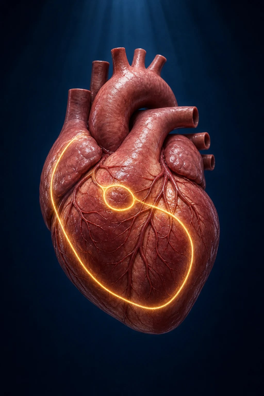
Supraventricular tachycardia (SVT) is an umbrella term for any abnormally rapid tachyarrhythmia that originates at or above the AV node (the bundle of His).[1] In practical, bedside ECG usage it refers to a **regular tachycardia with a narrow QRS complex (under 120 ms / under 3 small squares) and a rate usually between 150 and 250 bpm — explicitly excluding sinus tachycardia and atrial fibrillation (both narrow-complex but irregular, with a clear mechanism).[2]
The clinical skill in SVT is threefold and is what examiners probe: (1) recognise that a regular narrow-complex tachycardia is SVT until proven otherwise; (2) distinguish the AV-node-dependent re-entrant tachycardias (AVNRT, AVRT) — which respond to vagal manoeuvres and adenosine — from the AV-node-independent atrial tachycardias (focal atrial tachycardia, multifocal atrial tachycardia); and (3) never miss the two life-threatening mimics: pre-excited atrial fibrillation in WPW (where AV-nodal blockers kill) and ventricular tachycardia masquerading as SVT with aberrancy.[1][2]
The dominant mechanism is re-entry: a self-perpetuating circuit in which an impulse repeatedly re-enters and re-excites tissue faster than the sinus node. Re-entry requires three conditions — a circuit of two interconnected pathways, different conduction velocities and refractory periods between them, and a unidirectional block in one limb that initiates the loop. AVNRT (~60% of SVT) and AVRT (~30%) are both re-entrant and both AV-node-dependent; focal atrial tachycardia (~10%) is driven by enhanced automaticity or triggered activity in a single atrial focus and is AV-node-independent.[2]
Classification
SVT is best classified by the anatomical substrate of the circuit, because this determines the ECG, the response to manoeuvres and the long-term treatment.[1][2]
By mechanism and AV-node dependence: [1]
- AV-node-dependent (terminating with vagal manoeuvres/adenosine):
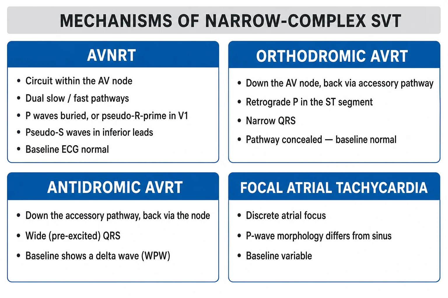
- AV nodal re-entrant tachycardia (AVNRT) — re-entry within the AV node using dual pathways. Commonest SVT (~50 to 60%). Typical (slow-fast) ~90%, atypical (fast-slow, slow-slow) ~10%.
- AV re-entrant tachycardia (AVRT) — re-entry using the AV node plus an accessory pathway connecting atrium to ventricle (the bundle of Kent). ~30% of SVT. Subdivided into orthodromic (narrow QRS, commonest) and antidromic (broad QRS, rarer, dangerous).
- AV-node-independent (atrial origin, adenosine reveals the diagnosis rather than terminating):
- Focal atrial tachycardia — a single rapidly-firing atrial focus (automaticity/triggered activity). ~10%.
- Multifocal atrial tachycardia (MAT) — three or more distinct P-wave morphologies; classically in COPD.
- Sinus node re-entrant tachycardia — re-entry within the sinus node; sudden onset/offset unlike sinus tachycardia. [1]
By accessory-pathway (pre-excitation) syndromes: [1]
- Wolff-Parkinson-White (WPW) syndrome — manifest accessory pathway (bundle of Kent): short PR (under 120 ms), delta wave (slurred upstroke), widened QRS in sinus rhythm. Syndrome = pathway plus symptomatic tachyarrhythmia. Carries a small risk of sudden cardiac death via pre-excited AF degenerating to VF.
- Concealed accessory pathway — pathway conducts only retrogradely (ventricle to atrium); no delta wave in sinus rhythm, but causes orthodromic AVRT.
- Permanent junctional reciprocating tachycardia (PJRT) — a slowly-conducting concealed accessory pathway near the AV node causing an incessant long-RP tachycardia; a classic cause of tachycardiomyopathy.
- Lown-Ganong-Levine (LGL) — short PR with normal QRS (no delta); historically attributed to a James fibre bypass tract; now regarded as a AV-nodal-conduction variant rather than a true accessory pathway. [1]
AVNRT
- Circuit WITHIN the AV node (dual slow/fast pathways)
- Commonest SVT (~60%)
- Female predominance, age 20-50
- Short RP tachycardia (RP under 70 ms)
- Pseudo R' in V1 / pseudo S in inferior leads
- Frog sign (cannon A waves) on JVP
AVRT (orthodromic)
- Anterograde via AV node, retrograde via accessory pathway
- Narrow QRS (uses normal His-Purkinje)
- WPW: delta wave + short PR in sinus rhythm
- Long RP tachycardia (RP over 70 ms)
- Concealed pathway = no delta wave
- Risk of pre-excited AF -> VF if manifest pathway
AVRT (antidromic)
- Anterograde via accessory pathway, retrograde via AV node
- BROAD-complex tachycardia
- All ventricles pre-excited
- Easily mistaken for ventricular tachycardia
- Associated with WPW; higher risk
- Treat as VT-with-aberrancy territory: avoid AV-nodal blockers
Focal atrial tachycardia
- Single rapidly-firing atrial focus (automaticity/triggered activity)
- AV-node-INDEPENDENT
- Adenosine causes AV block but the atrial focus keeps firing
- P-wave morphology localises the focus
- Can be incessant -> tachycardiomyopathy
- Beta-blocker, flecainide, ablation
MAT
- Three or more distinct P-wave morphologies
- Classically in COPD / hypoxaemia / theophylline
- Irregularly irregular - mimic of AF
- Cardioversion ineffective; treat the lungs
- Rate control: beta-blocker (cautious), verapamil
Epidemiology & Risk Factors
SVT is the commonest symptomatic tachyarrhythmia in young adults without structural heart disease. The estimated prevalence is around 2 to 2.5 per 1000, with an incidence of about 35 per 100 000 person-years; women are affected about twice as often as men.[1][2]
AVNRT is the commonest subtype, has a female predominance (around 70%), and typically presents between ages 20 and 50. AVRT / WPW is more common in younger patients and in men, and often presents in childhood, adolescence or the early twenties; WPW has a population prevalence of about 0.1 to 0.3%.[2]
Risk factors and predisposing substrates: [1]
| Factor / substrate | Association |
|---|---|
| Female sex | AVNRT (2:1 female predominance) |
| Young age, otherwise healthy | AVRT / WPW; first presentation often under 30 |
| Congenital heart disease (especially Ebstein anomaly) | Accessory pathways, WPW (Ebstein: up to 25% have WPW) |
| Hypertrophic / dilated cardiomyopathy | Accessory pathways, atrial tachycardia |
| COPD / hypoxaemia / theophylline | Multifocal atrial tachycardia (MAT) |
| Post-surgical atrial scarring (Fontan, atrial septal defect repair) | Focal atrial / incisional atrial tachycardia |
| Digitalis toxicity | Atrial tachycardia with block (classic) |
| Alcohol, caffeine, nicotine, recreational stimulants | Trigger for paroxysms; not the substrate |
| Pregnancy | Increased frequency of SVT (especially in structural disease) |
| Stress, fatigue, anxiety | Common precipitants of identifiable episodes |
| Thyrotoxicosis | Can precipitate or worsen any tachyarrhythmia |
SVT — the numbers that matter
Pathophysiology
All clinically important SVT mechanisms converge on one principle: a wave-front that does not extinguish but instead circles repeatedly, re-exciting myocardium faster than the sinus node. The two dominant substrates — dual AV-nodal pathways and an accessory pathway — both satisfy the three classical conditions for re-entry: (1) an anatomical or functional circuit of two interconnected limbs; (2) different conduction velocities and refractory periods in the two limbs; and (3) a unidirectional block in one limb that allows the wave-front to travel only one way around the loop, returning to its origin after the first limb has recovered excitability.[2]
AVNRT — dual AV-nodal physiology. In most people the AV node has a single effective conduction route. In patients with AVNRT there are functionally two pathways through or around the AV node: [1]
- The slow pathway (alpha) — slower conduction, shorter refractory period.
- The fast pathway (beta) — faster conduction, longer refractory period. [1]
In sinus rhythm the impulse travels down the fast pathway and reaches the bundle of His first. A premature atrial beat (the trigger) arrives when the fast pathway is still refractory but the slow pathway has recovered — the beat therefore conducts down the slow pathway, reaching the lower common pathway late enough that the fast pathway has now recovered. The impulse then conducts retrogradely up the fast pathway back to the atrium, and if it arrives back at the top of the slow pathway after the slow pathway has recovered, the circuit self-perpetuates: this is typical (slow-fast) AVNRT, accounting for ~90% of cases. The atria and ventricles are activated almost simultaneously (the circuit is tiny), so retrograde P waves are buried in or just after the QRS — producing the classic pseudo R-prime in V1 and pseudo S in the inferior leads and the RP interval of under 70 ms.[2] In the rarer atypical forms the circuit runs in reverse: fast-slow (long RP with inverted P waves) and slow-slow.
AVRT — the accessory pathway. An accessory pathway (bundle of Kent) is a congenital muscular bridge connecting atrial myocardium directly to ventricular myocardium, bypassing the AV node. Because accessory-pathway tissue conducts faster than the AV node (and, unlike the AV node, conducts faster as heart rate rises), in sinus rhythm the impulse reaches the ventricle partly through the Kent bundle, pre-exciting a portion of the ventricle — this produces the delta wave (a slurred initial QRS upstroke), a short PR interval (under 120 ms), and a widened QRS (fusion of pre-excited and normally-conducted activation). This is Wolff-Parkinson-White pattern. WPW syndrome = the pattern plus symptomatic tachyarrhythmia. [1]
In orthodromic AVRT (commonest, narrow QRS) the re-entrant circuit runs anterogradely down the AV node (normal, narrow QRS activation) and retrogradely up the accessory pathway back to the atrium — so the QRS is narrow and the RP interval is longer (over 70 ms). In antidromic AVRT (rare, broad QRS) the impulse travels anterogradely down the accessory pathway (the whole ventricle is pre-excited, giving a broad QRS that mimics VT) and retrogradely up the AV node.[2]
The WPW danger — pre-excited atrial fibrillation. The real lethality of WPW is not the orthodromic circuit but atrial fibrillation conducting down the accessory pathway. Unlike the AV node (which slows and blocks at high rates), accessory-pathway tissue conducts very fast and without decrement, so AF impulses can bombard the ventricles at 250 to 300 bpm with broad QRS complexes, generating an irregular broad-complex tachycardia that can degenerate into ventricular fibrillation and sudden death. Any AV-nodal-blocking drug — adenosine, verapamil, diltiazem, beta-blockers, digoxin — is dangerous here: by blocking the AV node they force even more conduction down the accessory pathway, accelerating the ventricular rate and precipitating VF.[1][2]
Focal atrial tachycardia arises from enhanced automaticity or triggered activity (delayed after-depolarisations) in a single atrial focus, firing faster than the sinus node. It is AV-node-independent: the focus continues to fire regardless of what the AV node does, so adenosine produces transient AV block but does not terminate the atrial tachycardia — a diagnostic clue. Incessant focal atrial tachycardia is a leading cause of tachycardiomyopathy. Multifocal atrial tachycardia (MAT) is driven by multiple competing atrial foci, classically in the hypoxic, theophylline-treated COPD patient, and is recognised by three or more distinct P-wave morphologies in one lead with irregular RR intervals. [1]
The polyuria pearl. During SVT the atria contract against closed AV valves (the atrial and ventricular cycles nearly coincide), generating raised atrial pressure and atrial stretch. This releases atrial natriuretic peptide (ANP), producing the polyuria that classically follows termination of an episode — a high-yield exam clue to SVT (and absent in sinus tachycardia).[2]
Clinical Presentation
The hallmark of paroxysmal SVT is palpitations of abrupt onset and abrupt offset — patients often remember the exact second the episode began and the exact second it stopped (often with urination, a vagal trigger, or after a manoeuvre). Episodes last minutes to hours and recur unpredictably over years.[1]
Typical symptoms: [1]
- Palpitations — rapid, regular, "fluttering" or "pounding"; the patient may feel it in the neck.
- Lightheadedness, presyncope, frank syncope — from reduced cerebral perfusion; syncope suggests haemodynamic compromise or, in WPW, is a warning sign for sudden-death risk.
- Dyspnoea / air hunger.
- Chest tightness or chest pain — common even with normal coronaries (demand ischaemia from rapid rate); ischaemic-type pain mandates a lower threshold to cardiovert.
- Anxiety, sense of impending doom.
- Neck pulsations — the frog sign (prominent regular cannon A waves) visible in the neck when atria contract against closed AV valves; highly suggestive of AVNRT.
- Polyuria after termination — ANP release from atrial stretch; a classic discriminator from sinus tachycardia. [1]
During the episode the examination findings are: [1]
- A rapid, regular pulse at 150 to 250 bpm.
- A normal or low blood pressure; hypotension indicates instability.
- Jugular venous pulsations — in AVNRT, regular, prominent "cannon" A waves (the frog sign) as the atria beat against closed tricuspid valve.
- Heart sounds may be normal; the first heart sound can vary in intensity. [1]
Atypical presentations (must-know for examiners): [1]
- Elderly — may present with syncope, falls, dyspnoea, or exacerbation of heart failure rather than typical palpitations; rates can be higher and tolerance lower. Atrial tachycardia and AF are proportionally more common in older patients; AVNRT less so.
- Pregnancy — SVT frequency increases; episodes are poorly tolerated (higher circulating volume, cardiovascular stress); the safe drugs differ (see Special Populations).
- Children / infants — present with poor feeding, irritability, pallor, tachypnoea, and in infants heart failure if the tachycardia is incessant (think PJRT or concealed accessory pathway).
- Incessant SVT (focal atrial tachycardia, PJRT) — the patient may be unaware of "paroxysms"; presents insidiously with new heart failure or a reduced ejection fraction = tachycardiomyopathy, which is reversible with rate control or ablation.
- WPW with AF — irregular palpitations, pre-syncope, or collapse; the ECG shows an irregular broad-complex tachycardia — a medical emergency. [1]
Differential Diagnosis
A regular narrow-complex tachycardia narrows the differential, but several conditions must be distinguished because treatment diverges sharply.[1][2]
| Diagnosis | Distinguishing features |
|---|---|
| Sinus tachycardia | Rate usually under 150 bpm; gradual warm-up and warm-down (not abrupt); a normal P wave precedes every QRS with a constant PR; rate varies with autonomic tone / posture / volume status; an identifiable cause (fever, pain, hypovolaemia, anxiety, thyrotoxicosis, anaemia, pulmonary embolism). Vagal manoeuvres slow the rate transiently without terminating it. |
| Atrial fibrillation | Irregularly irregular pulse and RR intervals; no discrete P waves; adenosine produces AV block but does not terminate the arrhythmia. |
| Atrial flutter (common, 2:1) | Regular, typically 150 bpm with sawtooth flutter (F) waves best in II, III, aVF and V1; carotid sinus pressure / adenosine unmasks the flutter waves by slowing AV conduction. 1:1 conduction (300 bpm) is rare and dangerous. |
| Focal atrial tachycardia | A discrete abnormal P wave precedes each QRS; AV-node-independent (adenosine induces AV block but the atrial tachycardia persists); often long RP; can be incessant -> tachycardiomyopathy. |
| Multifocal atrial tachycardia (MAT) | Three or more P-wave morphologies, irregular RR, baseline chaos; classically COPD; do not cardiovert. |
| Junctional tachycardia / AV junctional ectopic tachycardia | Retrograde P waves buried in or just after QRS; usually seen post-cardiac surgery or in digoxin toxicity; lacks the abrupt on/off of AVNRT. |
| Ventricular tachycardia (broad-complex) | QRS over 120 ms, AV dissociation, capture/fusion beats, concordance; always treat a broad-complex tachycardia as VT until proven otherwise. AVRT-antidromic and pre-excited AF can mimic VT. |
| Inappropriate sinus tachycardia / POTS | Persistent sinus rate over 100 at rest without cause; differs structurally but confused clinically. |
The three "do-not-miss" mimics in any SVT assessment: [1]
- Sinus tachycardia from a serious cause (sepsis, hypovolaemia, pulmonary embolism, thyrotoxicosis) — giving adenosine is harmless but useless; find and treat the cause.
- Atrial flutter with 2:1 block at 150 bpm — adenosine transiently slows AV conduction and reveals the sawtooth waves, confirming the diagnosis; definitive treatment is rate control, rhythm control, anticoagulation as indicated, and often isthmus ablation.
- Pre-excited AF / antidromic AVRT (WPW) — irregular or broad-complex tachycardia where AV-nodal blockers are lethal; use flecainide or amiodarone or synchronised DC cardioversion. [1]
Clinical & Bedside Assessment
The bedside management of a stable SVT follows a fixed ladder. ABCDE first: secure the airway, give high-flow oxygen if hypoxic, attach monitoring, set up IV access, and take bloods.[1]
Assess stability — the single decision that branches management: [1]
- Unstable (any of): hypotension / shock, syncope or reduced GCS, ischaemic chest pain, acute heart failure, or very rapid rate compromising perfusion -> synchronised DC cardioversion (do not delay for adenosine).
- Stable: proceed to vagal manoeuvres then adenosine. [1]
Vagal manoeuvres increase vagal (parasympathetic) tone, transiently slowing AV-nodal conduction and breaking AVNRT and AVRT circuits: [1]
- Modified Valsalva manoeuvre (first-line, per the REVERT trial[3]): the patient is positioned semi-recumbent at 45 degrees, blows into a 10 mL syringe to generate about 40 mmHg pressure for 15 seconds (forced expiration against a closed glottis), and is then immediately laid flat with the legs passively raised to 45 degrees for 15 seconds. The postural modification nearly tripled the termination rate (43% vs 17%) in the REVERT trial.[3][4]
- Carotid sinus massage: with the patient supine, apply firm pressure in a circular motion for 5 to 10 seconds to the carotid bifurication (one side at a time). First auscultate for carotid bruits and avoid in patients with prior stroke/TIA or known carotid disease; have resuscitation equipment available. Rarely causes embolic stroke in the elderly.
- Cold-water face immersion / cold stimulus to the face (diving reflex) — particularly useful in children.
The frog sign — prominent regular cannon A waves in the jugular venous pulse — is a bedside clue highly suggestive of AVNRT (atria contracting against closed AV valves).[2]
Record a 12-lead ECG during any manoeuvre — vagal manoeuvres and adenosine may transiently slow AV conduction and unmask atrial activity (flutter waves, P-wave morphology), establishing the diagnosis even if they do not terminate the arrhythmia. [1]
Investigations
First-line (during the tachycardia): [1]
- 12-lead ECG — the single most important test. Assess rate, regularity, QRS width (under or over 120 ms), P-wave position and morphology, and the RP interval.[1]
- Continuous cardiac monitoring and a rhythm strip during adenosine.
- IV access and a 12-lead ECG printed before, during, and after termination.
- Bloods: U&E (potassium, magnesium), FBC, digoxin level if relevant, TSH to exclude thyrotoxicosis, and a troponin if there is chest pain or a prolonged episode (it will be demand-positive; do not attribute SVT to an ACS unless ST changes persist).
ECG analysis of a regular narrow-complex tachycardia — the RP interval method (RP interval = from onset of QRS to the next P wave):[2]
- Short RP tachycardia (RP under 70 ms): the P wave is buried in or just after the QRS.
- AVNRT (commonest) — look for pseudo R-prime in V1 and pseudo S in inferior leads (II, III, aVF); these disappear in sinus rhythm.
- Orthodromic AVRT with a rapidly-conducting retrograde pathway (occasionally short RP).
- Junctional tachycardia.
- Long RP tachycardia (RP over 70 ms): the P wave is clearly separated from the preceding QRS.
- Atypical AVNRT (fast-slow) — inverted P waves, long RP.
- Orthodromic AVRT / PJRT — the classic long-RP tachycardia; PJRT is incessant.
- Focal atrial tachycardia.
- Sinus node re-entrant tachycardia. [1]
ECG in sinus rhythm (after termination) — look for the substrate: [1]
- Wolff-Parkinson-White pattern: short PR (under 120 ms), delta wave (slurred upstroke of the QRS), broadened QRS, and repolarisation abnormalities (T-wave changes opposite to the delta-wave direction). Localise the accessory pathway by the delta-wave vector (e.g. left-sided pathways have a positive delta in V1; right-sided, negative).
- Concealed pathway — sinus-rhythm ECG is normal (pathway conducts only retrogradely); diagnosis is made at electrophysiology study. [1]
Advanced / confirmatory investigations: [1]
- Ambulatory ECG / event monitor / implantable loop recorder — to capture infrequent paroxysms and correlate symptoms with rhythm.
- Echocardiogram — to exclude structural heart disease (Ebstein anomaly, hypertrophic cardiomyopathy) and assess ventricular function (tachycardiomyopathy in incessant SVT).
- Electrophysiological study (EPS) — the gold standard: it maps the circuit, localises an accessory pathway, confirms dual AV-nodal physiology, and is performed immediately before catheter ablation in the same procedure.[2]
Severity / risk in WPW — interventional EPS: asymptomatic patients with a manifest WPW pattern on ECG may warrant risk-stratification EPS (shortest pre-excited RR interval in AF under 250 ms = high risk of sudden death), particularly in high-risk professions (pilots, competitive athletes).[1][2]
Management — Resuscitation
ABCDE first. Give high-flow oxygen only if hypoxic. Establish IV access, attach continuous cardiac monitoring and a defibrillator, and ensure resuscitation equipment (including atropine and a temporary pacemaker) is at the bedside before giving adenosine.[1][1]
The stability decision — unstable SVT needs electricity, not drugs: [1]
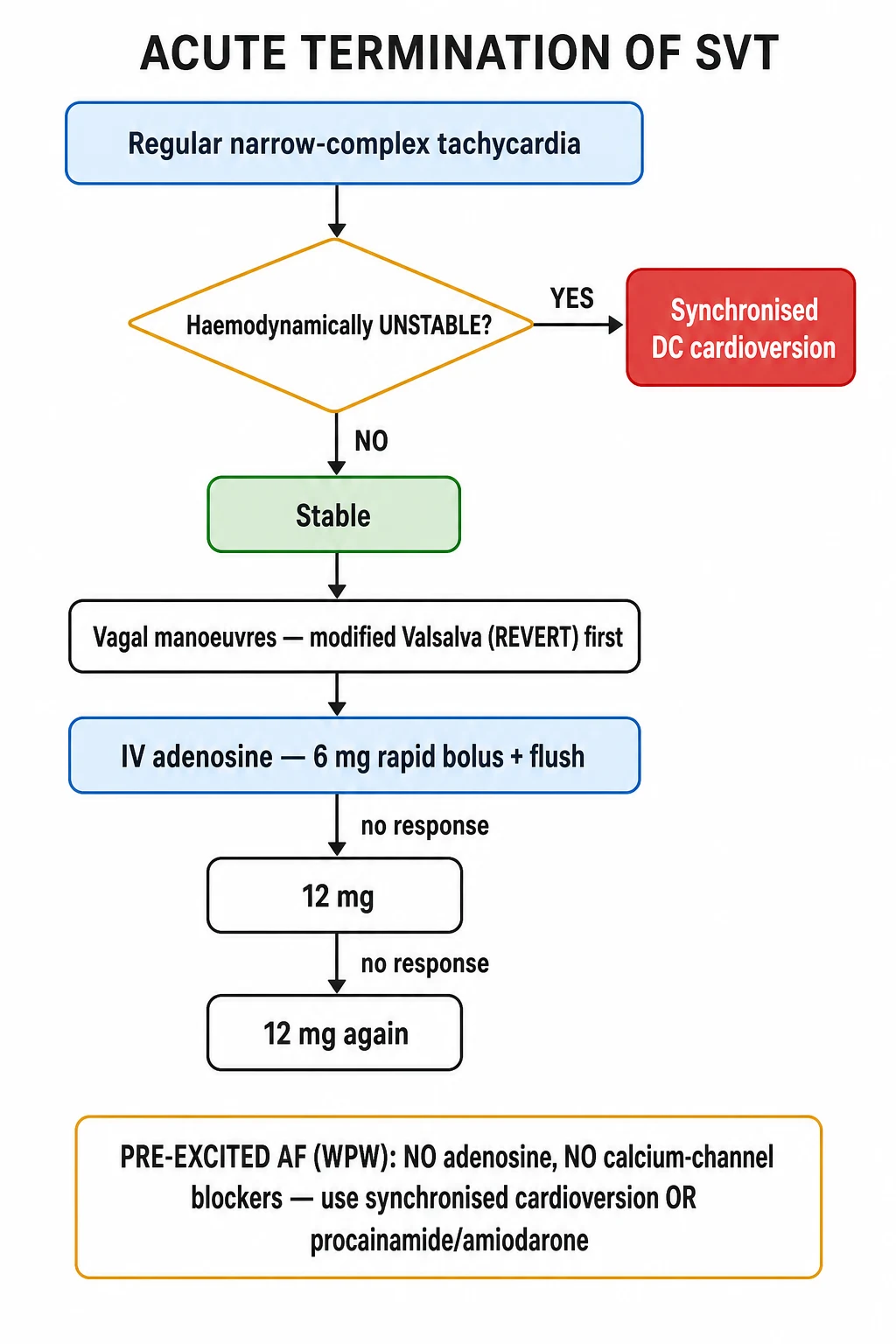
Synchronised DC cardioversion is first-line if the patient is haemodynamically unstable (any of: systolic BP under 90 mmHg / shock, syncope or reduced consciousness, ischaemic chest pain, acute heart failure, or signs of end-organ hypoperfusion). [1]
- Synchronised shock (sync to the R wave to avoid delivering energy on the T wave, which would cause VF).
- Start at 100 J biphasic (or 200 J monophasic); increase to 150 J, then 200 J as needed.
- Sedate the conscious patient first (e.g. midazolam 2.5 to 5 mg IV plus fentanyl 50 to 100 mcg IV, with anaesthetic support where available).
- Pre-cardioversion: give a brief run of IV adenosine if there is time and the patient is not crashing — it may terminate the rhythm and avoid a shock; but do not delay cardioversion in the genuinely unstable patient.[1]
For stable SVT, proceed directly to the vagal-manoeeuvre then adenosine ladder (below). If broad-complex or pre-excited AF, see Specific Subtypes — the ladder differs. [1]
Management — Definitive & Stepwise
The stable narrow-complex SVT ladder is fixed and examinable verbatim:[1][2][1]
Step 1 — Vagal manoeuvres (modified Valsalva first-line, or carotid sinus massage if bruits excluded).
- Modified Valsalva: 45 degrees semi-recumbent, 15-second forced strain to ~40 mmHg, then supine with legs raised to 45 degrees for 15 seconds (REVERT protocol).[3]
- Terminate if sinus rhythm is restored; record a 12-lead ECG.
Step 2 — Adenosine (if vagal manoeuvres fail).[1]
- Adenosine 6 mg as a rapid IV bolus through a large cannula, immediately followed by a 10 to 20 mL normal-saline flush, with the arm raised. Continuous ECG monitoring.
- If no response within 1 to 2 minutes, adenosine 12 mg rapid IV bolus (with flush).
- If still no response, a further adenosine 12 mg rapid IV bolus.
- Warn the patient before each dose: expect a flushing, a transient sense of doom, chest tightness, and brief dyspnoea — and possibly a few seconds of asystole — followed by return of rhythm. The effect lasts only 10 to 15 seconds because adenosine is taken up and deaminated within seconds.
- Mechanism: adenosine binds the A1 receptor, opening acetylcholine-sensitive potassium channels in the AV node, hyperpolarising AV-nodal cells and producing transient AV-nodal block — breaking the AVNRT/AVRT circuit.
- Contraindications: asthma and severe COPD (bronchospasm via A2B receptors); second- or third-degree AV block or sick sinus syndrome without a pacemaker; heart-transplant patients (denervated, hypersensitive — reduce dose to 3 mg or use verapamil).
- Drug interactions: dipyridamole potently inhibits adenosine uptake — reduce the adenosine dose to one-quarter (1.5 mg); theophylline / aminophylline / caffeine are competitive antagonists at the A1 receptor (expect a higher adenosine dose or failure); carbamazepine may potentiate adenosine-induced heart block. [1]
Step 3 — If adenosine is contraindicated or fails — a second-line agent: [1]
- IV verapamil 5 mg over 2 minutes (or 2.5 to 5 mg in the elderly); repeat 5 mg after 10 minutes if needed, maximum 15 mg. Never give IV verapamil to a patient already on a beta-blocker and never in WPW with pre-excited AF — risk of asystole/VF.
- IV beta-blocker: metoprolol 5 mg IV over 2 minutes (repeat up to 3 doses), or esmolol (a short-acting cardioselective agent, 500 mcg/kg loading over 1 minute then 50 mcg/kg/min) when titration is needed.
- IV diltiazem is an alternative calcium-channel blocker. [1]
Step 4 — If pharmacological cardioversion fails and the patient is stable — seek expert cardiology input, consider IV amiodarone 300 mg over 20 to 60 minutes (especially in heart failure or structural disease), or proceed to synchronised DC cardioversion under sedation. [1]
ADENOSINE — the safe-use checklist
ADENOSINE
Asthma/severe COPD - do NOT give (bronchospasm)
Dipyridamole blocks uptake - cut the dose to 1.5 mg
Continuous ECG recording during every dose
Warn about flushing, doom, brief asystole, dyspnoea
Brief dyspnoea/apnoea is expected and transient
Rapid 6/12/12 mg bolus followed by saline flush + arm raise
Theophylline antagonises; carbamazepine potentiates block
Never give AV-nodal blockers in pre-excited AF
Have verapamil/beta-blocker and DC cardioversion ready
Long-term management — definitive therapy:[1][2]
- Catheter ablation is curative and first-line for recurrent SVT. Radiofrequency (or cryoablation) energy destroys the critical limb of the circuit: slow-pathway ablation for AVNRT (success about 95%, small risk of AV block), accessory-pathway ablation for AVRT/WPW (success about 90 to 95%). Complications in under 1% (AV block requiring pacemaker, pericardial effusion, vascular access, phrenic-nerve injury). Ablation is particularly recommended in WPW to eliminate sudden-death risk.
- Pharmacological prophylaxis is reserved for patients who decline or fail ablation, or while awaiting it:
- Beta-blockers (e.g. metoprolol 25 to 100 mg twice daily, bisoprolol 2.5 to 10 mg daily) or verapamil 40 to 120 mg three times daily — first-line for infrequent, tolerable episodes.
- Flecainide 50 to 100 mg twice daily or propafenone 150 to 300 mg three times daily — for structurally normal hearts only (contraindicated in ischaemic or structural heart disease).
- Sotalol 40 to 160 mg twice daily (with QT monitoring).
- Amiodarone — reserved for refractory disease or structural heart disease; avoid long-term if possible (thyroid, pulmonary, hepatic, photosensitivity toxicity).
- Avoidance of triggers: caffeine, alcohol, nicotine, recreational stimulants; manage stress, anxiety, and sleep.
- The "pill-in-the-pocket" approach (single-dose oral therapy taken at the start of an episode, e.g. oral flecainide 3 mg/kg or diltiazem plus a beta-blocker) is an option for infrequent, haemodynamically tolerated episodes in patients with no structural heart disease. [1]
Specific Subtypes & Scenarios
- Typical AVNRT (slow-fast) — commonest SVT. Short RP, pseudo R-prime in V1, pseudo S inferiorly, frog sign. Vagal manoeuvres and adenosine terminate reliably. Definitive: slow-pathway catheter ablation (success ~95%).
- Orthodromic AVRT — narrow QRS, long RP, uses AV node anterogradely. May be due to a manifest (WPW) or concealed pathway. Treatment identical to AVNRT acutely; ablation of the accessory pathway is curative.
- Antidromic AVRT — broad QRS (all ventricles pre-excited); easily confused with ventricular tachycardia. Treat with caution — procainamide, flecainide, amiodarone, or DC cardioversion; avoid AV-nodal blockers. Ablation is definitive.
- Wolff-Parkinson-White syndrome with atrial fibrillation (pre-excited AF) — medical emergency. The ECG shows a rapid, irregular, broad-complex tachycardia with varying QRS morphology (varying degrees of pre-excitation) at rates 250 to 300 bpm. Never give adenosine, verapamil, diltiazem, beta-blockers, or digoxin — all force conduction down the accessory pathway and can precipitate VF. Use IV flecainide 2 mg/kg over 10 minutes (if no structural heart disease), IV amiodarone 300 mg, or synchronised DC cardioversion if unstable. Long-term: accessory-pathway ablation to eliminate sudden-death risk.[1][2]
- Focal atrial tachycardia — discrete abnormal P waves; AV-node-independent (adenosine causes AV block but the atrial focus keeps firing — diagnostic). Acute: beta-blocker or verapamil. Long-term: beta-blocker, flecainide, or catheter ablation of the focus.
- Multifocal atrial tachycardia (MAT) — three or more P-wave morphologies, irregular RR, in COPD/hypoxaemia. Cardioversion is ineffective. Treat the underlying lung disease, correct hypoxaemia and electrolytes; rate-control with a beta-blocker (cautiously, given COPD) or verapamil; stop theophylline.
- Permanent junctional reciprocating tachycardia (PJRT) — a concealed, slowly-conducting accessory pathway near the AV node causing an incessant long-RP tachycardia, often presenting in children/young adults with tachycardiomyopathy. Diagnosis: long-RP tachycardia with deeply inverted P waves (inferior/lateral). Treatment: catheter ablation of the pathway, after which the cardiomyopathy reverses.
- Sinus node re-entrant tachycardia — sudden onset/offset unlike sinus tachycardia; terminates with vagal manoeuvres/adenosine. Often in structural heart disease.
- SVT with aberrancy vs VT — any broad-complex tachycardia (QRS over 120 ms) in a patient with structural heart disease is VT until proven otherwise. Do not give verapamil (haemodynamic collapse). If uncertain, treat as VT (amiodarone or DC cardioversion).[2]
- Digitalis-toxic atrial tachycardia with block — a classic board/exam scenario; stop digoxin, correct potassium, give Digoxin-Fab antibody fragments if life-threatening; avoid cardioversion (can precipitate VF).
Complications & Pitfalls
Complications: [1]
- Syncope and falls — from transient cerebral hypoperfusion during episodes.
- Tachycardiomyopathy — reversible ventricular dysfunction from incessant SVT (focal atrial tachycardia, PJRT); resolves with rate control or ablation. The key teaching: always look for an incessant tachycardia in unexplained heart failure.[2]
- Sudden cardiac death — rare; occurs in WPW when pre-excited AF degenerates to VF. This is the rationale for offering ablation to symptomatic and selected high-risk asymptomatic WPW patients.
- Thromboembolism — rare in pure re-entrant SVT (atria contract), but rises if SVT degenerates to AF (anticoagulate by CHA2DS2-VASc).
- Demand ischaemia / infarction — a rapid SVT in coronary disease can precipitate type-2 MI; do not reflexively cath-lab a patient whose ST changes resolve with termination.
- Psychological — anxiety, avoidance behaviour, and impaired quality of life from unpredictable episodes.
Classic pitfalls (examiner favourites): [1]
- Treating a broad-complex tachycardia as "SVT with aberrancy" and giving verapamil — in VT this causes haemodynamic collapse. Broad-complex = treat as VT unless you can prove otherwise.
- Giving adenosine to an asthmatic — can precipitate severe bronchospasm.
- Giving AV-nodal blockers in pre-excited AF (WPW) — accelerates the accessory pathway and precipitates VF.
- Forgetting the adenosine saline flush / arm raise — adenosine is metabolised in seconds; without a rapid flush it never reaches the heart in effective concentration, and is wrongly declared "ineffective."
- Misdiagnosing atrial flutter (2:1, 150 bpm) as SVT — adenosine will unmask the sawtooth waves; definitive treatment differs.
- Missing WPW on the sinus-rhythm ECG after termination — always look for the delta wave and short PR, which dictate long-term strategy.
- Ignoring incessant tachycardia in a patient with new heart failure.
- Not reducing the adenosine dose in a patient on dipyridamole (profound asystole risk). [1]
Prognosis & Disposition
SVT is, in the great majority of cases, a benign arrhythmia with an excellent prognosis. Most patients are otherwise structurally normal; quality-of-life impairment (rather than mortality) drives treatment decisions.[1][2]
Disposition after an acute episode: [1]
- Terminated, stable, no red flags — can be discharged with ambulatory ECG monitoring, a clear safety-net, an outpatient ECG to look for WPW, and a cardiology referral to consider ablation.
- WPW pattern found — refer for cardiology / EPS and ablation (curative, eliminates sudden-death risk).
- Incessant SVT / tachycardiomyopathy — admit for rhythm/rate control and urgent ablation planning.
- Pre-excited AF / haemodynamic instability / failed cardioversion — admit (CCU/HDU), cardiology input.
- Inappropriate sinus tachycardia of uncertain cause — investigate and treat the underlying cause before discharge. [1]
Prognostic points: catheter ablation offers a >90 to 95% cure; recurrence after successful ablation is low (under 5 to 10%). Pharmacological therapy controls but does not cure. WPW sudden-death risk is low (~0.1 to 0.4% per year in symptomatic patients) but eliminated by accessory-pathway ablation, which is why symptomatic WPW is ablated. Asymptomatic WPW in low-risk individuals (especially if the pathway loses pre-excitation on exercise/EPS) can be observed.[1]
Special Populations
- Pregnancy — SVT frequency rises and episodes are less well tolerated. Adenosine is safe in pregnancy (short half-life, does not cross the placenta in effective amounts). DC cardioversion is safe. Beta-blockers: metoprolol is preferred (pregnancy category B; limited placental transfer); avoid atenolol (fetal growth restriction) and avoid amiodarone (fetal thyroid goitre/hypothyroidism) unless life-threatening. Verapamil is acceptable but second-line. Flecainide is used for fetal SVT and is acceptable in structurally normal maternal hearts. Catheter ablation is best deferred to after the first trimester (ideally postpartum) but can be done with lead-shielding and zero-fluoroscopy techniques if essential.[2]
- Children — present with poor feeding, irritability, pallor, tachypnoea; infants may be in heart failure from an incessant tachycardia (PJRT, concealed accessory pathway). Adenosine 0.1 mg/kg rapid IV (then 0.2 mg/kg, max single dose 12 mg). Vagal manoeuvres (ice to the face — diving reflex). Avoid verapamil in infants under 1 year (can cause refractory hypotension/asystole). Definitive: catheter ablation (typically deferred until age 4 to 5 unless refractory, due to smaller vascular access and AV-node proximity).[2]
- Elderly — atypical presentations (syncope, falls, dyspnoea, heart failure); atrial tachycardia and AF proportionally more common. Auscultate for carotid bruits before carotid sinus massage (embolic risk); consider modified Valsalva first. Lower starting doses of adenosine/verapamil; beware polypharmacy (digoxin toxicity -> atrial tachycardia with block).
- Asthma / severe COPD — adenosine contraindicated; use verapamil or a beta-blocker (cardioselective, with caution in COPD); modified Valsalva is safe.
- Heart-transplant patients — denervated hearts are exquisitely sensitive to adenosine (prolonged asystole); reduce the dose to 3 mg or use verapamil.
- Patients on dipyridamole or theophylline — see drug-interaction rules above (reduce or increase adenosine dose respectively).
- Structural / congenital heart disease (Ebstein anomaly, HCM, post-atrial-repair congenital) — higher incidence of accessory pathways and atrial tachycardia; manage with cardiology input; avoid flecainide in structural disease; ablation is preferred and often complex.
Evidence, Guidelines & Regional Differences
Landmark trials and guidance: [1]
- REVERT trial (Appelboam et al., Lancet 2015)[3] — the modified Valsalva (semi-recumbent strain + leg-lift) terminated SVT in 43% vs 17% with the standard Valsalva — it is now first-line across UK and ESC guidance. REVERT-2 (Chen et al., 2020)[4] confirmed and refined the technique.
- 2015 ACC/AHA/HRS Guideline for SVT (Page et al.)[1] — the US reference standard: vagal manoeuvres then adenosine; verapamil/beta-blocker as alternatives; catheter ablation first-line for recurrent disease and symptomatic WPW.
- 2019 ESC Guidelines for SVT (Brugada et al.)[2] — the European reference: algorithmic approach by QRS width and regularity; ablation preferred; risk-stratification EPS for asymptomatic WPW in selected patients.
Regional / examination deltas: [1]
- US (ACC/AHA/HRS 2015)[1] — modified Valsalva, adenosine, ablation; asymptomatic WPW: risk-stratification EPS considered, especially in high-risk professions.
- UK / Europe (ESC 2019, Resus Council UK)[2][1] — modified Valsalva promoted to first-line; ablation commissioned first-line; asymptomatic WPW managed conservatively unless high-risk.
- India (NMC / AIIMS practice) — adenosine availability and cardiac monitoring are universal in tertiary centres; catheter ablation available in major centres; for unstable SVT synchronised DC cardioversion is standard. In resource-limited settings carotid sinus massage and modified Valsalva are emphasised as zero-cost first steps.
- Controversies: (1) management of asymptomatic WPW — risk-stratification EPS vs conservative observation; (2) flecainide vs ablation as first long-term strategy in structurally normal hearts; (3) the safety of adenosine in mild/moderate COPD (recent data suggest it is safer than historically taught, but severe COPD and asthma remain contraindications).
Exam Pearls
- Regular, narrow-complex tachycardia 150 to 250 bpm = SVT (exclude sinus tachycardia and AF first).
- Abrupt onset and abrupt offset + polyuria after termination (ANP release from atrial stretch) = the classic SVT story.
- Frog sign (regular cannon A waves in the JVP) = AVNRT.
- Short RP (under 70 ms) = AVNRT (look for pseudo R-prime in V1, pseudo S inferiorly); long RP (over 70 ms) = orthodromic AVRT, PJRT, focal atrial tachycardia.
- Modified Valsalva first (REVERT: 45 degrees strain + leg-lift) — then adenosine 6 -> 12 -> 12 mg rapid IV bolus with saline flush and arm raise.
- Adenosine: warn about flushing, brief asystole, chest tightness, dyspnoea; contraindicated in asthma/severe COPD and heart-transplant patients (give 3 mg); reduce to 1.5 mg if on dipyridamole; theophylline antagonises (need higher dose).
- Verapamil 5 mg IV is the alternative if adenosine is contraindicated — but never in WPW-AF and never with a beta-blocker.
- Unstable SVT = synchronised DC cardioversion (100 J biphasic), not adenosine.
- WPW with pre-excited AF is an emergency: irregular broad-complex tachycardia, never give adenosine/verapamil/beta-blockers/digoxin — use flecainide/amiodarone or DC cardioversion.
- Broad-complex tachycardia = treat as VT until proven otherwise.
- WPW on sinus-rhythm ECG: short PR, delta wave, broad QRS; ablate to prevent sudden death.
- Incessant tachycardia + new heart failure = think tachycardiomyopathy (focal atrial tachycardia, PJRT); ablate.
- AVNRT is the commonest SVT (~60%); female predominance.
- PJRT = concealed slowly-conducting accessory pathway, long RP, incessant, tachycardiomyopathy.
- MAT = three or more P-wave morphologies, irregular, in COPD; treat the lungs, do not cardiovert.
- Catheter ablation is curative and first-line for recurrent SVT (success over 90 to 95%). [1]
Exam application bank (NEET-PG / INICET)
One-line answer
Supraventricular tachycardia (SVT) is a rapid, usually regular tachyarrhythmia originating at or above the atrioventricular (AV) node with a narrow QRS (under 120 ms) and a rate usually 150 to 250 bpm. The two re-entrant mechanisms dominate: AV nodal re-entrant tachycardia (AVNRT) — a circuit within the AV node using dual slow-fast pathways (commonest, ~60%) — and AV re-entrant tachycardia (AVRT) — a circuit using an accessory pathway bypassing the AV node, classically Wolff-Parkinson-White (WPW). Presents with paroxysmal palpitations of abrupt onset/offset, dyspnoea, lightheadedness and, characteristically, polyuria after termination. Management: if haemodynamically unstable — synchronised DC cardioversion; if stable — modified Valsalva manoeuvre, then IV adenosine 6 mg rapid bolus escalating to 12 mg then 12 mg. IV verapamil or a beta-blocker if adenosine is contraindicated. Catheter a [1]
Worked stems (answer without another resource)
Stem 1 — Classic presentation. Map symptoms to mechanism; name the first investigation and first treatment step with dose/route if drug therapy is standard. [1]
Stem 2 — Unstable / complicated. List red flags that force immediate resuscitation, theatre, ICU, antidote, or reperfusion — and what you do in the first 15 minutes. [1]
Stem 3 — Atypical group. Elderly, pregnancy, child, or immunocompromised: how presentation and thresholds change. [1]
Stem 4 — Differential trap. Name the three closest mimics and one discriminator for each. [1]
Stem 5 — Disposition. Who goes home with safety-netting, who is admitted, who needs HDU/ICU/theatre, and what follow-up is mandatory. [1]
Rapid viva checklist
- Definition + classification
- Pathophysiology chain
- Bedside signs / criteria
- Score with exact components (if any)
- Emergency bundle
- Definitive therapy with doses
- Complications of disease and of treatment
- Special populations
- Guideline/trial name if classic
- Three exam traps
Coverage self-check
If you cannot answer any stem above from this page alone, re-read the matching section — the page is intended to be self-sufficient for final-prof and NEET-PG/INICET questions on Supraventricular Tachycardia.
References
- [1]Page RL, Joglar JA, Caldwell MA, et al. 2015 ACC/AHA/HRS Guideline for the Management of Adult Patients With Supraventricular Tachycardia: A Report of the American College of Cardiology/American Heart Association Task Force on Clinical Practice Guidelines and the Heart Rhythm Society Circulation, 2016.PMID 26399663
- [2]Brugada J, Katritsis DG, Arbelo E, et al. 2019 ESC Guidelines for the management of patients with supraventricular tachycardiaThe Task Force for the management of patients with supraventricular tachycardia of the European Society of Cardiology (ESC) Eur Heart J, 2020.PMID 31504425
- [3]Appelboam A, Reuben A, Mann C, et al. Postural modification to the standard Valsalva manoeuvre for emergency treatment of supraventricular tachycardias (REVERT): a randomised controlled trial Lancet, 2015.PMID 26314489
- [4]Chen C, Chen Y, Bai J, et al. A multicenter randomized controlled trial of a modified Valsalva maneuver for cardioversion of supraventricular tachycardias Am J Emerg Med, 2020.PMID 31422858