Cardiology · Cardiology
Syncope
Also known as Fainting · Blackout · Transient loss of consciousness · Vasovagal syncope · Neurocardiogenic syncope · Carotid sinus syndrome
Syncope is a transient loss of consciousness (TLoC) due to transient global cerebral hypoperfusion, characterised by rapid onset, short duration, and spontaneous complete recovery without electrical brain injury. It must be separated from seizures and psychogenic collapse. The three mechanism-based categories are reflex (neurally-mediated, commonest in the young), orthostatic hypotension (drugs, volume depletion, autonomic failure), and cardiac (arrhythmic or structural — highest mortality). The initial evaluation — a focused history, eyewitness account, examination and a 12-lead ECG in every patient — yields a diagnosis in the majority. Reproduce the San Francisco Syncope Rule (CHEAT) and the Canadian Syncope Risk Score to risk-stratify; admit the high-risk group, manage vasovagal with lifestyle measures and countermanoeuvres, treat OH with volume/salt expansion and midodrine/fludrocortisone, and treat cardiac syncope by correcting the underlying arrhythmia or obstruction. Red flags: exertional, supine or unprovoked syncope, palpitations, abnormal ECG, structural heart disease, family history of sudden cardiac death (SCD) under 40.
On this page & tools
Your progress
Saved locally on this device.
Practise this topic
Exam tags
Red flags

Overview & Definition
Syncope is a symptom, defined as a transient loss of consciousness (TLoC) due to transient global cerebral hypoperfusion, characterised by rapid onset, short duration, and spontaneous complete recovery without the need for electrical or chemical cardioversion/cerebral resuscitation.[1] The definition contains four required features:[1]
- Sudden onset (seconds).
- Short duration (typically under a minute; complete recovery within minutes).
- Spontaneous, rapid, complete recovery — the patient returns to baseline mental status without postictal confusion.
- Loss of postural tone — the patient slumps or falls. [1]
The defining mechanism — transient global cerebral hypoperfusion — separates syncope from the other causes of TLoC (seizures, which are electrical cortical disturbance; and psychogenic pseudosyncope, which has no loss of consciousness). The clinical skill in syncope is not the recognition that an episode occurred but the mechanistic diagnosis: distinguishing the benign reflex and orthostatic causes (the majority) from the dangerous cardiac causes (the minority but the cause of excess mortality).[1][5]
The single most important investigation — mandatory in every syncope patient — is the 12-lead ECG, supplemented by a meticulous history and an eyewitness account.[1]
Classification
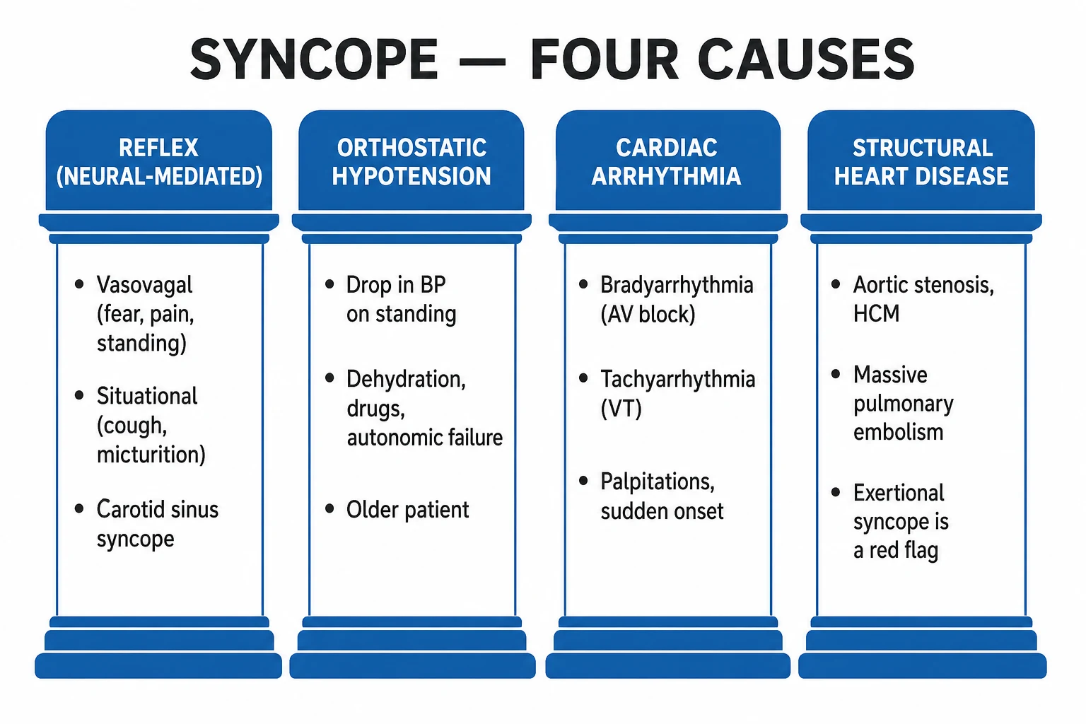
The ESC 2018 framework classifies syncope by mechanism into three groups:[1]
Reflex (neurally-mediated)
- **Commonest form overall and in the young.**
- Autonomic reflex triggered in susceptible individuals; cardiovascular response is paradoxical (bradycardia and/or vasodilation) despite a physiological trigger.
- Subtypes: **vasovagal** (orthostatic or emotional stress), **situational** (micturition, defaecation, swallowing, cough, post-prandial, weight-lifting), **carotid sinus syndrome**.
- **Prognosis**: benign in the young; injury from falls in the elderly.
Orthostatic hypotension (OH)
- **Failure of the normal standing pressor response.** Drop in SBP at least 20 mmHg or DBP at least 10 mmHg within 3 minutes of standing.
- Subtypes: **classical OH** (within 3 min), **initial OH** (within 15 s), **delayed OH** (after 3 min, sometimes up to 30 min).
- Causes: **non-neurogenic** (volume depletion, drugs, deconditioning) or **neurogenic** (autonomic failure — PAF, MSA, Parkinson's disease, diabetic autonomic neuropathy).
- **Prognosis**: intermediate; predicts cardiovascular events and mortality in older adults.
Cardiac (highest risk)
- Syncope due to a structural or arrhythmic cardiac cause — the **highest-mortality group**.
- **Arrhythmia** — bradycardia (sinus node dysfunction, AV block, Mobitz II, complete heart block, drug-induced), tachycardia (VT, SVT, AF with WPW, torsades).
- **Structural obstruction** — aortic stenosis, hypertrophic cardiomyopathy, mitral stenosis, pulmonary stenosis, pulmonary hypertension/embolism, cardiac tamponade, aortic dissection, atrial myxoma, prosthetic valve thrombosis.
- **Prognosis**: one-year mortality up to 30 percent; needs urgent work-up.
Syncope must also be separated from the other categories of transient loss of consciousness:[1]
- Seizure — cortical neuronal hypersynchrony; typically a longer postictal phase, aura, lateral tongue biting, postictal confusion.
- Psychogenic pseudosyncope — no true loss of consciousness; eyes closed, normal BP/heart rate/EEG during tilt, prolonged episodes, no injury from the fall.
- Rare causes of TLoC / collapse without true syncope: cataplexy (narcolepsy), drop attacks, vertebrobasilar TIA, subclavian steal, hypoglycaemia, hyperventilation with hypocapnia, sleep apnoea-induced hypoxia, deep venous thrombosis/PE presenting as syncope, and factitious disorders. [1]
Epidemiology & Risk Factors
Syncope is common. Lifetime prevalence is roughly 35–40 percent in the general population, with a bimodal age distribution: a first peak in adolescence and young adults (predominantly vasovagal) and a second, larger peak after age 70 (mixed: orthostatic, cardiac, carotid sinus).[1][5] It accounts for 1–3 percent of emergency department presentations and up to 6 percent of hospital admissions in some series.[6]
Reflex syncope predominates in young adults (especially women), whereas cardiac and orthostatic causes dominate in older adults — the cause shifts with age because structural heart disease, polypharmacy, autonomic failure, and carotid sinus hypersensitivity accumulate with age.[1]
Syncope — the high-yield numbers
Risk factors for a CARDIAC cause (the high-risk group):[1][2]
- Older age, male sex.
- Known structural heart disease (heart failure, previous MI, cardiomyopathy, valvular disease).
- Abnormal ECG (any conduction disease, ischaemia, channelopathy pattern, prior infarct, LVH/strain).
- Family history of premature sudden cardiac death (under 40 years).
- Syncope during exertion, while supine, or without prodrome; syncope followed by chest pain, breathlessness, or palpitations. [1]
Predisposing and precipitating factors for reflex syncope and OH:[1][5]
- Reflex: prolonged standing, warm/crowded environments, painful or visceral stimuli (venepuncture, venesection, hair-grooming, dental work), emotional distress, sight of blood, dehydration, recent alcohol.
- Orthostatic (non-neurogenic): volume depletion (haemorrhage, vomiting, diarrhoea, diuretic over-diuresis, dialysis), drugs (antihypertensives, alpha-blockers, nitrates, diuretics, dopamine agonists, tricyclics, antipsychotics, PDE-5 inhibitors), prolonged bed rest, deconditioning, pregnancy, post-prandial splanchnic pooling.
- Orthostatic (neurogenic / autonomic failure): pure autonomic failure, multiple system atrophy, Parkinson's disease (and dopaminergic therapy), dementia with Lewy bodies, diabetic autonomic neuropathy, amyloidosis, autoimmune autonomic ganglionopathy, paraneoplastic dysautonomia, hereditary sensory and autonomic neuropathies, post-vagal (the "cured" form of POTS). [1]
Drugs most commonly implicated in iatrogenic syncope in older adults (polypharmacy): antihypertensives (ACE inhibitors/ARBs, calcium-channel blockers), alpha-blockers (tamsulosin, doxazosin — common after a fall), diuretics (volume depletion), nitrates, tricyclic antidepressants, antipsychotics, dopamine agonists (for Parkinson's/RLS), PDE-5 inhibitors, opioids, and QT-prolonging drugs (causing torsades — macrolides, fluoroquinolones, antipsychotics, antiarrhythmics, methadone, ondansetron).[1]
Vasovagal recurrence rate is roughly 30 percent over three years in patients presenting to medical attention. The one-year mortality in cardiac syncope is up to 30 percent, compared with under 5 percent for unexplained syncope and under 1 percent for reflex syncope in young patients.[1]
Pathophysiology
Syncope occurs when cerebral perfusion falls below the autoregulation floor so that cerebral blood flow drops by about one-third, or cessation of flow for 6–10 seconds.[1] The mechanism is global cerebral hypoperfusion — unilateral carotid disease alone usually does not cause syncope (it causes focal TIA), because the contralateral carotid and the vertebrobasilar circulation maintain global flow.
The three mechanism groups differ in how they reduce cerebral perfusion:[1][5]
Reflex (neurally-mediated) syncope
The Bezold–Jarisch reflex is the prototype. In a susceptible individual, a trigger (prolonged standing, pain, emotion) produces a paradoxical autonomic response:[1]
- Prolonged standing → venous pooling → reduced venous return → reduced ventricular filling.
- The underfilled but vigorously contracting ventricle generates strong mechanoreceptor firing (via unmyelinated vagal C-fibres) — the heart signals "I am overfull" when it is actually empty.
- Afferent vagal firing to the medullary cardiovascular centre (nucleus tractus solitarius).
- The medullary response is sympathetic withdrawal + parasympathetic (vagal) activation.
- The efferent response has two limbs:
- Cardioinhibitory — vagal activation causes sinus bradycardia, AV block, even asystole (the "cardioinhibitory" type).
- Vasodepressor — sympathetic withdrawal causes peripheral vasodilation, fall in systemic vascular resistance and blood pressure (the "vasodepressor" type).
- Most episodes are mixed (both limbs); the resulting fall in cardiac output and blood pressure causes cerebral hypoperfusion and syncope. [1]
Vasovagal syncope proceeds in two phases: an initial presyncopal sympathoexcitation (tachycardia, pallor, sweating, narrowed pulse pressure as the body tries to maintain BP) and then the vasovagal cascade (sudden bradycardia and vasodilation, precipitous BP fall, collapse).[1]
The mechanism of situational syncope is analogous: a visceral afferent trigger (bladder distension in micturition, defaecation strain, swallowing, cough-induced raised intrathoracic pressure, post-prandial splanchnic pooling) drives vagal afferents, producing vagal override.[1]
Orthostatic hypotension
The normal standing pressor response is a baroreflex-mediated increase in heart rate, cardiac contractility and peripheral vascular resistance, preserving blood pressure on standing. Orthostatic hypotension is failure of this baroreflex mechanism.[5]
- Neurogenic OH — disease of the autonomic nervous system (autonomic ganglia, postganglionic sympathetic neurones): pure autonomic failure, multiple system atrophy, Parkinson's disease, dementia with Lewy bodies, diabetic autonomic neuropathy, amyloid autonomic neuropathy. There is failure of compensatory tachycardia on standing (HR rise under 15 bpm despite a profound BP drop). Supine hypertension is common (loss of nocturnal BP dipping).
- Non-neurogenic OH — hypovolaemia (haemorrhage, vomiting, diarrhoea, dialysis), drugs (antihypertensives, diuretics, alpha-blockers, nitrates), deconditioning/prolonged bed rest, pregnancy, post-prandial splanchnic pooling. The baroreflex is intact; HR rises briskly on standing (the POTS pattern: HR rise at least 30 bpm within 10 minutes of standing without a BP drop). [1]
Carotid sinus hypersensitivity
The carotid sinus baroreceptor is oversensitive in some older men. Mechanical stimulation (tight collar, head turning, shaving, tumour pressure, neck manipulation) triggers an exaggerated vagal (cardioinhibitory) and sympathetic-withdrawal (vasodepressor) response.[1] Three response types: cardioinhibitory (asystole over 3 s — dominant), vasodepressor (SBP drop at least 50 mmHg without bradycardia), mixed.
Cardiac syncope — why it is dangerous
Cardiac causes reduce cardiac output abruptly, producing a sudden fall in mean arterial pressure that cannot be compensated by baroreflexes in time:[1]
- Tachyarrhythmia (VT, torsades, AF with WPW, rapid AF): shortened diastolic filling time and loss of atrial contribution → fall in stroke volume.
- Bradyarrhythmia (sinus node dysfunction, Mobitz II, complete heart block, drug-induced): slow ventricular rate reduces cardiac output.
- Structural obstruction (severe aortic stenosis, HCM, MS, pulmonary hypertension, tamponade, atrial myxoma, pulmonary embolism): fixed obstruction to forward flow; the exertional syncope of aortic stenosis and HCM is pathognomonic — the peripheral vasodilation of exercise meets a fixed obstruction to cardiac output. [1]
The cardiac group carries the highest one-year mortality because the underlying disease (cardiomyopathy, channelopathy, severe structural disease, ischaemia) is itself lethal — syncope is the warning sign of imminent sudden cardiac death in HCM, ARVC, Brugada, long QT and CPVT.[1]
Hyperventilation and presyncope
Hypocapnia from hyperventilation causes cerebral vasoconstriction — cerebral blood flow falls by roughly 2 percent for every 1 mmHg drop in PaCO2. Anxiety-driven hyperventilation is a frequent cause of presyncope (and rarely true syncope) in young women, accompanied by perioral and digital tingling, carpopedal spasm, and a sense of air hunger.[5]
Clinical Presentation
The history and an eyewitness account are the single most important diagnostic tools — they yield a confident mechanism-based diagnosis in roughly half of all patients at the bedside.[1]
Reflex (vasovagal) syncope — the prototype benign presentation
Prodrome (seconds to minutes): pallor, nausea, light-headedness, diaphoresis (cold sweat), yawning, blurred or tunnel vision, warmth, abdominal discomfort, sighing. The patient often feels they are "going to faint".[1][5]
Triggers: prolonged standing (especially in warm, crowded environments), painful or emotional stimuli (venepuncture, blood drawing, instrumentation, dental work), sight of blood, hair-grooming, prolonged fasting, recent illness/dehydration.[1]
The episode: gradual loss of consciousness while upright (or rapidly upright); the patient slumps gracefully rather than crashing, may have brief multifocal myoclonic jerks (syncopal myoclonus — not seizure), is pale, sweating, with a thready pulse, and may be transiently incontinent.[1]
Recovery: rapid within seconds to a minute once horizontal; the patient may feel washed-out but returns rapidly to baseline mental status without postictal confusion. Prolonged recovery, confusion, or focal deficit suggests an alternative diagnosis. [1]
Situational syncope
Syncope during or immediately after a specific visceral act:[1]
- Micturition syncope — nocturnal, immediately after voiding (often in older men with prostatic obstruction; bladder decompression triggers vagal firing).
- Defaecation syncope — straining (often constipated elderly patients).
- Swallowing (deglutition) syncope — particularly with oesophageal disease (tumour, achalasia, diverticulum) or cardiac conduction disease.
- Cough syncope — paroxysms of coughing in patients with COPD/bronchitis; raised intrathoracic pressure reduces venous return and cardiac output.
- Post-prandial syncope — 30–60 minutes after a large meal, in older adults (splanchnic pooling).
- Hair-grooming syncope — head/neck manipulation in young women.
- Weight-lifting / brass-instrument player's syncope — raised intrathoracic pressure (Valsalva-type). [1]
Carotid sinus syndrome
Syncope in an older man precipitated by head turning, tight collar, shaving, tumour pressure on the neck, or neck manipulation; reproduction on carotid sinus massage (CSM).[1]
Orthostatic syncope
Light-headedness, blurring vision, weakness, palpitations on standing, resolving on sitting or lying back. Onset within 3 minutes of standing (classical OH) or after a longer interval (delayed OH). Context of volume depletion (vomiting, diarrhoea, haemorrhage, dialysis), drugs (recent antihypertensive titration, diuretic, alpha-blocker), or autonomic disease (Parkinson's, diabetes, MSA).[1][5]
Neurogenic OH features: lack of compensatory tachycardia, supine hypertension, worse after meals (post-prandial), in hot environments, after exercise, on rising in the morning, with concurrent urinary/gastrointestinal/sweating/erectile autonomic failure.[5]
Cardiac syncope — recognise these red flags
Cardiac syncope is sudden (often no prodrome), may occur during exertion or while supine, may be preceded by palpitations, chest pain, or breathlessness, and occurs in a patient with known structural heart disease, an abnormal ECG, or a family history of sudden cardiac death (SCD) under 40.[1][2]
- Exertional syncope — think aortic stenosis, hypertrophic cardiomyopathy, pulmonary hypertension, coronary anomaly, long-QT syndrome type 1 (LQT1), catecholaminergic polymorphic VT.
- Syncope on emotion or with auditory stimuli (sudden loud noise, alarm clock) — long-QT type 2 (LQT2).
- Syncope at rest or during sleep — long-QT type 3 (LQT3), Brugada syndrome, complete heart block, paediatric arrhythmia.
- Syncope while supine, without prodrome, in a patient with cardiomyopathy — ventricular tachycardia until proven otherwise.
- Syncope with chest pain and a widened mediastinum — acute aortic dissection, massive pulmonary embolism, cardiac tamponade.
- Syncope followed by breathlessness, hypoxia, and a gallop — massive pulmonary embolism, decompensated heart failure. [1]
Atypical presentations
- Elderly: a fall without clear recollection of warning, single unwitnessed collapse, or new confusion — the prodrome may be blunted and the cause is often multifactorial (OH + drugs + CSH + structural disease).[1]
- Diabetic patient: hypoglycaemia masquerades as syncope — always check a finger-prick glucose; autonomic neuropathy causes neurogenic OH.[1]
- Pregnant patient: supine syncope from aortocaval compression by the gravid uterus (turn her to the left lateral position); anaemia and vasodilation lower the threshold; rarely, peripartum cardiomyopathy in late pregnancy/peripartum.
- Athlete: syncope is never normal — exclude HCM, ARVC, Brugada, LQTS, CPVT, anomalous coronary artery, myocarditis.[1]
- Immunocompromised: opportunistic myocarditis, pericardial effusion/tamponade, drug-induced QT prolongation from P450-interacting agents (azoles, macrolides, antiretrovirals).
- Anticoagulated patient: a head strike during syncope carries a much higher risk of intracranial haemorrhage; image even after a seemingly minor fall.
Syncope vs seizure — bedside discrimiators
| Feature | Syncope | Seizure |
|---|---|---|
| Onset | Gradual, with prodrome | Sudden (or aura) |
| Trigger | Upright posture, exertion, visceral | Often none; flashing light, sleep deprivation |
| Colour | Pale | Flush/cyanosis |
| Tone | Floppy | Rigid then jerky |
| Duration of LOC | Seconds to a minute | Minutes |
| Recovery | Rapid, complete, no confusion | Slow, postictal confusion, drowsiness |
| Incontinence | Possible | Common |
| Tongue bite | Tip of tongue (clenched teeth on falling) | Lateral border of tongue (inside cheek) |
| Myoclonus | Brief, multifocal, synchronous with fall | Rhythmic, prolonged, then postictal |
| Post-event | Patient alert within minutes | Patient confused, amnesic for minutes-hours |
A witnessed lateral tongue bite is highly specific for seizure; an eyewitness description of rhythmic tonic-clonic activity with postictal confusion distinguishes a generalised seizure.[1]
Differential Diagnosis
The differential of transient loss of consciousness (TLoC) is the clinical lynchpin. The first step is to separate true syncope (cerebral hypoperfusion) from seizure and psychogenic pseudosyncope.[1][5]
A complete differential for TLoC: [1]
- Syncope (cerebral hypoperfusion) — reflex, orthostatic, cardiac (above).
- Seizure — generalised tonic-clonic, complex partial, absence; EEG/clinical features as above.
- Psychogenic pseudosyncope — conversion disorder; no true LOC, eyes closed, no injury, normal BP/HR/EEG during tilt, episodes may last many minutes.
- Hypoglycaemia — sweating, tremor, hunger, confusion; recovers with glucose; check capillary glucose in every patient.
- Hyperventilation with hypocapnia — perioral/digital tingling, carpopedal spasm; presyncope more than true syncope.
- Cataplexy — sudden loss of tone triggered by emotion (laugh, surprise) in a patient with narcolepsy; no loss of consciousness.
- Drop attack — sudden fall without LOC; vertebrobasilar, basilar migraine, cryptogenic.
- Vertebrobasilar TIA — focal posterior-circulation signs (diplopia, dysarthria, ataxia, bilateral visual loss); rarely pure syncope.
- Subclavian steal — subclavian stenosis proximal to the vertebral origin with arm exercise; weak/delayed radial pulse, arm blood pressure differential over 15 mmHg.
- Pulmonary embolism (massive) — syncope with hypoxia, dyspnoea, pleuritic pain, deep-vein thrombosis risk; acute right heart strain on ECG/echo.
- Subarachnoid haemorrhage — thunderclap headache, meningism; syncope can be the presenting feature.
- Aortic dissection — tearing chest/back pain, BP differential, widened mediastinum; syncope with neurological signs.
- Internal bleeding (GI, retroperitoneal, occult) — hypovolaemic syncope, postural drop, tachycardia.
- Carotid sinus syndrome — precipitated by neck manipulation (above). [1]
Differentiating features (≥3 distinguishing features per differential):[1]
- Vasovagal vs cardiac: vasovagal = standing/trigger, prodrome, pallor, rapid recovery, young/structurally normal; cardiac = exertion/supine, no prodrome, palpitations/chest pain, abnormal ECG, structural disease.
- OH vs vasovagal: OH = immediate on standing (within 3 min), drug/volume context, recovery on sitting, no prominent vagal prodrome; vasovagal = prodrome, trigger can be emotional/visceral, may be upright for some time before onset, prominent autonomic prodrome.
- Seizure vs syncope: lateral tongue bite, postictal confusion, prolonged myoclonus, incontinence, aura favour seizure (above table).
- Pulmonary embolism vs reflex: PE = hypoxia, dyspnoea, pleuritic chest pain, DVT risk, raised JVP, right-heart strain on ECG/echo.
- Aortic dissection vs reflex: tearing pain, BP differential, widened mediastinum on CXR, new murmur.
- Subarachnoid vs reflex: thunderclap headache, meningism, focal signs; CT brain diagnostic.
- Psychogenic pseudosyncope vs true syncope: eyes closed during episode, no fall in BP/HR, no pallor/sweating, prolonged episodes (many minutes), no injury from the fall, recall of the event by the "unconscious" patient. [1]
Differentials needing urgent work-up: pulmonary embolism, subarachnoid haemorrhage, aortic dissection, internal bleeding, myocardial infarction — any syncope with hypoxia, chest pain, BP differential, neurological signs, or haemodynamic instability.[1]
Clinical & Bedside Assessment
History: circumstances (posture, activity, environment, position), prodrome, triggers, recovery, frequency, injuries, comorbidities, drug list (with recent changes), family history (sudden death under 40, channelopathy, cardiomyopathy), and an eyewitness account.[1]
The "witnessed collapse" history — three key questions to the witness:[1]
- Were the patient's eyes open or closed during the episode?
- Was the patient responsive (could they be roused)?
- Was there any movement (twitching, jerking, head turning) and what colour was the patient? [1]
Examination: full cardiovascular (heart rate, BP both arms, postural BP, JVP, murmurs, signs of heart failure, radiofemoral delay, carotid bruits), respiratory, neurological (residual focal deficit, tongue), and a rectal/pelvic exam if occult bleeding suspected.[1]
Lying and standing blood pressure (active stand test, Canadian protocol)
Measure BP after 5 minutes supine, then immediately on standing (1 minute) and at 3 minutes (and up to 10 minutes if delayed OH suspected).[5]
- Classical orthostatic hypotension — SBP drop at least 20 mmHg or DBP drop at least 10 mmHg within 3 minutes of standing.[1]
- Initial OH — SBP drop at least 40 mmHg within 15 seconds of standing, with rapid spontaneous recovery.
- Delayed OH — meets the same threshold but after 3 minutes of standing (sometimes up to 30 minutes); common in older adults and autonomic failure.
- POTS — HR rise at least 30 bpm (40 bpm in adolescents) within 10 minutes of standing without a qualifying BP drop.
Carotid sinus massage (CSM) — technique
Performed in patients over 40 with unexplained syncope compatible with reflex cause.[1]
- Continuous ECG and non-invasive BP monitoring, IV access available.
- Supine AND upright positions (upright increases sensitivity).
- Right carotid first, then left — NEVER both sides simultaneously.
- Firm longitudinal massage of the carotid bifurcation (at the level of the cricoid cartilage) for 5–10 seconds.
- Contraindications: carotid bruit, history of TIA/stroke/MI in past 3 months, known significant carotid stenosis, audible bruit, recent neck surgery.
- Diagnostic (positive) response: asystole over 3 seconds and/or SBP drop at least 50 mmHg with reproduction of symptoms.
- Complications (~0.2%): transient ischaemic attack/stroke; minimise by screening for bruit and prior cerebrovascular disease. [1]
Bedside features suggesting structural cardiac disease
- Ejection systolic murmur radiating to the carotids — aortic stenosis.
- Ejection systolic murmur that increases with Valsalva and decreases with squatting — hypertrophic cardiomyopathy.
- Mid-diastolic rumble, opening snap, loud S1 — mitral stenosis.
- Systolic murmur louder on inspiration at upper-left sternal edge — pulmonary stenosis.
- Raised JVP, Kussmaul sign, muffled heart sounds, pulsus paradoxus — cardiac tamponade.
- Radio-femoral delay, weak femoral pulses — coarctation (rare adult presentation).
- Signs of heart failure (basal crackles, raised JVP, S3, peripheral oedema) — decompensated cardiac disease. [1]
Syncopal myoclonus vs epileptic myoclonus
Syncopal myoclonus is brief, multifocal, non-rhythmic, occurs at the moment of the fall, and stops with recumbency. Epileptic myoclonus is rhythmic, prolonged (over a minute), generalised, and followed by postictal depression.[1]
Investigations
Mandatory in every patient
- 12-lead ECG — mandatory in all syncope patients; abnormal in roughly 5 percent overall, much higher in cardiac-syncope subsets. Look for: sinus pause over 3 s, Mobitz II, complete heart block, bifascicular block (RBBB + LAFB/LPFB), alternating bundle branch block, Brugada pattern (coved ST elevation in V1–V3), QTc prolongation (over 460 ms in women, 470 ms in men), epsilon waves (ARVC), LVH with deep T inversions (HCM), WPW pattern (delta wave, short PR), pathologic Q waves (prior infarct), RVH/RBBB pattern of pulmonary hypertension (S1Q3T3), evidence of ischaemia, atrial fibrillation.[1][2]
- Capillary glucose — exclude hypoglycaemia in every patient.
- Postural BP (active stand) — as above.
First-line in the emergency setting
- Full blood count (anaemia → occult bleeding), U&E (volume status, renal dysfunction), troponin (if chest pain, structural disease, or risk; its role in unexplained syncope is debated), β-hCG in women of reproductive age (ectopic), D-dimer if PE suspected (high sensitivity, low specificity).[6]
- Echocardiogram — when structural heart disease is suspected or the ECG is abnormal; assesses LV/RV function, valvular disease, HCM, tamponade, pulmonary hypertension, myxoma.
- Chest X-ray — widened mediastinum (dissection), pulmonary oedema, pneumothorax, PE oligoaemia.
Risk-stratification scores — reproduced verbatim
San Francisco Syncope Rule (SFSR, Quinn 2004) — high-risk if any present (the CHEAT mnemonic):[2]
- C — History of Congestive Heart Failure
- H — Haematocrit under 30 percent
- E — ECG abnormal (any new or not previously documented change)
- A — Abnormal vital sign: shortness of breath OR systolic BP under 90 mmHg at triage
- T — Trouble breathing (shortness of breath) [1]
Sensitivity for serious 7-day outcomes ~96–98 percent (specificity modest ~62 percent); a negative SFSR reliably identifies low-risk patients suitable for discharge; a positive SFSR warrants observation/admission and work-up.[2]
Canadian Syncope Risk Score (CSRS, Thiruganasambandamoorthy 2016) — nine predictors, summed; predicts 30-day serious outcome:[3][4]
- Abnormal QRS/QTc ECG (+1)
- Cancer history (+1)
- Brugada ECG pattern (+1)
- Triage SBP 90–110 mmHg (+1)
- Triage home heart rate under 50 or over 90 bpm (+1)
- History of syncope without prodrome (+1)
- ED venous blood urea elevated (+1)
- ED systolic BP under 90 mmHg (+2)
- ED capillary glucose under 4.0 mmol/L (+1) [1]
Four risk bands (30-day serious outcome probability): [1]
| Total score | Risk band | 30-day serious outcome |
|---|---|---|
| –3 to +1 | Very low | under 0.7 percent |
| +2 to +3 | Low | under 2 percent |
| +4 to +5 | Medium | 2 to 5 percent |
| +6 to +7 | High | over 5 percent |
| +8 to +11 | Very high | over 15 percent |
The CSRS offers graduated risk rather than a binary high/low; very-low and low bands can usually be discharged; high and very-high bands warrant admission.[3][4]
EGSYS (Evaluation of Guidelines in Syncope Study) score — predicts a cardiac cause of syncope:[1]
- Palpitations preceding syncope (+3)
- Heart disease (+3)
- Abnormal ECG (+3)
- Syncope during effort (+2)
- Syncope in supine position (+2)
- Autonomic prodrome present (−1) [1]
A score over 3 indicates a likely cardiac cause; sensitivity ~95 percent for cardiac origin. [1]
OESIL score (Osservatorio Epidemiologico della Sincope nel Lazio) — predicts one-year mortality:[1]
- Age over 65
- Abnormal ECG
- History of cardiovascular disease
- Lack of prodrome [1]
Each present scores 1 (max 4); one-year mortality rises from 0 percent (score 0) to over 30 percent (score 4). [1]
Calgary Syncope Symptom Study diagnostic criteria for vasovagal syncope — a validated questionnaire combining: female sex or age under 60, diarrhoea, post-prandial syncope, prolonged standing/warm environment, prodrome (hot/warm, diaphoresis, nausea), and rapid complete recovery; achieves very high diagnostic accuracy without the need for tilt testing when the history is classical.[1]
Targeted investigations
- Tilt-table test — passive 60–70 degrees head-up tilt, with sublingual nitroglycerin provocation if passive phase negative; for recurrent unexplained syncope with suspected reflex or orthostatic cause. Reproduces the vasovagal reflex and quantifies the response (cardioinhibitory, vasodepressor, mixed) and the magnitude of OH.[1]
- Ambulatory ECG monitoring — chosen by symptom-episode frequency:
- Daily episodes → 24–48 h Holter.
- Weekly → external loop recorder (1–4 weeks).
- Monthly or less frequent → external patch recorder (up to 14–30 days) or implantable loop recorder (ILR) (subcutaneous, 2–3 years).
- ILR is the single most informative test in unexplained recurrent syncope with a negative initial work-up.[1]
- Echocardiogram — for any structural heart disease or abnormal ECG.
- Exercise ECG — for exertional syncope (coronary ischaemia, exercise-induced arrhythmia, LQT1).
- Electrophysiological study (EPS) — declining role; consider in patients with prior MI, unexplained syncope, and a non-diagnostic ECG/ambulatory monitor; identifies inducible VT or His-Purkinje disease.
- CT coronary angiography / coronary angiography — if ischaemia suspected.
- Cardiac MRI — for cardiomyopathy (HCM, ARVC, sarcoid, amyloid), suspected myocarditis, and the work-up of high-risk channelopathies.
- Brain imaging (CT/MRI) and EEG — NOT routinely indicated in uncomplicated syncope; reserved for focal signs, suspected seizure, head injury, or persistent altered mental status.[1]
- Carotid sinus massage — for unexplained syncope in the over-40s (technique above).
- Blood tests — FBC, U&E, glucose, troponin (if cardiac suspected), β-hCG (women), D-dimer and CTPA if PE suspected.
Biomarkers in risk-stratification
High-sensitivity troponin is useful in suspected acute coronary syndrome but has limited specificity in unexplained syncope. NT-proBNP/BNP may identify occult cardiac dysfunction. D-dimer is sensitive but not specific for PE. None of these substitutes for the history and ECG.[6]
Management — Resuscitation
ABCDE assessment for any patient who arrives unconscious or collapses in front of you.[1]
- Airway — protect with head-tilt/chin-lift, recovery position; clear obstruction.
- Breathing — assess for spontaneous respiration; high-flow oxygen if hypoxic.
- Circulation — check pulse. If no pulse, no breathing — start CPR/ALS; do NOT assume syncope.
- Disability — check capillary glucose immediately (treat hypoglycaemia: 15–20 g rapid carbohydrates, or 50 mL of 50 percent dextrose IV if unconscious, repeated as needed).
- Exposure — screen for injury (especially head strike), occult bleeding, sepsis. [1]
Time-critical bundle for suspected cardiac syncope
- Continuous ECG monitoring, telemetry bed.
- IV access, bloods: FBC, U&E, troponin, glucose, β-hCG in women.
- 12-lead ECG and review by a senior.
- Echocardiogram if structural disease suspected.
- Urgent cardiology review if arrhythmia or structural cause. [1]
Symptomatic bradycardia / asystole with syncope
- Atropine 0.5 mg IV, repeated every 3–5 minutes up to maximum 3 mg.[1]
- If ineffective: transcutaneous pacing (with analgesia/sedation), then transvenous pacing.
- Indications for emergent pacing: complete heart block, Mobitz II, alternating bundle branch block with syncope, sinus pause over 3 s, symptomatic sinus node dysfunction unresponsive to atropine.[1]
Volume resuscitation for hypovolaemic syncope
- Balanced crystalloid (Hartmann's or 0.9% saline) 250–500 mL bolus, reassess, repeat to clinical response.
- Identify and stop the source of bleeding (GI, retroperitoneal, ruptured ectopic). [1]
Syncope with a haemodynamically compromising arrhythmia
- VT with pulse: amiodarone 300 mg IV over 20–60 minutes then infusion 900 mg/24 h, OR synchronised DC cardioversion if unstable.
- Polymorphic VT / torsades: IV magnesium sulfate 2 g, correct electrolytes (K to 4.5–5.0, Mg over 2.0), stop culprit QT-prolonging drug; overdrive pacing if bradycardia-dependent.
- VT/VF cardiac arrest: defibrillation, ALS algorithm.
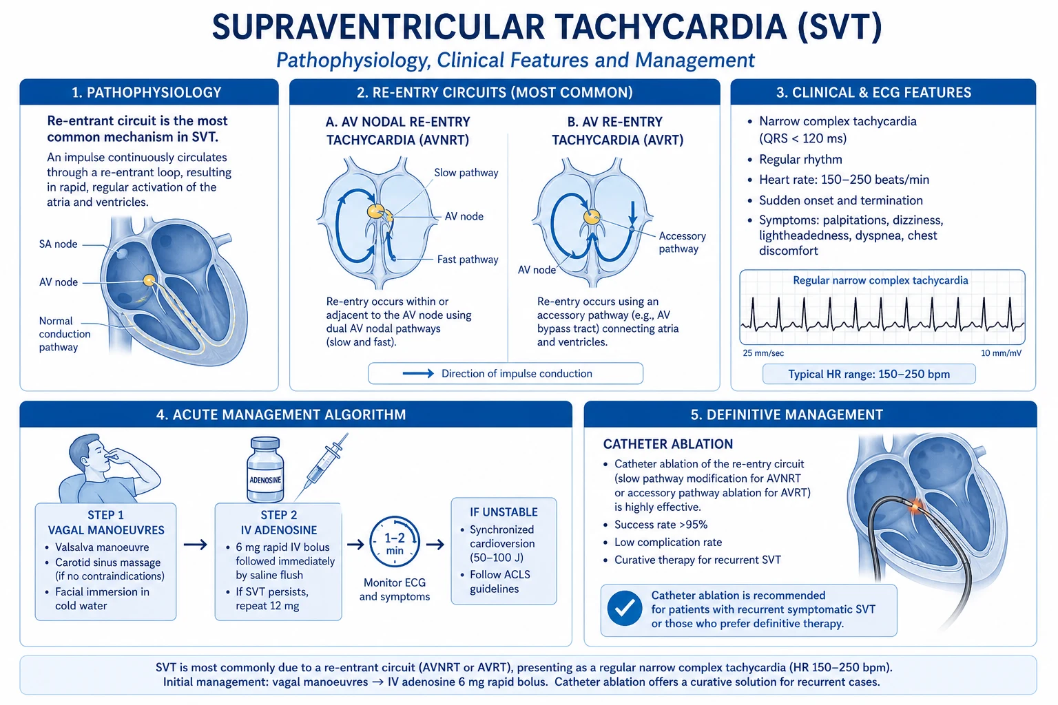
- Supraventricular tachycardia (SVT): vagal manoeuvres then adenosine 6 mg rapid IV push (then 12 mg, then 12 mg if needed); synchronised cardioversion if unstable.[1]
- AF with WPW, unstable: synchronised DC cardioversion; AVOID AV-nodal blockers (verapamil, diltiazem, beta-blockers, adenosine, digoxin) — they preferentially block the AV node and accelerate conduction down the accessory pathway, precipitating VF.
Management — Definitive & Stepwise
The ESC single-column diagnostic pathway: initial evaluation (history, examination, ECG, postural BP) → risk stratification (SFSR, CSRS) → targeted investigation OR empiric diagnosis → management of the underlying cause.[1]
ESC risk stratification — disposition guide
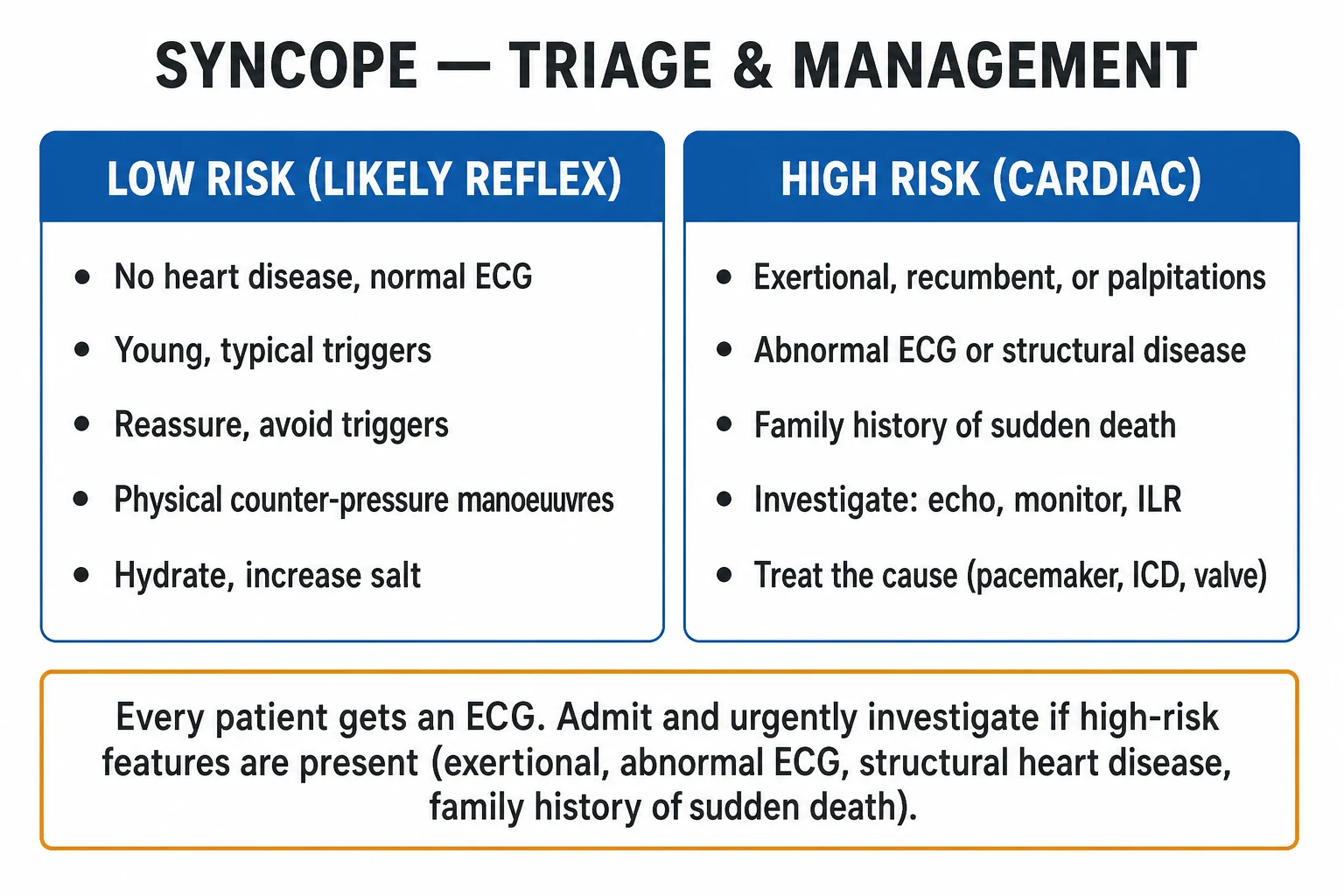
HIGH-risk features → admit (or ED observation) + cardiology work-up:[1]
- Syncope during exertion or while supine.
- Palpitations at the time of syncope.
- Family history of sudden cardiac death under 40.
- Abnormal ECG (any conduction disease, channelopathy pattern, ischaemia, prior infarct).
- Structural heart disease (heart failure, cardiomyopathy, valvular disease).
- Serious injury (e.g., fracture, internal bleeding, head injury).
- Incontinence without a clear reflex trigger (raise the possibility of seizure).
- Older patients with comorbidity, recurrent falls, or polypharmacy. [1]
LOW-risk features → can discharge with safety-net and primary-care follow-up:[1]
- Young, healthy, no structural heart disease.
- Clear vasovagal or orthostatic mechanism with prodrome.
- Normal ECG, normal examination.
- No injury, no recurrence.
- Negative SFSR and low CSRS band. [1]
Reflex (vasovagal) syncope
Lifestyle and non-pharmacological (first-line, most effective):[1]
- Trigger avoidance and education (reassurance of the benign prognosis in the young).
- Hydration 2–3 L/day and salt augmentation to 6–10 g/day if not hypertensive.
- Physical countermanoeuvres at first prodrome — leg crossing with tensing (squatting is even better), arm tensing with handgrip, bilateral fist clenching; these boost venous return and BP, often aborting the episode.
- Tilt training (prolonged standing against a wall, gradually increasing) — modest evidence, poor adherence. [1]
Pharmacotherapy (limited evidence; second-line): [1]
- Fludrocortisone 0.1–0.3 mg PO daily — mineralocorticoid-induced volume expansion; check potassium.
- Midodrine 2.5–10 mg PO TDS — alpha-1 agonist peripheral vasoconstrictor; useful in vasodepressor phenotype; avoid in hypertension, urinary retention.
- Beta-blockers — NOT recommended (multiple negative randomised trials: VPS II, POST).
- SSRIs (paroxetine, sertraline) — modest evidence; reserved for refractory cases.
- Pacemaker — only in severe recurrent cardioinhibitory vasovagal syncope with documented asystole on ILR; the ISSUE-3 trial showed benefit in selected patients, but VPS II, SYNPACE, and SPRITELY were largely negative — pacemaker is not routine.[1]
Carotid sinus syndrome
- Education and trigger avoidance (loose collars, gentle neck movement, avoid neck manipulation).
- Dual-chamber pacemaker for recurrent cardioinhibitory or mixed carotid sinus syndrome with documented asystole over 3 s; avoid pacing the pure vasodepressor type (no benefit).[1]
Orthostatic hypotension
Non-pharmacological (first-line): [1]
- Stop / reduce culprit drugs (antihypertensives, diuretics, alpha-blockers, dopaminergics, antipsychotics).
- Adequate hydration (2–3 L/day) and salt (6–10 g/day if not hypertensive).
- Compression stockings (waist-high) or abdominal binder (more effective for splanchnic pooling).
- Slow sit-to-stand, ankle-dorsiflexion before standing, split supine time over 2 minutes.
- Small frequent low-carbohydrate meals (avoid post-prandial hypotension).
- Avoid heat, hot baths, large meals, alcohol.
- Head-up tilt of the bed (10–20 degrees) — reduces nocturnal diuresis and supine hypertension.
- Counterpressure manoeuvres at first symptoms. [1]
Pharmacotherapy: [1]
- Fludrocortisone 0.1–0.3 mg PO daily — volume expansion; monitor potassium and for supine hypertension/oedema.
- Midodrine 2.5–10 mg PO TDS — alpha-1 agonist; first-line symptomatic agent; give the last dose by mid-afternoon to limit supine hypertension at night.
- Droxidopa (L-DOPS) 100–600 mg PO TDS — synthetic noradrenaline precursor; useful in neurogenic OH.
- Pyridostigmine 30–60 mg PO TDS — enhances ganglionic neurotransmission (mild effect, less supine hypertension).
- Manage supine hypertension — head-up tilt sleeping, evening short-acting antihypertensive (e.g., hydralazine, captopril), avoid bedtime midodrine.[1][5]
Cardiac syncope — treat the cause
- Arrhythmia:
- Sinus node dysfunction / AV block — permanent pacemaker (dual-chamber for AV block; AAIR/DDDR for sinus node dysfunction with intact AV conduction).
- Mobitz II, complete heart block, alternating BBB with syncope — pacemaker (or in selected high-grade block, consider aetiology: ischaemia, drugs, infiltrative disease).
- SVT — catheter ablation (curative for AVNRT, AVRT, focal atrial tachycardia, atrial flutter).
- AF — rate or rhythm control, anticoagulation by CHA2DS2-VASc.
- VT — ICD (secondary prevention if structural heart disease), catheter ablation for scar-related VT, treat ischaemia/cardiomyopathy.
- Channelopathies (LQTS, Brugada, CPVT, ARVC) — ICD + disease-specific measures (beta-blocker for LQTS/CPVT; avoid fever and sodium-channel blockers in Brugada; avoid QT-prolonging drugs in LQTS).
- Drug-induced QT prolongation — stop culprit, correct K/Mg, monitor.
- Structural obstruction:
- Severe aortic stenosis — aortic valve replacement (surgical or transcatheter, TAVI in higher-risk patients).
- Hypertrophic cardiomyopathy with obstruction — beta-blocker ± disopyramide; septal reduction therapy (myectomy or alcohol septal ablation); ICD for high-risk patients.
- Mitral stenosis — percutaneous balloon mitral valvuloplasty or surgical repair/replacement.
- Pulmonary embolism — anticoagulation; systemic thrombolysis or catheter-directed thrombolysis for massive PE with shock; embolectomy in selected cases.
- Pulmonary hypertension — disease-specific vasodilator therapy (endothelin antagonist, PDE-5 inhibitor, prostacyclin, sGC stimulator).
- Cardiac tamponade — emergency pericardiocentesis.
- Atrial myxoma — surgical resection.
- Aortic dissection — surgical (type A) or medical (type B) management.[1]
Implantable loop recorder — when?
ILR is the right next step in unexplained recurrent syncope with a negative initial work-up (normal ECG, echo, tilt-test), particularly when an arrhythmic cause is suspected (older patient, structural disease, infrequent episodes). It is also used in suspected epilepsy refractory to therapy (to exclude cardiac syncope masquerading as seizures), and in cryptogenic stroke to detect occult AF.[1]
Driving advice (UK DVLA / Indian RTO)
Group 1 (private car/motorbike licence):[1]
- Isolated unexplained syncope with low-risk features — no driving restriction.
- Vasovagal syncope with a clear trigger — no restriction; advise on trigger avoidance.
- Unexplained syncope with high-risk features — 6 months off driving (or until a cause is found and treated).
- Cardiac syncope — depends on the cause and treatment; usually 4 weeks after successful treatment, 6 months if untreated. [1]
Group 2 (HGV/PSV): stricter — 3 years off or permanent, depending on cause. [1]
Indian RTO follows comparable principles: a person with uncontrolled syncope should not drive; after definitive treatment, the patient must be reviewed and certified fit before resuming. [1]
Specific Subtypes & Scenarios
- Vasovagal syncope in the young athlete — never assume benign. Exclude HCM, ARVC, anomalous coronary, LQTS, CPVT, Brugada with ECG, echocardiogram, exercise ECG, and (if any doubt) cardiac MRI; once excluded, return-to-play with education, hydration, and counterpressure manoeuvres.[1]
- Syncope in the elderly — multifactorial (OH, polypharmacy, CSH, structural disease, autonomic failure); high rate of "falls" that are actually syncope with amnesia; lower threshold to admit and review drugs.[1]
- Situational syncope — manage each (above): micturition syncope (sit to void at night), defaecation (avoid straining, treat constipation), swallowing (treat oesophageal disease), cough (treat bronchospasm), post-prandial (small frequent low-carbohydrate meals, avoid rapid standing), hair-grooming (avoid tight head positions), weight-lifting/brass-instrument (avoid sustained Valsalva).
- Syncope in hypertrophic cardiomyopathy — syncope is a major sudden-death risk marker; compute the HCM SCD-5-year risk score and consider ICD.
- Syncope in aortic stenosis — exertional syncope is a symptom of severe disease warranting aortic valve replacement.
- Syncope in Brugada syndrome — syncope is a high-risk feature prompting ICD.
- Syncope in long QT syndrome — LQT1 exertional (swimming), LQT2 auditory/emotional (alarm clock), LQT3 sleep; beta-blocker (propranolol/nadolol) ± ICD in high-risk patients; avoid QT-prolonging drugs (www.crediblemeds.org).
- Syncope in CPVT — exercise/emotion-triggered bidirectional/polymorphic VT in a structurally normal heart; beta-blocker (nadolol) ± flecainide ± ICD (and left cardiac sympathetic denervation in refractory cases).
- Syncope in pulmonary embolism (massive) — hypoxia, dyspnoea, deep-vein thrombosis risk; acute right-heart strain on ECG (S1Q3T3, T-wave inversion V1–V3, right-axis deviation, RBBB) and echo; systemic thrombolysis if haemodynamically unstable.
- Syncope in pulmonary hypertension — exertional; assess with echo and right-heart catheterisation; disease-specific vasodilator therapy.
- Syncope in cardiac tamponade — Beck triad (hypotension, raised JVP, muffled heart sounds), pulsus paradoxus; emergency pericardiocentesis.
- Syncope in pregnancy — common vasovagal + supine aortocaval compression; left lateral tilt position, hydration, treat anaemia; exclude ectopic pregnancy (β-hCG, pelvic ultrasound); peripartum cardiomyopathy (late pregnancy to months postpartum).
- Syncope after device implantation — pacemaker malfunction (lead dislodgement, oversensing, battery depletion), ICD shock (VT/VF treated), lead fracture; interrogate the device.
- Psychogenic pseudosyncope — conversion disorder; eyes closed, normal BP/HR/EEG during tilt, prolonged episodes, no injury; multidisciplinary care with psychology/CBT; avoid unnecessary cardiac tests once the diagnosis is established.[1]
Complications & Pitfalls
Complications of syncope: [1]
- Fall-related injury — fractures (especially hip in the elderly, wrist, skull), head injury, intracranial haemorrhage (especially in anticoagulated patients), dental/facial trauma, soft-tissue injury.
- Recurrent syncope and injury — loss of confidence, fear of leaving home, activity restriction, depression.
- Driving and occupational impact — restriction per DVLA/RTO, loss of livelihood (especially Group 2 licence holders, pilots, heavy machinery operators).
- Psychological morbidity — anxiety, impaired quality of life.
- Loss of consciousness-related — incontinence, tongue biting (tip), myoclonic jerks. [1]
Complications of procedures: [1]
- Carotid sinus massage — transient ischaemic attack/stroke (~0.2 percent), transient arrhythmia (mitigate by screening for bruit/stenosis, never both sides).
- Tilt-table testing — provoked prolonged asystole, hypotension, nausea/vomiting; rare.
- Implantable loop recorder — infection, haematoma, lead/device migration. [1]
Classic diagnostic pitfalls:[1][6]
- Mislabelling seizures as syncope and vice versa — focus on the postictal state and lateral tongue bite.
- Missing cardiac syncope with a 'normal' ECG — a single 12-lead can be normal in intermittent arrhythmia; use ambulatory monitoring or ILR if suspicion is high.
- Over-investigating with brain CT/EEG in uncomplicated vasovagal syncope — these are low-yield and costly.
- Missing orthostatic hypotension from drugs — the most common cause in older adults; always do a careful drug review.
- Missing drug-induced long QT — check the ECG and the drug list (macrolides, fluoroquinolones, antipsychotics, antiarrhythmics, methadone, ondansetron, antifungals via P450 inhibition).
- Attributing syncope to 'vertebrobasilar insufficiency' without evidence — true vertebrobasilar TIA is rare and almost always has focal posterior-circulation signs; isolated syncope is almost never vertebrobasilar.
- Dismissing elderly falls as 'mechanical' — many "mechanical falls" are unwitnessed syncope; lower threshold for cardiac work-up.
- Relying on the San Francisco Syncope Rule alone — high sensitivity but limited specificity; combine with the CSRS for graduated risk.
- Misattributing massive pulmonary embolism to "vasovagal" — check oxygenation, look for DVT risk and right-heart strain.
- Misattributing subarachnoid haemorrhage to "vasovagal" — ask about thunderclap headache. [1]
Prognosis & Disposition
Prognosis is determined by the mechanism and the underlying disease, not by syncope itself:[1]
- Reflex syncope — benign in the young (one-year mortality under 1 percent); in the elderly, recurrent syncope causes falls and injury. Tends to remit in adolescents and young adults.
- Orthostatic syncope — intermediate prognosis; predicts cardiovascular events and mortality in older adults (a marker of frailty and autonomic disease).
- Cardiac syncope — highest mortality (one-year up to 30 percent); the underlying disease (cardiomyopathy, channelopathy, severe structural disease, ischaemia) drives prognosis. Syncope in HCM, ARVC, Brugada, LQTS, CPVT is a major sudden-death risk marker.
- Unexplained syncope — intermediate risk; ILR often reveals an arrhythmic cause. [1]
Risk scores inform disposition:[1]
- SFSR positive (any of CHF history, Hct under 30, abnormal ECG, SBP under 90, SOB) → admit / observe.
- CSRS very low/low band → discharge with safety-net.
- CSRS high / very high band → admit for telemetry and work-up.
- OESIL score 3–4 → high one-year mortality — intensive work-up and follow-up. [1]
Admission for telemetry is indicated when the ECG is abnormal, structural heart disease is present, the syncope was exertional/supine/unprovoked, there is a family history of SCD, the patient is haemodynamically unstable, or the cause remains unexplained after initial work-up.[1]
Follow-up after a low-risk syncope: GP review, safety-net to return if recurrence, chest pain, palpitations, or new symptoms, no driving for 4 weeks if isolated unexplained, driving restriction per code for cardiac syncope.[1]
Special Populations
- Paediatric syncope — most often vasovagal or breath-holding in toddlers; ECG is mandatory to exclude long QT syndrome (family history of sudden death, deafness, drowning, exertional syncope), hypertrophic cardiomyopathy, congenital heart disease, anomalous coronary artery, post-PDA ligation. A paediatric cardiology referral is required if the ECG is abnormal or there is exertional syncope.
- Pregnancy — common benign vasovagal + supine aortocaval compression; turn to left lateral tilt, hydrate, treat anaemia; exclude ectopic pregnancy (β-hCG, pelvic US); rarely, peripartum cardiomyopathy (late pregnancy to months postpartum).
- Elderly — polypharmacy review is paramount; address frailty and falls; treat supine hypertension complicating OH therapy; accept modest BP targets; lower threshold for cardiac work-up.
- Immunocompromised — opportunistic cardiac disease (myocarditis, pericardial effusion/tamponade), dehydration, drug interactions (P450 interactions prolonging QT — azoles, macrolides, antiretrovirals).
- Anticoagulated patient — higher risk of intracranial haemorrhage with head injury; image the head even after a seemingly minor strike if on warfarin/DOAC; weigh risk-benefit of continuing anticoagulation.
- Athletes — mandate cardiology review to exclude HCM, ARVC, Brugada, CPVT, LQTS, coronary anomaly, myocarditis; pre-participation ECG; syncope is never normal in an athlete.[1]
- Diabetic patient — hypoglycaemia (always check capillary glucose), autonomic neuropathy (neurogenic OH — lack of compensatory tachycardia, gastroparesis, erectile dysfunction, sweating abnormalities), and cardiac disease.
Evidence, Guidelines & Regional Differences
The ESC 2018 Syncope Guideline is the international reference.[1] Key updates versus the 2009 edition: (1) emphasis on the quality of initial evaluation — a careful history, examination, ECG, and postural BP yield a diagnosis in the majority; (2) explicit risk stratification to separate low-risk (discharge) from high-risk (admit) patients; (3) preferred use of the implantable loop recorder in unexplained syncope over exhaustive non-invasive testing; (4) avoidance of unnecessary tests — particularly routine brain imaging and EEG; (5) clarity on the limited role of pacing and pharmacotherapy in reflex syncope.
The ACCF/AHA/HRS 2017 US guideline emphasises a similar pathway but differs in practice: greater use of ED observation units for risk-stratification, more caution on hospital admission (resource-sensitive), and explicit attention to the cost-effectiveness of testing.[1]
NICE NG109 (UK) Transient loss of consciousness emphasises the initial assessment, the 12-lead ECG in every patient, and the high-risk features that prompt specialist/cardiology referral; recommends against routine EEG/CT brain in uncomplicated syncope; supports the ED-based Syncope Evaluation Unit model. [1]
Controversies: [1]
- Beta-blockers for vasovagal syncope — multiple negative randomised trials (VPS II, POST); not recommended.[1]
- Permanent pacing for vasovagal syncope — VPS II, SYNPACE negative; ISSUE-3 and SPRITELY showed selected benefit in patients with documented asystole on ILR; pacing is not routine but reserved for severe recurrent cardioinhibitory vasovagal syncope.
- CSRS vs SFSR — the SFSR is a binary rule with high sensitivity / low specificity; the CSRS offers graduated risk and is now preferred in many centres.[3][4]
Regional variations (India): [1]
- Limited access to ILR and tilt-table testing in much of the country; echocardiography-driven work-up for cardiac syncope.
- High prevalence of rheumatic heart disease (mitral stenosis causing exertional presyncope/syncope) and conduction disease from ischaemic and Chagas-like cardiomyopathies in some regions.
- Resource-aware algorithms: ECG + careful history + targeted echo first; reserve tilt and ILR for recurrent unexplained syncope at tertiary centres.
- Driving restrictions follow the Indian RTO: patients with uncontrolled syncope are not permitted to drive; fitness to drive is certified after definitive treatment. [1]
Exam Pearls
Syncope — CHEAT (San Francisco Syncope Rule high-risk)
CHEAT
History of congestive heart failure
Hct below 30 percent on venous blood
Any new or not-previously-documented change
SBP under 90 mmHg at triage
Shortness of breath — admit/observe
Reflex syncope — countermanoeuvres
STAND
Squatting (best) — boosts venous return
Cross and tense the legs (standing)
Bilateral fist clench + arm tensing
Avoid driving/high-risk activities until treated
Drink 2–3 L/day and add salt 6–10 g/day
- Red flags for cardiac syncope — exertional, supine, no prodrome, palpitations, chest pain, abnormal ECG, structural heart disease, family history of SCD under 40.
- Tongue biting — lateral = seizure; tip = syncope (clenched teeth on falling).
- Postictal confusion favours seizure; rapid complete recovery favours syncope.
- Pallor, sweating, gradual slump favours vasovagal; sudden collapse, no warning favours cardiac.
- ECG findings to know cold: complete heart block, Mobitz II, alternating BBB, bifascicular block + syncope, sinus pause over 3 s, Brugada pattern, QTc over 460 ms (F) or 470 ms (M), epsilon waves (ARVC), LVH with deep T inversions (HCM), WPW delta wave, S1Q3T3 (PE), RVH (pulmonary hypertension).
- Carotid sinus massage: 5–10 seconds, right then left, never both, diagnostic = asystole over 3 s and/or SBP drop at least 50 mmHg; contraindicated with bruit, recent TIA/stroke/MI, significant carotid stenosis.
- Orthostatic hypotension: SBP drop at least 20 mmHg or DBP drop at least 10 mmHg within 3 minutes of standing.
- A normal ECG does NOT exclude cardiac syncope — but a normal ECG and a clear vasovagal story in a young, structurally normal patient is reassuring.
- Exertional syncope = aortic stenosis, HCM, pulmonary hypertension, coronary anomaly, LQT1, CPVT — until proven otherwise.
- LQTS by trigger — LQT1 exertional/swimming; LQT2 auditory/emotional (alarm clock); LQT3 sleep/rest.
- Pacemaker in vasovagal syncope — only severe recurrent cardioinhibitory with documented asystole on ILR (POST/VPS II/SYNPACE/SPRITELY); beta-blockers are NOT recommended.
- Midodrine and fludrocortisone for both refractory vasovagal and orthostatic hypotension — know the doses (midodrine 2.5–10 mg TDS; fludrocortisone 0.1–0.3 mg daily).
- Implantable loop recorder is the single most informative test in unexplained recurrent syncope.
- Routine brain CT/EEG in uncomplicated syncope is NOT indicated.
- Aortic stenosis with syncope — valve replacement; HCM with syncope — compute SCD risk and consider ICD.
- Atropine for symptomatic bradycardia: 0.5 mg IV, repeat every 3–5 minutes, max 3 mg; transcutaneous/transvenous pacing if ineffective. [1]
Exam application bank (NEET-PG / INICET)
One-line answer
Syncope is a transient loss of consciousness (TLoC) due to transient global cerebral hypoperfusion, characterised by rapid onset, short duration, and spontaneous complete recovery without electrical brain injury. It must be separated from seizures and psychogenic collapse. The three mechanism-based categories are reflex (neurally-mediated, commonest in the young), orthostatic hypotension (drugs, volume depletion, autonomic failure), and cardiac (arrhythmic or structural — highest mortality). The initial evaluation — a focused history, eyewitness account, examination and a 12-lead ECG in every patient — yields a diagnosis in the majority. Reproduce the San Francisco Syncope Rule (CHEAT) and the Canadian Syncope Risk Score to risk-stratify; admit the high-risk group, manage vasovagal with lifestyle measures and countermanoeuvres, treat OH with volume/salt expansion and midodrine/fludrocort
Worked stems (answer without another resource)
Stem 1 — Classic presentation. Map symptoms to mechanism; name the first investigation and first treatment step with dose/route if drug therapy is standard. [1]
Stem 2 — Unstable / complicated. List red flags that force immediate resuscitation, theatre, ICU, antidote, or reperfusion — and what you do in the first 15 minutes. [1]
Stem 3 — Atypical group. Elderly, pregnancy, child, or immunocompromised: how presentation and thresholds change. [1]
Stem 4 — Differential trap. Name the three closest mimics and one discriminator for each. [1]
Stem 5 — Disposition. Who goes home with safety-netting, who is admitted, who needs HDU/ICU/theatre, and what follow-up is mandatory. [1]
Rapid viva checklist
- Definition + classification
- Pathophysiology chain
- Bedside signs / criteria
- Score with exact components (if any)
- Emergency bundle
- Definitive therapy with doses
- Complications of disease and of treatment
- Special populations
- Guideline/trial name if classic
- Three exam traps
Coverage self-check
If you cannot answer any stem above from this page alone, re-read the matching section — the page is intended to be self-sufficient for final-prof and NEET-PG/INICET questions on Syncope.
References
- [1]Brignole M, Moya A, de Lange FJ, Deharo JC, Elliott PM, Fanciulli A, et al. [Design and implementation of real-time continuous glucose monitoring instrument] Sheng Wu Yi Xue Gong Cheng Xue Za Zhi, 2017.PMID 29761993
- [2]Quinn JV, Stiell IG, McDermott DA, Sellers KL, Kohn MA, Wells GA. Biomedicine. An end to the prion debate? Don't count on it Science, 2004.PMID 15286333
- [3]Thiruganasambandamoorthy V, Kwong K, Wells GA, Sivilotti MLA, Mukarram M, Rowe BH, et al. Vitamin D production in UK Caucasian and South Asian women following UVR exposure J Steroid Biochem Mol Biol, 2016.PMID 27016471
- [4]Thiruganasambandamoorthy V, Rowe BH, Sivilotti MLA, et al. Spreading of perturbations in myosin group kinetics along actin filaments Proc Natl Acad Sci U S A, 2019.PMID 31405981
- [5]Parry SW, Tan MP. Gender Differences in Personality across the Ten Aspects of the Big Five Front Psychol, 2011.PMID 21866227
- [6]Solbiati M, Bozzano V, Dipaola F, et al. A 13-Year Retrospective Study of Basal Cell Carcinoma in a Canadian Dermatology Practice: A Comparison Between Anatomical Location and Histopathologic Subtypes J Cutan Med Surg, 2016.PMID 26631769