cardiology · cardiology
Venous Thromboembolism
Also known as Deep vein thrombosis · Pulmonary embolism · DVT · PE · Thrombosis and embolism · VTE
Venous thromboembolism (VTE) is the combined disease of deep vein thrombosis (DVT) and pulmonary embolism (PE) — a single pathophysiological continuum in which a thrombus, most often originating in the deep veins of the lower limb, propagates or embolises through the right heart into the pulmonary arterial tree. It is the third commonest cardiovascular disease after acute MI and stroke, with an annual incidence of 1 to 2 per 1000 adults, rising sharply after the age of 70. The pathophysiological substrate is Virchow's triad — venous stasis, endothelial injury and hypercoagulability — and a single episode may be provoked (recent surgery, immobility, cancer, pregnancy, oestrogen, hospitalisation) or unprovoked (idiopathic, often the first signal of an occult cancer or thrombophilia). DVT presents with unilateral leg swelling, pain, pitting oedema, warmth and erythema along the deep venous distribution; proximal (iliofemoral) DVT carries a 50 percent risk of embolisation if untreated, whereas distal (calf) DVT carries a much lower embolic risk. PE presents with pleuritic chest pain, dyspnoea, tachycardia and tachypnoea; massive PE (about 5 percent — hypotension, syncope, cardiac arrest) has a mortality of over 30 percent untreated, falling to 6 to 8 percent with thrombolysis; submassive (intermediate-risk) PE (about 25 percent — RV strain on echo or CT, raised troponin/BNP) has a 30-day mortality of 3 to 15 percent; low-risk PE is normotensive without RV dysfunction. Diagnosis uses the Wells score for clinical probability, D-dimer (rule-out in low/moderate probability), compression ultrasound for DVT, and CT pulmonary angiography as the gold standard for PE. Treatment is prompt anticoagulation — DOACs (apixaban, rivaroxaban, dabigatran, edoxaban) first-line for the majority (Konstantinides 2019 ESC, Witt 2018 ASH), LMWH for cancer-associated VTE and pregnancy (still preferred in some cancer settings per Agnelli 2020 ADAM VTE), with systemic thrombolysis (alteplase 100 mg over 2 hours, or 0.6 mg/kg over 15 minutes in arrest) for massive / high-risk PE and rescue/catheter-directed thrombolysis for selected intermediate-risk PE (PEITHO). The complications are chronic thromboembolic pulmonary hypertension (CTEPH, incidence about 2 to 4 percent after PE — Pengo), post-thrombotic syndrome (up to 50 percent after proximal DVT), recurrent VTE (about 10 percent at 1 year, 25 percent at 5 years after unprovoked VTE), and anticoagulant-associated bleeding (major bleed rate 1 to 2 percent per year on DOACs).
On this page & tools
Your progress
Saved locally on this device.
Exam tags
Red flags
Overview & Definition
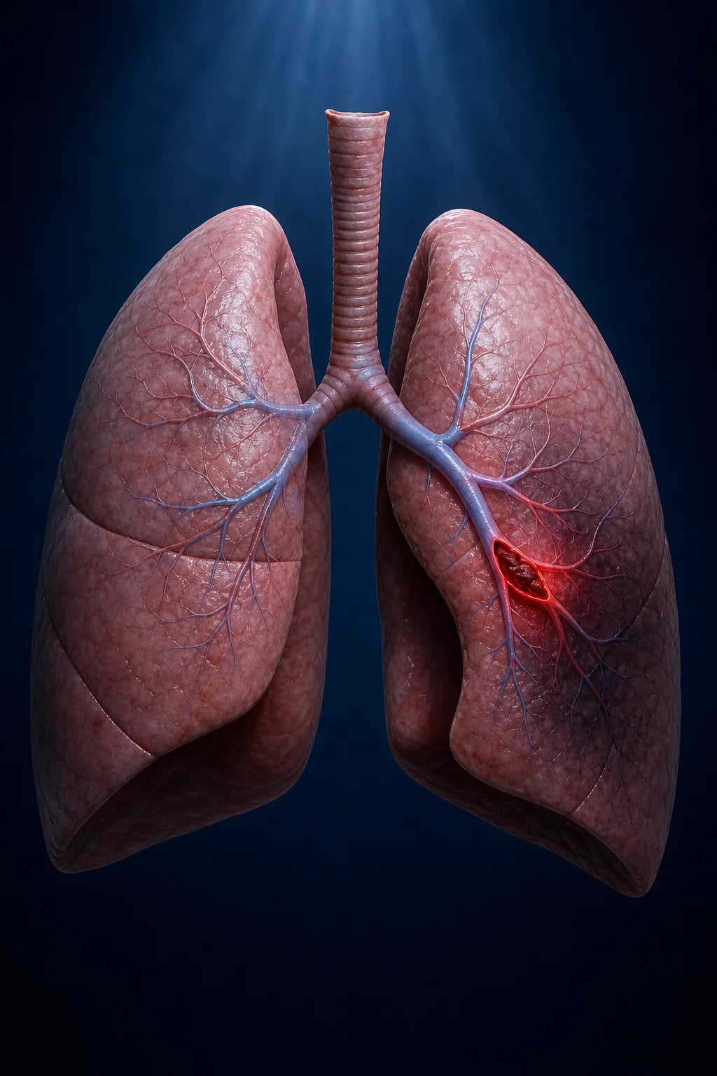
Venous thromboembolism (VTE) is the umbrella clinical syndrome that encompasses deep vein thrombosis (DVT) and its principal complication, pulmonary embolism (PE). It is best understood not as two separate diseases but as a single pathophysiological continuum in which a thrombus, characteristically originating in the deep veins of the lower limb, may remain confined locally (causing DVT), propagate proximally, or embolise through the right heart into the pulmonary arterial tree (causing PE).[1][3][11]
The clinical importance of VTE lies in its scale, lethality, and preventability. VTE is the third commonest cardiovascular disease after acute MI and stroke, with an annual incidence of 1 to 2 per 1000 adults, rising sharply after the age of 70 to about 5 to 7 per 1000 per year. In adults over 80 the lifetime prevalence approaches 10 percent. Acute PE is responsible for an estimated 300,000 deaths per year in Europe and 60,000 to 100,000 in the United States — many of them in patients who were already hospitalised. Up to 60 percent of hospital-associated VTE events are preventable with appropriate pharmacological thromboprophylaxis, which is the cornerstone of the in-hospital "VTE-bundle" (risk assessment on admission, LMWH or DOAC for at-risk patients, ambulation).[3]
The exam-relevant concepts in VTE are (1) recognising the unified pathophysiology (Virchow's triad) so that DVT and PE are treated as one disease with predictable natural history; (2) applying clinical probability scores (Wells DVT, Wells PE, PERC) before testing, because D-dimer is a rule-out test whose positive predictive value is far too low to "rule-in"; (3) identifying the high-risk clinical scenarios — massive PE, proximal DVT, recurrent unprovoked VTE, cancer-associated VTE, pregnancy — because each demands a specific management plan; (4) choosing between DOACs (apixaban, rivaroxaban, dabigatran, edoxaban), LMWH, and warfarin with attention to the specific patient factors (renal function, drug interactions, pregnancy, cancer); (5) recognising the complications — CTEPH, post-thrombotic syndrome, recurrent VTE, anticoagulant bleeding, HIT — which drive long-term outcomes as much as the acute event itself.[1][2][11]
Definition (precise). VTE = objectively confirmed DVT, PE, or both, in any venous distribution. DVT = thrombotic occlusion of a deep vein (most often infrainguinal — tibial, peroneal, popliteal, femoral — but also iliac, IVC, pelvic/gonadal, axillary/subclavian, cerebral [cerebral venous sinus thrombosis], portal/splanchnic, renal). PE = lodgement of a venous thrombus in the pulmonary arterial tree, characterised in life by acute right-ventricular overload, hypoxaemia and (in massive PE) cardiogenic-obstructive shock.[1]
Classification
VTE is classified by anatomical location, by clinical severity, by provocation (provoked vs unprovoked), by underlying pathophysiology, and by temporal pattern (incident vs recurrent) — these classifications are not academic: they drive the treatment choice and duration.[1][2]
Anatomical — DVT (proximal vs distal)
- **Distal (calf) DVT** = infra-popliteal (peroneal, tibial, soleal, gastrocnemial veins); roughly half of all lower-limb DVT; risk of embolisation ~5 to 10 percent if untreated
- **Proximal DVT** = popliteal, femoral or iliac (the knee-to-groin veins and above); the embologenic DVT; risk of embolisation up to 40 to 50 percent if untreated — the group that mandates anticoagulation
- **Iliac (iliofemoral) DVT** = proximal, large, with the highest risk of PE, the highest risk of post-thrombotic syndrome, and the rare complication of phlegmasia cerulea dolens — may need catheter-directed thrombolysis
- **Upper-extremity DVT** (subclavian/axillary/jugular) — usually catheter-related or Paget-Schroetter (thoracic outlet syndrome); 10 to 20 percent of DVT; growing problem in cancer patients with indwelling catheters
Anatomical — PE (massive vs submassive vs low-risk)
- **High-risk (massive) PE** = PE with **haemodynamic instability** — SBP below 90 mmHg or shock (about 5 percent of PE); mortality ~30 percent untreated; falls to 6 to 8 percent with thrombolysis; criterion for systemic thrombolysis (alteplase 100 mg IV over 2 h)
- **Intermediate-risk (submassive) PE** = normotensive with **evidence of RV dysfunction** (echo: RV dilatation/hypokinesis; CT: RV/LV ratio over 1.0) **or myocardial injury** (troponin/BNP elevation); about 25 to 40 percent of PE; mortality 3 to 15 percent; consider thrombolysis or catheter-directed therapy in selected
- **Low-risk PE** = normotensive, no RV dysfunction, no myocardial injury, low-risk on PESI/sPESI (about 50 percent of PE); mortality below 1 percent; anticoagulation alone, often outpatient-managed
- **Anatomical lobar**: lobar, segmental, subsegmental; saddle PE lodges at the bifurcation of the main pulmonary artery and is a form of high-risk PE
Provoked vs unprovoked
- **Provoked VTE** = an identifiable transient or persistent major provoking factor within the preceding 3 months — surgery, trauma, hospitalisation, immobility (over 3 days), pregnancy/postpartum (within 6 weeks), oestrogen (combined oral contraceptive, HRT), or active cancer
- **Unprovoked VTE** = no identifiable provoking factor; ~30 to 40 percent of VTE; associated with a 10 to 15 percent rate of occult-cancer detection in the year after diagnosis and a 5 to 10 percent rate of inherited thrombophilia
- **Persistent risk factor** = active cancer, inflammatory bowel disease, myeloproliferative neoplasm, paroxysmal nocturnal haemoglobinuria, ongoing immobilisation — drives prolonged/extended anticoagulation
- **Recurrent VTE** = a second VTE event in a patient with prior VTE; risk after stopping anticoagulation is roughly 10 percent at 1 year and 25 percent at 5 years for unprovoked events
By underlying mechanism
- **Provoked by stasis** — immobility, long-haul travel (over 6 h), heart failure, post-operative state
- **Provoked by endothelial injury** — surgery, trauma, central venous catheters, IV drug use (IVDU may also cause septic pulmonary emboli and right-heart endocarditis)
- **Provoked by hypercoagulability** — inherited (factor V Leiden, prothrombin G20210A, protein C/S/antithrombin deficiency), acquired (antiphospholipid syndrome, cancer, pregnancy, oestrogen, nephrotic syndrome, paroxysmal nocturnal haemoglobinuria, inflammatory bowel disease)
- **Septic emboli and tumour emboli** — distinct entities, often produce fever and peripheral lesions (Janeway lesions, Osler nodes); non-anticoagulation pathways dominate treatment
The classification that drives the acute decision is PE severity (massive / intermediate / low), which integrates haemodynamics plus an assessment of RV status by imaging (echo, CT) and biomarkers (troponin, BNP). The classification that drives the duration of anticoagulation is provoked vs unprovoked vs recurrent vs cancer-associated.[1][2]
Epidemiology & Risk Factors
VTE is a common, recurrent, lethal and often hospital-acquired disease. Global burden: roughly 10 million VTE events per year worldwide; annual incidence in adults 1 to 2 per 1000, rising to 5 to 7 per 1000 over 70 years and 10 to 15 per 1000 over 80. Acute PE is the commonest preventable cause of in-hospital death in many health systems, and roughly half of all VTE events are associated with a recent hospitalisation.[3]
Demographic risk factors: age (exponential risk increase after 60), male sex (slightly higher incidence), Black ethnicity (about 1.5-fold higher risk, partly Factor VIII and von Willebrand factor), obesity (BMI over 30 — roughly 2-fold risk), prior VTE (the dominant predictor of recurrence — about 8-fold), varicose veins.[3]
Genetic / thrombophilia risk factors: factor V Leiden (most common — 5 percent of Caucasians; heterozygous 4 to 7-fold risk, homozygous 20-fold), prothrombin G20210A (2 to 3 percent of Caucasians — 3 to 4-fold risk), protein C / protein S / antithrombin deficiency (rare, but high-risk — up to 10-fold). Combined defects or antiphospholipid syndrome carry the highest lifetime risk. [1]
Acquired risk factors (the in-hospital "VTE bucket"): surgery (especially orthopaedic — hip/knee replacement, hip fracture; abdominal, pelvic and cancer surgery — risk highest in the first 4 weeks post-op), trauma / spinal cord injury, hospitalisation and immobility (over 3 days bed-rest), active cancer (4 to 7-fold risk — particularly pancreatic, gastric, brain, ovarian, lung; 15 to 20 percent of all VTE), chemotherapy and anti-angiogenic agents (bevacizumab, lenalidomide, thalidomide, L-asparaginase), central venous catheters (upper-extremity DVT), pregnancy and postpartum (5-fold risk; highest in the immediate 6 weeks postpartum), oestrogen (combined oral contraceptive — 3 to 4-fold risk; falls to 1-fold with progestogen-only; HRT — 2 to 3-fold), inflammatory bowel disease, nephrotic syndrome, myeloproliferative neoplasms (polycythaemia vera, essential thrombocythaemia — paradoxically predispose to both thrombosis and bleeding), paroxysmal nocturnal haemoglobinuria (terminal complement-mediated hypercoagulability). [1]
Temporary transient risk factors: long-haul travel (over 6 hours — modest, but well-publicised; the "economy-class syndrome"), dehydration, acute medical illness (pneumonia, COVID-19, sepsis — the COVID-19 pandemic produced a measurable surge in hospital-associated VTE).[3][1]
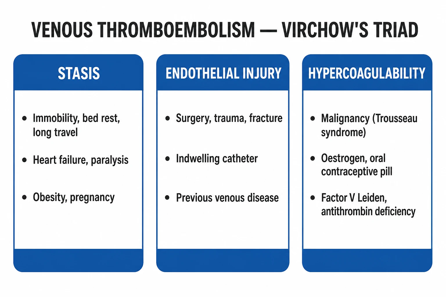
The fundamental substrate is Virchow's triad (1856) — venous stasis, endothelial injury, hypercoagulability — and most clinical risk factors act on one or more of these three axes. The bedside test for any patient is to ask, "Which axis(es) of Virchow's triad am I operating on here, and is the risk transient or persistent?" — this directly informs the duration of anticoagulation.[1]
Venous thromboembolism — key numbers
Pathophysiology
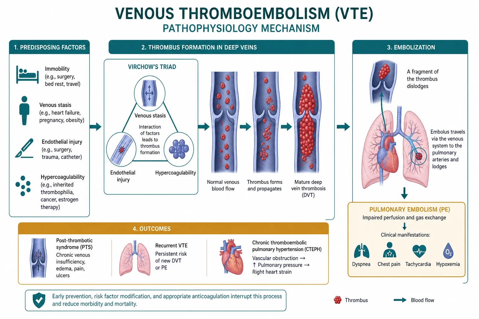
VTE is the thrombotic manifestation of Virchow's triad. Understanding each axis is essential because each clinical risk factor acts predominantly on one or more axis, and each pharmacological treatment interrupts one or more axis.[1][3]
1. Venous stasis. Slow flow in the calf veins (especially at the valve cusps) prolongs the contact time between activated coagulation factors and the endothelium, and reduces the washout of activated clotting factors by the liver. Stasis is the dominant factor in immobility (post-operative, long-haul travel, hospitalisation, heart failure), obesity (low-flow state), and pregnancy (aortocaval compression plus venous dilatation). The valves themselves are the typical nidus for thrombus formation — the valve cusp is the most common site of origin of calf DVT. [1]
2. Endothelial injury. The endothelium is normally antithrombotic, antiplatelet and profibrinolytic (heparan sulphate, thrombomodulin, nitric oxide, prostacyclin, tissue plasminogen activator). Injury exposes subendothelial collagen and tissue factor, activates platelets, and triggers the coagulation cascade. Endothelial injury dominates post-operative VTE (especially orthopaedic — direct venous trauma plus tourniquet plus immobility), central venous catheter insertion, IV drug use (a major cause of septic DVT and right-heart endocarditis), and trauma / fracture. [1]
3. Hypercoagulability — inherited and acquired. Thrombophilia raises the baseline activity of the coagulation cascade or reduces the activity of natural anticoagulant pathways. Inherited: factor V Leiden (resistance to activated protein C — about 5 percent of Caucasians), prothrombin G20210A (raised prothrombin levels), protein C / S / antithrombin deficiency. Acquired: cancer (tissue factor-bearing microparticles; inflammation; compression of pelvic veins; chemotherapy-related), antiphospholipid syndrome (lupus anticoagulant, anticardiolipin, anti-β2-glycoprotein-I antibodies — a prothrombotic state paradoxically manifesting in vitro as prolonged aPTT), oestrogen-induced hepatic synthesis of clotting factors (fibrinogen, factors VII, X), pregnancy (raised clotting factors, decreased protein S, venous compression), nephrotic syndrome (loss of antithrombin and protein S in urine), myeloproliferative neoplasms, PNH. [1]
Thrombus formation. The classical model is the vessel-wall injury → platelet adherence → platelet activation → coagulation cascade activation → fibrin deposition sequence (Virchow, then refined by Davie, Ratnoff and Macfarlane, and then the modern cell-based model). In veins, however, the platelet contribution is small relative to the fibrin/red-cell "red clot" pattern (in contrast to the platelet-rich "white" arterial thrombus). The thrombus grows layered (lines of Zahn) — alternating pale (platelets/fibrin) and dark (red cells) bands — anchored at a valve cusp and propagating in the direction of flow. [1]
Embolisation. A fragment — most often the free-floating proximal tail of a propagating DVT — breaks off, travels through the right heart, and lodges in the pulmonary arterial tree. The larger the embolus, the more proximal the occlusion: a large embolus may straddle the bifurcation of the main pulmonary artery (saddle embolus) and produce immediate right-ventricular afterload failure (massive PE). Multiple smaller emboli produce sum-of-parts occlusion.[3]
Why the right ventricle fails in massive PE. A normal RV cannot generate a systolic pressure above about 50 mmHg. Acute obstruction of more than about 50 percent of the pulmonary arterial bed raises pulmonary artery pressure above this, RV afterload rises acutely, RV stroke volume falls, and the thin-walled right ventricle dilates and fails — the LV is underfilled (displaced interventricular septum, reduced LV preload, falling cardiac output, hypotension, shock). The same pathophysiology underpins the RV strain pattern on echo (RV dilatation, hypokinesis, septal flattening — "D-sign") and the CT finding of RV/LV ratio greater than 1.0. Compensatory neurohormonal activation (sympathetic tachycardia, RV ischaemia) raises troponin and BNP.[1][3]
Clinical Presentation
The clinical face of VTE depends on anatomical distribution and severity. Many events are silent or atypical, particularly in the elderly, in post-operative patients and in those with chronic cardiorespiratory disease.[1]
DVT — symptoms and signs. At least half of all calf-vein DVTs are clinically silent. When symptomatic, the classical pentad is: [1]
- Unilateral leg swelling — the most sensitive sign; the asymmetry (more than 3 cm difference in calf circumference at 10 cm below the tibial tuberosity) is part of the Wells score; pitting oedema confined to the symptomatic leg.
- Pain — deep, dull, often in the calf on dorsiflexion of the foot (Homan's sign — sensitive but non-specific); palpable cord along the line of the deep venous system (the thrombosed vein).
- Erythema and warmth — the skin overlying the thrombus is visibly erythematous and warm to the touch; cyanosis or duskiness in iliofemoral DVT.
- Tenderness — on palpation of the calf or thigh deep veins; palpable cord in superficial thrombophlebitis (different condition).
- Prominent superficial veins (collaterals) — a sign of chronic deep venous obstruction (the superficial veins dilate to bypass). [1]
Phlegmasia cerulea dolens and phlegmasia alba dolens are the dramatic extreme of iliofemoral DVT: the entire leg becomes massively swollen, painful, dusky (cerulea dolens), then pale and pulseless (alba dolens) — venous gangrene is the dreaded endpoint; this is the one circumstance where catheter-directed thrombolysis is considered. [1]
A bilateral leg swelling is more often due to right heart failure, nephrotic syndrome, IVC obstruction or compression than bilateral DVT. [1]
PE — symptoms and signs. The classical syndrome is: [1]
- Dyspnoea (tachypnoea) — the commonest symptom; in the PIOPED study, dyspnoea and tachypnoea were present in about 70 to 80 percent of PE.
- Pleuritic chest pain — sharp, worse on inspiration; reflects pulmonary infarction or pleural irritation.
- Tachycardia — sinus tachycardia (heart rate over 100/min) is the commonest sign; over 110/min is part of the Wells score.
- Cough and haemoptysis — the latter is a Wells item; classically a feature of pulmonary infarction.
- Syncope or pre-syncope — the most distinctive symptom of massive PE; signals acute right-ventricular failure with low cardiac output.
- Hypotension and shock — the definition of massive / high-risk PE (SBP under 90 mmHg or needing vasopressors); the bedside examination shows cool peripheries, oliguria, cyanosis, raised JVP. [1]
The physical signs of PE are also non-specific: tachypnoea, tachycardia, low-grade fever, clear lung fields (or pleuritic rub, or localised crackles), accentuation of the pulmonary component of the second heart sound (P2 loud), right ventricular heave, raised JVP, a fourth heart sound. A unilateral pleural rub is classically heard in pulmonary infarction. S1Q3T3 pattern (S wave in lead I, Q wave in lead III, inverted T in lead III) is a classic ECG clue — found in about 10 to 15 percent of massive PE, not sensitive. [1]
Massive / submassive / low-risk PE by clinical features.
- Massive (high-risk) PE: SBP below 90 mmHg or shock; syncope; cardiac arrest; cool peripheries. Mortality ~30 percent untreated.
- Submassive (intermediate-risk) PE: normotensive with RV strain (echocardiographic or CT evidence of RV dysfunction) OR raised cardiac biomarkers (troponin, BNP). Mortality 3 to 15 percent.
- Low-risk PE: normotensive, no RV strain, no biomarker elevation, low PESI / sPESI score. Mortality below 1 percent. [1]
Atypical presentations (high-yield):
- In the elderly — a fall, an unexplained persistent tachycardia, an unexplained drop in functional status, a worsening of chronic heart failure or COPD, "another admission with pneumonia", confusion.
- In pregnancy — pleuritic chest pain, tachycardia and mild hypoxia may be attributed to dyspnoea of pregnancy; a threshold of suspicion is needed.
- In post-operative patients — a low-grade fever, an unexplained tachycardia and hypoxia on day 3 to 5 after major orthopaedic or abdominal surgery.
- Recurrent VTE — unilateral leg swelling or pleuritic pain in someone with previous VTE is recurrent VTE until proven otherwise.[1][3]
Differential Diagnosis
VTE has a wide differential on both sides of the circulation. The first task is to think DVT vs a mimicker in a swollen leg, and PE vs a mimicker in acute dyspnoea and pleuritic chest pain — because each mimicker has a different treatment and the anticoagulation of VTE is not benign.[1]
DVT mimickers
- **Cellulitis / erysipelas** — diffuse erythema, warmth, tenderness; often with fever; the **swelling is usually bilaterally acute and the erythema is sharply demarcated with lymphangitis**; ultrasound excludes co-existing DVT
- **Ruptured Baker's (popliteal) cyst** — sudden onset of calf pain and swelling; **ultrasound shows the cyst** and a normal deep venous system; commonly misdiagnosed as DVT but the history is typically acute-on-chronic with knee arthritis
- **Musculoskeletal calf pain / gastrocnemius tear** — trauma, focal muscle tenderness, normal Doppler ultrasound
- **Chronic venous insufficiency / post-thrombotic syndrome** — bilateral, chronic, with skin changes (lipodermatosclerosis, haemosiderin, varicose veins); history of previous DVT establishes the diagnosis
- **Superficial thrombophlebitis** — palpable tender cord in the territory of the great or small saphenous vein; **DVT must be excluded** if the thrombophlebitis extends to within 5 cm of the saphofemoral junction (then treat as DVT)
- **Lymphoedema / cellulitis / pelvic mass / IVC compression** — bilateral in pelvic or IVC obstruction; unilateral in lymphoedema; classically non-pitting after a chronic phase
- **Heart failure, nephrotic syndrome, cirrhosis, pregnancy** — bilateral leg swelling, no unilateral tenderness, ultrasound clarifies
PE mimickers
- **Pneumonia / pneumonitis / COVID-19** — fever, productive cough, focal consolidation, ground-glass (COVID); PE may co-exist (especially in hospitalised COVID-19) — the threshold for CTPA in unexplained hypoxia must be low
- **Pneumothorax / pleural effusion** — unilateral pleuritic chest pain, hyper-resonance (pneumothorax) or stony dullness (effusion), tracheal deviation; CXR discriminates
- **Acute coronary syndrome** — central chest pain, ECG changes (ST elevation/depression, T-wave inversion), positive troponin; massive PE may also have ECG changes and a mildly elevated troponin — the combination of dyspnoea + chest pain + raised D-dimer raises PE above ACS
- **Acute aortic dissection** — severe tearing chest/back pain, **asymmetric pulses, BP differential, widened mediastinum on CXR**; CT aortogram distinguishes
- **Pericarditis / myopericarditis** — sharp retrosternal chest pain relieved by sitting forward, pericardial rub, diffuse ST elevation and PR depression; troponin may be mildly elevated
- **Acute decompensated heart failure** — known HF, S3 gallop, bilateral crackles, raised JVP, raised BNP; PE may co-exist in unexplained decompensation
- **Anxiety, hyperventilation, panic attack** — the highest rate of inappropriate CTPA; PERC rule applied at triage can safely avoid testing in low-probability presentations
- **Rib fracture, musculoskeletal chest pain, herpes zoster** — pain is reproducible on palpation, dermatomal, with normal vitals
When to dual-test for VTE + mimicker
- **Unexplained pleuritic chest pain + hypoxia + raised D-dimer in pregnancy** — PE investigation with D-dimer and CTPA (CT pulmonary angiogram with shielding of the maternal breast can be performed in pregnancy with a dose-reduction protocol; MRI/lung scintigraphy V/Q scan alternative)
- **Outpatient pleuritic chest pain + bleeding risk + raised D-dimer** — V/Q SPECT may be preferred over CTPA (less radiation, less nephrotoxicity)
- **Calf swelling + incarcerated groin hernia / pelvic mass** — Doppler USS for DVT plus CT pelvis for the mass
- **Post-operative PE vs pneumonia** — CXR, CTPA, and microbiology — the standard CTPA protocol will show an infarct, an effusion, and a consolidation
- **Suspected CTEPH after PE** — V/Q scan is the **first-line screening test** (segmental unmatched perfusion defects); CTPA may miss surgically accessible disease
The single best discriminator is to ask "does the swelling involve the deep venous system, and does the chest pain involve the pulmonary arterial tree?" — Doppler ultrasound and CT pulmonary angiography answer those questions respectively with high sensitivity and specificity. [1]
Clinical & Bedside Assessment
The bedside assessment is the clinical probability, not the diagnosis. Probability scores determine whether the D-dimer rules out, the CTPA/U.S. rules in, and whether empirical anticoagulation is started while waiting for a definitive test.[1]
Wells score for DVT (Wells et al., Lancet 1997; revised 2001) — the most widely validated clinical probability score for DVT: [1]
- Active cancer (within 6 months or palliative) — 1
- Paralysis, paresis or recent plaster immobilisation of the lower extremity — 1
- Bedridden for 3 days or major surgery within 12 weeks — 1
- Localised tenderness along the distribution of the deep venous system — 1
- Entire leg swollen — 1
- Calf swelling more than 3 cm compared to the asymptomatic side (measured 10 cm below the tibial tuberosity) — 1
- Pitting oedema confined to the symptomatic leg — 1
- Collateral superficial veins (non-varicose) — 1
- Alternative diagnosis as likely as DVT — −2 (subtracts points) [1]
Interpretation: Wells score ≥ 3 = high probability (about 75 percent prevalence of DVT); 1 to 2 = moderate; ≤ 0 = low probability (about 5 percent prevalence). High probability → Doppler ultrasound; moderate → D-dimer first, ultrasound if D-dimer positive; low → D-dimer first, ultrasound if D-dimer positive. D-dimer of the appropriate assay (age-adjusted for over-50s) below the cutoff with low probability → DVT excluded. [1]
Wells score for PE (Wells et al., Thromb Haemost 2000; revised 2001): [1]
- Clinical signs of DVT (leg swelling, pain with palpation of the deep veins) — 3
- PE the most likely diagnosis (or equally likely) to alternative — 3
- Heart rate over 100/min (tachycardia) — 1.5
- Immobilisation (bed rest over 3 days) or surgery in the previous 4 weeks — 1.5
- Previous DVT/PE — 1.5
- Haemoptysis — 1
- Active cancer (within 6 months or palliative) — 1 [1]
Interpretation: Wells > 6 = high probability (about 60 percent PE prevalence), 2 to 6 = moderate, less than 2 = low. High or moderate → D-dimer (rule out) or directly to CTPA. Low → D-dimer, CTPA only if positive. The simplified dichotomised version (PE likely / PE unlikely) is used in many emergency departments: "PE likely" combines moderate and high. [1]
PERC rule (Kline, Acad Emerg Med 2004) — designed to avoid unnecessary D-dimer testing in low-probability presentations. A patient is PERC-negative when ALL 8 criteria are absent: [1]
- Age under 50
- Pulse (heart rate) below 100/min
- SaO2 at least 95 percent on room air
- No unilateral leg swelling
- No haemoptysis
- No surgery or trauma within 4 weeks
- No prior DVT or PE
- No oestrogen use [1]
If PERC-negative AND clinical probability is low, PE can be excluded without a D-dimer — saving a test in about 20 percent of patients presenting with possible PE. [1]
Homan's sign — calf pain on forced dorsiflexion of the foot — has high specificity but poor sensitivity. It is largely historical; modern practice uses Doppler ultrasound rather than clinical signs alone. [1]
Bedside assessment of PE severity: [1]
- Vital signs — heart rate, blood pressure, respiratory rate, oxygen saturation, temperature.
- JVP — raised in massive / submassive PE (right-heart failure).
- Cardiac examination — loud P2, right ventricular heave, fourth heart sound, a third heart sound is ominous.
- Pulmonary — clear lungs (no pulmonary oedema); unilateral rub / infarction.
- Lower limbs — look for signs of DVT in either leg; the asymmetric swollen leg is the bedside clue that the dyspnoea is embolic.
- Mottled skin, cool peripheries — circulatory collapse (high-risk PE).
- Syncope or pre-syncope — high-risk PE until CTPA / echo excludes. [1]
The PESI (Pulmonary Embolism Severity Index) and the simplified sPESI add independent prognostic information: age over 80, cancer, chronic cardiopulmonary disease, heart rate over 110, SBP below 100, oxygen saturation below 90 percent. sPESI 0 = low mortality (about 1 percent) — candidate for outpatient management.[1][2]
Investigations
The test ladder for VTE depends on the anatomical target and the clinical probability.[1][2]
D-dimer — a fragment of cross-linked fibrin released during fibrinolysis; highly sensitive (98 percent) but poorly specific. A normal D-dimer in a low- or moderate-probability patient essentially rules out VTE (negative likelihood ratio less than 0.05). A positive D-dimer is not diagnostic (raised in malignancy, infection, pregnancy, post-operative state, advanced age). Age-adjusted cutoff (age × 10 ng/mL for over-50s) improves specificity in the elderly without losing sensitivity. Modern high-sensitivity D-dimer assays (VIDAS, Innovance, STA-Liatest) — when combined with a low-risk score — have rule-out thresholds with sensitivities over 99 percent. [1]
Doppler compression ultrasound of the lower-limb veins — the imaging standard for DVT. Non-compressibility of the vein under probe pressure is the diagnostic criterion; flow augmentation, colour flow and spectral Doppler augment the assessment. Sensitivity for proximal DVT is over 95 percent; distal (calf) DVT is harder to image but usually less clinically significant (lower embolic risk; can be serially monitored if Wells is low and isolated distal DVT is found). One-shot examination of the symptomatic leg is sufficient in low-probability presentations; serial ultrasound (1 week) is used when D-dimer is positive but the first ultrasound is negative. [1]
CT pulmonary angiography (CTPA) — the gold standard for PE. Multidetector CT identifies a filling defect within the pulmonary arterial tree down to the subsegmental level. Sensitivity roughly 83 percent at the gold standard (PIOPED II), specificity 96 percent. CTPA has now largely replaced V/Q scintigraphy because it is faster, more widely available, gives an alternative diagnosis (pneumonia, effusion, dissection), and quantifies RV strain (RV/LV ratio). Risks: radiation (one CTPA ≈ 5 to 7 mSv, less than V/Q), iodinated contrast (allergy, AKI — particularly in CKD), breast cancer risk in young women, and overdiagnosis of subsegmental PE.[1]
V/Q scintigraphy — the functional lung scan, useful in pregnancy (lower foetal radiation than CTPA in some protocols) and in young patients with contraindications to iodinated contrast. A normal V/Q scan excludes PE; a high-probability V/Q scan (multiple segmental unmatched perfusion defects) is diagnostic of PE. Many V/Q scans are intermediate-probability — in pregnancy, SPECT V/Q is preferred. [1]
Pulmonary angiography — the historical gold standard, now rarely performed (replaced by CTPA). [1]
Echocardiography — bedside, non-invasive, immediately actionable in suspected massive PE. RV dilatation and hypokinesis (RV end-diastolic area > LV), septal flattening (D-sign), McConnell's sign (RV free-wall akinesis with apical sparing), tricuspid regurgitation, PASP elevation are the bedside findings. A normal echocardiogram essentially excludes massive PE in the hands of an experienced sonographer. Echo also identifies the differential of obstructive shock (tamponade) and other mimics.[3]
Cardiac biomarkers — troponin I/T (RV micro-infarction is the source; raised in submassive and massive PE; prognostic for 30-day mortality), BNP and NT-proBNP (raised in submassive / massive PE — RV stretch; prognostic). Both are independent prognostic variables in haemodynamically stable PE and are part of the ESC risk stratification (intermediate-high if both echo and biomarker are positive). [1]
ABG — hypoxaemia, hypocapnia (respiratory alkalosis from hyperventilation) is the typical pattern in PE; hypoxia with a normal A-a gradient is unusual and may suggest an alternative diagnosis. End-tidal CO2 is often reduced (ETCO2 below 28 mmHg in massive PE). [1]
Thrombophilia screening — only in selected patients after provoked unprovoked events, with the intention of changing the duration or choice of anticoagulation. Screen in young (< 50), unprovoked VTE; recurrent VTE; unusual site (mesenteric, cerebral venous sinus); family history of VTE; recurrent foetal loss. Testing is best performed at least 4 weeks after stopping anticoagulation (acute-phase changes confound factor VIII, antithrombin, protein S). The panel includes factor V Leiden, prothrombin G20210A, protein C, protein S, antithrombin, lupus anticoagulant, anticardiolipin, anti-β2-glycoprotein-I, homocysteine. [1]
Other investigations as dictated by the differential: ECG (excludes ACS, shows S1Q3T3, right-bundle-branch block, T-wave inversion V1 to V3), CXR (pneumothorax, effusion, consolidation, widened mediastinum for dissection), D-dimer / troponin / BNP / renal function / liver function / coagulation screen / CBC (thrombocytosis, polycythaemia, leukaemic white count), anti-Xa levels (in patients on LMWH with extremes of weight or renal function), upper-limb Doppler / CT venogram for catheter-related upper-extremity DVT, transthoracic / transoesophageal echo for RV strain and PFO (paradoxical embolus).[1][2]
VTE investigations — outcomes
Management — Resuscitation
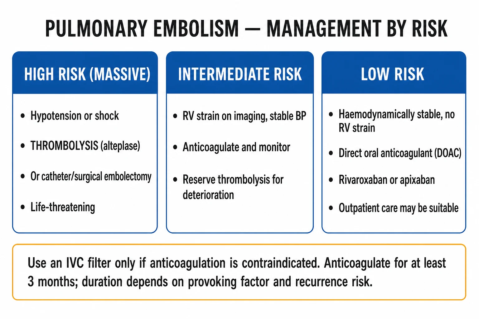
Massive / high-risk PE is a resuscitation emergency. The patient is dying from acute right-ventricular failure; the only interventions proven to reduce mortality are immediate systemic thrombolysis (or, if thrombolysis is absolutely contraindicated, surgical embolectomy / catheter-directed therapy). The principles: acknowledge the diagnosis on haemodynamics alone in the deteriorating patient — do not delay treatment for CTPA if the patient is in shock, RV strain is unequivocal on echo, and alternative causes (tamponade, tension pneumothorax, acute MI) have been excluded at the bedside.[1][3][10]
ABCDE and oxygen:
- High-flow oxygen to target SpO2 94 to 98 percent; intubate and ventilate if tiring or comatose — note that PE patients on IPPV deteriorate rapidly because sedation and positive pressure reduce preload (already compromised); the threshold is high.
- Two large-bore cannulae, arterial line, central venous access, urinary catheter — same as any resuscitation.
- ECG, bedside echo, ABG, lactate, set of troponin and BNP — for risk stratification and to confirm the diagnosis at the bedside. [1]
Volume resuscitation — cautious. A small 250 mL crystalloid bolus over 15 minutes is reasonable if the patient is not visibly overloaded; excess fluids worsen RV overload and precipitate arrest. The patient is preload-insensitive because the failure is afterload. [1]
Vasoactive support while arranging thrombolysis. Noradrenaline is the first-line vasopressor — it raises MAP, supports RV coronary perfusion, and modestly improves RV inotropy without major change in pulmonary vascular resistance. Dobutamine at 5 to 15 mcg/kg/min may improve RV contractility but lowers BP and is best combined with noradrenaline. Avoid pure systemic vasodilators (GTN, sodium nitroprusside) — they lower RV coronary perfusion and accelerate collapse.[1]
Systemic thrombolysis (massive / high-risk PE).
- Alteplase 100 mg IV infusion over 2 hours (the standard regimen); or accelerated regimen 0.6 mg/kg IV over 15 minutes — used in cardiac arrest / peri-arrest PE (the European Society of Cardiology-bolused regimen), with a maximum of 50 mg.
- Indication — haemodynamically unstable PE (SBP below 90 mmHg or shock) without absolute contraindication. Do NOT delay for CTPA if the diagnosis is supported by echo.
- Relative contraindications — recent surgery, recent GI bleed, intracranial neoplasm, recent stroke/TIA (< 3 to 6 months absolute), active bleeding, severe uncontrolled hypertension.
- Bleeding risk — about 2 to 3 percent intracranial haemorrhage; 13 percent major bleeding overall.
- Concurrent anticoagulation — UFH is generally given at the start; stop the UFH infusion during the alteplase 2 h infusion and resume once the fibrinogen and clinical bleeding risk allow. [1]
Catheter-directed thrombolysis (CDT) and mechanical thrombectomy — for high-risk PE if systemic thrombolysis is absolutely contraindicated, or for selected intermediate-high-risk PE where systemic thrombolysis bleeding risk is thought high or where expertise is available (ultrasound-facilitated catheter-directed alteplase 5 to 10 mg per lung over hours, or suction thrombectomy devices — FlowTriever, Indigo). The PEITHO-3 and other trials are refining which intermediate-risk patients benefit; current ESC recommends considering CDT in intermediate-high PE if expertise is available.[1][10]
Surgical embolectomy — the bilateral pulmonary artery thrombectomy on cardiopulmonary bypass, performed by an experienced cardiac surgeon. Indicated when thrombolysis has failed or is absolutely contraindicated, and as a salvage in cardiac arrest. [1]
ECMO (VA-ECMO) — bridging to recovery or surgery — has emerged as a salvage for refractory cardiogenic shock / arrest from massive PE in experienced centres. [1]
Empirical anticoagulation at the bedside. In a stable patient with intermediate or high probability, start LMWH (enoxaparin 1 mg/kg SC BD) while arranging confirmatory imaging — bleeding risk if no contraindication is very low. [1]
Management — Definitive & Stepwise
The definitive management of VTE is prompt anticoagulation, risk-stratified duration, and prevention of complications. The escalation ladder for severe PE is anticoagulation alone (low-risk) → anticoagulation + monitoring (intermediate-low) → rescue CDT/IVC filter (intermediate-high) → systemic thrombolysis or surgical embolectomy (massive).[1][2]
Step 1 — Anticoagulation, first-line (DOACs)
The 2019 ESC and 2018 ASH guidelines converge on DOACs first-line for the majority of VTE patients without cancer or pregnancy.[1][2]
Apixaban
- **Apixaban 10 mg BD orally for 7 days**, then **5 mg BD indefinitely** (AMPLIFY trial — NEJM 2013: non-inferior to enoxaparin/warfarin with less major bleeding)
- Direct factor Xa inhibitor; oral; fixed dose; **no INR monitoring**
- **Cautions**: CrCl under 15 ml/min (avoid or use warfarin), age over 80 with body weight under 60 kg or creatinine over 1.5 mg/dL (2 of 3 → reduce to 2.5 mg BD per AMPLIFY)
- **Bleeding management**: andexanet alfa in major bleeding if anti-Xa activity is the target of reversal
- **Drug interactions**: strong dual CYP3A4/P-gp inhibitors (ketoconazole, ritonavir) → avoid; strong inducers (rifampicin, carbamazepine, phenytoin) → avoid
- Approved for VTE treatment; extended treatment supported by AMPLIFY-EXT (2.5 mg or 5 mg BD vs placebo — 80 percent reduction in recurrence)
Rivaroxaban
- **Rivaroxaban 15 mg BD orally for 21 days** then **20 mg daily** for the maintenance phase (EINSTEIN-DVT NEJM 2010 and EINSTEIN-PE NEJM 2012: non-inferior to enoxaparin/VKA with similar bleeding profile)
- Direct factor Xa inhibitor; oral; fixed dose; taken with food (the 15 mg and 20 mg tablets have better bioavailability with food)
- **Cautions**: CrCl under 15 ml/min → avoid; child-Pugh B/C → avoid; CrCl 15 to 50 → caution
- **Bleeding management**: andexanet alfa (under licence/expert use)
- **Drug interactions**: azole antifungals, HIV protease inhibitors → avoid; inducers (rifampicin, carbamazepine, phenytoin, St John's wort) → avoid
- The most-used DOAC in the peri-operative orthopaedic VTE prophylaxis setting
Dabigatran
- **Dabigatran 150 mg BD orally** (after a **minimum 5-day lead-in with LMWH** — because dabigatran was studied as a step-on to heparin in RE-COVER, NEJM 2009: non-inferior to warfarin, similar major bleeding, lower clinically relevant non-major bleeding)
- Direct thrombin (factor IIa) inhibitor; oral; capsule must be swallowed whole, kept in original packaging
- **Cautions**: CrCl under 30 ml/min → avoid; dyspepsia common (10 to 15 percent); risk of GI bleeding slightly higher than apixaban in some meta-analyses
- **Bleeding management**: idarucizumab 5 g IV in two doses — **specific, immediate reversal** for life-threatening bleeding or emergency surgery; PCC as alternative
Edoxaban
- **Edoxaban 60 mg daily orally** (after 5-day LMWH lead-in) for treatment of VTE — Hokusai-VTE NEJM 2013: non-inferior to warfarin, similar bleeding
- Reduce to **30 mg daily** if CrCl 15 to 50 ml/min, body weight under 60 kg, or concomitant P-glycoprotein inhibitor
- **Caution**: contraindicated if CrCl under 15 ml/min; sparse data in high-risk ASA or cancer subgroup
- **Bleeding management**: andexanet alfa (under licence/expert use); PCC as alternative
DOAC choice — practical synthesis (2018 ASH / 2019 ESC). [1]
- First-line across most non-cancer, non-pregnant adults is apixaban or rivaroxaban (no lead-in, fixed-dose, lower bleeding profile vs warfarin in pooled data).
- Rivaroxaban has the largest body of evidence in post-operative orthopaedic prophylaxis; the 15 mg BD for 21 days loading regimen should be taught explicitly.
- Dabigatran is reserved for patients in whom idarucizumab reversal is desirable (urgent surgery, life-threatening bleed) — the only DOAC with a specific antidote widely in use.
- Edoxaban is widely used in cancer-associated VTE in Japan and parts of Europe. [1]
Step 2 — LMWH (special indications)
LMWH (enoxaparin 1 mg/kg BD SC; dalteparin 200 IU/kg daily SC; tinzaparin 175 IU/kg daily SC) remains first-line in pregnancy and in cancer-associated VTE (although recent data challenge the latter). [1]
- Pregnancy and lactation — DOACs are contraindicated (insufficient safety data; dabigatran rivaroxaban and edoxaban are teratogenic in animal studies; LMWH does not cross the placenta). Dalteparin, enoxaparin and tinzaparin are safe throughout pregnancy.
- Cancer-associated VTE (active cancer) — LMWH was traditionally preferred (CLOT trial, Lee 2003). The recent ADAM VTE trial (Agnelli NEJM 2020) showed apixaban non-inferior to dalteparin for cancer-associated VTE, with a higher rate of major GI bleeding in GI / GU cancers. Current ASH and ASCO: either LMWH or apixaban or rivaroxaban (with caution in GI / GU cancers) is acceptable; LMWH still preferred in GI / GU cancer, very high bleeding risk, thrombocytopenia, or drug interactions.
- Severe renal impairment (CrCl below 30 ml/min) — LMWH accumulates; use unfractionated heparin (UFH) infusion (initially 18 IU/kg/h, titrate to aPTT 1.5 to 2.5× control) when rapid reversibility is needed.
- Thrombocytopenia / HIT — switch to argatroban, bivalirudin or danaparoid in HIT. [1]
Step 3 — Warfarin and unfractionated heparin
Warfarin (a vitamin K antagonist) is no longer first-line for VTE but retains a role in: mechanical heart valves (DOACs contraindicated), severe CKD (eGFR below 15), APLS with arterial events (DOACs are inferior to warfarin in this group — TRAPS trial), patient preference / availability.[2]
Regimen — start warfarin 5 to 10 mg daily (lower in elderly, frail, liver disease); start LMWH concurrently on day 1, continue LMWH for at least 5 days AND until INR is in therapeutic range (2.0 to 3.0) on two consecutive measurements (because the early procoagulant fall in proteins C and S can cause warfarin-induced skin necrosis if heparin is omitted). Target INR 2.0 to 3.0 (target 2.5). [1]
Unfractionated heparin (UFH) — IV infusion, 18 IU/kg/h (70 IU/kg bolus in acute PE), titrate to aPTT 1.5 to 2.5× control or anti-Xa 0.3 to 0.7 IU/mL. UFH is used when: renal failure (no accumulation), high bleeding risk (short half-life — protamine reversal), haemodynamic instability / imminent thrombolysis (immediate onset), HIT history (avoid UFH). [1]
Step 4 — IVC filter (when anticoagulation is contraindicated)
Retrievable IVC filters are used when anticoagulation is absolutely contraindicated (active major bleed), or in selected patients with recurrent PE despite adequate anticoagulation. They do not prevent progression of DVT and are associated with a higher long-term DVT risk. Removed once anticoagulation can be resumed (typically within 3 to 6 months).[2]
Stepwise Protocol — Initial, Transition, Long-Term
The acute-and-chronic sequence is single-protocol and risk-stratified.[1][2]
Initial (first 7 to 21 days) — acute treatment
- **Apixaban 10 mg BD × 7 days then 5 mg BD** OR **rivaroxaban 15 mg BD × 21 days then 20 mg daily** (single-drug, no lead-in heparin)
- **Dabigatran 150 mg BD** OR **edoxaban 60 mg daily after 5 days LMWH** (parenteral lead-in required)
- **LMWH (enoxaparin 1 mg/kg BD SC)** — mainline in pregnancy, cancer (particularly GI/GU), severe CKD when UFH not required
- **UFH infusion 18 IU/kg/h titrated** — imminent thrombolysis, end-stage renal failure, very high bleeding risk
- Massive PE: add systemic thrombolysis (alteplase 100 mg IV over 2 h, or 0.6 mg/kg IV over 15 min in arrest) on top of heparin
Transition (day 7 to 21)
- **Apixaban 10 mg BD for 7 days → 5 mg BD** (continuous single-drug regimen)
- **Rivaroxaban 15 mg BD × 21 days → 20 mg daily** (the date-of-step-down is fixed)
- **Dabigatran / edoxaban** — switch from LMWH lead-in (after at least 5 days) to oral DOAC at therapeutic dose
- **Warfarin** — overlap LMWH and warfarin for at least 5 days AND until INR 2.0 to 3.0 on two consecutive checks
Long-term (months to indefinite)
- **Provoked VTE (transient risk factor)**: **3 months** (then stop)
- **First unprovoked VTE**: **at least 3 to 6 months**, but most patients extend to **indefinite** with annual reassessment (recurrence risk ~30 percent in 5 years if provoked, 10 percent per year after unprovoked if first 3 months uneventful)
- **Recurrent unprovoked VTE**: **indefinite** (extended evidence strongest for apixaban and rivaroxaban; AMPLIFY-EXT — 2.5 mg or 5 mg BD reduced recurrence ~80 percent vs placebo)
- **Cancer-associated VTE**: **at least 6 months**, often extended as long as cancer is active / on treatment. LMWH, apixaban and rivaroxaban all reasonable (caution in GI/GU cancers)
- **Provoked VTE with persistent risk factor** (IBD, nephrotic syndrome, myeloproliferative) — **indefinite** with annual reassessment
Provoked vs Unprovoked — the duration framework
- **Provoked (transient, reversible — surgery, immobilisation, pregnancy, oestrogen)** — **3 months** is enough; recurrent risk ~5 percent/year after stopping; do not screen for cancer or thrombophilia routinely
- **Provoked by persistent risk factor (cancer, IBD, ongoing immobility)** — **extended** as long as the factor persists
- **Unprovoked VTE** — recurrence risk roughly 10 percent/year after stopping anticoagulation; routine cancer screen at presentation (history, examination, CXR, bloods, age-appropriate cancer screening) is cost-effective. Inherited thrombophilia screening in selected (< 50, family history, unusual site)
- **Recurrent VTE on anticoagulation** — investigate for progression, breakthrough, malignancy; consider dose escalation / switch to LMWH; add IVC filter if anticoagulation absolutely contraindicated
Specific Subtypes & Scenarios
Cancer-associated VTE
- **Incidence ~15 to 20 percent of all VTE**; particularly common in pancreatic, gastric, brain, ovarian and lung cancers
- **First 3 to 6 months: LMWH or apixaban / rivaroxaban** (careful in GI/GU cancers — higher bleeding; ADAM VTE, SELECT-D)
- **At least 6 months**; indefinite if cancer is active and on treatment
- **Recurrence despite anticoagulation** — LMWH (higher dose, 1.5 mg/kg daily), add IVC filter, consider switching to fondaparinux
- **Avoid**: DOACs in GI/GU cancers due to bleeding; LMWH if end-stage CKD; warfarin for many cancer patients due to interaction with chemotherapy and nutrition
Pregnancy and postpartum
- **5-fold increase** in VTE risk, especially in the immediate postpartum (highest in the first 6 weeks)
- **DOACs contraindicated** (no safety data, teratogenic in animal studies)
- **LMWH throughout pregnancy** (enoxaparin 1 mg/kg BD SC or dalteparin 5,000 IU SC daily) — does not cross placenta, no teratogenicity
- **Warfarin is teratogenic** (warfarin embryopathy at 6 to 12 weeks); UFH for term / peri-delivery
- **Postpartum**: continue LMWH for at least 6 weeks post-delivery (and at least 12 weeks total), bridging to warfarin or DOAC if needed; breastfeeding-safe anticoagulants are LMWH and warfarin
Thrombophilia / family history
- **Factor V Leiden** (heterozygous, 4 to 7-fold risk; homozygous, 20-fold), **prothrombin G20210A** (3 to 4-fold), **protein C/S / antithrombin deficiency** (rare, high-risk)
- **Antiphospholipid syndrome** — recurrent VTE (arterial, venous, microvascular), recurrent foetal loss, thrombocytopenia; requires warfarin INR 2.5 to 3.5 long-term; DOACs inferior to warfarin in arterial/mixed APLS (TRAPS)
- **Myeloproliferative neoplasms** — phlebotomy + aspirin + cytoreduction in high-risk PV/ET
- **PNH** — complement inhibitor (eculizumab / ravulizumab) reduces VTE rate by over 80 percent
Post-operative VTE
- **Major orthopaedic surgery** (hip / knee replacement, hip fracture) — VTE risk 40 to 60 percent untreated; prophylaxis with **LMWH or DOAC** for 14 to 35 days
- **General abdominal / pelvic surgery** — risk 15 to 30 percent untreated; prophylaxis indicated in over-40s with additional risk factors
- **Extended prophylaxis** in cancer surgery up to 4 weeks; the **enhanced recovery after surgery (ERAS) protocols** emphasise early ambulation + mechanical prophylaxis
- **Mechanical prophylaxis** (intermittent pneumatic compression, graduated compression stockings) — useful in surgical or bleeding-risk patients as an adjunct to pharmacological prophylaxis
IVDU and septic emboli
- **Trousseau syndrome** — migratory thrombophlebitis and recurrent VTE in mucin-secreting adenocarcinoma (pancreatic, gastric, lung); treat with LMWH
- **Right-heart endocarditis and septic pulmonary emboli** in IVDU; organisms: S. aureus, Candida, HACEK; the emboli are septic, not bland — antibiotics, not anticoagulation, are primary; only anticoagulate if vegetations or intracardiac thrombus (controversial)
- **Lemierre's syndrome** — septic thrombophlebitis of the internal jugular vein with septic emboli from Fusobacterium in young patients; prolonged antibiotics, usually not anticoagulated
Recurrent VTE
- **Definition**: a new VTE event after completing appropriate anticoagulation, or **breakthrough VTE on anticoagulation**
- **On anticoagulation**: check adherence; consider drug interactions; measure anti-Xa if on LMWH; test for occult malignancy (CT abdomen/pelvis) in recurrent unprovoked breakthrough
- **Off anticoagulation**: stratified by provocation — provoked (3-month course again? no — usually extend), unprovoked (extend to indefinite; consider apixaban 2.5 mg BD per AMPLIFY-EXT)
- **Special case**: distal DVT in a patient with proximal DVT on anticoagulation — switch to LMWH, exclude propagation/extension
Complications & Pitfalls
Chronic complications of VTE
- **CTEPH (chronic thromboembolic pulmonary hypertension)** — 2 to 4 percent of PE (Pengo NEJM 2004); a progressive pulmonary hypertension driven by organised, fibrotic thrombus in the pulmonary arterial tree
- **CTEPH presents** with insidious dyspnoea, exercise intolerance and signs of right-heart failure months to years after PE; **V/Q scan is the screening test of choice** (segmental unmatched perfusion defects); confirmatory imaging is **pulmonary angiography ± pulmonary angioscopy**
- **Treatment** — **pulmonary endarterectomy** (curative in 70 to 80 percent); **balloon pulmonary angioplasty** inoperable group; **riociguat** for inoperable / residual CTEPH; lifelong anticoagulation either way
- **Post-thrombotic syndrome (PTS)** — chronic limb pain, swelling, heaviness, skin change (lipodermatosclerosis) and, in 5 to 10 percent, **venous ulceration** after proximal DVT; pathophysiology = valvular incompetence and outflow obstruction; **prevention with adequate anticoagulation and graduated compression stockings early**; treatment with graded compression, ulcer care, venotonic drugs (horse-chestnut seed extract in mild cases)
Anticoagulant complications
- **Major bleeding on DOAC** — 1 to 2 percent per year; specifically reversible with **andexanet alfa** for factor Xa inhibitors, **idarucizumab for dabigatran**; 4-factor PCC for apixaban / rivaroxaban; emergency haemostasis; the bleeding should be presumed modifiable with dose reduction / non-modifiable
- **Warfarin-induced skin necrosis** — a rare but devastating complication (1 in 5,000), typically 3 to 10 days after starting warfarin, in breasts / thighs / buttocks; driven by the **rapid fall in protein C (and protein S)** before factors II, IX, X fall — paradoxical thrombosis of cutaneous venules
- **Warfarin-induced skin necrosis prophylaxis** — start LMWH or UFH concurrently; load warfarin slowly (5 mg, not 10 mg in patients with high risk for deficiency); avoid in known protein C deficiency; switch back to LMWH
- **Heparin-induced thrombocytopenia (HIT)** — a prothrombotic antibody-mediated reaction 5 to 14 days after heparin exposure; **platelet count fall over 50 percent, new thrombosis, or skin necrosis at injection site** — the 4T score (Thrombocytopenia, Timing, Thrombosis, other causes) guides pre-test probability; **stop ALL heparin (including LMWH and flushes) and start argatroban (bivalirudin / fondaparinux as alternatives); warfarin alone is contraindicated** — it causes venous limb gangrene in HIT
Specific catastrophic scenarios
- **Phlegmasia cerulea dolens** — extensive iliofemoral DVT with arterial compromise; **blue, swollen, painful leg**; threatened venous gangrene; treat with catheter-directed thrombolysis + heparin; orthopaedic / vascular surgical opinion
- **Upper-extremity DVT with thoracic outlet syndrome (Paget-Schroetter)** — young men after effort; catheter-directed thrombolysis followed by first-rib resection
- **Cerebral venous sinus thrombosis (CVST)** — DVT in cerebral dural sinuses; **alternative anticoagulation should be continued for 3 to 12 months**; **DOACs are now first-line** after the RESPECT-CVT trial
- **Mesenteric vein thrombosis, hepatic vein (Budd-Chiari) or portal vein thrombosis** — often related to cancer, cirrhosis, or thrombophilia; anticoagulation ± catheter-directed therapy
Classic pitfalls (the errors that harm patients): [1]
- Treating a swollen leg as cellulitis without an ultrasound — the bedside miss of a DVT in a leg with subtle redness, warmth and pain.
- Anchoring on the Wells score and missing an atypical presentation in the elderly, post-operative patient, or in those with chronic cardiorespiratory comorbidity (the Wells score is most useful for its negative, not its positive, side).
- D-dimer in a high-probability patient — D-dimer rules out only when the pre-test probability is low; the test is not a discharge tool in a high-probability patient.
- CTPA in early pregnancy for the wrong indication — a radiation dose can be reduced but should not be withheld when indicated.
- Anchoring on a Wells-negative, D-dimer-low PE — if the clinician still strongly suspects PE, pursue imaging; clinical gestalt adds information to the score.
- Treating HIT with warfarin alone — warfarin alone in acute HIT causes venous limb gangrene; switch to argatroban / bivalirudin / fondaparinux first.
- Starting warfarin without bridging LMWH — risk of warfarin-induced skin necrosis in protein C deficiency and rebound hypercoagulability.
- Stopping DOAC at 3 months in every patient — most unprovoked VTE and all cancer-associated VTE need longer treatment.
- Failing to screen for cancer after unprovoked VTE — age-appropriate cancer screening reduces cancer-related mortality; add PSA, CT abdomen/pelvis, etc. as appropriate.
- Failure to consider catheter-directed thrombolysis in iliofemoral DVT — high-PTS risk; some centres offer early CDT in iliofemoral DVT. [1]
Prognosis & Disposition
The natural history of VTE depends on the severity of the acute event, the cause, the choice and duration of anticoagulation, and the patient's underlying comorbidities. Short-term and long-term outcomes diverge in the modern DOAC era.[1][11]
Acute mortality. [1]
- Massive (high-risk) PE — short-term mortality 30 to 50 percent untreated, falling to 6 to 8 percent with systemic thrombolysis in contemporary registries.
- Submassive (intermediate-risk) PE — 30-day mortality 3 to 15 percent.
- Low-risk PE — 30-day mortality below 1 percent.
- DVT alone — mortality low with anticoagulation; 30-day mortality reflects underlying comorbidities.
- In-hospital — the contemporary all-cause 30-day mortality for haemodynamically stable PE is 1 to 2 percent with DOAC anticoagulation. [1]
Recurrence. [1]
- Provoked VTE — recurrence ~5 percent / year after stopping anticoagulation at 3 months.
- Unprovoked VTE — recurrence ~10 percent at 1 year, 20 to 25 percent at 5 years, 30 to 35 percent at 10 years after stopping anticoagulation.
- Cancer-associated VTE — recurrence ~10 to 15 percent / year despite anticoagulation (higher with warfarin), ~5 percent / year on LMWH or apixaban.
- Extended DOAC — apixaban and rivaroxaban extended therapy reduce recurrent VTE by ~80 percent (AMPLIFY-EXT, EINSTEIN-EXT). [1]
Long-term complications. [1]
- CTEPH — incidence 2 to 4 percent after PE (Pengo, NEJM 2004) — higher after massive PE; V/Q scan at 3 to 6 months if symptoms persist.
- PTS — ~50 percent after proximal DVT, 5 to 10 percent severe.
- Anticoagulant bleeding — ~1 to 2 percent major bleed / year on DOACs; 2 to 4 percent on warfarin; higher in elderly, CKD, cancer, bleeding history. [1]
Disposition. [1]
- Massive PE — ICU / coronary care unit with haemodynamic monitoring.
- Submassive (intermediate) PE — monitored bed with telemetry and access to thrombolysis / CDT.
- Low-risk PE — outpatient management is now established for carefully selected haemodynamically stable patients with low PESI / sPESI score, low bleeding risk, good social support; contemporary trials (HoPE, MERCURY PE) show no increase in adverse events.
- DVT (distal) — outpatient management for most distal DVTs with low bleeding risk (serial ultrasound at 1 week off anticoagulation is an alternative).
- DVT (proximal) — hospitalisation is not required in most stable patients; prompt outpatient LMWH / DOAC is the standard.[1][2]
Special Populations
Pregnancy
- **Diagnosis** — D-dimer rises in pregnancy; clinical decision tools preferred; Doppler ultrasound first-line for suspected DVT; CTPA or V/Q SPECT for suspected PE (low radiation protocols available)
- **Treatment** — LMWH throughout pregnancy (enoxaparin 1 mg/kg BD SC); stop 24 h before delivery; restart 4 to 6 h post-delivery; do NOT use warfarin (teratogenic); DOACs contraindicated
- **Postpartum** — continue LMWH for at least 6 weeks and at least 12 weeks total; LMWH and warfarin are breastfeeding-safe
Cancer
- **LMWH, apixaban or rivaroxaban** acceptable (ASH, ASCO); caution in GI / GU cancers (bleeding); LMWH still preferred in selected (very high bleeding risk, platelet below 50, GI/GU cancer)
- **Duration** — at least 6 months, often extended as long as the cancer is active
- **Catheter-related upper-extremity DVT** — symptomatic treatment and anticlotting of the catheter if needed; consider line removal if infected
Elderly and frail
- **Age-adjusted D-dimer** improves specificity without losing sensitivity; the **PERC rule** is particularly useful in the elderly
- **DOAC dose reduction** in elderly (apixaban 2.5 mg BD in 2 of 3: age > 80, weight < 60 kg, Cr > 1.5 mg/dL per AMPLIFY)
- **CrCl calculation** (Cockcroft-Gault) is required for DOAC dosing and contraindication; renal function declines post-discharge in 10 to 15 percent of the elderly
- **Fall risk** — major fall is rarely a contraindication to anticoagulation because the 5 percent annual risk of major bleeding with normal anticoagulation is outweighed by the much higher VTE risk; shared decision-making is the rule
CKD and ESRD
- **CrCl 30 to 50 ml/min** — apixaban / rivaroxaban / dabigatran / edoxaban usable with caution (rivaroxaban reduce dose to 15 mg daily below 50)
- **CrCl under 30 ml/min** — dabigatran avoid; rivaroxaban / edoxaban avoid; **apixaban** usable in mild-moderate renal impairment with caution; **warfarin or UFH** preferred in ESRD and on dialysis
- **Contrast-induced AKI** — hydration with IV crystalloid; **stop nephrotoxic agents**; CTPA with low-contrast protocols or V/Q scan in CKD stage 4 (eGFR below 30)
Paediatrics
- **Rare in children**; bilateral leg swelling in a child is rarely DVT
- **Most common cause** — central venous catheter (line-related upper-extremity DVT) in children with cancer
- **Treatment** — LMWH first-line (dalteparin or enoxaparin); LMWH anti-Xa monitoring required (target 0.5 to 1.0 IU/mL); recent approval of rivaroxaban for paediatric VTE (EINSTEIN-Jr)
- **Thrombophilia screening** in children with provoked unprovoked VTE; rare in cases of central-line DVT
Evidence, Guidelines & Regional Differences
Key guidelines: [1]
- 2019 ESC Guidelines for the diagnosis and management of acute pulmonary embolism (Konstantinides et al., European Heart Journal 2019) — the European reference; integrates risk stratification, DOAC, thrombolysis, catheter-directed therapy.[1]
- 2018 ASH Guidelines for management of VTE (Witt et al., Blood Advances 2018) — the most comprehensive systematic review; covers prophylaxis, treatment and special populations.[2]
- AHA Scientific Statement on massive and submassive PE (Jaff et al., Circulation 2011) — the American reference on PE severity.[3]
- ASH 2020 Guidelines on Treatment of VTE in Cancer — the ADAM-VTE and SELECT-D evidence base.
- American College of Chest Physicians (CHEST) guidelines — the historical reference for VTE treatment duration.
Landmark trials and statements every exam candidate must know: [1]
- AMPLIFY (Agnelli et al., NEJM 2013) — apixaban 10 mg BD × 7 days → 5 mg BD vs enoxaparin/warfarin in 5,395 acute VTE patients. Apixaban was non-inferior for recurrent VTE/VTE-related death (2.3 percent vs 2.7 percent) with significantly less major plus clinically relevant non-major bleeding (4.3 percent vs 9.7 percent) — apixaban now DOAC of choice for many.[4]
- AMPLIFY-EXT (Agnelli et al., NEJM 2013) — apixaban 2.5 mg or 5 mg BD vs placebo for 12 months after 6 to 12 months of anticoagulation for VTE. Recurrent VTE / death 1.7 percent vs 11.6 percent (2.5 mg) and 1.7 percent vs 11.6 percent (5 mg) — 80 percent risk reduction with both doses; the 2.5 mg BD dose has the bleeding profile of placebo — the lowest-bleeding extended option.[5]
- RE-COVER (Schulman et al., NEJM 2009) — dabigatran 150 mg BD after 5-day LMWH lead-in vs warfarin in acute VTE. Dabigatran was non-inferior for recurrent VTE (2.4 percent vs 2.1 percent) with similar major bleeding (1.6 vs 1.9 percent) and significantly less clinically relevant bleeding (5.6 vs 8.8 percent).[6]
- EINSTEIN-DVT (EINSTEIN Investigators, NEJM 2010) — rivaroxaban 15 mg BD × 21 days → 20 mg daily vs enoxaparin/warfarin in 3,449 DVT patients. Rivaroxaban non-inferior for recurrent VTE (2.1 percent vs 3.0 percent) with similar major bleeding (0.8 vs 1.2 percent) — establishes rivaroxaban as a single-drug acute regimen (no lead-in heparin).[7]
- EINSTEIN-PE (EINSTEIN-PE Investigators, NEJM 2012) — rivaroxaban vs enoxaparin/warfarin in 4,832 PE patients. Rivaroxaban non-inferior for recurrent VTE (2.1 percent vs 1.8 percent) with less major bleeding (1.1 vs 2.2 percent) — extends the rivaroxaban indication to PE.[8]
- PEITHO (Meyer et al., NEJM 2014) — tenecteplase + heparin vs heparin alone in 1,006 normotensive intermediate-risk PE. Tenecteplase reduced haemodynamic decompensation (2.6 percent vs 5.6 percent) but with major bleeding including stroke (2.0 percent vs 0.6 percent) and no net mortality benefit — established that thrombolysis in intermediate-risk PE is not routinely first-line; CDT or close monitoring is the modern alternative.[10]
- ADAM VTE (Agnelli et al., NEJM 2020) — apixaban vs dalteparin in 287 cancer-associated VTE. Apixaban non-inferior for recurrent VTE (6.8 percent vs 10.6 percent) but with higher major bleeding in GI / GU cancers — opens the door to DOAC in cancer VTE with caution.[9]
- Pengo (NEJM 2004) — incidence of CTEPH after PE: 1 percent at 6 months, 3.1 percent at 1 year, 3.8 percent at 2 years — the severity-stratified baseline for CTEPH surveillance and the trigger for V/Q scan follow-up.[11]
Regional deltas: [1]
- US (AHA/CHEST) — DOACs first-line per most updated CHEST guidelines; the JO563 and ASH frameworks align with the European. Apex and Magellan for thromboprophylaxis. V/Q scan for CTEPH screening.
- Europe (ESC) — KonMari dominated 2019 ESC PE guideline — risk-stratified algorithm, DOAC priority, CDT in intermediate-risk. Pulmonary endarterectomy is the standard for CTEPH; balloon pulmonary angioplasty inoperable group.
- India — DVT-Pac practice points: warfarin still widely used due to cost; DOACs increasingly available; DVT investigation under-resourced (limited Doppler USS and CTPA); infective pathology (TB, sepsis) is a competing risk in the differential; high prevalence of IVDU-related right-heart endocarditis and septic emboli.
- Low-resource settings — LMWH or UFH first; warfarin when INR monitoring is feasible; post-operative prophylaxis is often mechanical (intermittent pneumatic compression, graduated compression stockings) when pharmacological prophylaxis is unavailable.
- Antiphospholipid syndrome — warfarin target INR 2.5 to 3.5; TRAPS showed rivaroxaban failure in arterial / mixed APLS. [1]
Exam Pearls
- VTE = DVT + PE = Virchow triad (stasis + endothelial injury + hypercoagulability). DVT is the originator, PE is the killer, both are the same disease. [1][3]
- Risk-stratify PE by haemodynamics + RV dysfunction + troponin / BNP — high (massive) → systemic thrombolysis (alteplase 100 mg IV over 2 h or 0.6 mg/kg over 15 min in arrest); intermediate (submassive) → anticoagulation ± rescue CDT; low → DOAC, often outpatient.
- Wells score is a probability tool, not a diagnostic tool — Wells low + D-dimer below the cutoff (using age-adjusted for over-50s) → PE / DVT excluded. Wells moderate to high → image.
- DOACs first-line for non-cancer, non-pregnant VTE — apixaban 10 mg BD × 7 d then 5 mg BD; rivaroxaban 15 mg BD × 21 d then 20 mg daily. LMWH in cancer-associated (especially GI/GU), pregnancy, severe CKD.
- Provoked VTE: 3 months. First unprovoked: 6 months to indefinite (most patients extended with annual reassessment). Cancer: at least 6 months, often indefinite. Recurrent unprovoked: indefinite (apixaban 2.5 mg BD or rivaroxaban 20 mg daily per AMPLIFY-EXT / EINSTEIN-EXT).
- Treat the patient, not the scan — CTPA may catch subsegmental PE in patients who would have anticoagulated anyway; do not let a borderline-positive subsegmental PE drive indefinite anticoagulation without weighing bleeding risk.
- CTEPH — 2 to 4 percent after PE (Pengo NEJM 2004); unexplained dyspnoea after PE → V/Q scan.
- HIT — platelets fall over 50 percent, or new thrombosis, or skin necrosis at injection site, 5 to 14 days after heparin; stop heparin (including LMWH and flushes); start argatroban/bivalirudin/fondaparinux; never start warfarin alone.
- Warfarin-induced skin necrosis — 3 to 10 days after starting warfarin in protein C/S deficiency; always bridge with LMWH for at least 5 days AND until INR in therapeutic range.
- The four big DOAC trials the examiner tests — AMPLIFY (apixaban), AMPLIFY-EXT (apixaban extended), RE-COVER (dabigatran), EINSTEIN-DVT and EINSTEIN-PE (rivaroxaban).
- DOAC reversal — idarucizumab for dabigatran (specific, immediate; 5 g IV); andexanet alfa for factor Xa inhibitors (apixaban, rivaroxaban, edoxaban); PCC for non-specific reversal.
- The euphemism "anticoagulate pending confirmation" is the bedside default when DVT / PE is suspected and there is no bleeding risk — first-dose parenteral LMWH or DOAC while imaging is arranged.
- Always remember the differential: cellulitis, Baker's cyst, musculoskeletal pain, lymphoedema for DVT; pneumonia, pneumothorax, MI, aortic dissection, pericarditis, anxiety for PE. A normal D-dimer in a low-probability patient excludes PE; a raised D-dimer in a high-probability patient does NOT exclude it.
Causes of provoked VTE — mnemonic
HITS
Recent admission, immobility > 3 days, post-op (orthopaedic, abdominal, pelvic, cancer surgery)
Long-haul travel, paralysis, heart failure, plus IV drug use (Septic PE and right-heart endocarditis)
Inherited (FV Leiden, protein C/S, antithrombin) / acquired (cancer, APLS, pregnancy, oestrogen)
Venous catheters, fractures, surgical scars, endothelial injury
Exam application bank (NEET-PG / INICET)
One-line answer
Venous thromboembolism (VTE) is the combined disease of deep vein thrombosis (DVT) and pulmonary embolism (PE) — a single pathophysiological continuum in which a thrombus, most often originating in the deep veins of the lower limb, propagates or embolises through the right heart into the pulmonary arterial tree. It is the third commonest cardiovascular disease after acute MI and stroke, with an annual incidence of 1 to 2 per 1000 adults, rising sharply after the age of 70. The pathophysiological substrate is Virchow's triad — venous stasis, endothelial injury and hypercoagulability — and a single episode may be provoked (recent surgery, immobility, cancer, pregnancy, oestrogen, hospitalisation) or unprovoked (idiopathic, often the first signal of an occult cancer or thrombophilia). DVT presents with unilateral leg swelling, pain, pitting oedema, warmth and erythema along the deep venous
Worked stems (answer without another resource)
Stem 1 — Classic presentation. Map symptoms to mechanism; name the first investigation and first treatment step with dose/route if drug therapy is standard. [1]
Stem 2 — Unstable / complicated. List red flags that force immediate resuscitation, theatre, ICU, antidote, or reperfusion — and what you do in the first 15 minutes. [1]
Stem 3 — Atypical group. Elderly, pregnancy, child, or immunocompromised: how presentation and thresholds change. [1]
Stem 4 — Differential trap. Name the three closest mimics and one discriminator for each. [1]
Stem 5 — Disposition. Who goes home with safety-netting, who is admitted, who needs HDU/ICU/theatre, and what follow-up is mandatory. [1]
Rapid viva checklist
- Definition + classification
- Pathophysiology chain
- Bedside signs / criteria
- Score with exact components (if any)
- Emergency bundle
- Definitive therapy with doses
- Complications of disease and of treatment
- Special populations
- Guideline/trial name if classic
- Three exam traps
Coverage self-check
If you cannot answer any stem above from this page alone, re-read the matching section — the page is intended to be self-sufficient for final-prof and NEET-PG/INICET questions on Venous Thromboembolism.
References
- [1]Konstantinides SV, Meyer G, Becattini C, et al. The 2019 ESC Guidelines on the Diagnosis and Management of Acute Pulmonary Embolism Eur Heart J, 2019.PMID 31697840
- [2]Witt DM, Nieuwlaat R, Clark NP, et al. American Society of Hematology 2018 guidelines for management of venous thromboembolism: optimal management of anticoagulation therapy Blood Adv, 2018.PMID 30482765
- [3]Jaff MR, McMurtry MS, Archer SL, et al. Management of massive and submassive pulmonary embolism, iliofemoral deep vein thrombosis, and chronic thromboembolic pulmonary hypertension: a scientific statement from the American Heart Association Circulation, 2011.PMID 21422387
- [4]Agnelli G, Buller HR, Cohen A, et al. Oral apixaban for the treatment of acute venous thromboembolism N Engl J Med, 2013.PMID 23808982
- [5]Agnelli G, Buller HR, Cohen A, et al. Apixaban for extended treatment of venous thromboembolism N Engl J Med, 2013.PMID 23216615
- [6]Schulman S, Kearon C, Kakkar AK, et al. Dabigatran versus warfarin in the treatment of acute venous thromboembolism N Engl J Med, 2009.PMID 19966341
- [7]EINSTEIN Investigators Oral rivaroxaban for symptomatic venous thromboembolism N Engl J Med, 2010.PMID 21128814
- [8]EINSTEIN-PE Investigators Oral rivaroxaban for the treatment of symptomatic pulmonary embolism N Engl J Med, 2012.PMID 22449293
- [9]Agnelli G, Becattini C, Meyer G, et al. Apixaban for the Treatment of Venous Thromboembolism Associated with Cancer N Engl J Med, 2020.PMID 32223112
- [10]Meyer G, Vicaut E, Danays T, et al. Fibrinolysis for patients with intermediate-risk pulmonary embolism N Engl J Med, 2014.PMID 24716681
- [11]Pengo V, Lensing AWA, Prins MH, et al. Incidence of chronic thromboembolic pulmonary hypertension after pulmonary embolism N Engl J Med, 2004.PMID 15163775