cardiology · cardiology
Ventricular Tachyarrhythmias
Also known as Ventricular tachycardia · VT · Monomorphic VT · Polymorphic VT · Torsades de pointes · TdP · Ventricular fibrillation · VF · Bidirectional VT · Ventricular flutter · Outflow-tract VT · Fascicular VT · Belhassen VT · Catecholaminergic polymorphic VT · CPVT · Electrical storm
Ventricular tachyarrhythmias are rapid rhythms originating below the bundle of His that produce a broad QRS complex (over 120 ms) and span a continuum from monomorphic ventricular tachycardia (VT) through polymorphic VT (including torsades de pointes) to ventricular fibrillation (VF). Sustained VT with pulse is treated with IV amiodarone 300 mg if stable, or synchronised DC cardioversion if unstable; pulseless VT and VF are shockable rhythms managed by the ALS algorithm (immediate unsynchronised defibrillation, CPR, adrenaline 1 mg every 3 to 5 min, amiodarone 300 mg after the third shock). The 2022 ESC / 2017 AHA/ACC/HRS guidelines govern long-term management: implantable cardioverter-defibrillator (ICD) for secondary prevention in survivors of VT/VF, and primary prevention in LV ejection fraction under 35% more than 40 days after MI and 3 months after revascularisation (MADIT-II, SCD-HeFT). Torsades is treated by stopping the offending drug and giving IV magnesium sulfate 2 g. The single highest-yield exam rule: any broad-complex tachycardia at over 100 bpm in a patient with structural heart disease is VT until proven otherwise; AV dissociation, fusion beats and capture beats are pathognomonic.
On this page & tools
Your progress
Saved locally on this device.
Exam tags
Red flags
Overview & Definition
Ventricular tachyarrhythmias are abnormally fast heart rhythms originating from below the bundle of His — that is, from the ventricular myocardium, the His-Purkinje system, or the ventricular conduction tissue.[1][2] They are unified by a single ECG feature: a broad QRS complex (over 120 ms / over 3 small squares), because ventricular depolarisation does not use the normal rapidly-conducting His-Purkinje network. The spectrum ranges from non-sustained self-terminating runs of VT through sustained VT to the terminal rhythms of ventricular flutter and ventricular fibrillation (VF) — the leading cause of sudden cardiac death (SCD).[1]
The clinical skill the examiner probes in this topic has four parts: [1]
- Recognise that a broad-complex tachycardia at over 100 bpm is VT until proven otherwise, particularly in any patient with prior MI, cardiomyopathy, or structural heart disease.
- Distinguish VT from its mimics: SVT with aberrancy, antidromic AVRT, pre-excited AF in WPW, paced rhythm, and artefact — using AV dissociation, capture/fusion beats, concordance and the Brugada/Vereckei algorithms.
- Stratify stability and apply the right algorithm: pulseless VT/VF → ALS shockable; sustained VT with pulse, unstable → synchronised cardioversion; sustained VT with pulse, stable → IV antiarrhythmic; torsades → magnesium.
- Stratify long-term SCD risk and apply the ICD primary-prevention criteria (EF under 35% more than 40 days post-MI, more than 3 months post-revascularisation, NYHA II–III) versus secondary prevention (survivor of VT/VF).[1][2]
The most important mind-set — repeated until it becomes reflex — is: broad-complex tachycardia in a structurally abnormal heart is VT until proven otherwise; treat accordingly, never give verapamil, never assume SVT-with-aberrancy.[2]
Classification
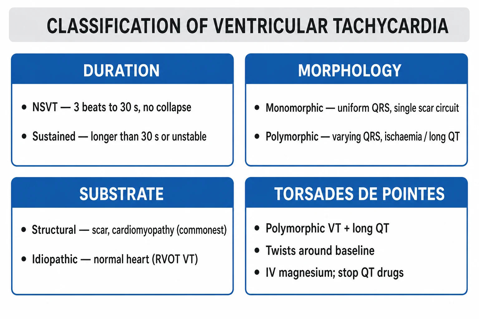
Ventricular tachyarrhythmias are classified along three axes — duration (sustained vs non-sustained), morphology (monomorphic vs polymorphic), and presence of a pulse (which determines the emergency algorithm).[1]
By duration: [1]
- Non-sustained VT (NSVT) — three or more consecutive ventricular beats at over 100 bpm, lasting under 30 seconds and not causing haemodynamic collapse. Often an incidental finding on ambulatory monitoring. Prognostic significance depends on substrate: in structural heart disease it predicts SCD; in the structurally normal heart it is usually benign.
- Sustained VT — VT lasting over 30 seconds, OR any VT requiring termination (cardioversion/defibrillation/antiarrhythmic) because of haemodynamic compromise. Always pathological; demands acute management and SCD-risk stratification. [1]
By QRS morphology: [1]
- Monomorphic VT — uniform QRS morphology from beat to beat, indicating a single re-entrant circuit (usually through a fixed scar) or a single automatic focus. The classical post-MI VT.
- Polymorphic VT — varying QRS morphology, indicating multiple foci or an unstable wandering re-entrant circuit. Torsades de pointes is the polymorphic VT that occurs in the setting of a prolonged QT interval, with the characteristic twisting of the QRS axis around the baseline.
- Bidirectional VT — alternating QRS polarity (often alternating right and left axis), classically in digoxin toxicity and catecholaminergic polymorphic VT (CPVT). [1]
By haemodynamic consequence (the decisive clinical split): [1]
- VT with a pulse — perfusion is present; apply the synchronised cardioversion vs antiarrhythmic decision.
- Pulseless VT — no perfusion; treat identically to VF in the ALS shockable-rhythm algorithm. [1]
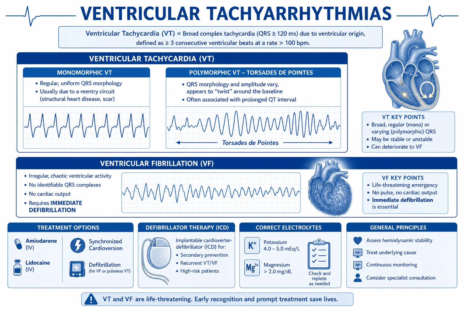
Monomorphic VT
- Uniform broad QRS morphology beat-to-beat
- Single re-entrant circuit through a fixed scar (~80% post-MI)
- Regular rate 100-250 bpm
- Most common sustained VT in structural heart disease
- Stable: amiodarone 300 mg IV; unstable: synchronised DC cardioversion
- Long-term: ICD if EF under 35% or scar-related substrate
Polymorphic VT
- Varying QRS morphology, irregular rate
- Causes: ischaemia, long QT (torsades), CPVT, Brugada, electrolyte
- Torsades: twisting of QRS axis around baseline + long QT
- Treat the cause: stop offending drug, correct K/Mg, treat ischaemia
- IV magnesium sulfate 2 g for torsades regardless of Mg level
- Avoid all QT-prolonging drugs; raise heart rate with isoprenaline/overdrive pacing
Torsades de pointes
- Polymorphic VT in the setting of prolonged QT (over 500 ms) or QTc over 470 ms (M)/480 ms (F)
- Twisting of the QRS axis around the baseline - sine-wave appearance
- Triggered by early after-depolarisations (EADs) in M cells
- Stop ALL QT-prolonging drugs; correct K to over 4.5; Mg 2 g IV
- Isoprenaline or overdrive pacing to heart rate over 100
- Long-term: beta-blocker for congenital LQTS; ICD for high-risk LQTS or recurrent TdP
Bidirectional VT
- Alternating QRS polarity (RBBB with alternating left/right axis)
- Pathognomonic for digoxin toxicity and CPVT
- In digoxin toxicity: check digoxin and K, give Digibind (Fab fragments)
- In CPVT: exertion/emotion-triggered, structurally normal heart, normal QT
- CPVT first-line: nadolol; left cardiac sympathetic denervation
- Differentiate from artefact and Bigeminy
Ventricular fibrillation
- Chaotic, disorganised ventricular activity; no organised QRS
- No pulse, no cardiac output - cardiac arrest rhythm
- Leading cause of sudden cardiac death (ischaemic heart disease)
- SHOCKABLE rhythm - immediate UNSYNCHRONISED defibrillation
- CPR 30:2, adrenaline 1 mg every 3-5 min, amiodarone 300 mg after 3rd shock
- Post-arrest: coronary angiography if cardiac cause, TTM 32-36 degrees C
Idiopathic VT (normal heart)
- ~10% of all VT, structurally normal heart, good prognosis
- RVOT VT: LBBB/inferior axis, adenosine-sensitive, exercise/stress-triggered
- Fascicular (Belhassen) VT: RBBB/left-axis, verapamil-sensitive, re-entry in left posterior fascicle
- Both amenable to catheter ablation (>90% cure)
- Do NOT implant ICD; treat with ablation or beta-blocker
- Differentiate from scar-related VT by structural assessment (echo/MRI)
Epidemiology & Risk Factors
Sudden cardiac death, of which the ventricular tachyarrhythmias are the dominant mechanism, accounts for 300,000 to 400,000 deaths per year in the United States alone and has an incidence of roughly 50 to 100 per 100,000 per year in developed nations.[1] Approximately 80% of SCD is due to ventricular tachyarrhythmia (VF or pulseless VT); the rest is bradyasystole or pulseless electrical activity.[2]
Substrate-specific incidence: [1]
- Ischaemic heart disease is by far the dominant substrate — roughly 80% of SCD occurs in patients with coronary disease, usually as VF or scar-related monomorphic VT in the months to years after MI. Acute ischaemia at the moment of arrhythmia accounts for about 20 to 30% of out-of-hospital VF.
- Cardiomyopathies (dilated, hypertrophic, arrhythmogenic right-ventricular) account for 10 to 15% of SCD.
- Channelopathies (long QT syndrome, Brugada syndrome, CPVT) account for 1 to 2% but are over-represented in young, structurally normal hearts and in athletes.
- Idiopathic VF / early repolarisation syndromes account for the remainder in structurally normal hearts. [1]
Risk factors cluster into four groups:[1][2]
| Risk category | Specific examples |
|---|---|
| Structural heart disease | Prior MI with scar (dominant), LV ejection fraction under 35%, dilated cardiomyopathy, hypertrophic cardiomyopathy (especially with maximal wall thickness over 30 mm), arrhythmogenic right-ventricular cardiomyopathy, cardiac sarcoidosis, myocarditis, infiltrative cardiomyopathy (amyloid), valvular cardiomyopathy (aortic stenosis, mitral prolapse), adult congenital heart disease (post-repair tetralogy of Fallot) |
| Acquired channelopathy | QT-prolonging drugs (antiarrhythmics, antibiotics, antipsychotics, methadone, antiemetics), electrolyte disturbance (hypokalaemia, hypomagnesaemia, hypocalcaemia), severe bradycardia, acute ischaemia |
| Inherited channelopathy | Long QT syndrome (KCNQ1, KCNH2, SCN5A), Brugada syndrome (SCN5A), catecholaminergic polymorphic VT (RyR2, CASQ2), short QT syndrome, early repolarisation syndrome |
| Provocants and triggers | Sympathetic surges (exercise, emotion, cocaine, amphetamine), electrolyte disturbance (diuretic-induced hypokalaemia), digoxin toxicity, myocardial ischaemia, alcohol binge, sleep deprivation, post-operative state, sympathetic stimulation in heart-failure decompensation |
LV ejection fraction is the single most powerful predictor of SCD in structural heart disease — and is the dominant criterion for primary-prevention ICD.[4][8]
[1]Ventricular tachyarrhythmias — the numbers that matter
Pathophysiology
Ventricular tachyarrhythmias arise by four fundamental mechanisms, each with its own substrate, trigger, and characteristic ECG.[1][2]
Mechanism 1 — Scar-related re-entry (the commonest sustained VT)
Roughly 80% of sustained VT in structural heart disease is driven by re-entry through a fixed anatomical circuit formed by surviving myocyte channels within a healed infarct scar or fibrotic cardiomyopathic substrate.[2] The scar provides the substrate; an extrasystole (often a ventricular premature beat from a different site) provides the trigger. The wavefront enters the scar at one entrance, travels slowly through a protected channel of surviving myocardium (the diastolic pathway, conducting so slowly that the rest of the ventricle has time to recover excitability), exits at a fixed site, and re-enters — completing a self-sustaining loop. Because the exit site and the path through the scar are fixed, every beat has the same QRS morphology — this is monomorphic VT. The clinical correlate is critical: monomorphic VT in a patient with prior MI = scar-related re-entry until proven otherwise.
Re-entry requires the three classical conditions: (1) an anatomical or functional circuit of two interconnected limbs; (2) different conduction velocities and refractory periods between the two limbs; and (3) a unidirectional block in one limb that allows the wavefront to travel only one way around the loop, returning to its origin after the first limb has recovered excitability. In scar-related VT the protected channel within the scar is the slow-conducting diastolic pathway, while the normal myocardium outside the scar is the fast limb. [1]
Mechanism 2 — Enhanced (abnormal) automaticity
A single ventricular focus depolarises faster than the sinus node and takes over the rhythm. This is the mechanism of outflow-tract VT (especially RVOT) and of digoxin-toxic VT. Automaticity is not dependent on a re-entrant circuit, which is why it does not terminate with overdrive pacing or vagal manoeuvres. The trigger is usually a catecholamine surge (exercise, emotion) in idiopathic RVOT VT; in digoxin toxicity the trigger is Na-K ATPase inhibition → intracellular Na rise → increased Na/Ca exchange → Ca overload → delayed after-depolarisations (DADs). The classical end-result of digoxin toxicity is bidirectional VT with alternating QRS polarity. [1]
Mechanism 3 — Triggered activity from delayed after-depolarisations (DADs)
Triggered activity is the mechanism underlying catecholamine-sensitive RVOT VT and digoxin-toxic bidirectional VT. The cellular mechanism is intracellular Ca overload during the action potential, leading to spontaneous Ca release from the sarcoplasmic reticulum (SR) after repolarisation. The Na/Ca exchanger then extrudes this Ca, generating a net inward (depolarising) current — the delayed after-depolarisation (DAD). If a DAD reaches threshold potential during diastole, it fires off an action potential — and if this recurs, the rhythm self-perpetuates as VT. DADs are amplified by anything that raises intracellular Ca: sympathetic stimulation, digoxin (Na-K ATPase inhibition), high heart rates, ischaemia. The therapeutic corollary is that beta-blockers and verapamil (Ca-channel block) work; adenosine transiently blocks the focus by adenosine-A1 receptor-mediated suppression of cAMP, which is why RVOT VT is adenosine-sensitive (a diagnostic and therapeutic clue). [1]
Mechanism 4 — Triggered activity from early after-depolarisations (EADs) — torsades de pointes
The pathophysiology of torsades de pointes centres on prolongation of the action-potential duration (APD) in mid-myocardial (M) cells and Purkinje cells — the cellular equivalent of QT prolongation on the surface ECG.[14][15] Prolongation of phase 3 repolarisation is most often caused by block of the rapidly activating delayed rectifier potassium current (IKr), encoded by KCNH2 (hERG). QT-prolonging drugs (macrolides, fluoroquinolones, antipsychotics, methadone, ondansetron, sotalol, amiodarone, haloperidol, certain antihistamines) almost universally block IKr. The prolonged plateau phase allows reactivation of L-type calcium channels, generating a depolarising current during phase 2 or 3 — the early after-depolarisation (EAD). The EAD triggers an extrasystole (typically from the Purkinje network), which falls on the vulnerable phase of the preceding T wave (R-on-T) and initiates a polymorphic VT with twisting of the QRS axis — the eponymous torsades de pointes. The twisting axis reflects the transmural dispersion of repolarisation (epicardium repolarises faster than mid-myocardial cells, generating a window of vulnerable heterogeneity).
Hypokalaemia, hypomagnesaemia, hypocalcaemia, bradycardia, female sex, structural heart disease and genetic long QT syndrome all predispose by prolonging repolarisation further.[14] The therapy follows directly: stop the offending drug, give IV magnesium (which suppresses EADs by blocking Ca entry), raise the heart rate with isoprenaline or overdrive pacing (which shortens repolarisation), correct potassium to over 4.5 mmol/L.[15]
Ventricular fibrillation — multiple wavelet re-entry
VF is the chaotic, disorganised electrical activity of the ventricles with no organised QRS, no coordinated contraction, no cardiac output. The dominant mechanism is the multiple wavelet hypothesis of Moe and colleagues: a large re-entrant wavefront continuously fragments into multiple daughter wavelets that wander, mutually annihilate, and re-form, never settling into a stable circuit. Modern mapping has refined this to the mother-rotor hypothesis — a single high-frequency stable rotor that breaks down into fibrillatory conduction. Either way, the practical message is unchanged: VF is not a perfusing rhythm, it is a cardiac arrest rhythm, and only defibrillation (a massive synchronised shock that simultaneously depolarises the entire myocardium and extinguishes all wavelets, allowing the sinus node to resume) terminates it.[1]
The channelopathies — molecular VT/VF substrates
Long QT syndrome — mutations in the KCNQ1 gene (LQT1, ~35% — IKs loss of function), KCNH2/hERG (LQT2, ~30% — IKr loss of function) or SCN5A (LQT3, ~10% — late inward Na gain of function that prolongs repolarisation). Triggers vary by genotype: LQT1 — swimming, exertion; LQT2 — auditory stimuli, postpartum; LQT3 — rest, sleep. Treatment: beta-blocker (especially nadolol or propranolol) for all; left cardiac sympathetic denervation for breakthrough; ICD for syncope despite beta-blocker, prior cardiac arrest, or high-risk genotypes (QTc over 500 ms).[1]
Brugada syndrome — loss-of-function mutation in SCN5A (the cardiac sodium channel, ~20 to 30% of cases) reduces the inward Na current, which disproportionately shortens the action-potential dome in the right ventricular epicardium (which is more dependent on the transient outward current Ito).[16] The transmural dispersion of repolarisation sets up phase-2 re-entry: an extrasystole from the depolarised region initiates polymorphic VT/VF. Classically a male in his 30s–40s with syncope or nocturnal agonal respiration; ECG shows coved-type ST elevation in V1–V3 (type 1). Treatment is ICD for symptomatic or spontaneous-type-1 patients; quinidine (blocks Ito) and isoprenaline for electrical storm.[16]
Catecholaminergic polymorphic VT (CPVT) — mutation in the cardiac ryanodine receptor RyR2 (autosomal dominant, ~60%) or calsequestrin CASQ2 (autosomal recessive) causes diastolic Ca leak from the SR during sympathetic activation, generating DADs and bidirectional or polymorphic VT.[1] The classical presentation is exercise- or emotion-induced bidirectional VT in a child or young adult with a structurally normal heart and normal resting ECG. Treatment: beta-blocker (nadolol first-line), left cardiac sympathetic denervation, flecainide, ICD for breakthrough.[2]
Why AV dissociation, fusion beats, and capture beats exist in VT
When a ventricular focus fires faster than the sinus node, the ventricles beat independently of the atria — the sinus node still fires, the atria still depolarise, but the AV node is refractory (still recovering from the previous ventricular beat), so most sinus impulses are blocked. Occasionally: [1]
- A fusion beat arises when a sinus impulse manages to reach the AV node just as a ventricular beat is being generated — both depolarise part of the ventricle, producing a QRS that is a hybrid of the two morphologies (intermediate width and shape).
- A capture beat arises when a sinus impulse gets through the AV node and conducts normally through the His-Purkinje system, fully 'capturing' the ventricle for one beat — producing a normal narrow QRS within the broad-complex tachycardia. [1]
Both fusion and capture beats are pathognomonic for VT — they prove that the atria and the ventricles are running independently (which can only happen in a rhythm of ventricular origin).[1][2]
Clinical Presentation
The clinical presentation of ventricular tachyarrhythmia is determined by whether a pulse is present and by the haemodynamic consequence. [1]
Sustained VT with a pulse — the typical presentation
The patient is usually awake but symptomatic: rapid palpitations, dyspnoea, lightheadedness, presyncope or frank syncope, ischaemic-type chest pain (VT at high rate increases myocardial oxygen demand while decreasing coronary perfusion), and signs of low cardiac output (pallor, sweating, cool peripheries, oliguria). Examination shows a rapid regular pulse at 100 to 250 bpm with a broad QRS on the monitor, variable first heart sound intensity (AV dissociation alters the mitral/tricuspid closure timing beat-to-beat), cannon A waves in the JVP (the right atrium occasionally contracts against a closed tricuspid valve), and signs of poor perfusion (hypotension, confusion).[2]
Pulseless VT/VF — the cardiac arrest presentation
The patient presents as sudden collapse with loss of consciousness, no pulse, no breathing, no response. There may be a brief preceding palpitation, but most patients have no warning. No time for history — the diagnosis is on the rhythm monitor. Time is myocardium and brain: every minute of delay to defibrillation reduces survival by ~10%.[1]
Atypical presentations — examiner favourites
- Elderly patient — may present with syncope alone, falls, new confusion, or exacerbation of heart failure rather than the textbook palpitations. The broad-complex tachycardia on the monitor is often misread as 'SVT with aberrancy'. Maintain a high index of suspicion: syncope in a patient with prior MI or cardiomyopathy is VT until proven otherwise.
- Post-MI patient — new broad-complex tachycardia within hours to weeks of MI is scar-related or ischaemic VT; do not attribute to a benign cause.
- Pregnant patient — palpitations may be attributed to the physiological tachycardia of pregnancy; sustained VT in pregnancy is rare but demands urgent treatment (most antiarrhythmics and synchronised cardioversion are safe).
- Athlete — exercise- or emotion-induced syncope or sudden collapse in a young athlete raises the question of HCM, ARVC, long QT, Brugada, CPVT. All demand a 12-lead ECG, echocardiogram, exercise test, and family-history evaluation. Competitive sport is contraindicated until the cause is identified.
- Torsades de pointes — classically presents with syncope in a patient on a QT-prolonging drug (macrolide, fluoroquinolone, antipsychotic, methadone, antiemetic), or with electrolyte disturbance (diuretic-induced hypokalaemia), or in congenital long QT syndrome (often first manifest at puberty or in the postpartum period). Episodes are usually self-terminating but can degenerate to VF.
- Brugada syndrome — male in his 30s–40s, syncope or nocturnal agonal respiration, family history of sudden death; ECG shows coved ST elevation in V1–V3. Fever is a well-recognised precipitant of VF in Brugada and warrants urgent cooling and monitoring.
- CPVT — child or young adult with exercise-induced syncope or seizure-like activity (cerebral hypoperfusion), structurally normal heart, normal resting ECG. Diagnosis is by exercise test, which reproduces the bidirectional/polymorphic VT.
- Digoxin-toxic patient — nausea, vomiting, visual disturbance (yellow-green halos, xanthopsia), confusion, and on ECG a bidirectional VT or atrial tachycardia with AV block. The bidirectional VT is the high-yield exam clue: bidirectional VT + a patient on digoxin = digoxin toxicity → check level, give Digibind. [1]
Differential Diagnosis
A broad-complex tachycardia (QRS over 120 ms at over 100 bpm) has a small but critical differential — distinguishing VT from its mimics is the most-tested skill in this topic.[1][2]
| Diagnosis | Distinguishing features |
|---|---|
| Ventricular tachycardia (dominant diagnosis) | Broad QRS over 120 ms; rate 100–250 bpm; AV dissociation (independent P waves), fusion beats, capture beats — all pathognomonic; concordance in precordial leads; extreme axis deviation; predominance of negative QRS in lead aVR; occurs in prior MI, cardiomyopathy, structural heart disease |
| SVT with aberrancy (rate-related BBB or pre-existing BBB) | Broad QRS but the rhythm originates at or above the AV node; no AV dissociation; the QRS morphology matches a typical bundle-branch-block pattern (rsR' in V1 for RBBB, rS in V1 with wide R in V6 for LBBB); response to vagal manoeuvres or adenosine terminates the tachycardia |
| Antidromic AVRT (WPW) | Anterograde conduction down an accessory pathway → broad pre-excited QRS that closely mimics VT; usually in a patient with known WPW; regular rate 150–250 bpm |
| Pre-excited atrial fibrillation in WPW | Irregular broad-complex tachycardia; varying QRS morphology with intermittent delta waves; the irregularity is the key clue; NEVER give AV-nodal blockers (adenosine, verapamil, beta-blockers, digoxin) — risk of VF |
| SVT with rate-related bundle branch block | A narrow-complex tachycardia that becomes broad at high rates; the QRS morphology is a typical BBB pattern |
| Paced rhythm with tracking | Ventricular-paced rhythm at high rates can mimic VT; the pace spike before each QRS is diagnostic |
| ECG artefact (tremor, tooth-brushing, Parkinsonian tremor) | Regular underlying normal QRS is visible buried within the apparent VT; the 'rate' is implausibly high; the patient is haemodynamically well despite the alarming monitor; pulse oximetry remains normal |
The single most important rule: broad-complex tachycardia at over 100 bpm in a patient with structural heart disease is VT until proven otherwise. Misdiagnosis as 'SVT with aberrancy' and giving verapamil causes haemodynamic collapse.[2]
The Brugada algorithm (4-step) and Vereckei aVR algorithm (single-lead) are the two bedside ECG algorithms to distinguish VT from SVT-with-aberrancy.[11]
Brugada algorithm for WCT — the 4 steps
RSRS
Step 1 - absence of an RS complex in ALL precordial leads (V1-V6) = VT (sensitivity high)
Step 2 - if RS present, R-to-S nadir over 100 ms in any precordial lead = VT
Step 3 - AV dissociation (capture/fusion beats, independent P waves) = VT
Step 4 - morphology criteria in V1-V2 and V6 for VT vs BBB (Brugada morphology criteria)
Vereckei aVR algorithm — 4 simple rules
AaVR
If lead aVR shows an initial R (positive) wave = VT
If initial r or q wave over 40 ms in aVR = VT
If there is a notch on the descending limb of a negative onset QRS in aVR = VT
If ventricular activation velocity (vertical) over 1/terminal (horizontal) ratio under or equal 1 in aVR = VT
Clinical & Bedside Assessment
The bedside assessment has one decisive branch — stability.[1][2]
First — ABCDE and the monitor. Secure the airway, give high-flow oxygen if hypoxic, attach continuous cardiac monitoring and defibrillator pads to the chest (anterior–apical), establish two large-bore IV cannulae, and take bloods (U&E including Mg and Ca, FBC, troponin, digoxin level if relevant, TFTs, toxicology). [1]
Second — the stability decision. A sustained VT with pulse is unstable if any of: [1]
- Systolic blood pressure under 90 mmHg or shock
- Syncope or reduced GCS
- Ischaemic chest pain
- Acute heart failure / pulmonary oedema
- Signs of end-organ hypoperfusion (oliguria, mottling, altered mental status) [1]
Unstable → synchronised DC cardioversion immediately (do not delay for antiarrhythmics). [1]
Stable → IV antiarrhythmic while preparing for cardioversion if deterioration. [1]
Third — get a 12-lead ECG during the tachycardia if at all possible (often not possible in unstable patients). Look for the three pathognomonic signs of VT: [1]
- AV dissociation — independent P waves 'marching through' the tachycardia
- Capture beats — occasional normal narrow QRS within the broad-complex tachycardia
- Fusion beats — QRS of intermediate morphology (hybrid of normal and VT) [1]
Apply the Brugada or Vereckei aVR algorithms to differentiate VT from SVT-with-aberrancy.[11]
Fourth — bedside clinical clues: [1]
- Cannon A waves in the JVP — the right atrium occasionally contracts against a closed tricuspid valve (because the AV node is refractory), producing intermittent large 'cannon' waves in the neck — a bedside clue to AV dissociation and therefore VT.
- Variable intensity of S1 — beat-to-beat variation in mitral/tricuspid closure timing alters the first heart sound — another bedside AV-dissociation clue.
- Carotid sinus massage — may transiently unmask atrial activity (vagal tone blocks AV conduction); never use as the primary diagnostic test in a broad-complex tachycardia — VT can degenerate with vagal manoeuvres. [1]
Investigations
First-line (acute presentation): [1]
- 12-lead ECG — the single most important test. Assess rate, regularity, QRS width, morphology, axis, P-wave timing, AV dissociation, capture/fusion beats, concordance, and the Brugada/Vereckei criteria. For torsades, look for prolonged QT and twisting of the QRS axis; for Brugada, look for coved ST elevation in V1–V3.
- Bloods — U&E (potassium, magnesium, calcium), FBC, troponin (acute ischaemia as cause or consequence), digoxin level if relevant, TFTs (thyrotoxicosis precipitates), toxicology (cocaine), glucose, ABG (hypoxia, acidosis), lactate (perfusion).
- Chest X-ray — heart size, pulmonary oedema, pneumothorax, lines/tubes. [1]
The two ECG algorithms for VT vs SVT-with-aberrancy: [1]
Brugada algorithm (4-step):[2]
- Absence of RS complex in ALL precordial leads (V1–V6) → VT (stop).
- R-to-S nadir over 100 ms in any precordial lead → VT (stop).
- AV dissociation (capture/fusion beats, independent P waves) → VT (stop).
- Morphology criteria in V1–V2 and V6 — for VT the QRS does not match a typical bundle-branch-block pattern (e.g. absence of rsR' in V1 for RBBB, or notching on the downstroke of S in V1) → VT. [1]
If all four steps are negative, the diagnosis is SVT with aberrancy. Sensitivity for VT is approximately 99%. [1]
Vereckei aVR algorithm (single-lead, simpler):[11]
Diagnose VT if any one of the following is present in lead aVR: [1]
- Initial R wave (positive deflection)
- Initial r or q wave over 40 ms
- Notch on the descending limb of a negative-onset QRS
- Ventricular activation-velocity ratio (vi/vt) under or equal to 1 — the ratio of vertical deflection in the first 40 ms to the vertical deflection in the last 40 ms of the QRS [1]
Sensitivity ~90%, specificity ~80%; far simpler and quicker than Brugada at the bedside. [1]
Second-line (substrate characterisation — once stabilised): [1]
- Transthoracic echocardiography — LV ejection fraction (the dominant ICD criterion), regional wall-motion abnormalities (scar, ischaemia), structural substrate (HCM with maximal wall thickness over 30 mm, ARVC with RV dilation/dyskinesia, dilated cardiomyopathy, valvular disease, infiltrative cardiomyopathy).
- Coronary angiography if ischaemia suspected (most patients with new VT/VF in the setting of risk factors warrant urgent angiography — ischaemia is the commonest precipitant).
- Cardiac MRI — late gadolinium enhancement to characterise the scar (subendocardial pattern = ischaemic; mid-wall = non-ischaemic, myocarditis, sarcoidosis, DCM; epicardial = ARVC, sarcoid); functional RV assessment for ARVC; tissue characterisation for sarcoidosis, amyloidosis, HCM.
- Electrophysiology study (EPS) — programmed ventricular stimulation to induce VT, map the circuit, identify the critical isthmus/exit site, and plan ablation. EPS is less used diagnostically than historically (modern imaging and ECG criteria suffice) but central to ablation planning.
- Ambulatory ECG / event monitor / implantable loop recorder — to detect NSVT, quantify arrhythmia burden, and guide ICD programming.
- Exercise test — to provoke CPVT (bidirectional/polymorphic VT on exercise), and to unmask ischaemia.
- Genetic testing and family screening — for long QT syndrome (KCNQ1, KCNH2, SCN5A, KCNE1, KCNE2), Brugada syndrome (SCN5A), CPVT (RyR2, CASQ2), ARVC (PKP2, DSG2, DSC2, JUP), and familial screening of first-degree relatives of SCD victims under 40. [1]
Management — Resuscitation
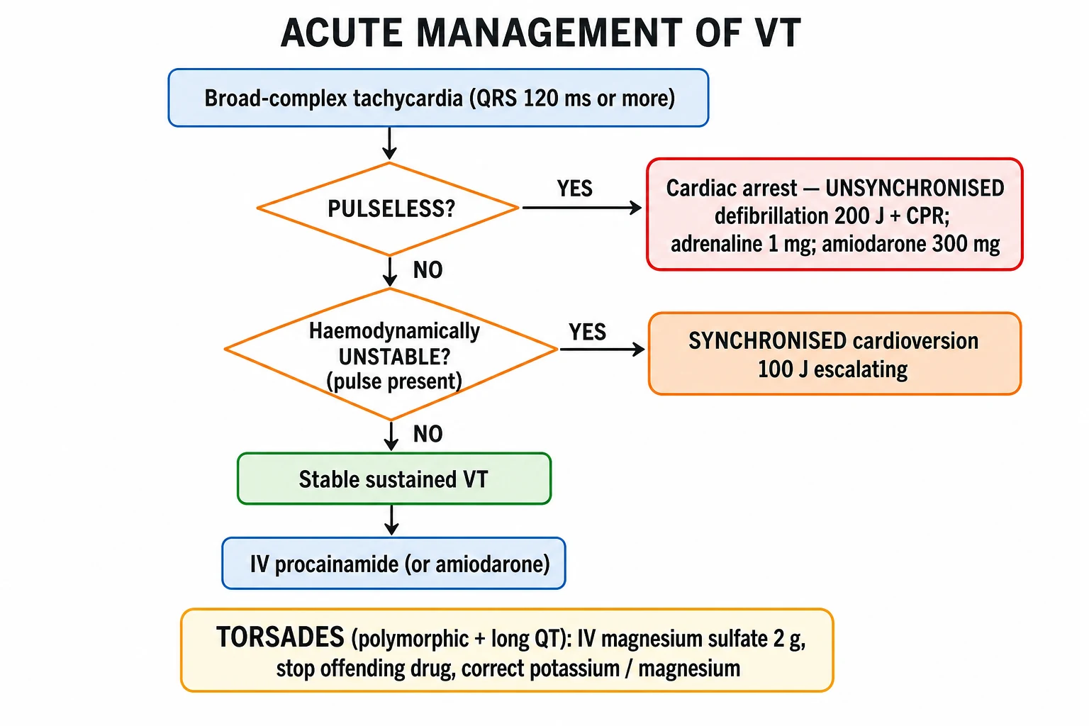
The acute management of ventricular tachyarrhythmia is dictated by whether a pulse is present.[1][2]
Pulseless VT and VF — the ALS shockable-rhythm algorithm
Both are shockable rhythms and managed identically. Immediate action saves lives; every minute of delay to defibrillation reduces survival by ~10%. [1]
- Confirm cardiac arrest — unresponsive, no pulse, no breathing.
- Start high-quality CPR — 30 compressions to 2 ventilations, rate 100 to 120 per minute, depth 5 to 6 cm, allow full chest recoil, minimise interruptions.
- Attach defibrillator pads (anterior–apical) and analyse rhythm.
- If shockable rhythm (VF/pulseless VT): deliver one UNSYNCHRONISED shock — biphasic 150 to 200 J (or monophasic 360 J).
- Immediately resume CPR for 2 minutes without rhythm check (CPR maintains coronary and cerebral perfusion during the post-shock period).
- Reassess rhythm after 2 minutes; if still shockable, deliver a second shock of equal or escalating energy.
- Resume CPR for 2 minutes and give adrenaline 1 mg IV (every 3 to 5 min — i.e. every other cycle from this point).
- Reassess rhythm; if still shockable, deliver a third shock, then resume CPR for 2 minutes and give amiodarone 300 mg IV bolus.
- Reassess rhythm; if still shockable, deliver a fourth shock, resume CPR, give another adrenaline 1 mg.
- Reassess rhythm; if still shockable, deliver a fifth shock, resume CPR, and give amiodarone 150 mg IV (further dose).
- Continue the cycle of CPR–shock–drug until either ROSC, the rhythm becomes non-shockable (convert to PEA/asystole algorithm), or resuscitation is discontinued.
- Reversible causes — the 4 Hs and 4 Ts:
- Hypoxia — oxygenate, secure airway
- Hypovolaemia — IV fluids, treat bleeding
- Hypo-/hyperkalaemia — check and correct (hypokalaemia → KCl; hyperkalaemia → calcium gluconate 10 mL of 10%, insulin-dextrose, salbutamol)
- Hypothermia — rewarm
- Thrombosis (coronary or pulmonary) — consider thrombolysis if PE suspected; urgent angiography if cardiac cause
- Tension pneumothorax — needle decompression, then chest drain
- Tamponade (cardiac) — pericardiocentesis
- Toxins — antidotes (naloxone, digoxin Fab, β-blocker for theophylline, etc.) [1]
Post-ROSC care: [1]
- Targeted temperature management (TTM) — maintain core temperature 32 to 36 °C for 24 hours in comatose post-arrest patients (improves neurological outcome).
- Optimise oxygenation and ventilation — target SpO2 94 to 98%, normocapnia (avoid hyperoxia and hypocapnia, both worsen neurological outcome).
- 12-lead ECG — to look for STEMI; if ST-elevation, urgent coronary angiography ± PCI.
- Haemodynamic optimisation — mean arterial pressure over 65 mmHg, urine output over 0.5 mL/kg/h.
- Investigate the cause — ischaemia, electrolyte, channelopathy, drug toxicity. [1]
Sustained VT with a pulse
If unstable (hypotension, syncope, ischaemic chest pain, acute heart failure, reduced GCS): [1]
- Synchronised DC cardioversion — 100 J biphasic escalating to 200 J if needed. Synchronise to the R wave to avoid delivering energy on the T wave (which would cause VF). Pre-sedate if time permits (e.g. midazolam 2.5 to 5 mg IV ± fentanyl 50 to 100 µg IV), but do not delay cardioversion in the unstable patient.[1]
If stable: IV antiarrhythmic (below), with cardioversion on standby.[1][2]
Management — Definitive & Stepwise
Stable sustained monomorphic VT
First-line: IV amiodarone 300 mg in 20 to 60 mL of 5% glucose over 20 to 60 minutes (slower than bolus to avoid hypotension), followed by an infusion of 900 mg over 24 hours (total 1.2 g in the first 24 hours).[1] Amiodarone is preferred in structural heart disease and heart failure because it is minimally negatively inotropic and has long-term safety data in this population.[9][10]
Alternative: IV lidocaine 50 to 100 mg (1 to 1.5 mg/kg) over 2 minutes, repeat up to a maximum of 3 mg/kg in 1 hour. Less effective than amiodarone in shock-resistant VF (ALIVE trial showed amiodarone superior)[12] but useful as second-line or in patients where amiodarone is contraindicated.
If pharmacological cardioversion fails or the patient deteriorates: synchronised DC cardioversion (100 J biphasic escalating). [1]
Procainamide (15 mg/kg at 20 mg/min, then infusion) is an alternative class Ia agent used in some centres and is the first-line agent in the stable wide-complex tachycardia algorithm of the 2010 AHA guidelines (it terminates both VT and SVT-with-aberrancy, so it side-steps the diagnostic dilemma).[1]
Torsades de pointes — the management is distinct
Torsades is treated differently from other VT because its mechanism (EAD-driven triggered activity in prolonged repolarisation) is fundamentally different.[14][15]
- Stop ALL QT-prolonging drugs (consult a QT-drug list — CredibleMeds; common culprits include sotalol, amiodarone, quinidine, procainamide, macrolides, fluoroquinolones, methadone, haloperidol, droperidol, certain antipsychotics, ondansetron, tacrolimus).
- IV magnesium sulfate 2 g (8 mmol) over 10 minutes — regardless of baseline Mg — magnesium suppresses EADs by inhibiting Ca influx; it works even when serum Mg is normal.[14][15]
- Correct potassium to over 4.5 mmol/L — hypokalaemia prolongs QT and worsens torsades.
- Raise the heart rate to over 100 bpm — this shortens the action potential and shortens the QT. Options: isoprenaline infusion (titrate from 1 to 4 µg/min, contraindicated in ischaemic torsades) or overdrive pacing (transvenous or transcutaneous atrial/ventricular pacing at 100 to 120 bpm).
- Identify and remove the trigger — drug, electrolyte disturbance, bradycardia, acute ischaemia.
- Long-term — for acquired LQTS: avoid all QT-prolonging drugs; check electrolytes. For congenital LQTS: beta-blocker (nadolol or propranolol) first-line; left cardiac sympathetic denervation for breakthrough; ICD for syncope despite beta-blocker, prior cardiac arrest, or QTc over 500 ms.
Idiopathic VT (structurally normal heart)
- RVOT VT (LBBB/inferior axis) — adenosine-sensitive; IV adenosine 6 mg escalating to 12 mg then 12 mg (terminates by adenosine-A1-mediated cAMP suppression), or IV verapamil 5 mg, or IV beta-blocker. Definitive: catheter ablation, curative in over 90%.
- Fascicular (Belhassen) VT (RBBB/left-axis, re-entry in the left posterior fascicle) — verapamil-sensitive; IV verapamil 5 mg over 2 minutes; definitive: catheter ablation, curative in over 85%. [1]
Long-term prevention of SCD — the ICD
The implantable cardioverter-defibrillator (ICD) is the cornerstone of long-term SCD prevention. It detects VT/VF and delivers anti-tachycardia pacing (ATP) or a shock to terminate the arrhythmia.[1][2]
ICD indications: [1]
Secondary prevention (survivors of VT/VF): [1]
- Survivors of cardiac arrest due to VT/VF, where the cause is not reversible (i.e. not acute ischaemia, drug toxicity, electrolyte disturbance, or early post-MI) — ICD indicated.
- Spontaneous sustained VT in structural heart disease — ICD indicated.
- Syncope with inducible sustained VT/VF on EPS — ICD indicated. [1]
Primary prevention (no prior event, but high risk): [1]
- LV ejection fraction under 35%, NYHA II–III, more than 40 days after MI, more than 3 months after revascularisation, optimal medical therapy for at least 3 months (MADIT-II, SCD-HeFT criteria).[4][8]
- LV ejection fraction under 40%, prior MI, NSVT on monitoring, and inducible VT on EPS that is not suppressible by class I antiarrhythmics (MADIT-I criteria).[3]
- LV ejection fraction under 40%, prior MI, NSVT, inducible VT on EPS (MUSTT criteria).[5]
- Non-ischaemic DCM, EF under 36%, NSVT or PVCs (DEFINITE criteria — significant reduction in arrhythmic death).[6]
- Hypertrophic cardiomyopathy with one major SCD risk factor (maximal wall thickness over 30 mm, family history of sudden death, prior unexplained syncope, NSVT, abnormal BP response to exercise).
- Arrhythmogenic right-ventricular cardiomyopathy with sustained VT/VF, or with high-risk features (syncope, extensive disease).
- Long QT syndrome with syncope despite beta-blocker, or prior cardiac arrest, or high-risk genotype (QTc over 500 ms).
- Brugada syndrome with spontaneous type 1 ECG and syncope or sustained VT.
- CPVT with syncope or sustained VT on beta-blocker.
ICD is NOT implanted: [1]
- Within 40 days of MI — DINAMIT showed no mortality benefit and excess non-arrhythmic death (the LV may yet recover).[7]
- Within 3 months of CABG/PCI — the LV may recover.
- Within 3 months of starting optimal medical therapy in newly diagnosed non-ischaemic DCM — re-assess EF before deciding.
- NYHA IV symptoms (refractory heart failure) — unless CRT-D planned or transplant-bridging.
- Life expectancy under 1 year (comorbidity, frailty).
- Incessant VT or electrical storm — ablate or stabilise first, then ICD.
Antiarrhythmic drugs for long-term suppression
- Beta-blockers (metoprolol, bisoprolol, carvedilol, nadolol) — first-line in HCM, LQTS, CPVT, post-MI; reduce sympathetic triggering of arrhythmia.
- Amiodarone — for VT storm, adjunctive to ICD, in refractory idiopathic VT. Toxicity is the limiting factor: pulmonary fibrosis (baseline CXR + PFTs), thyroid (hypo- and hyper-thyroidism — TFTs 6-monthly), hepatic (LFTs 6-monthly), corneal microdeposits, photosensitivity (grey-blue skin discolouration), peripheral neuropathy, bradycardia, QT prolongation (but low torsades risk).
- Sotalol — class II + class III (IKr-blocking) action; effective but torsadogenic — QT must be monitored; contraindicated in severe HF, hypokalaemia, and women (longer baseline QT).
- Mexiletine — oral class Ib (lidocaine analogue), useful adjunct in long-QT type 3 (LQT3) and HCM.
- Quinidine — blocks Ito, useful in Brugada syndrome (normalises ECG and suppresses VF) and short QT syndrome. [1]
Catheter ablation
Catheter ablation is the definitive therapy for idiopathic VT (RVOT, fascicular — cure rate over 85 to 90%) and an adjunct in scar-related VT (reduces VT recurrence and ICD shocks by ~75% but is not curative — multiple circuits and unmappable channels remain).[2] Indications: idiopathic VT with recurrent symptoms, scar-related VT with frequent ICD therapies, electrical storm, bundle-branch re-entry VT.
Electrical storm
Electrical storm is defined as 3 or more separate episodes of sustained VT, VF, or appropriate ICD therapies within 24 hours.[2] It is a medical emergency with in-hospital mortality of 25 to 50%. Management ladder:
- Sedation and analgesia — propofol, benzodiazepines, fentanyl; reduces sympathetic surge.
- Intubation and mechanical ventilation if unstable.
- IV amiodarone 300 mg then infusion 900 mg/24 h + IV beta-blocker (esmolol/metoprolol) — combined sympatholysis and antiarrhythmic.
- Deep anaesthesia (propofol infusion) refractory cases.
- Mechanical circulatory support — IABP or VA-ECMO if cardiogenic shock.
- Urgent catheter ablation of the triggering focus / scar substrate.
- Stellate ganglion block or left cardiac sympathetic denervation in refractory cases.
- Identify and treat reversible precipitants — ischaemia, electrolyte, infection, drug withdrawal/overdose. [1]
Specific Subtypes & Scenarios
- Monomorphic scar-related VT (post-MI) — fixed re-entry circuit through infarct scar; uniform broad QRS; treat per sustained-VT algorithm; long-term ICD; ablation adjunctive.
- Torsades de pointes — polymorphic VT + long QT; stop drug, magnesium 2 g, raise heart rate; long-term: beta-blocker for congenital LQTS; ICD for high-risk LQTS.
- Bidirectional VT — alternating QRS polarity; classical for digoxin toxicity (give Digibind / digoxin Fab fragments) and CPVT (beta-blocker first-line).
- RVOT VT — LBBB/inferior axis, adenosine-sensitive, structurally normal heart; ablation curative in over 90%.
- Fascicular (Belhassen) VT — RBBB/left-axis, verapamil-sensitive, structurally normal heart; ablation curative in over 85%.
- Bundle-branch re-entry VT — typically in dilated cardiomyopathy; circuit through the His-Purkinje system; ablation of the right bundle branch is curative.
- CPVT — exercise-induced bidirectional/polymorphic VT in structurally normal heart with normal resting ECG; beta-blocker (nadolol), left cardiac sympathetic denervation, flecainide, ICD for breakthrough.
- Brugada syndrome — male in 30s–40s, syncope or nocturnal agonal respiration; coved ST elevation in V1–V3; ICD for symptomatic or spontaneous-type-1; quinidine and isoprenaline for electrical storm; fever is a precipitant — cool aggressively.[16]
- Long QT syndrome — beta-blocker (nadolol/propranolol) for all; ICD for syncope despite beta-blocker, prior cardiac arrest, or QTc over 500 ms; avoid QT-prolonging drugs; genotype-specific triggers (LQT1 swimming/exertion; LQT2 auditory/postpartum; LQT3 sleep/rest).
- Idiopathic VF / early repolarisation syndrome — recurrent VF in structurally normal heart with J-point elevation in inferolateral leads; ICD for survivors; quinidine adjunctive; ablation of the Purkinje trigger in selected cases.
- Ventricular flutter — regular sine-wave appearance at 250 to 300 bpm; treated as VF — immediate defibrillation.
- Pulseless VT — treated identically to VF in the ALS shockable-rhythm algorithm.
Complications & Pitfalls
Complications: [1]
- Sudden cardiac death — the dominant feared outcome; up to 25% of structural-heart-disease VT degenerates to VF within 1 year if untreated.
- Syncope and traumatic injury from syncope.
- Tachycardiomyopathy from incessant VT — especially focal/incessant idiopathic VT (RVOT, fascicular) — reversible with ablation.
- Cardiogenic shock and acute heart failure from sustained VT.
- Anoxic brain injury after cardiac arrest — function of downtime and quality of resuscitation; TTM mitigates.
- Post-arrest multi-organ failure.
- ICD-related complications — inappropriate shocks (typically for AF with rapid rate, sinus tachycardia, or T-wave oversensing), pocket infection, lead fracture/failure, pneumothorax, cardiac tamponade from perforation, Twiddler's syndrome.
- Psychological morbidity from ICD shocks — depression, anxiety, post-traumatic stress.
- Amiodarone toxicity — pulmonary fibrosis, thyroid dysfunction, hepatic, corneal microdeposits, photosensitivity, peripheral neuropathy. [1]
Classic pitfalls (examiner favourites): [1]
- Treating a broad-complex tachycardia as 'SVT with aberrancy' and giving verapamil — in VT this causes haemodynamic collapse. Broad-complex = treat as VT unless you can prove otherwise.
- Misdiagnosing pre-excited AF in WPW as 'irregular broad-complex VT' — pre-excited AF needs flecainide/amiodarone or DC cardioversion, NOT AV-nodal blockers (which precipitate VF).
- Missing torsades as the cause of syncope in a patient on a QT-prolonging drug — measure the QT, give magnesium.
- Implanting an ICD within 40 days of acute MI — DINAMIT showed no mortality benefit and excess non-arrhythmic death; wait at least 40 days.[7]
- Using sotalol in heart failure or hypokalaemia — pro-arrhythmic (torsades); requires QT under 500 ms.
- Forgetting to look for digoxin toxicity in bidirectional VT — the answer is Digibind, not amiodarone.
- Forgetting that amiodarone itself prolongs the QT and can precipitate torsades in susceptible patients (especially with other QT drugs).
- Assuming 'electrocardiographic artefact' is VT — tremor, tooth-brushing, and Parkinsonian tremor can mimic polymorphic VT; check for a normal underlying QRS and a well patient.
Prognosis & Disposition
The prognosis is substrate-dependent.[1][2]
By substrate: [1]
- Idiopathic VT (structurally normal heart) — excellent prognosis; catheter ablation is curative; SCD risk is minimal.
- Scar-related VT (post-MI) — significant SCD risk; ICD reduces mortality by 23 to 60% (secondary prevention) and 28% (primary prevention, SCD-HeFT).[8]
- Cardiomyopathic VT — moderate-to-high SCD risk; ICD indicated if EF under 35%.
- Channelopathic VT/VF (LQTS, Brugada, CPVT) — variable; ICD in high-risk phenotypes.
- Electrical storm — in-hospital mortality 25 to 50%; one-year mortality after storm is high.
LV ejection fraction is the single most powerful predictor of SCD in structural heart disease, and the dominant ICD-implant criterion. [1]
Disposition after an acute episode: [1]
- Terminated, stable, no red flags, no structural heart disease — outpatient ECG, ambulatory ECG, cardiology referral.
- Sustained VT in structural heart disease — admission for SCD-risk stratification; ICD evaluation; consider ablation.
- Survivor of cardiac arrest — ITU admission, TTM, coronary angiography, work-up for cause, ICD before discharge if non-reversible.
- Electrical storm — ITU admission, sedation, urgent ablation, ICD therapy evaluation. [1]
Prognostic points: ICD reduces mortality by roughly 28% in primary prevention (SCD-HeFT)[8] and 23 to 60% in secondary prevention. Catheter ablation reduces VT recurrence and ICD shocks by ~75% but is adjunctive, not curative, in scar-related disease. Recurrence after successful idiopathic-VT ablation is under 10%.
Special Populations
- Pregnancy — sustained VT in pregnancy is treated the same as in the non-pregnant patient. Synchronised DC cardioversion is safe in all trimesters. IV lidocaine is safe. IV amiodarone should be avoided in the first trimester (fetal thyroid goitre, hypothyroidism, neurotoxicity) but used if life-threatening; sotalol is generally avoided. Beta-blockers — metoprolol is preferred (limited placental transfer); avoid atenolol (fetal growth restriction). Verapamil is acceptable second-line. Catheter ablation with zero-fluoroscopy / lead-shielding can be performed if essential (best in the second trimester). New sustained VT in pregnancy should prompt work-up for peripartum cardiomyopathy (a structural substrate) and long QT syndrome (often first manifests in the postpartum period).[2]
- Paediatric VT — commonest substrates are post-operative tetralogy of Fallot (re-entry VT around the RVOT patch), congenital long QT syndrome, CPVT, myocarditis, and anomalous coronary artery. Weight-based dosing of all antiarrhythmics. ICD implantation is technically feasible even in small children; subcutaneous ICDs and epicardial systems expand options.
- Elderly — comorbidity-weighted decision to implant ICD (life-expectancy under 1 year is a contraindication). Drug-drug interactions and QT prolongation are common (multiple medications, renal impairment).
- Athletes — HCM, ARVC, long QT, Brugada, CPVT are the leading causes of SCD in young athletes; competitive sport is contraindicated in ICD recipients per ESC 2022 guidelines.[2]
- Renal failure — electrolyte derangement (hyperkalaemia from any cause) precipitates VT; dialysis-related fluid/electrolyte shifts are potent triggers. Amiodarone is not renally cleared (no dose adjustment); sotalol is renally cleared and accumulates in renal failure (dose-adjust or avoid); lidocaine is hepatically metabolised.
- Heart-transplant recipients — denervation hypersensitivity (adenosine causes prolonged asystole), graft vasculopathy (diffuse coronary disease predisposes to VF), and acute rejection all predispose to VT/VF. Avoid adenosine (prolonged asystole); use amiodarone for stable VT.
- Patients with congenital heart disease — post-repair tetralogy of Fallot has a high incidence of VT (re-entry around the RVOT patch); ICD and ablation are the mainstays.
Evidence, Guidelines & Regional Differences
Landmark trials
- MADIT-I (Moss et al., NEJM 1996)[3] — first major primary-prevention ICD trial. Post-MI, EF under 35%, NSVT, inducible VT not suppressible by IV procainamide → ICD vs conventional therapy. 54% relative reduction in mortality; established the role of ICD in high-risk primary prevention.
- MUSTT (Buxton et al., NEJM 1999)[5] — coronary disease, EF under 40%, NSVT, inducible VT. EPS-guided ICD therapy reduced arrhythmic death by 76% and overall mortality by 55% versus no antiarrhythmic therapy; EP-guided drug therapy alone was no better than no treatment.
- MADIT-II (Moss et al., NEJM 2002)[4] — post-MI, EF under 30% (no requirement for NSVT or inducibility) → ICD vs conventional therapy. 31% relative reduction in mortality. Established EF under 30% as a primary-prevention ICD criterion independent of arrhythmia markers.
- DEFINITE (Kadish et al., NEJM 2004)[6] — non-ischaemic DCM, EF under 36%, PVCs/NSVT → ICD vs no ICD. Significant reduction in arrhythmic death; all-cause mortality borderline significant (p=0.08). Established ICD role in non-ischaemic DCM.
- DINAMIT (Hohnloser et al., NEJM 2004)[7] — prophylactic ICD implanted 6 to 40 days after acute MI, EF under 35%, impaired HRV. Reduced arrhythmic death but no overall mortality benefit (excess non-arrhythmic death). Established the 'no ICD within 40 days of MI' rule.
- SCD-HeFT (Bardy et al., NEJM 2005)[8] — NYHA II–III HF, EF under 35% (ischaemic and non-ischaemic) → ICD vs amiodarone vs placebo. ICD reduced all-cause mortality by 23%; amiodarone showed no benefit (and possible harm in NYHA III). Anchored EF under 35% as the dominant primary-prevention criterion for both ischaemic and non-ischaemic aetiologies.
- CAMIAT (Cairns et al., Lancet 1997)[9] — amiodarone post-MI with frequent PVCs; reduced arrhythmic death but not all-cause mortality.
- EMIAT (Julian et al., Lancet 1997)[10] — amiodarone post-MI with LV dysfunction; reduced arrhythmic death but not all-cause mortality. Together CAMIAT and EMIAT established that amiodarone is adjunctive, not a substitute for ICD in structural-heart-disease VT.
- ALIVE (Dorian et al., NEJM 2002)[12] — amiodarone vs lidocaine for shock-resistant VF. Survival to hospital admission: amiodarone 22.8% vs lidocaine 12.0%. Established amiodarone as first-line antiarrhythmic in shockable-rhythm cardiac arrest.
- ROC-ALPS (Kudenchuk et al., NEJM 2016)[13] — amiodarone, lidocaine, or placebo in out-of-hospital cardiac arrest. No overall survival-to-discharge benefit, but a significant survival benefit in witnessed arrests (where the rhythm was shockable on EMS arrival). Cautioned against universal use of these drugs while supporting their use in witnessed shockable arrest.
- Vereckei aVR algorithm (Heart Rhythm 2008)[11] — single-lead (aVR) algorithm to differentiate VT from SVT-with-aberrancy in wide-complex tachycardia; sensitivity ~90%, simpler than Brugada.
- Banai & Tzivoni, 1993[14] and Keren & Tzivoni, 1991[15] — defined the magnesium-sulfate-first treatment of torsades de pointes that survives in every modern guideline.
Guidelines
- 2017 AHA/ACC/HRS Guideline for Management of Patients With Ventricular Arrhythmias and the Prevention of Sudden Cardiac Death[1] — North American standard.
- 2022 ESC Guidelines for the Management of Patients With Ventricular Arrhythmias and the Prevention of Sudden Cardiac Death[2] — European standard; harmonises with 2017 AHA/ACC/HRS in most respects.
Regional / examination deltas
[1]- US (ACC/AHA/HRS 2017)[1] — primary-prevention ICD at EF under 35%, more than 40 days post-MI; secondary prevention ICD in survivors of VT/VF; procainamide is the preferred first-line for stable wide-complex tachycardia (terminates both VT and SVT-with-aberrancy, side-stepping the diagnostic dilemma).
- Europe (ESC 2022)[2] — broadly aligned with AHA/ACC/HRS; sotalol de-emphasised because of pro-arrhythmic risk; catheter ablation given more prominent role in scar-related VT (especially for ICD recipients with VT storm).
- India (NEET-PG / INICET focus) — algorithm-driven questions dominate: identify the rhythm, know the shockable-rhythm algorithm verbatim, know the ICD criteria, recognise torsades (Mg 2 g), recognise bidirectional VT (digoxin), know the Brugada/Vereckei criteria, and know that EF under 35% is the cut-off for primary-prevention ICD.
Exam Pearls
- Any broad-complex tachycardia (QRS over 120 ms) at over 100 bpm in a patient with structural heart disease is VT until proven otherwise — never give verapamil, never assume SVT-with-aberrancy.
- AV dissociation, fusion beats and capture beats are pathognomonic for VT.
- Pulseless VT = treat as VF — immediate unsynchronised defibrillation in the ALS shockable-rhythm algorithm.
- Sustained VT with pulse + stable → IV amiodarone 300 mg over 20 to 60 min, then 900 mg over 24 h (total 1.2 g in first 24 h). Alternative: lidocaine 50 to 100 mg (1 to 1.5 mg/kg) IV bolus.
- Sustained VT with pulse + unstable → synchronised DC cardioversion (100 J biphasic escalating to 200 J).
- Adrenaline 1 mg IV every 3 to 5 min (alternate cycles); amiodarone 300 mg after the 3rd shock, 150 mg after the 5th.
- Torsades de pointes: polymorphic VT + long QT — stop the QT drug, IV magnesium sulfate 2 g (regardless of baseline Mg), correct K to over 4.5, raise heart rate with isoprenaline or overdrive pacing.
- Bidirectional VT in a patient on digoxin = digoxin toxicity — check digoxin level and K, give Digibind (digoxin Fab fragments).
- Idiopathic VT (structurally normal heart): RVOT VT (LBBB/inferior axis, adenosine-sensitive, ablation curative) vs fascicular VT (RBBB/left-axis, verapamil-sensitive, ablation curative).
- ICD primary prevention: EF under 35%, more than 40 days post-MI, more than 3 months post-revascularisation, more than 3 months on optimal medical therapy (MADIT-II / SCD-HeFT criteria).
- ICD NOT implanted within 40 days of MI (DINAMIT — no mortality benefit) or within 3 months of CABG/PCI.
- Amiodarone toxicity: pulmonary fibrosis (baseline CXR), thyroid (hypo and hyper — TFTs 6-monthly), corneal microdeposits, photosensitivity (grey-blue skin), hepatitis, peripheral neuropathy, QT prolongation.
- Sotalol: contraindicated in severe HF (pro-arrhythmic); requires QT under 500 ms.
- Vereckei aVR algorithm: initial R wave in aVR = VT (single-lead, simplest bedside algorithm).
- Brugada algorithm step 1: absence of RS complex in ALL precordial leads = VT.
- Electrical storm = 3 or more episodes of VT/VF in 24 h — amiodarone + beta-blocker + deep sedation, urgent ablation.
- CPVT = exercise-induced bidirectional VT in a structurally normal heart with normal resting ECG — beta-blocker (nadolol) first-line, ICD for breakthrough.
- Brugada syndrome = coved ST elevation in V1–V3, male in 30s–40s, syncope or nocturnal agonal respiration; fever is a precipitant — cool aggressively; ICD for symptomatic/spontaneous type 1; quinidine and isoprenaline for storm.
- Targeted temperature management: 32 to 36 °C for 24 h in comatose post-arrest patients.
- CPR: 30 compressions : 2 breaths, rate 100 to 120 per minute, depth 5 to 6 cm, full recoil, minimise interruptions. [1]
Exam application bank (NEET-PG / INICET)
One-line answer
Ventricular tachyarrhythmias are rapid rhythms originating below the bundle of His that produce a broad QRS complex (over 120 ms) and span a continuum from monomorphic ventricular tachycardia (VT) through polymorphic VT (including torsades de pointes) to ventricular fibrillation (VF). Sustained VT with pulse is treated with IV amiodarone 300 mg if stable, or synchronised DC cardioversion if unstable; pulseless VT and VF are shockable rhythms managed by the ALS algorithm (immediate unsynchronised defibrillation, CPR, adrenaline 1 mg every 3 to 5 min, amiodarone 300 mg after the third shock). The 2022 ESC / 2017 AHA/ACC/HRS guidelines govern long-term management: implantable cardioverter-defibrillator (ICD) for secondary prevention in survivors of VT/VF, and primary prevention in LV ejection fraction under 35% more than 40 days after MI and 3 months after revascularisation (MADIT-II, SCD-H [1]
Worked stems (answer without another resource)
Stem 1 — Classic presentation. Map symptoms to mechanism; name the first investigation and first treatment step with dose/route if drug therapy is standard. [1]
Stem 2 — Unstable / complicated. List red flags that force immediate resuscitation, theatre, ICU, antidote, or reperfusion — and what you do in the first 15 minutes. [1]
Stem 3 — Atypical group. Elderly, pregnancy, child, or immunocompromised: how presentation and thresholds change. [1]
Stem 4 — Differential trap. Name the three closest mimics and one discriminator for each. [1]
Stem 5 — Disposition. Who goes home with safety-netting, who is admitted, who needs HDU/ICU/theatre, and what follow-up is mandatory. [1]
Rapid viva checklist
- Definition + classification
- Pathophysiology chain
- Bedside signs / criteria
- Score with exact components (if any)
- Emergency bundle
- Definitive therapy with doses
- Complications of disease and of treatment
- Special populations
- Guideline/trial name if classic
- Three exam traps
Coverage self-check
If you cannot answer any stem above from this page alone, re-read the matching section — the page is intended to be self-sufficient for final-prof and NEET-PG/INICET questions on Ventricular Tachyarrhythmias.
References
- [1]Al-Khatib SM, Stevenson WG, Ackerman MJ, et al. 2017 AHA/ACC/HRS Guideline for Management of Patients With Ventricular Arrhythmias and the Prevention of Sudden Cardiac Death: A Report of the American College of Cardiology/American Heart Association Task Force on Clinical Practice Guidelines and the Heart Rhythm Society J Am Coll Cardiol, 2018.PMID 29097296
- [2]Zeppenfeld K, Tfelt-Hansen J, de Riva M, et al. 2022 ESC Guidelines for the management of patients with ventricular arrhythmias and the prevention of sudden cardiac death Eur Heart J, 2022.PMID 36017572
- [3]Moss AJ, Hall WJ, Cannom DS, et al. Improved survival with an implanted defibrillator in patients with coronary disease at high risk for ventricular arrhythmia. Multicenter Automatic Defibrillator Implantation Trial Investigators N Engl J Med, 1996.PMID 8960472
- [4]Moss AJ, Zareba W, Hall WJ, et al. Prophylactic implantation of a defibrillator in patients with myocardial infarction and reduced ejection fraction N Engl J Med, 2002.PMID 11907286
- [5]Buxton AE, Lee KL, Fisher JD, et al. A randomized study of the prevention of sudden death in patients with coronary artery disease. Multicenter Unsustained Tachycardia Trial Investigators N Engl J Med, 1999.PMID 10601507
- [6]Kadish A, Dyer A, Daubert JP, et al. Prophylactic defibrillator implantation in patients with nonischemic dilated cardiomyopathy N Engl J Med, 2004.PMID 15152060
- [7]Hohnloser SH, Kuck KH, Dorian P, et al. Prophylactic use of an implantable cardioverter-defibrillator after acute myocardial infarction N Engl J Med, 2004.PMID 15590950
- [8]Bardy GH, Lee KL, Mark DB, et al. Amiodarone or an implantable cardioverter-defibrillator for congestive heart failure N Engl J Med, 2005.PMID 15659722
- [9]Cairns JA, Connolly SJ, Roberts R, et al. Randomised trial of outcome after myocardial infarction in patients with frequent or repetitive ventricular premature depolarisations: CAMIAT. Canadian Amiodarone Myocardial Infarction Arrhythmia Trial Investigators Lancet, 1997.PMID 9078198
- [10]Julian DG, Camm AJ, Frangin G, et al. Randomised trial of effect of amiodarone on mortality in patients with left-ventricular dysfunction after recent myocardial infarction: EMIAT. European Myocardial Infarct Amiodarone Trial Investigators Lancet, 1997.PMID 9078197
- [11]Vereckei A, Duray G, Szenasi G, et al. New algorithm using only lead aVR for differential diagnosis of wide QRS complex tachycardia Heart Rhythm, 2008.PMID 18180024
- [12]Dorian P, Cass D, Schwartz B, et al. Amiodarone as compared with lidocaine for shock-resistant ventricular fibrillation N Engl J Med, 2002.PMID 11907287
- [13]Kudenchuk PJ, Brown SP, Daya M, et al. Amiodarone, Lidocaine, or Placebo in Out-of-Hospital Cardiac Arrest N Engl J Med, 2016.PMID 27557314
- [14]Banai S, Tzivoni D. Drug therapy for torsade de pointes J Cardiovasc Electrophysiol, 1993.PMID 8269292
- [15]Keren A, Tzivoni D. Torsades de pointes: prevention and therapy Cardiovasc Drugs Ther, 1991.PMID 1854660
- [16]Antzelevitch C, Brugada P, Borggrefe M, et al. Brugada syndrome: recent advances and controversies Curr Cardiol Rep, 2008.PMID 18715534