Dermatology · Medicine
Acanthosis nigricans
Also known as Acanthosis nigricans (AN) · AN · Velvety hyperpigmentation
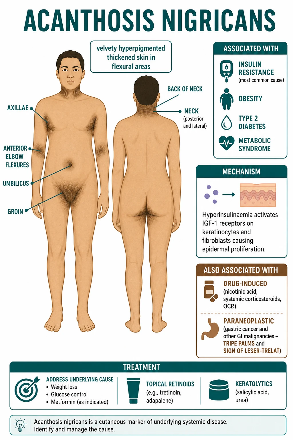
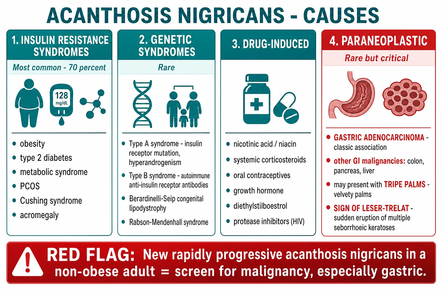
Acanthosis nigricans (AN) = hyperpigmented, velvety, thickened skin in flexural areas (axillae, neck, groin, umbilicus). Most commonly associated with insulin resistance (obesity, type 2 diabetes, metabolic syndrome, PCOS). Mechanism: hyperinsulinaemia activates IGF-1 receptors on keratinocytes and fibroblasts → epidermal proliferation and papillomatosis. Other causes: drug-induced (nicotinic acid, corticosteroids, OCP, growth hormone, protease inhibitors), genetic syndromes (Type A/B insulin resistance, Rabson-Mendenhall, Berardinelli-Seip lipodystrophy), and paraneoplastic (gastric adenocarcinoma is the classic association, also GI lymphoma, liver, breast, lung, ovary). Red flag: rapidly progressive, extensive AN in a non-obese adult — with or without tripe palms and the sign of Leser-Trélat — mandates urgent screening for GI malignancy. Treatment: address the underlying cause (weight loss most effective; metformin; GLP-1 receptor agonists), stop causative drugs; symptomatic topical retinoids (tretinoin 0.025%), keratolytics (salicylic acid 6%, urea 20%), and procedural options (dermabrasion, laser).
On this page & tools
Your progress
Saved locally on this device.
Exam tags
Red flags
Overview and Definition
Acanthosis nigricans is a cutaneous marker rather than a primary disease. It presents as symmetric, velvety, hyperpigmented, thickened plaques in flexural and intertriginous skin — most characteristically the axillae, posterior and lateral neck, and groin, but also the umbilicus, antecubital and popliteal fossae, inframammary folds, and the anogenital region.[1][2] Although the pigmentation is striking, the colour change is driven mainly by epidermal thickening and papillomatosis rather than increased melanin; histology shows only mild basal hyperpigmentation against a backdrop of marked acanthosis and hyperkeratosis.[7]
The clinical importance of AN lies in what it signals about the patient's metabolic and oncological state. In most patients it is a visible marker of insulin resistance — obesity, type 2 diabetes mellitus (T2DM), the metabolic syndrome, polycystic ovary syndrome (PCOS), and a cluster of endocrinopathies in which compensatory hyperinsulinaemia drives keratinocyte proliferation through insulin-like growth factor 1 (IGF-1) receptors.[7][6] In a minority it is drug-induced (nicotinic acid, systemic corticosteroids, oral contraceptives, growth hormone, HIV protease inhibitors),[11][1] in rare families it is a genetic/syndromic disorder of severe insulin resistance,[8] and in perhaps one in a thousand cases it is paraneoplastic — most classically heralding gastric adenocarcinoma, often together with tripe palms and the sign of Leser-Trélat.[3][9][10]
This dual personality — a benign cosmetic marker in the obese adolescent and a lethal warning sign in the non-obese adult — is the central teaching point of the topic. Every clinician must be able to recognise the morphological triad, work through the metabolic differential, and know exactly when to abandon reassurance and arrange urgent upper gastrointestinal endoscopy. [1]
Classification
The Curth classification, refined over subsequent decades, divides AN by aetiology into eight clinical categories.[3][1] The division that matters most at the bedside is benign (any aetiology that is not cancer — by far the majority) versus malignant (paraneoplastic), because the tempo, distribution, and prognosis diverge sharply.
A short summary of the eight Curth categories follows, because examiners test the list:[3][2]
- Obesity-associated (synonymous with "benign acquired") — by far the most common; resolves with weight loss.
- Syndromic / insulin-resistance — Type A (genetic INSR defect), Type B (auto-antibodies against the insulin receptor), Rabson-Mendenhall, Berardinelli-Seip lipodystrophy, Crouzon/severe craniosynostosis syndromes.
- Benign familial (hereditary) — autosomal dominant, onset in childhood, no metabolic abnormality.
- Malignant (paraneoplastic) — abrupt onset, generalized, mucosal involvement; gastric adenocarcinoma in roughly half, with other GI, liver, breast, lung, ovarian, and haematological malignancies also reported.
- Acral AN — on the dorsa of hands and feet of otherwise healthy patients; no systemic cause.
- Unilateral (naevoid) AN — a localized epidermal naevus following lines of Blaschko; not metabolic.
- Drug-induced AN — nicotinic acid, systemic corticosteroids, oral contraceptives and oestrogens, growth hormone, protease inhibitors, and occasionally fusidic acid and cobalt.
- Mixed — a patient with, for example, obesity and a tumour; classification is by the dominant clinical driver. [1]
Epidemiology and Risk Factors
The prevalence of AN tracks the prevalence of obesity and insulin resistance in a population. In community surveys of overweight and obese adolescents and adults, AN is found in anywhere from roughly one-quarter to two-thirds, and its presence correlates strongly with body mass index (BMI), waist circumference, fasting insulin, and a family history of T2DM.[1][7] In lean adults the prevalence is far lower — around 5–7% — and a lean patient with new AN should prompt the search for an alternative driver (endocrine, drug, or malignant).[2]
- Obesity and central adiposity — the single dominant risk factor; the relationship is dose-dependent with BMI.
- Family history of type 2 diabetes — AN is a recognised early marker of insulin resistance in children of diabetic parents.
- Ethnicity — prevalence is higher in Hispanic, African, South Asian, and Native American populations, paralleling the higher prevalence of insulin resistance in these groups. This is not a difference in disease susceptibility so much as a difference in the metabolic substrate; clinicians in these populations should expect to see AN frequently and should treat it as a screening sign.
- Endocrinopathy — T2DM, PCOS, Cushing's syndrome, acromegaly, hypothyroidism, and Addison's disease can all produce AN through varying degrees of insulin resistance or growth-factor excess.
- Drug exposure — nicotinic acid is the classical culprit; systemic steroids, OCPs, growth hormone, and HIV protease inhibitors are well described.[11]
- Malignancy — a rare but critical risk factor; adults over 40 with new, rapidly progressive, extensive AN warrant paraneoplastic screening.
- Darker skin phototype (Fitzpatrick IV–VI) — AN is more visually apparent and often more cosmetically distressing in pigmented skin; it is also more common because of the higher background prevalence of insulin resistance.
Pathophysiology
The unifying mechanism across most forms of AN is growth-factor-driven proliferation of keratinocytes and dermal fibroblasts, producing papillomatosis, acanthosis, and hyperkeratosis. The dominant growth factor differs by aetiology.[1][7]
Insulin resistance and the IGF-1 receptor
In insulin-resistant states, the pancreas compensates for receptor-level resistance by secreting more insulin, producing hyperinsulinaemia. Insulin and IGF-1 share structural homology, and at supraphysiological concentrations insulin spills over to bind and activate IGF-1 receptors on the surface of keratinocytes and dermal fibroblasts.[7] This drives epidermal proliferation, papillomatosis, and dermal collagen condensation — the triad that produces the characteristic velvety, thickened plaque. The same logic explains why AN is associated with conditions of insulin excess or resistance (obesity, T2DM, PCOS, Cushing's, acromegaly, hypothyroidism) but is not a feature of type 1 diabetes, where absolute insulin deficiency rather than resistance is the problem.[6]
The pathway can be sketched as a five-step cascade:[7][4]
- Insulin resistance at peripheral receptor level →
- Compensatory hyperinsulinaemia (the pancreas secretes more to hold glucose) →
- Excess insulin spillover onto IGF-1 receptors on keratinocytes/fibroblasts →
- Proliferation — papillomatosis, acanthosis, hyperkeratosis →
- The clinically apparent velvety, hyperpigmented, thickened plaque. [1]
Malignant acanthosis nigricans
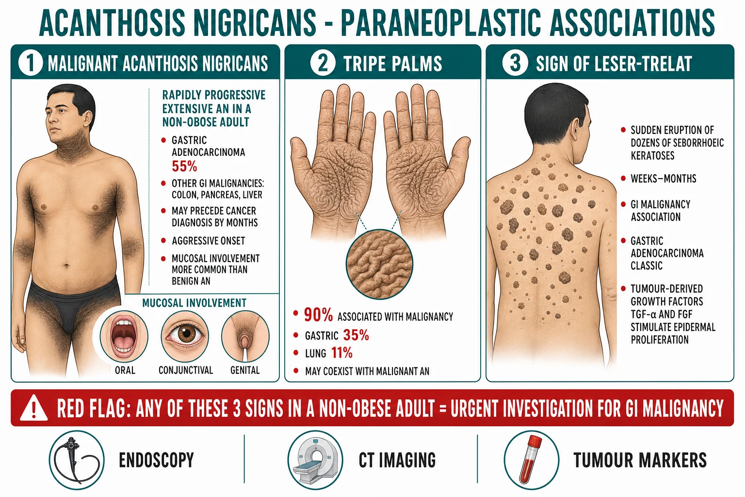
In paraneoplastic AN the keratinocyte proliferation is driven not by insulin but by tumour-derived growth factors, most importantly transforming growth factor-alpha (TGF-α) and fibroblast growth factors (FGF), which act on epidermal growth factor receptors (EGFR) to produce explosive, generalized proliferation.[1][9] This explains the abrupt tempo, the involvement of non-flexural skin, and the mucosal and palmar involvement (tripe palms) that distinguish malignant from benign AN. The tumour may secrete these factors directly, or, as is increasingly recognised, the EGFR signalling pathway may be upregulated indirectly by tumour-derived cytokines.[9]
Drug-induced AN
Nicotinic acid (niacin) is the prototype drug cause, and the mechanism is thought to be a direct stimulant effect on epidermal growth-factor signalling rather than insulin; the lesions resolve on withdrawal of the drug.[11] Systemic corticosteroids and growth hormone work mainly through induced insulin resistance (steroids) or direct growth-factor signalling (GH/IGF-1 axis), and protease inhibitors produce a lipodystrophy/insulin-resistance syndrome.[1]
Syndromic AN
In the Type A syndrome of insulin resistance, loss-of-function mutations in the INSR gene (encoding the insulin receptor) produce severe receptor-level resistance, profound hyperinsulinaemia, AN, ovarian hyperandrogenism, and (in women) virilisation.[8] The Type B syndrome is caused by auto-antibodies against the insulin receptor and overlaps with other autoimmune disease. The rare Rabson-Mendenhall syndrome and Donohue syndrome (leprechaunism) are progressively more severe recessive INSR mutations, while Berardinelli-Seip congenital lipodystrophy produces AN through near-total absence of adipose tissue and severe insulin resistance.[8]
Histopathology
Histology is rarely required but is characteristic when obtained: papillomatosis (finger-like dermal projections), hyperkeratosis, and acanthosis (epidermal thickening), with only mild basal-layer hyperpigmentation — confirming that the visible "hyperpigmentation" is largely an optical effect of thickened, undulating epidermis rather than true melanin excess.[7]
Why the same lesion arises from different drivers
The unifying insight is that three different growth-factor systems converge on the keratinocyte to produce the same histological lesion. In insulin resistance, the driver is insulin spilling onto the IGF-1 receptor; in malignancy, it is tumour-derived TGF-α and FGF acting on the EGFR; in growth-hormone excess and steroid therapy, it is the GH/IGF-1 axis and induced insulin resistance respectively; and in the genetic INSR defects (Type A, Rabson-Mendenhall, lipodystrophy) it is sustained hyperinsulinaemia driven by receptor loss. Each path converges on a final common pathway of keratinocyte and fibroblast proliferation with papillomatosis and hyperkeratosis.[7][8] This convergence explains why the clinical lesion is identical regardless of cause, why the differential hinges on tempo and distribution rather than morphology, and — crucially for management — why treating the upstream driver (weight loss, metformin, tumour resection, drug withdrawal) improves the skin while treating the skin alone does not.
Clinical Presentation
The lesion of AN is one of the most instantly recognisable in dermatology. Three morphological descriptors define it.[1][2]
- Velvety texture — the surface is soft, smooth, and finely papillated, feeling like velvet to the touch, the result of dermal papillomatosis.
- Hyperpigmentation — brown to grey-brown or almost black discoloration. The colour is partly melanin and partly an optical consequence of the thickened, undulating epidermis; it is not a primary disorder of melanocytes.
- Thickening — the skin is visibly and palpably thickened, often with skin tags (acrochordons) embedded within or adjacent to the plaque. [1]
Site distribution
The characteristic sites, in approximate descending order of frequency, are:[1][4]
- Axillae — the single most common site.
- Posterior and lateral neck — produces the so-called "dirty neck" (or "neck that won't wash clean"), which can be intensely stigmatising.
- Groin and inguinal folds.
- Umbilicus.
- Antecubital and popliteal fossae.
- Knuckles, elbows, and extensor surfaces — especially in dark-skinned patients with diabetes.
- Inframammary folds, perianal and anogenital skin, and the mucosal surfaces (oral mucosa, lips, tongue, conjunctiva, oesophagus, pharynx). [1]
Symptoms
AN is usually asymptomatic. It may be mildly pruritic, particularly in intertriginous areas where secondary maceration or candidal overgrowth supervenes, and it can be malodorous when moist. The dominant complaint is cosmetic: patients (and parents of affected children) are distressed by the appearance and by the false perception of poor hygiene, particularly when the neck is heavily involved. [1]
Associated cutaneous findings
- Skin tags (acrochordons) are frequently co-located within or at the edge of the plaque and share the same insulin-resistance substrate.
- Seborrhoeic keratoses — a sudden eruption of multiple seborrhoeic keratoses together with AN defines the sign of Leser-Trélat, a recognised paraneoplastic marker.[10]
- Tripe palms (acanthosis palmaris) — a velvety, rugose, moss-like thickening of the palmar skin, with exaggerated dermatoglyphics; when AN and tripe palms coexist, roughly nine in ten patients have an underlying malignancy.[9]
- Inflammation and maceration of intertriginous plaques is common in obese patients.
Features that suggest malignant (paraneoplastic) AN
The clinical features that should trigger a paraneoplastic work-up are the cornerstone of safe practice:[1][9]
- Rapid onset and progression over weeks to months, rather than the years-long slow evolution of obesity-associated AN.
- Extensive distribution beyond the flexures, with involvement of non-intertriginous skin.
- Mucosal involvement — oral mucosa, lips, tongue, and pharynx are far more commonly affected in malignant than in benign AN.
- Tripe palms and/or sign of Leser-Trélat.
- Onset in a non-obese adult, especially over the age of forty, with weight loss and GI symptoms.
- AN that precedes or accompanies a known malignancy, or that flares with tumour recurrence (a paraneoplastic marker of disease activity).[9]
Atypical presentations and pitfalls
Examiners test atypical presentations deliberately, and several are worth memorising. Unilateral (naevoid) AN presents in childhood as a localised velvety plaque along the lines of Blaschko, most often on the torso or neck; it has no metabolic significance and is managed cosmetically. Acral AN appears on the dorsa of the hands and feet of otherwise well adults and is similarly benign. Mucosal AN — velvety change on the lips, buccal mucosa, tongue, conjunctiva, or oesophagus — is uncommon in obesity-associated disease and should heighten suspicion of a paraneoplastic driver, particularly when accompanied by tripe palms.[9]
In dark-skinned patients (Fitzpatrick IV–VI), AN is more visually striking and more easily mistaken for Addisonian pigmentation or post-inflammatory change; the velvety thickening on palpation and the flexural distribution are the discriminating features. In adolescents, AN over the knuckles and elbows can be mislabelled as "grime" or poor hygiene, generating significant bullying; recognising it as a metabolic sign rather than a cleanliness problem is part of the therapeutic encounter.[2] In pregnancy, new AN reflects the insulin resistance of gestation and warrants screening for gestational diabetes rather than reassurance alone. Finally, a patient with known cancer who develops new AN during follow-up should be investigated for recurrence — the skin disease is a sensitive surrogate marker of tumour activity.[9]
Differential Diagnosis
The differential of velvety, pigmented, flexural change is finite and examinable. Each mimic has a distinguishing feature.[1][2]
| Differential | Distinguishing feature from AN |
|---|---|
| Confluent and reticulated papillomatosis (CARP, Gougerot-Carteaud) | Reticulated, net-like pattern over the upper trunk and interscapular area rather than flexural; often younger, non-obese patients; responds to minocycline |
| Post-inflammatory hyperpigmentation (PIH) | Follows a documented inflammatory dermatosis (eczema, lichen planus); no velvety thickening or papillomatosis; histology shows basal melanin with normal epidermal architecture |
| Ichthyosis (especially acquired ichthyosis) | Generalised dry, scaly, fish-like scaling; no flexural predilection (often flexure-sparing); acquired form may itself be paraneoplastic |
| Pemphigus vegetans | Vegetating, crusted, malodorous plaques in intertriginous areas; positive Nikolsky; oral involvement; biopsy shows acantholysis and suprabasal clefting |
| Linear epidermal naevus (linear AN/naevoid AN) | Unilateral, follows lines of Blaschko, present from childhood; no metabolic or systemic association |
| Tinea versicolor (pityriasis versicolor) | Fine scale, positive potassium hydroxide microscopy,fluoresce with Wood's lamp; hypopigmented or light-brown macules rather than velvety thickening |
| Addison's disease pigmentation | Generalised, with predilection for sun-exposed sites, palmar creases, oral mucosa, and recent scars; hypotension and hyponatraemia; no velvety thickening |
The single most commonly confused entity is CARP, which can look superficially similar but is reticulated rather than flexural, occurs in younger, leaner patients, and responds dramatically to oral minocycline — a useful therapeutic discriminator if the diagnosis is in doubt. [1]
The reasoning that separates the mimics is mechanical. Ask first whether the lesion is truly thickened and velvety (AN, CARP, pemphigus vegetans) or flat pigment only (PIH, Addison's, pityriasis versicolor) — the former implies epidermal proliferation, the latter pigment deposition. Among the proliferative mimics, ask next about distribution: AN favours the flexures symmetrically, CARP favours the upper trunk and interscapular area in a reticulated net, pemphigus vegetans produces moist vegetating plaques with a positive Nikolsky sign and oral involvement, and a linear epidermal naevus is unilateral and Blaschkoid from childhood. Finally, ask about tempo and context: an acquired ichthyosis in an adult is itself a recognised paraneoplastic marker (often lymphoma) and warrants investigation in its own right, while Addisonian pigmentation is generalised, involves the palmar creases and oral mucosa, and is accompanied by hypotension, fatigue, and hyponatraemia.[1] When clinical doubt persists after this structured assessment, a punch biopsy for histology and direct immunofluorescence resolves most cases.
Clinical and Bedside Assessment
There is no named sign specific to AN, but a focused assessment answers three questions in sequence: (1) Is this AN? (2) What is the metabolic background? (3) Is this malignant?[1][4]
General inspection. Note the distribution and morphology of the lesions, the BMI and body habitus (calculate BMI and measure waist circumference), and the presence of skin tags, seborrhoeic keratoses, and stigmata of Cushing's or acromegaly. [1]
Site examination. Inspect all flexural sites — axillae, neck (especially posterior and lateral), groins, umbilicus, antecubital and popliteal fossae, inframammary folds, and anogenital skin. Examine the oral mucosa, lips, and tongue (mucosal involvement favours malignancy), and the palms (velvety thickening = tripe palms). Look specifically for a sudden eruption of seborrhoeic keratoses (sign of Leser-Trélat). [1]
Systemic examination. Palpate for lymphadenopathy, hepatomegaly (liver disease and hepatic tumours), abdominal masses (gastric cancer), and signs of endocrine disease (moon facies, buffalo hump, abdominal striae of Cushing's; prognathism, frontal bossing, large hands of acromegaly; goitre; clitoromegaly/virilisation in Type A insulin resistance). Measure blood pressure (metabolic syndrome). [1]
Bedside anthropometry. Document BMI and waist circumference. A waist circumference above 94 cm in men or 80 cm in women (Europid cut-offs; lower in South and East Asians) together with AN marks a high metabolic risk even before laboratory confirmation.[6]
Investigations
Investigations are directed by the clinical context — a child with obesity and a long-standing AN needs a metabolic work-up, whereas a non-obese adult with rapidly progressive extensive AN needs paraneoplastic screening. Skin biopsy is rarely required; the diagnosis is clinical.[1]
First-line metabolic work-up
For every patient with newly diagnosed AN, the metabolic screen is mandatory.[7][6]
- Fasting plasma glucose and HbA1c — to detect prediabetes (HbA1c 5.7–6.4%) or diabetes (HbA1c ≥6.5%, fasting glucose ≥7.0 mmol/L).
- Fasting insulin and HOMA-IR (Homeostatic Model Assessment of Insulin Resistance) — HOMA-IR is calculated as (fasting insulin in mIU/L × fasting glucose in mmol/L) ÷ 22.5. Insulin resistance is conventionally defined by a HOMA-IR above approximately 2.5–3.0 in Europid populations, with lower thresholds (~2.0) often applied in South and East Asian populations because of their greater insulin resistance at any given BMI.[6]
- Lipid panel — fasting total cholesterol, HDL, LDL, and triglycerides. The metabolic syndrome pattern is high triglycerides and low HDL.
- Oral glucose tolerance test (OGTT) — recommended in children and adolescents with AN and obesity to detect impaired glucose tolerance not captured by fasting tests.[2]
Interpreting the metabolic screen. A fasting insulin above roughly 75 pmol/L or a HOMA-IR above 2.5–3.0 (lower in South and East Asian populations) confirms significant insulin resistance and explains the cutaneous findings in the absence of overt diabetes. Document the full metabolic syndrome criteria alongside the HOMA-IR, because AN is one of its most reliable cutaneous markers: central obesity (waist circumference above 94 cm in Europid men, 80 cm in Europid women; 90/80 cm in South Asians), triglycerides at or above 1.7 mmol/L, HDL below 1.0 mmol/L in men or 1.3 mmol/L in women, blood pressure at or above 130/85 mmHg, and fasting glucose at or above 5.6 mmol/L; three of five defines the syndrome.[6] In children and adolescents, where adult cut-offs do not apply, age- and sex-specific percentile thresholds for BMI, waist circumference, and lipids are used, and the OGTT (75 g glucose load, 2-hour sample) is particularly valuable because impaired glucose tolerance is common and fasting tests are often normal.[2] A negative HOMA-IR in a typical-looking AN should prompt a search for a drug cause or, in the right context, a paraneoplastic driver.
Endocrine screen (selected)
When an endocrinopathy is suspected on clinical grounds:[4]
- Thyroid function tests (TSH, free T4) — hypothyroidism.
- 9 a.m. cortisol and 24-hour urinary free cortisol or dexamethasone suppression test — Cushing's syndrome.
- IGF-1 and growth hormone — acromegaly.
- Testosterone, LH, FSH, oestradiol, and pelvic ultrasound — PCOS in women with hyperandrogenism.
- Anti-insulin receptor antibodies and INSR sequencing — Type B and Type A insulin resistance syndromes when suspected.[8]
Paraneoplastic work-up (critical)
For a patient with rapidly progressive, extensive, mucosal, or tripe-palm-associated AN in whom malignancy is suspected, urgent investigation is mandatory:[1][9]
- Upper gastrointestinal endoscopy — the single highest-yield test, because gastric adenocarcinoma accounts for roughly half of paraneoplastic AN.
- CT chest, abdomen, and pelvis with contrast — to stage the tumour and identify non-gastric primaries (lung, liver, ovary, colon, pancreas).
- Tumour markers — carcinoembryonic antigen (CEA), carbohydrate antigen 19-9 (CA 19-9), and alpha-fetoprotein, recognising their limited sensitivity but utility in monitoring.
- Mammography and pelvic ultrasound — for breast and ovarian primaries.
- Repeat endoscopy if the first is negative but suspicion remains, because small or early gastric cancers may be missed. [1]
Skin biopsy
Reserved for cases where the diagnosis is in doubt (e.g., suspected CARP, pemphigus vegetans, or lichen planus). Findings are papillomatosis, hyperkeratosis, and acanthosis with mild basal hyperpigmentation — a pattern that confirms AN but does not distinguish benign from malignant variants.[7]
Management — Approach
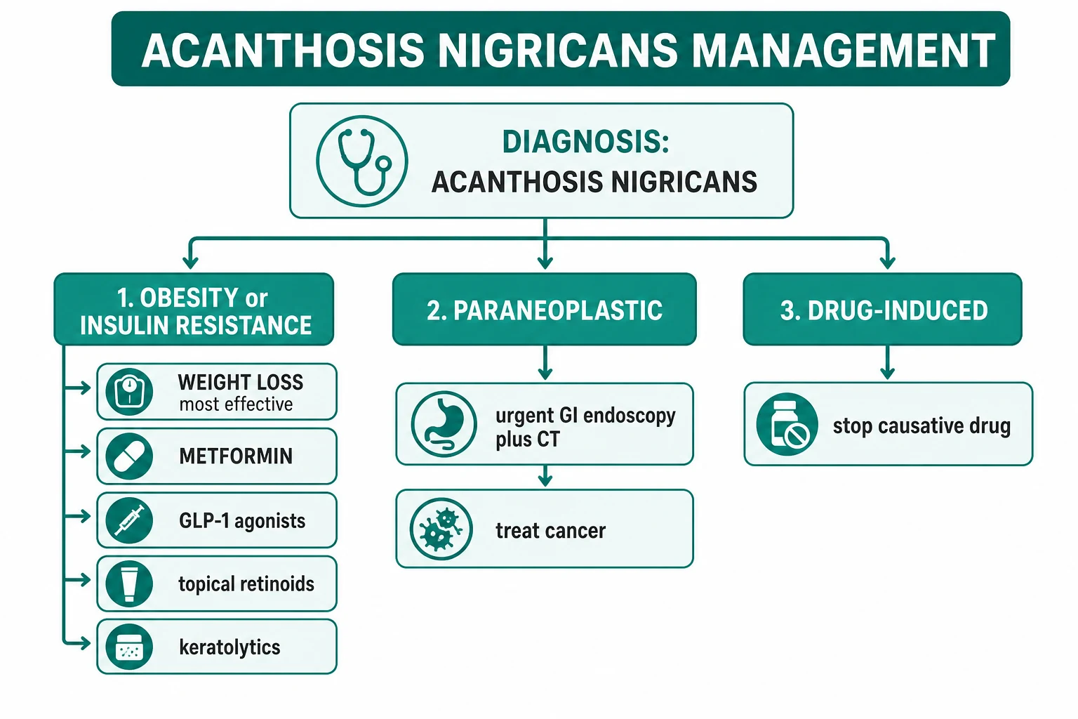
There is no specific cure for the lesion of AN itself. Management is entirely directed at the underlying cause, with topical and procedural measures reserved for residual cosmetic concerns. The three management pathways — insulin resistance, paraneoplastic, and drug-induced — run in parallel and answer three different questions.[1][7]
Step 1 — Establish and treat the underlying cause
This is the single most important step. Treatment of the cause improves the lesion; symptomatic measures improve only its appearance. [1]
Step 2 — Symptomatic topical and procedural therapy
For residual cosmetic concern after the cause has been addressed, the following are useful.[1][7]
Step 3 — Patient education and prevention
Explain that AN is a marker of insulin resistance, not a disease in its own right; that it will improve with weight loss, exercise, and glycaemic control; and that it is not contagious, not the result of poor hygiene, and not a sign of skin cancer. This counselling is often the most therapeutic intervention in a distressed adolescent.[2]
Management — Insulin-Resistance / Obesity-Associated AN
This is the most common pathway and the one with the strongest evidence.[7][6]
- Weight loss is the most effective intervention. A loss of even 5–10% of body weight reduces insulin resistance, lowers circulating insulin, and visibly lightens and thins the plaques. Caloric restriction combined with at least 150 minutes per week of moderate aerobic exercise, plus resistance training, is the foundation.[1]
- Metformin (typically 500 mg twice daily, titrated to 1 g twice daily with meals) improves insulin sensitivity by reducing hepatic gluconeogenesis and is first-line pharmacotherapy in T2DM, prediabetes, and PCOS-associated AN; improvements in AN have been demonstrated.[7]
- GLP-1 receptor agonists (semaglutide, liraglutide, tirzepatide) produce substantial weight loss and improve insulin sensitivity; emerging case series report improvement in AN severity and are a logical escalation for obese patients with AN and T2DM or obesity.[5]
- Glycaemic optimisation — control of HbA1c to under 7% reduces chronic hyperinsulinaemia.
- Insulin in T2DM — note that exogenous insulin can worsen AN by recreating the hyperinsulinaemic drive; where possible prefer insulin-sensitisers (metformin) and weight loss over escalating insulin doses in patients with prominent AN.[6]
OBESITY treatment ladder for AN
W-LOSS
5–10% body weight — the single most effective intervention
Diet + ≥150 min/week aerobic + resistance exercise
500–1000 mg BD; insulin sensitiser, first-line
Weight loss + insulin sensitivity; emerging evidence
Tretinoin 0.025%, salicylic acid 6%, urea 20%
Management — Malignant (Paraneoplastic) AN
The only effective treatment for the skin disease is treatment of the underlying tumour.[1][9] AN that regresses after successful tumour resection and flares with recurrence serves as a useful surrogate tumour marker worth documenting during oncological follow-up.[9]
- Urgent upper GI endoscopy and CT staging within two weeks of clinical suspicion; biopsy of any lesion.
- Surgical resection with curative intent where the tumour is operable (gastrectomy for gastric adenocarcinoma; resection of lung, ovarian, renal, or colonic primaries).
- Chemoradiotherapy for advanced or unresectable disease; AN may regress with effective tumour control and flare with recurrence, providing a useful tumour marker.[9]
- Symptomatic topicals (retinoids, keratolytics) for comfort and appearance.
- Realistic prognosis — most paraneoplastic AN presents with advanced disease; median survival is often measured in months. Honest communication with the patient and family is part of the management.
Management — Drug-Induced AN
Stop, switch, or reduce the causative drug; lesions resolve over weeks to months.[11][1]
- Nicotinic acid (niacin) — the most common drug cause; switch lipid-lowering strategy (statin ± ezetimibe, or a PCSK9 inhibitor) where feasible.
- Systemic corticosteroids — taper where possible; topical steroids for the underlying disease often suffice.
- Oral contraceptives/oestrogens — consider progestogen-only or non-hormonal alternatives.
- Growth hormone — review the indication; AN usually reflects effective (or excessive) therapy.
- Protease inhibitors — switch within the HIV antiretroviral class to a non-protease-inhibitor regimen. [1]
Specific Subtypes and Syndromes
Type A insulin resistance syndrome
A rare, autosomal dominant or recessive disorder caused by heterozygous or compound heterozygous loss-of-function mutations in the INSR gene.[8] It presents in young, lean, often black or Asian women with severe insulin resistance, hyperinsulinaemia, marked AN, ovarian hyperandrogenism (hirsutism, acne, oligomenorrhoea, virilisation, clitoromegaly), and acanthosis. Management is metformin, insulin-sensitisers, androgen suppression, and lifestyle; prognosis depends on the degree of beta-cell preservation.[8]
Type B insulin resistance syndrome
Caused by auto-antibodies against the insulin receptor, typically in middle-aged women with other autoimmune disease (SLE, Sjögren's) or a lymphoproliferative disorder. Severe insulin resistance, hyperglycaemia (or paradoxical hypoglycaemia from agonist antibody effects), widespread AN, and sometimes acanthosis of the hands and face. Treatment is immunosuppression (corticosteroids, cyclophosphamide, rituximab) plus insulin-sensitisers; spontaneous remission can occur.[1]
Rabson-Mendenhall and Donohue (leprechaunism) syndromes
Progressively more severe recessive INSR mutations. Rabson-Mendenhall features AN, pineal hyperplasia, dental and nail dysplasia, and precocious puberty; Donohue (leprechaunism) is the most extreme, with elfin facies, hirsutism, lipodystrophy, and death in infancy from refractory diabetes.[8]
Berardinelli-Seip congenital lipodystrophy
Near-total absence of adipose tissue from birth produces severe insulin resistance, hypertriglyceridaemia, hepatic steatosis, and prominent AN. Management is metabolic (diet, metformin, leptin analogue metreleptin in selected cases).[8]
Benign familial (hereditary) AN
Autosomal dominant, onset in early childhood, no associated metabolic abnormality, no progression; reassurance is all that is required.[2]
Acral and unilateral (naevoid) AN
Acral AN affects the dorsa of the hands and feet in otherwise well patients. Unilateral/naevoid AN is a localised epidermal naevus along Blaschko's lines, present from childhood; treatment is cosmetic (keratolytics, laser).[2]
Complications and Pitfalls
The complications of AN are the complications of its underlying cause, plus a small set of lesion-specific problems.[1]
- Missed malignancy — the cardinal pitfall. Failing to recognise the paraneoplastic variant in a non-obese adult with rapidly progressive disease delays diagnosis of an often-advanced cancer. The corrective is a low threshold for endoscopy when the tempo, distribution, or mucosal involvement is atypical.[9]
- Cardiovascular disease and type 2 diabetes — AN is a visible marker of insulin resistance and the metabolic syndrome; the long-term risk is atherosclerotic disease and diabetes, not the skin lesion itself.[6]
- Cosmetic and psychosocial morbidity — particularly the "dirty neck" in adolescents and dark-skinned patients; bullying, low self-esteem, and social withdrawal are under-recognised.[2]
- Secondary infection and maceration of intertriginous plaques in obese patients, with malodour and candidal overgrowth.
- Excess insulin therapy in T2DM can paradoxically worsen AN; insulin-sensitisers are preferred when AN is prominent.[6]
- Diagnostic error — confusing AN with CARP (which responds to minocycline), with pemphigus vegetans (life-threatening), or with post-inflammatory hyperpigmentation. Skin biopsy resolves doubt in atypical cases.[1]
Prognosis and Disposition
Prognosis is dictated entirely by the underlying cause.[1][9]
- Obesity/insulin-resistance-associated AN is benign and chronic; it improves steadily with weight loss (typically visible within months), glycaemic control, and exercise. It is cosmetically distressing but not life-threatening.
- Drug-induced AN resolves completely within weeks to months of stopping the causative agent.
- Benign familial AN is lifelong and stable; no treatment is needed beyond reassurance.
- Syndromic AN (Type A/B, lipodystrophy) is persistent and mirrors the severity of the underlying insulin resistance; control of metabolism improves but rarely clears the lesions.[8]
- Malignant AN has a poor prognosis, dictated by the underlying tumour. Most cases present with advanced malignancy; median survival is often in the order of months. Effective tumour control may regress the skin disease, providing a surrogate tumour marker.[9]
Disposition. A patient with benign AN is managed in primary care or dermatology with a metabolic work-up and lifestyle advice; referral to endocrinology is appropriate for PCOS, Cushing's, acromegaly, or suspected insulin-resistance syndromes. A patient with suspected paraneoplastic AN is referred urgently (within two weeks) for gastroenterology review and endoscopy.[9]
Prevention
Because the dominant form of AN is obesity- and insulin-resistance-associated, primary prevention is the prevention of obesity and type 2 diabetes:[6]
- Maintain a healthy body weight (BMI 18.5–24.9 kg/m²; lower targets in South and East Asian populations).
- Regular aerobic and resistance exercise (≥150 minutes per week).
- A diet low in refined carbohydrate and saturated fat, with adequate fibre.
- Screen at-risk individuals (family history of T2DM, PCOS, gestational diabetes) with fasting glucose and HbA1c. [1]
Drug-induced AN is prevented by choosing alternatives to nicotinic acid and high-dose systemic corticosteroids where feasible, and by recognising protease-inhibitor lipodystrophy in HIV therapy. [1]
Malignant AN is not preventable, but early recognition and urgent endoscopy in the appropriate clinical context is the closest thing to secondary prevention — it offers the only realistic chance of curative resection.[9]
Special Populations
Children and adolescents
AN is common in obese children and adolescents and is a recognised early marker of insulin resistance in this group. The appropriate response is not reassurance that it is "just cosmetic" but a focused metabolic work-up (BMI, fasting glucose and insulin, HOMA-IR, lipids, OGTT), family counselling, and a structured weight-management programme. The cosmetic distress — particularly the "dirty neck" — is real and deserves acknowledgement; topical retinoids and keratolytics have a role once metabolic management is under way.[2]
Paediatric counselling pearls. Frame AN for the child and parents as a visible warning light, not a disease — it signals that the body is handling glucose less efficiently than it should and that weight management and activity now will prevent type 2 diabetes later. Avoid language that blames the child for "not washing"; the "dirty neck" is a metabolic sign and is neither contagious nor a hygiene problem. Set a concrete goal — a 5–10% reduction in BMI percentile over six to twelve months through family-based lifestyle change — and review at intervals, because visible lightening of the lesion is itself motivating and confirms adherence. Screen first-degree relatives for type 2 diabetes, because a strong family history is common and modifies the child's long-term risk.[2]
Pregnancy
AN may appear or worsen in pregnancy, driven by the physiological insulin resistance of late gestation and by weight gain; it often regresses post-partum. New AN in pregnancy should also prompt screening for gestational diabetes (oral glucose tolerance test at 24–28 weeks) and for PCOS if hyperandrogenism is present.[4]
Darker skin phototypes (Fitzpatrick IV–VI)
AN is more visually apparent and more cosmetically distressing in pigmented skin and is also more prevalent because of the higher background rate of insulin resistance in many of these populations. Topical retinoids and keratolytics are equally effective but post-inflammatory hyperpigmentation from irritant topicals can be more prominent; start at low concentration and titrate.[2]
Patients on long-term corticosteroids or HIV protease inhibitors
Recognise drug-induced AN early; co-ordinate with the prescribing specialty (rheumatology, infectious diseases) to switch or de-escalate where possible rather than adding cosmetic topicals in isolation.[11]
Evidence, Guidelines, and Regional Differences
The evidence base for AN management is largely observational and from case series; there are no large randomised trials for topical or systemic therapy. The strongest evidence supports weight loss and metformin for insulin-resistance-associated AN, with emerging evidence for GLP-1 receptor agonists.[5][7]
Insulin resistance cut-offs are population-specific. The HOMA-IR threshold defining insulin resistance is conventionally above 2.5–3.0 in Europid populations, but lower thresholds (~2.0) are recommended in South and East Asian populations, who develop insulin resistance and metabolic complications at lower BMIs. Waist-circumference cut-offs similarly differ: Europid men ≥94 cm and women ≥80 cm; South Asian men ≥90 cm and women ≥80 cm (IDF). This matters when interpreting a metabolic work-up in an AN patient.
RANZCP/RACP and Diabetes Australia endorse HbA1c-based screening and lifestyle intervention; paraneoplastic AN with GI symptoms is referred for endoscopy under urgent cancer-pathway pathways consistent with the local state or national cancer-agency criteria.
Controversies. The role of GLP-1 receptor agonists and tirzepatide in AN specifically (as opposed to obesity and T2DM generally) is being defined; early case series are encouraging but randomised data are awaited.[5] The clinical utility of HOMA-IR (versus fasting insulin or simply clinical recognition of metabolic syndrome) is debated; many clinicians treat the metabolic phenotype rather than chase the HOMA-IR number.
Exam Pearls
PARANEOPLASTIC triad mnemonic
3T-L
Malignant acanthosis nigricans — abrupt, extensive, mucosal
Velvety palmar thickening — ~90% malignant when AN coexists
Sudden eruption of seborrhoeic keratoses — Leser-Trélat
Gastric adenocarcinoma is the classic primary — urgent endoscopy
Self-test: A 55-year-old thin man presents with new, rapidly progressive velvety pigmentation of the axillae, neck, palms, and oral mucosa over 8 weeks, plus a 6 kg weight loss. What is the next best step?
This is a textbook paraneoplastic AN presentation: thin (non-obese) adult, abrupt onset, mucosal and palmar (tripe palms) involvement, weight loss. The next best step is urgent upper gastrointestinal endoscopy with biopsy plus CT chest/abdomen/pelvis to identify and stage gastric adenocarcinoma. Topical retinoids and metformin are inappropriate and would delay diagnosis.
Red Flags
Exam application bank (NEET-PG / INICET)
One-line answer
Acanthosis nigricans (AN) = hyperpigmented, velvety, thickened skin in flexural areas (axillae, neck, groin, umbilicus). Most commonly associated with insulin resistance (obesity, type 2 diabetes, metabolic syndrome, PCOS). Mechanism: hyperinsulinaemia activates IGF-1 receptors on keratinocytes and fibroblasts → epidermal proliferation and papillomatosis. Other causes: drug-induced (nicotinic acid, corticosteroids, OCP, growth hormone, protease inhibitors), genetic syndromes (Type A/B insulin resistance, Rabson-Mendenhall, Berardinelli-Seip lipodystrophy), and paraneoplastic (gastric adenocarcinoma is the classic association, also GI lymphoma, liver, breast, lung, ovary). Red flag: rapidly progressive, extensive AN in a non-obese adult — with or without tripe palms and the sign of Leser-Trélat — mandates urgent screening for GI malignancy. Treatment: address the underlying cause (weight
Worked stems (answer without another resource)
Stem 1 — Classic presentation. Map symptoms to mechanism; name the first investigation and first treatment step with dose/route if drug therapy is standard. [1]
Stem 2 — Unstable / complicated. List red flags that force immediate resuscitation, theatre, ICU, antidote, or reperfusion — and what you do in the first 15 minutes. [1]
Stem 3 — Atypical group. Elderly, pregnancy, child, or immunocompromised: how presentation and thresholds change. [1]
Stem 4 — Differential trap. Name the three closest mimics and one discriminator for each. [1]
Stem 5 — Disposition. Who goes home with safety-netting, who is admitted, who needs HDU/ICU/theatre, and what follow-up is mandatory. [1]
Rapid viva checklist
- Definition + classification
- Pathophysiology chain
- Bedside signs / criteria
- Score with exact components (if any)
- Emergency bundle
- Definitive therapy with doses
- Complications of disease and of treatment
- Special populations
- Guideline/trial name if classic
- Three exam traps
Coverage self-check
If you cannot answer any stem above from this page alone, re-read the matching section — the page is intended to be self-sufficient for final-prof and NEET-PG/INICET questions on Acanthosis nigricans.
References
- [1]Das A, Datta D, Kassir M, et al. Acanthosis nigricans: A review J Cosmet Dermatol, 2020.PMID 32516476
- [2]Leung AKC, Lam JM, Barankin B, et al. Acanthosis Nigricans: An Updated Review Curr Pediatr Rev, 2022.PMID 36698243
- [3]Curth HO. Acanthosis nigricans Birth Defects Orig Artic Ser, 1971.PMID 5173298
- [4]Lause M, Kamboj A, Fernandez Faith E. Dermatologic manifestations of endocrine disorders Transl Pediatr, 2017.PMID 29184811
- [5]Lal K, Herringshaw E. The Use of GLP-1 Agonists in the Management of Cutaneous Disease J Clin Aesthet Dermatol, 2024.PMID 39263264
- [6]Abate MCMO, Aroucha PMT, Nóbrega DVMD, et al. Cutaneous manifestations of diabetes mellitus: a narrative review Einstein (Sao Paulo), 2025.PMID 40105573
- [7]Hermanns-Lê T, Scheen A, Piérard GE. Acanthosis nigricans associated with insulin resistance : pathophysiology and management Am J Clin Dermatol, 2004.PMID 15186199
- [8]Moller DE, Flier JS. Detection of an alteration in the insulin-receptor gene in a patient with insulin resistance, acanthosis nigricans, and the polycystic ovary syndrome (type A insulin resistance) N Engl J Med, 1988.PMID 2460770
- [9]Lam CPM, Chan MWM. Metastatic adenocarcinoma of the stomach presenting as malignant acanthosis nigricans and tripe palms: a case report Hong Kong Med J, 2023.PMID 37489275
- [10]André R, Laffitte E, Abosaleh M. Sign of Leser-Trélat and Cutaneous T-Cell Lymphoma: A Rare Association Dermatopathology (Basel), 2018.PMID 29998101
- [11]Stals H, Vercammen C, Peeters C. Acanthosis nigricans caused by nicotinic acid: case report and review of the literature Dermatology, 1994.PMID 8075456