Dermatology · Medicine
Acne fulminans
Also known as Acne fulminans · Acne maligna · Acute febrile ulcerative acne conglobata
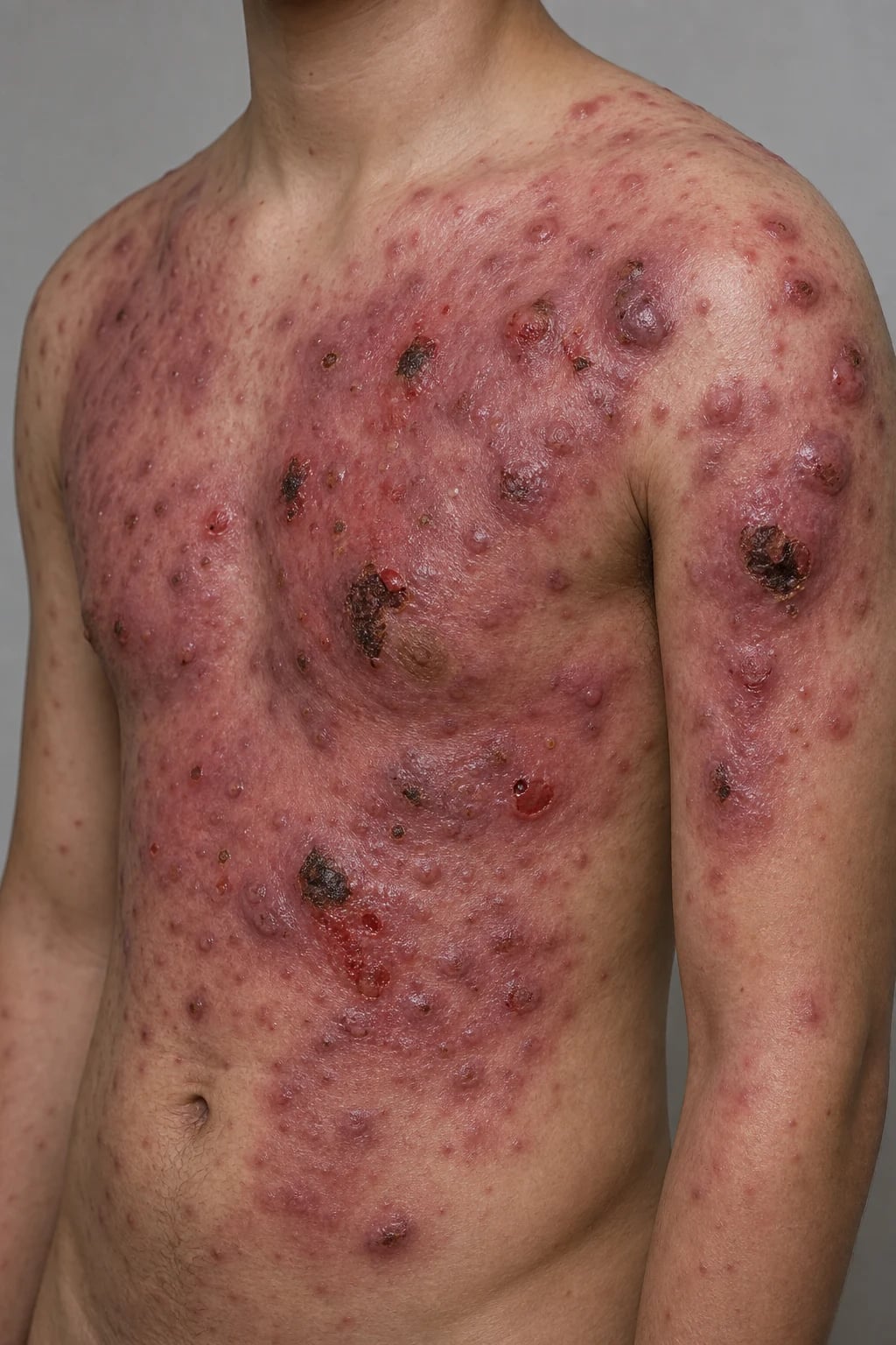
Acne fulminans is the most severe form of acne, characterised by sudden onset of large, painful, ulcerative nodules with necrotic haemorrhagic crusts on the chest, back and face, accompanied by systemic symptoms (fever, arthralgia, myalgia) and laboratory abnormalities (leukocytosis, elevated ESR/CRP). It primarily affects adolescent males. The pathophysiology is a Type III hypersensitivity / immune-complex reaction to Cutibacterium acnes. Treatment is a dermatological emergency: systemic corticosteroids FIRST (prednisolone 0.5-1 mg/kg/day), then low-dose isotretinoin (0.5 mg/kg/day) introduced after 1-2 weeks and gradually uptitrated; isotretinoin alone can worsen the condition.
On this page & tools
Your progress
Saved locally on this device.
Exam tags
Red flags
Definition & Classification
Acne fulminans (AF), historically termed acute febrile ulcerative acne conglobata and acne maligna, is the most severe variant of inflammatory acne. It is defined by an explosive, sudden onset of large, painful, ulcerative nodules covered with necrotic, haemorrhagic crusts on the chest, back and (less prominently) the face, accompanied by systemic symptoms (fever, arthralgia, myalgia, weight loss, malaise) and laboratory abnormalities (leukocytosis with neutrophilia, elevated ESR and CRP, mild anaemia of chronic disease).[1][2][3]
The hallmark that separates AF from every other form of acne is the combination of cutaneous ulceration and systemic inflammation — fever and raised inflammatory markers are absent in uncomplicated acne vulgaris and even in acne conglobata. AF sits at the apex of the acne severity spectrum and represents a true dermatological emergency because of rapid scarring, systemic upset and the severe psychological impact of sudden disfigurement in an adolescent.[6]
The spectrum of acne severity
| Severity tier | Hallmark | Systemic features | Onset |
|---|---|---|---|
| Acne vulgaris | Comedones ± inflammatory papules/pustules | None | Gradual, chronic |
| Severe nodulocystic acne | Painful deep nodules and cysts; no ulceration | None | Gradual |
| Acne conglobata | Coalescing double comedones, abscesses, sinus tracts; draining burrows | None | Chronic, indolent |
| Acne fulminans | Ulcerative nodules with necrotic haemorrhagic crusts | Fever, arthralgia, myalgia, leukocytosis | Sudden, over days–weeks |
The crucial distinction at the bedside: acne conglobata is chronic, indolent and lacks systemic symptoms, whereas acne fulminans is acute, ulcerative and systemically inflammatory.[2][6]
The follicular occlusion tetrad
Acne fulminans overlaps with the follicular occlusion tetrad, a group of conditions driven by occlusion and rupture of the pilosebaceous unit with a neutrophilic, scarring inflammatory response: [1]
The follicular occlusion tetrad — the '4 Hs' mnemonic
SAPHO and the autoinflammatory syndromes
SAPHO syndrome (Synovitis, Acne, Pustulosis, Hyperostosis, Osteitis) is the key systemic overlap: severe acne (including AF) is associated with osteolytic and hyperostotic bone lesions, anterior chest-wall arthritis and palmar-plantar pustulosis. The PAPA / PAPASH / PASH / PsAPASH family of autoinflammatory syndromes (driven by PSTPIP1/CD2BP1 mutations and IL-1β overproduction) places acne fulminans on a spectrum with pyoderma gangrenosum and sterile pyogenic arthritis — the rationale for IL-1 blockade (anakinra, canakinumab) in refractory disease.[7][8]
Epidemiology & Risk Factors
Acne fulminans is rare: it accounts for well under one percent of all acne presentations and probably affects on the order of a few hundred new patients per year in large Western populations. Despite this rarity it is heavily examined because its management is a high-yield exception to the usual acne rules.[2][3]
Why adolescent males?
- Androgen-driven sebum production — at puberty, testosterone and dihydrotestosterone enlarge the sebaceous gland and increase sebum output, the substrate for Cutibacterium acnes proliferation. Males produce more sebum than females at this age.
- Androgen-driven CD4+ T-cell response to C. acnes is more intense in males.
- The pilosebaceous follicle in adolescent males on the trunk is large, deep and sebum-rich, predisposing to deep follicular rupture when antigen load surges.[1]
Recognised triggers
| Trigger | Mechanism |
|---|---|
| Testosterone / anabolic-androgenic steroids (bodybuilding, sport) | Massive sebum and follicular hyperproliferation; abrupt antigen surge |
| Initiation of oral isotretinoin (especially high starting dose) | Rapid follicular alteration → sudden release of C. acnes antigen into dermis → immune-complex storm[4] |
| Infections (upper respiratory, streptococcal) | Immune priming |
| High-protein / bodybuilding supplements (whey protein, leucine, BCAAs, androgenic precursors) | Androgenic and insulin-like growth factor-1 (IGF-1) drive to sebaceous glands[5] |
| Psychological stress | HPA-axis-mediated sebum and cytokine up-regulation |
| Testosterone therapy for delayed puberty or hypogonadism | Same as anabolic steroids |
The bodybuilding/sports-supplement history is mandatory in any adolescent male presenting with severe acne: whey protein, creatine and over-the-counter androgenic precursors ("testosterone boosters") are increasingly recognised precipitants.[5]
Pathophysiology
Acne fulminans is best understood as an explosive, immune-mediated hypersensitivity reaction to Cutibacterium acnes (formerly Propionibacterium acnes) antigens, rather than as a simple bacterial infection. Three complementary mechanisms explain the clinical phenotype: [1]
1. Type III (immune-complex) hypersensitivity
Circulating immune complexes of C. acnes antigen, IgG/IgM antibody and complement form in the bloodstream and deposit in tissue. Complement activation generates C5a, a potent neutrophil chemoattractant. This same mechanism accounts for the systemic symptoms — fever, arthralgia, myalgia and (occasionally) erythema nodosum — which are the hallmarks distinguishing AF from purely cutaneous acne. The deposition of immune complexes in bone, synovium and skin drives the osteolytic lesions, sacroiliitis and cutaneous ulceration respectively.[1][2]
2. Intense neutrophilic infiltration and tissue destruction
C5a, IL-8 (CXCL8) and leukotriene B4 recruit massive numbers of neutrophils into the dermis around the ruptured follicle. In a hypersusceptible host these neutrophils fail to clear C. acnes effectively and instead degranulate, releasing lysosomal enzymes (elastase, collagenase, proteases) that destroy the follicular wall, dermal collagen and overlying epidermis, producing the characteristic ulceration with necrotic haemorrhagic crust. This is why cultures are sterile — the inflammation is driven by the host immune response, not by bacterial overgrowth.[1][2]
3. The autoinflammatory (IL-1) axis
A subset of AF (particularly the PAPA/PAPASH overlap) is driven by mutations in PSTPIP1/CD2BP1, which produce excessive IL-1β via inflammasome activation. IL-1β amplifies neutrophil recruitment, drives osteitis and fever, and links AF to the autoinflammatory syndromes. This pathway explains why IL-1 blockade (anakinra, canakinumab) works in refractory disease, and why TNF-α inhibitors are also effective.[7][8]
Why isotretinoin can itself precipitate AF
Isotretinoin rapidly remodels the pilosebaceous unit — it shrinks the sebaceous gland, normalises follicular keratinisation and dramatically reduces sebum. In a susceptible host this sudden follicular alteration releases a large bolus of C. acnes antigen into the dermis, triggering the immune-complex cascade described above. High starting doses (≥1 mg/kg/day) and pre-existing severe inflammation increase the risk; this is the mechanistic basis for the cardinal rule: never start isotretinoin alone in acne fulminans.[4][6]
Clinical Presentation
The clinical picture is dramatic and unfolds over days to weeks — classically in an adolescent boy with mild pre-existing acne who deteriorates explosively. [1]
Cutaneous features
- Onset: sudden, often over hours to days; a dramatic deterioration from mild acne.
- Morphology: large, tender, inflammatory nodules and plaques that rapidly ulcerate and develop necrotic, haemorrhagic crusts. The crusting is extensive and adherent; removal reveals a ragged, bleeding ulcer base.
- Distribution: chest, back, shoulders and (less prominently) face. The lower face and jawline may be spared or minimally involved; comedones are less conspicuous than in ordinary acne.
- Pain: lesions are exquisitely tender; the patient often cannot lie on his back or wear a shirt.
- Scarring: severe atrophic, keloidal and hypertrophic scarring is inevitable and develops rapidly.[1][2]
Systemic features
[1]Musculoskeletal features
- Osteolytic bone lesions — most characteristically in the clavicle (especially the medial end) and sternum; also long bones and spine. These produce localised bone pain, swelling and warmth, and may be the presenting complaint.[9]
- Sacroiliitis — may mimic ankylosing spondylitis; HLA-B27 negative in most cases.
- Enthesopathy and myopathy — contribute to the myalgia and stiffness.
- Pathological fracture is a rare complication of extensive osteolysis.[9]
Time course and systemic associations
- The eruption peaks over one to three weeks; without treatment, lesions continue to appear and ulcerate for many weeks, and systemic symptoms persist.
- Proteinuria and microscopic haematuria are occasionally seen and reflect immune-complex deposition in the kidney (a forme fruste of the same hypersensitivity).
- The systemic upset and the visible disfigurement together frequently trigger a psychological crisis — depression, withdrawal, school refusal and suicidal ideation are common and must be actively screened for.[6]
Atypical presentations
- Females — rarer; should prompt a search for hyperandrogenism (PCOS, congenital adrenal hyperplasia, adrenal or ovarian tumour).
- Adults — consider an exogenous androgen or testosterone therapy as the trigger.
- Post-isotretinoin flare — a patient started on isotretinoin for severe nodulocystic acne who flares with systemic symptoms within the first weeks has isotretinoin-induced acne fulminans; isotretinoin must be stopped or reduced and corticosteroids introduced.[4]
- Post-testosterone — adolescents or men on testosterone replacement or anabolic steroids for bodybuilding.
Differential Diagnosis
A complete differential separates conditions with overlapping cutaneous morphology from those with overlapping systemic features. [1]
Other differentials
- Dissecting cellulitis of the scalp — boggy, nodular, sinus-tract-forming scalp lesion in a young Black male; part of the follicular occlusion tetrad; lacks the systemic features of AF.
- Sweet syndrome (acute febrile neutrophilic dermatosis) — tender erythematous plaques/pseudovesicles on the upper body; fever, leukocytosis, arthralgia; classically associated with malignancy (AML), IBD and drugs; dense dermal neutrophilic infiltrate on biopsy; steroid-responsive. Distinguished from AF by the absence of follicular/ulcerative acneiform lesions.
- Staphylococcal furunculosis/carbuncle — tender abscesses; positive culture; lacks the conglobate acneiform background and the polyarticular arthralgia.
- Atypical infection (atypical mycobacteria, disseminated fungal infection such as sporotrichosis, blastomycosis) — considered in the immunocompromised or endemic-area patient; biopsy and tissue culture essential.
- Behcet disease / bowel-associated dermatitis-arthritis syndrome — oral and genital aphthae, pathergy, erythema nodosum-like lesions, bowel symptoms; sterile pustules but not ulcerative acne.
- Severe nodulocystic acne without AF — no systemic symptoms, no fever, normal WBC/ESR/CRP; the single most important exclusion.[2][6]
The key discriminator at the bedside is the presence of systemic symptoms (fever, arthralgia, myalgia) plus raised inflammatory markers (leukocytosis, ESR/CRP) in AF — their absence points to acne conglobata or severe nodulocystic acne. [1]
Clinical & Bedside Assessment
The diagnostic triad
Acne fulminans — the diagnostic triad ('F.U.S.')
Examination
- Skin: count and map the ulcerative nodules; photograph for monitoring; assess for secondary infection (surrounding cellulitis, pus, lymphangitis).
- Musculoskeletal: palpate the sternoclavicular joints, clavicles, sternum and sacroiliac joints for tenderness, swelling and warmth; examine all peripheral joints for synovitis; assess range of movement.
- Abdomen: hepatosplenomegaly.
- General: temperature, weight (and recent weight loss), lymphadenopathy, blood pressure.
- Shins: erythema nodosum. [1]
Comorbidity and risk-factor screening
- Substance and supplement history — anabolic-androgenic steroids, testosterone "boosters", whey protein, creatine, branched-chain amino acids (BCAAs). This is frequently concealed; ask directly and non-judgementally.[5]
- Drug history — recent isotretinoin initiation; testosterone therapy.
- Mental health — screen for depression, anxiety, school refusal and suicidal ideation using a validated tool (PHQ-A, HADS). The sudden disfigurement is psychologically devastating in adolescents; this is a mandatory, not optional, part of the assessment.[6]
- Sexual/reproductive — in females, menstrual history, hirsutism, acne distribution (PCOS screen); in any female potentially treated with isotretinoin, contraception and a pregnancy test are mandatory.
Severity and response monitoring
- Lesion count and serial photographs are the practical bedrock.
- Fever chart, CRP/ESR trend, WBC — the inflammatory markers fall rapidly with corticosteroids and are excellent objective response measures.
- GAGS (Global Acne Grading System) and CADI (Cardiff Acne Disability Index) for quality of life may be used but are not diagnostic.[3]
Investigations
Acne fulminans is a clinical diagnosis confirmed by the combination of cutaneous morphology, systemic symptoms and the characteristic laboratory pattern. Investigations serve to support the diagnosis, exclude mimics and stage systemic involvement. [1]
Blood tests
| Test | Typical finding | Purpose |
|---|---|---|
| Full blood count | Leukocytosis with neutrophilia; mild anaemia of chronic disease; normal/elevated platelets | Supports diagnosis; baseline |
| ESR and CRP | Markedly elevated (ESR often >50 mm/h; CRP often >50 mg/L) | Supports diagnosis; tracks response |
| Liver function tests | Mild transaminitis occasionally; baseline before isotretinoin | Exclude hepatitis; baseline |
| U&E, creatinine | Usually normal; occasional proteinuria/microscopic haematuria | Exclude renal immune-complex involvement |
| Fasting lipids, glucose, HbA1c | Baseline before isotretinoin and corticosteroid | Isotretinoin/steroid baseline |
| Beta-hCG (females) | Must be negative before isotretinoin | Mandatory |
| Blood cultures | Sterile | Exclude bacteraemia/sepsis |
| Autoinflammatory/autoimmune panels (ANCA, ANA, RF, immune complexes, complement) | Usually normal; low complement in active immune-complex disease | Only if overlap syndrome suspected |
| PSTPIP1 sequencing | Mutations in PAPA/PAPASH | Only if autoinflammatory syndrome suspected |
Microbiology
- Skin swabs and tissue cultures are typically sterile — the inflammation is immune-complex-mediated, not infective. Cultures are sent to exclude secondary staphylococcal or streptococcal infection (which can supervene on ulcerated skin) and to guide adjunctive antibiotics.[1]
- Send bacterial, mycobacterial and fungal cultures if an infective mimic is plausible (immunocompromised patient, endemic exposure).
Skin biopsy
A biopsy is not required in classic cases but is indicated when:
- The diagnosis is uncertain.
- Pyoderma gangrenosum, Sweet syndrome or an infective process must be excluded.
- An overlap/autoinflammatory syndrome is suspected. [1]
Histology shows follicular rupture with a dense perifollicular and mid-dermal neutrophilic infiltrate, necrosis of the follicular wall, haemorrhage, and a mixed inflammatory infiltrate extending into the subcutis — a non-specific but supportive picture. Neutrophilic dermatoses (Sweet, pyoderma gangrenosum) are excluded by the presence of folliculocentricity and pilosebaceous units at the centre of the inflammation.[2]
Imaging for musculoskeletal disease
Any patient with bone or joint pain must be imaged: [1]
- X-ray of the clavicle, sternum and symptomatic long bones — may show osteolytic lesions, periosteal reaction, sclerosis or osteitis; the medial clavicle is the classical site.[9]
- MRI — more sensitive; shows bone marrow oedema, osteitis and soft-tissue inflammation earlier than X-ray. Preferred for sacroiliitis and early disease.
- Whole-body bone scintigraphy (Tc-99m) — useful to map multifocal osteitis in suspected SAPHO overlap.
- CT — occasionally used to define cortical destruction in refractory osteolytic lesions.
Management — Resuscitation
Acne fulminans is a dermatological emergency. The goals of immediate management are: (1) suppress the life-disrupting immune-complex inflammation, (2) prevent the rapid and permanent scarring that occurs within days to weeks, (3) relieve pain and systemic upset, and (4) address the psychological crisis. [1]
The cardinal rule: corticosteroids FIRST
The rationale: corticosteroids broadly suppress the immune-complex cascade, complement activation, neutrophil recruitment and cytokine release that define AF. They bring fever and arthralgia under control within 24 to 72 hours and halt the appearance of new ulcerative lesions, buying the time needed to introduce isotretinoin safely later. [1]
Supportive and resuscitative measures
- Admission is warranted for: severe systemic upset, inability to eat or sleep, suspected secondary infection or sepsis, extensive ulceration requiring nursing care, or suicidal ideation. Most patients can be managed as outpatients with close follow-up.
- Analgesia and antipyresis — paracetamol 1 g QDS; avoid NSAIDs where possible (they may worsen acne and, in combination with steroids, raise gastrointestinal risk), though short courses are acceptable for severe bone pain.
- Wound care for ulcerated lesions — gentle cleansing with saline or a dilute antiseptic; non-adherent dressings; hydrocolloid or foam dressings for exudative ulcers; debridement of necrotic crust only when loosely adherent. Avoid aggressive debridement (pathergy is not a feature of AF, but traumatic debridement delays healing).
- Psychological first aid — explain the diagnosis and prognosis honestly, involve the family, and arrange prompt adolescent mental-health input for any sign of depression or suicidal ideation. The cosmetic impact is the patient's chief concern and must be taken seriously.[6]
- Exclude infection — send blood and skin cultures; start empirical anti-staphylococcal antibiotic (e.g. flucloxacillin) only if secondary infection is clinically evident.
Management — Definitive & Stepwise
The definitive regimen is a sequential combination of systemic corticosteroid then low-dose isotretinoin, with careful tapering and monitoring. This approach is endorsed by the evidence-based recommendations of Greywal et al. (JAAD 2017), which remain the most widely cited guidance in the absence of randomised trial data.[6]
Stepwise algorithm
STEP 1 — Corticosteroid induction (Weeks 0 to 4) [1]
- Oral prednisolone 0.5 to 1.0 mg/kg/day (maximum ~60 to 80 mg/day) in a single morning dose or split BD.
- Continue for 4 weeks (or until systemic symptoms resolved and no new lesions for at least 2 weeks).
- Fever and arthralgia typically resolve within days; cutaneous inflammation within 1 to 2 weeks.[2][6]
STEP 2 — Introduce low-dose isotretinoin (Weeks 2 to 4) [1]
- Once the inflammation is controlled (no new lesions, falling CRP, afebrile), add oral isotretinoin at a LOW starting dose of 0.5 mg/kg/day (some experts start at 0.25 to 0.5 mg/kg/day).[6]
- Co-prescribe the corticosteroid (do not stop it yet) — the steroid cover prevents an isotretinoin-induced flare.
STEP 3 — Taper corticosteroid, uptitrate isotretinoin (Weeks 4 to 8 and beyond) [1]
- Taper prednisolone slowly — reduce by 5 to 10 mg every 1 to 2 weeks once isotretinoin is established; the taper typically takes 2 to 3 months to avoid a rebound flare.
- Uptitrate isotretinoin gradually toward the standard target of 0.5 to 1.0 mg/kg/day, guided by tolerance and response. [1]
STEP 4 — Continue isotretinoin to cumulative target (Months 2 to 6) [1]
- Continue isotretinoin to a total cumulative dose of 120 to 150 mg/kg (a standard 4 to 6 month course).
- The cumulative dose is the best predictor of long-term remission.[3][8]
Adjunctive and supportive therapies
| Therapy | Indication | Detail |
|---|---|---|
| Topical benzoyl peroxide wash (2.5 to 5 percent) | Adjunctive; reduces C. acnes load | Gentle; avoid irritant topicals (retinoids, AHAs) over active ulceration |
| Oral antibiotic (doxycycline 100 mg BD, erythromycin 500 mg BD, or azithromycin pulse) | Documented or suspected secondary infection; C. acnes load reduction | Avoid tetracyclines with isotretinoin (benign intracranial hypertension risk); macrolide preferred if co-prescribed |
| Intralesional triamcinolone (2.5 to 5 mg/mL) | Individual painful nodules | Dilute; inject into the lesion, not surrounding skin |
| Wound dressings | Ulcerated lesions | Non-adherent foam/hydrocolloid; saline cleansing |
| Psychological support / SSRI | Depression, anxiety, suicidal ideation | CAMHS/adolescent mental health referral |
Biologic and advanced therapy for refractory disease
A minority of patients are refractory to, or intolerant of, the corticosteroid + isotretinoin regimen, or relapse on tapering. The recent literature (Taudorf 2024; Wozna 2024) supports off-label biologic therapy on the basis of case reports and small series — there are no randomised trials.[7][8]
Therapies to avoid as monotherapy
- Isotretinoin alone at presentation — the cardinal error; precipitates/worsens the flare.[4]
- Potent topical retinoids or irritant topicals over actively ulcerated skin — exacerbate inflammation.
- Aggressive surgical debridement or dermabrasion during active inflammation — delayed healing, scarring.
Monitoring during therapy
- Lipids (fasting triglycerides, cholesterol), LFTs — monthly during isotretinoin; isotretinoin raises triglycerides and transaminases.
- Glucose, blood pressure, weight, bone density (if prolonged steroids) — corticosteroid monitoring.
- Mood and suicidality — isotretinoin and the disease itself both carry mood risks; active surveillance.
- Infection — dual steroid + isotretinoin immunosuppression; have a low threshold for cultures.
- Pregnancy (females on isotretinoin) — strict contraception; monthly beta-hCG; iPLEDGE-equivalent programme where applicable. [1]
Management of scarring
Scarring is inevitable and is the principal long-term morbidity. Scar treatment is deferred until the disease is quiescent (usually ≥6 to 12 months after completion of isotretinoin): [1]
- Atrophic scars — fractional CO2 laser, microneedling, subcision, cross technique with trichloroacetic acid for ice-pick scars, hyaluronic acid fillers.
- Hypertrophic/keloidal scars — intralesional triamcinolone (10 to 40 mg/mL every 4 to 6 weeks), silicone gel sheeting, pulsed dye laser, 5-fluorouracil injections.
- Post-inflammatory hyperpigmentation — sun protection, hydroquinone (in adult skin), azelaic acid, retinoids (post-isotretinoin).[6]
Specific Subtypes & Scenarios
Isotretinoin-induced acne fulminans
A patient started on isotretinoin for severe nodulocystic acne who, within the first weeks, develops a flare with ulcerative nodules, fever, arthralgia and leukocytosis has isotretinoin-induced acne fulminans — well documented even at low starting doses.[4]
Management:
- Reduce or STOP isotretinoin.
- Start oral prednisolone 0.5 to 1 mg/kg/day and continue until inflammation is controlled.
- Reintroduce isotretinoin at a low dose (0.25 to 0.5 mg/kg/day) once inflammation is quiescent, with continued steroid cover and gradual uptitration as above.[4][6]
Testosterone / anabolic-steroid-induced acne fulminans
Bodybuilders and athletes using testosterone or anabolic-androgenic steroids may develop a fulminant acneiform eruption indistinguishable from idiopathic AF. [1]
Management:
- Cessation of the offending agent is central — without it, standard therapy is less effective and relapse is likely.
- Standard prednisolone → low-dose isotretinoin regimen as above.
- Doping-control / sports-medicine liaison where relevant; substitute non-androgenic training support.[5]
Acne fulminans with prominent osteoarticular disease (SAPHO overlap)
When bone pain, sacroiliitis or clavicular osteitis dominate, consider SAPHO syndrome. Management adds: [1]
- NSAIDs (first-line for pain/osteitis; caution with concurrent steroids).
- Bisphosphonates (pamidronate, zoledronic acid) for refractory hyperostosis/osteitis and bone pain.
- TNF-α inhibitors (infliximab, adalimumab) for refractory osteoarticular disease.[7][9]
Autoinflammatory syndromes (PAPA, PAPASH, PASH, PsAPASH)
When AF coexists with pyoderma gangrenosum, pyogenic arthritis or hidradenitis, suspect a PSTPIP1-associated autoinflammatory syndrome. Management is multidisciplinary (dermatology, rheumatology, genetics) and centres on IL-1 blockade (anakinra, canakinumab), often with TNF-α inhibitors, in addition to isotretinoin for the acne component.[7]
Female patients
- Rule out hyperandrogenism — PCOS (free testosterone, LH/FSH, DHEAS, 17-OH progesterone), congenital adrenal hyperplasia, and adrenal/ovarian tumour if clinical suspicion.
- Spironolactone 50 to 200 mg/day is useful for androgen-driven acne but is not a treatment for the acute fulminant flare (steroids + isotretinoin are).
- Pregnancy prevention is mandatory with isotretinoin — two reliable forms of contraception, monthly beta-hCG; isotretinoin is absolutely teratogenic.[3]
Resource-limited settings
Where biologics are unavailable or unaffordable, the steroid + low-dose isotretinoin regimen remains the workhorse and is highly effective for the great majority of patients. Pulsed oral azithromycin (500 mg daily for 3 days every 2 to 4 weeks) is a reasonable adjunct when tetracyclines are contraindicated (co-prescribed with isotretinoin) or unavailable.[8]
Complications & Pitfalls
Cutaneous complications
- Severe atrophic, keloidal and hypertrophic scarring — inevitable; the principal long-term morbidity; early treatment limits but does not prevent it.
- Post-inflammatory hyperpigmentation — especially in darker skin types.
- Contractures over joints with extensive ulceration (rare).[6]
Musculoskeletal complications
- Osteolytic bone lesions of clavicle and sternum; chronic osteitis; pathological fracture (rare).
- Sacroiliitis and enthesitis; overlap with ankylosing spondylitis in a minority.
- Bone lesions usually heal over months once inflammation is controlled.[9]
Psychological complications
- Depression, anxiety, social isolation, school refusal and suicidal ideation — the sudden disfigurement in an adolescent is devastating. Mental-health screening and support are mandatory, not optional. Both the disease and isotretinoin have been linked to mood disturbance; active surveillance is essential.[6]
Key therapeutic pitfalls
The five pitfalls in acne fulminans ('P.I.T.A.S.')
Other pitfalls
- Missing secondary infection — sterile cultures are the rule but secondary S. aureus infection can supervene on ulcerated skin; send cultures and treat empirically if cellulitis is present.
- Overlooking osteolytic bone lesions — any bone pain in AF warrants imaging; clavicular osteolysis may precede the skin eruption.
- iPLEDGE / contraception failures — isotretinoin is absolutely teratogenic; in the adolescent population, ensure the contraception and pregnancy-testing programme is rigorously followed.
- Steroid adverse effects — weight gain, glucose intolerance, mood disturbance, adrenal suppression; mitigate with the shortest effective course and a structured taper. [1]
Prognosis & Disposition
Response to treatment
- Fever and systemic symptoms typically resolve within 24 to 72 hours of starting corticosteroids.
- Cutaneous inflammation (new lesions, pain, crusting) settles over 1 to 3 weeks.
- Complete clearance of active disease usually requires the full 4 to 6 month isotretinoin course.[2][8]
Scarring
Scarring is inevitable and severe — atrophic, keloidal and hypertrophic. Early treatment limits the extent but does not prevent scarring. Scar revision is undertaken 6 to 12 months after disease quiescence.[6]
Bone lesions
Osteolytic lesions usually resolve over months as the inflammation is controlled; chronic osteitis and pathological fracture are uncommon. Sacroiliitis may persist in a minority (SAPHO overlap).[9]
Relapse
Relapse occurs if isotretinoin is stopped prematurely, the steroid taper is too rapid, or an ongoing trigger (anabolic steroids, dietary supplements) persists. A full cumulative dose of isotretinoin (120 to 150 mg/kg) is the best protection against relapse.[8]
Long-term outlook
- Systemic disease — excellent prognosis with appropriate therapy; no permanent systemic sequelae in most patients.
- Cutaneous scarring — the dominant long-term morbidity; lifelong.
- Mortality — rare; reported in association with uncontrolled sepsis from secondary infection, or with suicide.[2]
Disposition
- Outpatient management is appropriate for most patients with close (weekly initially) dermatology follow-up.
- Admission for severe systemic upset, extensive ulceration requiring nursing care, suspected sepsis or secondary infection, or suicidal ideation.
- Specialist referral — all patients should be under the care of a dermatologist; rheumatology and adolescent mental-health input as indicated. [1]
Special Populations
Females
- Rule out hyperandrogenism (PCOS, congenital adrenal hyperplasia, adrenal/ovarian neoplasm).
- Spironolactone and combined oral contraceptive are useful for androgen-driven background acne but do not treat the acute fulminant flare.
- Isotretinoin contraception programme is absolute — two reliable contraceptive methods, monthly beta-hCG, and participation in iPLEDGE or its regional equivalent.[3]
Children (prepubertal)
- Rare; should prompt evaluation for an autoinflammatory syndrome (PAPA, PAPASH) and for premature adrenarche / androgen excess.
- Weight-based dosing of prednisolone and isotretinoin.
- Multidisciplinary care with paediatric dermatology, paediatric rheumatology and clinical genetics.[7]
Immunocompromised patients
- Higher risk of genuine secondary infection; biopsy and tissue culture to exclude atypical infection (mycobacteria, fungi) before committing to immunosuppression.
- Lower threshold for adjunctive anti-staphylococcal cover. [1]
Adolescents and mental health
- The cosmetic impact is the patient's principal concern; address it directly and empathetically.
- Screen for depression and suicidality at every visit (PHQ-A or HADS).
- Involve CAMHS / adolescent mental health early; school liaison to support attendance.
- Family involvement and peer support improve adherence and outcome.[6]
Elite athletes
- Identify and counsel on anabolic-androgenic steroid cessation; doping-control liaison where relevant.
- Substitute non-androgenic training and nutrition support (dietitian input to replace whey protein/BCAA supplements with non-androgenic alternatives). [1]
Evidence, Guidelines & Regional Differences
Landmark evidence
There are no randomised controlled trials in acne fulminans — the disease is too rare. All recommendations rest on expert consensus, narrative and systematic reviews, case series and case reports.[6][8]
- Greywal et al. (JAAD 2017) — the most widely cited evidence-based recommendations for the management of AF and its variants; endorses the corticosteroid-first, low-dose-isotretinoin-second approach as the standard of care.[6]
- Greydanus et al. (2023) — a systematic review of AF and its multiple associated factors, covering epidemiology, triggers (anabolic steroids, isotretinoin, supplements) and management.[1]
- Thung et al. (2023) — a narrative review of AF summarising the clinical features, pathophysiology and treatment ladder.[2]
- Taudorf et al. (JDDG 2024) — a clinical and literature review of TNF-α inhibitor treatment of AF, establishing the biologic evidence base for refractory disease.[7]
- Wozna et al. (Frontiers in Medicine 2024) — a recent case report and literature review of AF treatment.[8]
- Jemec and Rasmussen (JAAD 1989) — the classic case report and review of the bone lesions of AF, establishing clavicular and sternal osteolysis as characteristic.[9]
- Patel et al. (2020) — a key case report of AF induced by a low dose of isotretinoin, reinforcing the rule against isotretinoin monotherapy.[4]
- United States (AAD/JAAD) — the Greywal evidence-based recommendations underpin practice; biologics are available and increasingly used early for refractory disease; iPLEDGE governs isotretinoin prescribing.
- Europe (EADV/JDDG) — the steroid + isotretinoin backbone is universal; biologic access varies by country; the Taudorf (JDDG) review informs TNF-inhibitor use.
- United Kingdom (NICE/BAD) — cost-aware systemic therapy; biologics via specialist commissioning; emphasis on psychological support and scar management within the NHS.
- India / South Asia — prednisolone + low-dose isotretinoin is the workhorse; biologics are rarely accessible or affordable; pulsed azithromycin and conventional systemic therapy dominate; cultural awareness of supplement use (gym culture, protein supplements) is rising.
- Universal — the cardinal rule (corticosteroids FIRST) is invariant across regions.
Controversies
- Optimal steroid duration and taper — there is no trial-defined schedule; consensus favours 4 weeks of induction followed by a 2 to 3 month taper.
- Role and timing of biologics — increasingly used, but evidence is case-report level; some experts reserve biologics for true refractory disease, others use them earlier in severe osteoarticular or autoinflammatory overlap.
- Causality of dietary supplements — whey protein, BCAAs and creatine are reported triggers, but causality is difficult to establish; the systematic review by Greydanus notes the strength of the association.[1][5]
- Mood and isotretinoin — the debate over whether isotretinoin independently causes depression/suicidality persists; in AF the disease itself is a major mood risk, so active surveillance is the safe position regardless of the underlying debate.[6]
Exam Pearls
Exam application bank (NEET-PG / INICET)
One-line answer
Acne fulminans is the most severe form of acne, characterised by sudden onset of large, painful, ulcerative nodules with necrotic haemorrhagic crusts on the chest, back and face, accompanied by systemic symptoms (fever, arthralgia, myalgia) and laboratory abnormalities (leukocytosis, elevated ESR/CRP). It primarily affects adolescent males. The pathophysiology is a Type III hypersensitivity / immune-complex reaction to Cutibacterium acnes. Treatment is a dermatological emergency: systemic corticosteroids FIRST (prednisolone 0.5-1 mg/kg/day), then low-dose isotretinoin (0.5 mg/kg/day) introduced after 1-2 weeks and gradually uptitrated; isotretinoin alone can worsen the condition. [1]
Worked stems (answer without another resource)
Stem 1 — Classic presentation. Map symptoms to mechanism; name the first investigation and first treatment step with dose/route if drug therapy is standard. [1]
Stem 2 — Unstable / complicated. List red flags that force immediate resuscitation, theatre, ICU, antidote, or reperfusion — and what you do in the first 15 minutes. [1]
Stem 3 — Atypical group. Elderly, pregnancy, child, or immunocompromised: how presentation and thresholds change. [1]
Stem 4 — Differential trap. Name the three closest mimics and one discriminator for each. [1]
Stem 5 — Disposition. Who goes home with safety-netting, who is admitted, who needs HDU/ICU/theatre, and what follow-up is mandatory. [1]
Rapid viva checklist
- Definition + classification
- Pathophysiology chain
- Bedside signs / criteria
- Score with exact components (if any)
- Emergency bundle
- Definitive therapy with doses
- Complications of disease and of treatment
- Special populations
- Guideline/trial name if classic
- Three exam traps
Coverage self-check
If you cannot answer any stem above from this page alone, re-read the matching section — the page is intended to be self-sufficient for final-prof and NEET-PG/INICET questions on Acne fulminans.
Acne fulminans — the 'fulminant' exam checklist
References
- [1]Greydanus DE, et al. Acne fulminans and its multiple associated factors: a systematic review Eur J Dermatol, 2023.PMID 38465543
- [2]Thung I, et al. [Translated article] Acne Fulminans: A Narrative Review Actas Dermosifiliogr, 2023.PMID 37506824
- [3]Tanzi EL, et al. Acne fulminans G Ital Dermatol Venereol, 2020.PMID 33084268
- [4]Patel P, et al. Acne fulminans induced by a low dose isotretinoin: case report and review of the literature Dermatol Online J, 2020.PMID 33423422
- [5]Kanellea X, et al. Acne related to dietary supplements Dermatol Online J, 2020.PMID 32941710
- [6]Greywal T, Zaenglein AL, Baldwin HE, et al. Evidence-based recommendations for the management of acne fulminans and its variants J Am Acad Dermatol, 2017.PMID 28619551
- [7]Taudorf EH, Jensen MB, Bouazzi D, et al. Tumor necrosis factor-α inhibitor treatment of acne fulminans - a clinical and literature review J Dtsch Dermatol Ges, 2024.PMID 38128111
- [8]Wozna J, Korecka K, Stepka J, et al. Acne fulminans treatment: case report and literature review Front Med (Lausanne), 2024.PMID 39139785
- [9]Jemec GB, Rasmussen I. Bone lesions of acne fulminans. Case report and review of the literature J Am Acad Dermatol, 1989.PMID 2521643