Dermatology · Medicine
Acne keloidalis nuchae (folliculitis keloidalis nuchae)
Also known as Acne keloidalis nuchae (AKN) · Folliculitis keloidalis nuchae · Folliculitis nuchae · Sycosis nuchae · Dermatitis papillaris capillitii
Acne keloidalis nuchae (AKN) is a chronic scarring folliculitis of the nape of the neck and lower occipital scalp that produces firm keloidal papules, coalescent keloidal plaques, tufted hairs, sinus tracts and permanent scarring alopecia. It almost exclusively affects young men of African descent with tightly curled hair and is triggered by close clipping of the nape, friction from collars and helmets, and ingrown hairs. Despite the misleading name, AKN is neither true acne (no sebaceous gland or Cutibacterium acnes role) nor a true keloid pathologically — the keloidal appearance reflects a chronic foreign-body granulomatous fibrosing reaction to intra-dermal hair fragments. Management is a stepwise ladder: trigger avoidance and topical retinoid/antibiotic; intralesional triamcinolone 10–40 mg/mL; oral doxycycline or rifampicin 300 mg BD + clindamycin 300 mg BD for 10–12 weeks; long-pulsed Nd:YAG laser hair removal; and surgical excision down to the fascia for tumoural disease. Laser hair removal is the key to preventing recurrence.
On this page & tools
Your progress
Saved locally on this device.
Exam tags
Red flags
Overview & Definition
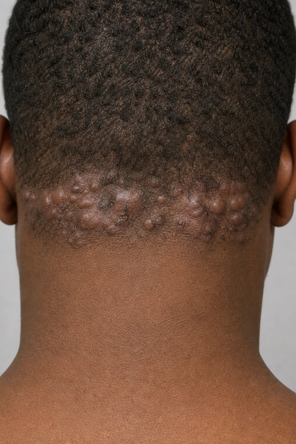
Acne keloidalis nuchae (AKN), also called folliculitis keloidalis nuchae, folliculitis nuchae, sycosis nuchae and historically dermatitis papillaris capillitii, is a chronic, progressive, scarring folliculitis of the nape of the neck and lower occipital scalp that produces firm keloidal papules and plaques, tufted hair bundles (polytrichia), sinus tracts, abscesses and ultimately permanent scarring (cicatricial) alopecia.[1][4]
The name is doubly misleading and is the source of the most common exam trap: [1]
- It is NOT true acne. There is no role for sebaceous gland dysfunction and no role for Cutibacterium acnes (formerly Propionibacterium acnes). The word "acne" entered the name only because the early papulopustular stage superficially resembles acne.
- It is NOT a true keloid pathologically. Although the plaques look and feel keloidal (firm, bossellated, symmetric, extending beyond the original follicle), histology shows a chronic foreign-body granulomatous and fibrosing reaction to intra-dermal hair shaft fragments, not the disorganised type-III collagen deposition of a true keloid.[1][5]
For this reason the modern preferred name is folliculitis keloidalis nuchae, which correctly frames the disease as a primary inflammatory folliculitis with secondary keloidal scarring rather than a primary keloid.[1][3]
AKN is classified within the primary cicatricial (scarring) alopecias as a lymphocytic/fibrosing subtype and overlaps clinically and pathogenetically with folliculitis decalvans and the broader follicular occlusion tetrad (hidradenitis suppurativa, dissecting cellulitis of the scalp, acne conglobata, pilonidal sinus).[3][10]
Classification
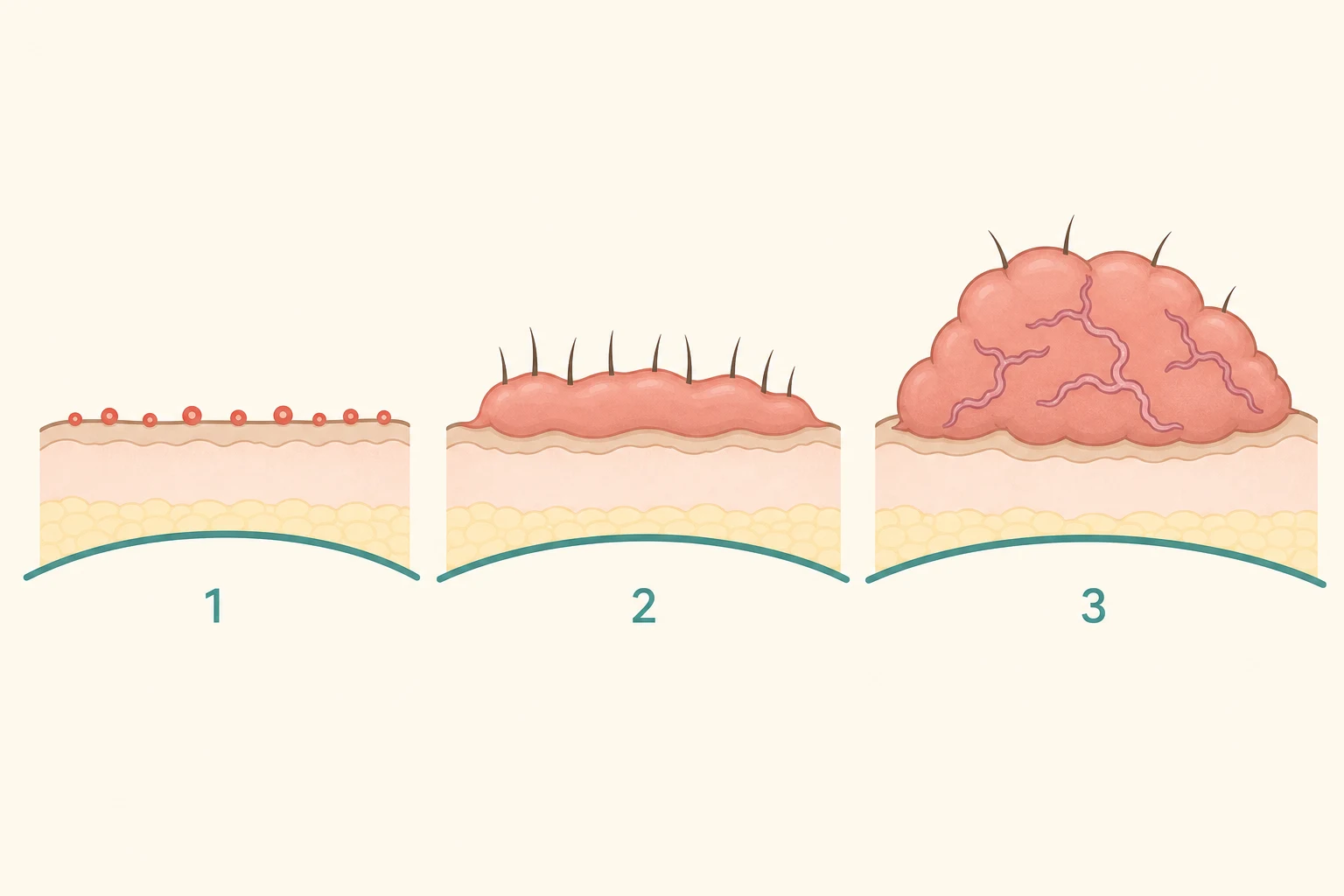
AKN is classified clinically by stage of evolution, which directly dictates therapy:[1][7]
A second classification axis is by dominant lesion type, useful when choosing the surgical technique: papular, plaque, nodular, tumoural (keloidal), and sinus-subtype (predominantly abscess and sinus tracts).[7][8]
Epidemiology & Risk Factors
Demographic. AKN almost exclusively affects young adult men of African descent, with onset typically between 14 and 25 years of age. The male-to-female ratio is approximately 20:1. The disease is uncommon in white, Hispanic and Asian populations and rare in women, but it can occur in any ethnicity or sex when the predisposing hair/skin type and mechanical triggers coincide.[1][6]
Hair and skin type. The strongest predisposing factor is tightly curled ("woolly") nuchal hair, characteristic of people of sub-Saharan African descent. The acute angle of emergence and the tight curl mean that a closely clipped hair tip easily re-enters or pierces the skin. Skin of colour is also a strong independent risk factor for keloidal-type scarring in response to any follicular inflammation.[1][11]
Mechanical triggers. [1]
- Close clipping of the nape with barbershop clippers or a razor — the dominant precipitant; the shorter the cut and the sharper the blade, the higher the risk.
- Friction and pressure from tight shirt collars, helmet straps (motorcycle, bicycle, construction, military), backpacks, headrests and tight braids.
- Traction and chemical relaxers in women who also keep the nape closely trimmed.[12]
Behavioural and occupational. Athletes, military recruits, police officers and motorcyclists are over-represented because of helmets and close-cropped haircuts.[6]
Co-morbid and iatrogenic. AKN is part of the follicular occlusion spectrum, so look for hidradenitis suppurativa, dissecting cellulitis of the scalp, acne conglobata and pilonidal sinus. Isolated reports associate severe AKN with ciclosporin and other immunosuppressants post-transplant, and with anabolic steroid use.[1]
Genetics. Familial clustering is reported but no single locus has been reproducibly identified; familial keloidal diathesis is a risk modifier.[1]
Pathophysiology
The lesion begins with a mechanism essentially identical to pseudofolliculitis barbae but at the nape rather than the beard.[1][11]
Step 1 — Close clipping of tightly curled nuchal hair. Clippers cut the hair shaft obliquely, producing a sharp tip. Because the hair is tightly curled and the follicle is curved, the cut hair retracts below the skin surface. [1]
Step 2 — Extrafollicular or transfollicular penetration. As the hair continues to grow, the sharp tip either penetrates outward through the follicular wall into the dermis (transfollicular) or curves back and pierces the skin surface adjacent to the follicle (extrafollicular). Either way, keratin and hair-shaft protein are deposited inside the dermis, where they are treated as foreign material.[1][5]
Step 3 — Foreign-body granulomatous reaction. The immune system mounts a granulomatous foreign-body reaction around the hair fragment, with multinucleated giant cells, histiocytes/macrophages, lymphocytes and plasma cells. Acute neutrophilic folliculitis may coexist when Staphylococcus aureus superinfects. [1]
Step 4 — Fibrosis and follicular destruction. Chronic inflammation drives dense keloidal-type fibrosis, with destruction of the hair follicle, the sebaceous gland and the arrector pili muscle, producing the firm keloidal plaque and permanent scarring (cicatricial) alopecia characteristic of advanced disease.[1]
Why the nape and not elsewhere? The nuchal scalp has a uniquely dense population of curved hair follicles, is constantly subjected to friction and occlusion from collars, helmets and headrests, and is the site most often shaved shortest by barbers. These factors combine to produce the characteristic nape-localised disease.[1]
Why is it "keloid-like" but not a true keloid? A true keloid is a benign fibroproliferative tumour of disorganised type-III collagen that extends beyond the original wound, recurs after excision, and has no follicular trigger. In AKN, the fibrosis is driven by and centred on a ruptured hair follicle, histology shows the offending hair shaft and granulomatous inflammation, and the lesion is trigger-dependent (cessation of clipping halts progression). This explains why trigger avoidance and laser hair removal are central to treatment.[1][9]
Role of Staphylococcus aureus. Secondary staphylococcal infection is common in the papulopustular and sinus stages and contributes to purulence, pain and abscess formation; it is not the primary driver, which is mechanical.[1][6]
Clinical Presentation
Site. AKN is confined to the nape of the neck and the lower occipital scalp, almost always in a horizontal band along and just below the occipital hairline. It does not involve the face, chest, back or beard — distinguishing it immediately from acne vulgaris and pseudofolliculitis barbae. Bilateral and symmetric. Extension upward onto the occipital scalp or across the whole nape occurs in advanced disease.[1][6]
Lesion morphology (by stage). [1]
- Stage I — papular/pustular. Small (2–5 mm) firm follicular papules and tiny pustules in the nuchal hairline; may be mistaken for ordinary folliculitis or acne. Often itchy.
- Stage II — plaque. Firm, dome-shaped, smooth, skin-coloured to hyperpigmented keloidal papules that enlarge and coalesce into a keloidal plaque running across the nape. Tufted hairs (polytrichia) — multiple hair shafts emerging from a single dilated follicular opening — are characteristic. Occasional small crusts and broken hair shafts.
- Stage III — tumoural/keloidal. A large, lobulated, bossellated keloidal mass with discharging sinus tracts, abscesses, fistulae and scarring alopecia. The plaque is hard, fixed to deeper tissues and may be tender.[1][7]
Symptoms. Many patients are asymptomatic apart from the cosmetic concern. When present, symptoms include pruritus, pain (especially with abscess/sinus), discharge of pus or keratinaceous material, foul odour from chronically infected sinuses, and bleeding from trauma. The cosmetic and psychological burden is often the dominant complaint.[6]
Course. Chronic, indolent, progressive and relapsing over years to decades. Spontaneous resolution is rare; without intervention most patients progress to permanent scarring alopecia. The course is punctuated by acute infective flares.[1]
Atypical presentations. In women (rare), the disease is often milder and may mimic acne vulgaris or folliculitis decalvans; chemical relaxers and traction contribute. In lighter skin types (Asian, Hispanic, white men with curly hair) the keloidal quality is less obvious and the diagnosis is more easily missed. In immunosuppressed patients (transplant, HIV) the disease may be more severe and refractory.[1]
Differential Diagnosis
The differential is one of the highest-yield exam areas. The key is site + follicular origin + keloidal quality.[1][3]
Other considerations. [1]
- Acne vulgaris — face, chest, back; comedones; sebaceous; never the nape alone.
- Tinea capitis / tinea barbae — scaly annular patch with hair loss; KOH positive; fungal culture positive; not keloidal.
- Discoid lupus erythematosus — well-demarcated erythematous plaques with follicular plugging, carpet-tack scale and scarring; biopsy shows interface dermatitis; immunofluorescence a lupus band.
- Basal cell carcinoma — pearly papule with rolled border and telangiectasia; on the nape would be solitary; biopsy diagnostic.
- Squamous cell carcinoma (Marjolin's ulcer) — non-healing ulcer within a long-standing AKN plaque; biopsy mandatory.[1]
- Acne necrotica (nuchal) / necrotising lymphocytic folliculitis — small necrotic papules with umbilication and scarring; less keloidal.
- Impetigo — honey-coloured crusts; acute; not keloidal; resolves with antibiotics.
Clinical & Bedside Assessment
Diagnosis is clinical. A confident bedside diagnosis rests on site (nape of neck), morphology (keloidal follicular papules and plaques with tufted hairs) and demographic (young man of African descent with tightly curled, closely clipped hair).[1][6]
History. Ask specifically about: [1]
- Hair-cutting habits: how short the nape is clipped, frequency, use of clippers vs razor, whether the barber "fades" the nape to skin level.
- Mechanical exposure: helmets (motorcycle, construction, sports, military), tight collars, backpacks, headrests, traction braids.
- Symptoms: itch, pain, discharge, bleeding, odour.
- Cosmetic and psychological impact: screen with the DLQI.
- Family history of keloids or similar scalp disease.
- Follicular occlusion tetrad: axillary, inguinal, gluteal or submammary boils (hidradenitis suppurativa); boggy nodules elsewhere on the scalp (dissecting cellulitis); severe conglobate acne; pilonidal sinus.
- Sexual/medical history if HIV or immunosuppression is plausible (severe or atypical AKN). [1]
Examination. Inspect the whole scalp, beard, axillae, groins and gluteal cleft (follicular occlusion tetrad). Palpate the plaque to assess firmness, fixity, fluctuation (abscess) and sinus tracts. Use dermoscopy to demonstrate tufted hairs (polytrichia), perifollicular scale, dilated follicular openings, broken hair shafts and white structureless areas of fibrosis.[1]
Severity assessment. There is no universally adopted severity score (unlike PASI for psoriasis). Use the clinical stage (I–III), the approximate area of nuchal involvement, the presence of sinus tracts/abscess and the DLQI to grade and to justify escalation.[7]
Investigations
Investigations are usually unnecessary. AKN is a clinical diagnosis. Reserve tests for atypical, severe, complicated or refractory disease.[1][4]
Skin biopsy (4 mm punch) when: [1]
- The morphology or site is atypical and a mimicker (discoid lupus, BCC, sarcoidosis) is plausible.
- There is a non-healing ulcer within a long-standing plaque — to exclude squamous cell carcinoma (Marjolin's ulcer).[1]
- The disease is refractory to standard therapy.
Histology. A ruptured or destroyed hair follicle, a naked hair shaft within the dermis surrounded by a granulomatous infiltrate with multinucleated foreign-body giant cells, lymphocytes, histiocytes and plasma cells, plus dense keloidal-type fibrosis and loss of sebaceous glands and follicles (cicatricial alopecia). The findings are diagnostic but not always distinguishable from folliculitis decalvans, hence the overlap debate.[3]
Bacterial swab (culture and sensitivity) from any pus or discharging sinus — typically grows Staphylococcus aureus (including MRSA in endemic areas); occasionally Gram-negatives in chronic sinus disease. Guides antibiotic choice for infective flares.[1]
Fungal studies (KOH, fungal culture) only if tinea capitis is in the differential — negative in AKN. [1]
Dermoscopy. Tufted folliculitis (multiple hairs from one ostium), perifollicular hyperkeratosis, white structureless areas of scarring, broken and dystrophic hair shafts. [1]
Blood tests before systemic therapy or surgery: FBC, U&E, LFTs, HIV and hepatitis B/C serology if surgery is planned; glucose/HbA1c if long-term corticosteroid or immunosuppressive therapy is contemplated; FBC, U&E and LFTs at baseline and during the rifampicin + clindamycin combination.[10]
Management — Resuscitation
AKN is not usually a dermatological emergency, but several scenarios require prompt action.[1]
Acute abscess. A fluctuant, painful swelling within an AKN plaque is treated by incision and drainage (or punch drainage for recurrent sinuses), a bacterial swab, and a 7–10 day course of an anti-staphylococcal antibiotic guided by local sensitivities (e.g. flucloxacillin 500 mg QDS oral, or clindamycin 300 mg QDS if MRSA is suspected/confirmed). Analgesia with paracetamol ± a short course of NSAID. [1]
Severe pain or systemic symptoms (fever, cellulitis extending beyond the nape) warrant same-day assessment, intravenous antibiotics and consideration of incision and drainage. [1]
Psychological distress. The cosmetic burden is significant; offer reassurance, an explicit management plan and, where appropriate, psychological support or counselling. Screen for depression and anxiety.[6]
Urgent referral to dermatology or a scalp surgery service for: a non-healing ulcer (possible Marjolin's ulcer), extensive tumoural disease suitable for excision, refractory disease despite first- and second-line therapy, or diagnostic uncertainty. [1]
Management — Definitive & Stepwise
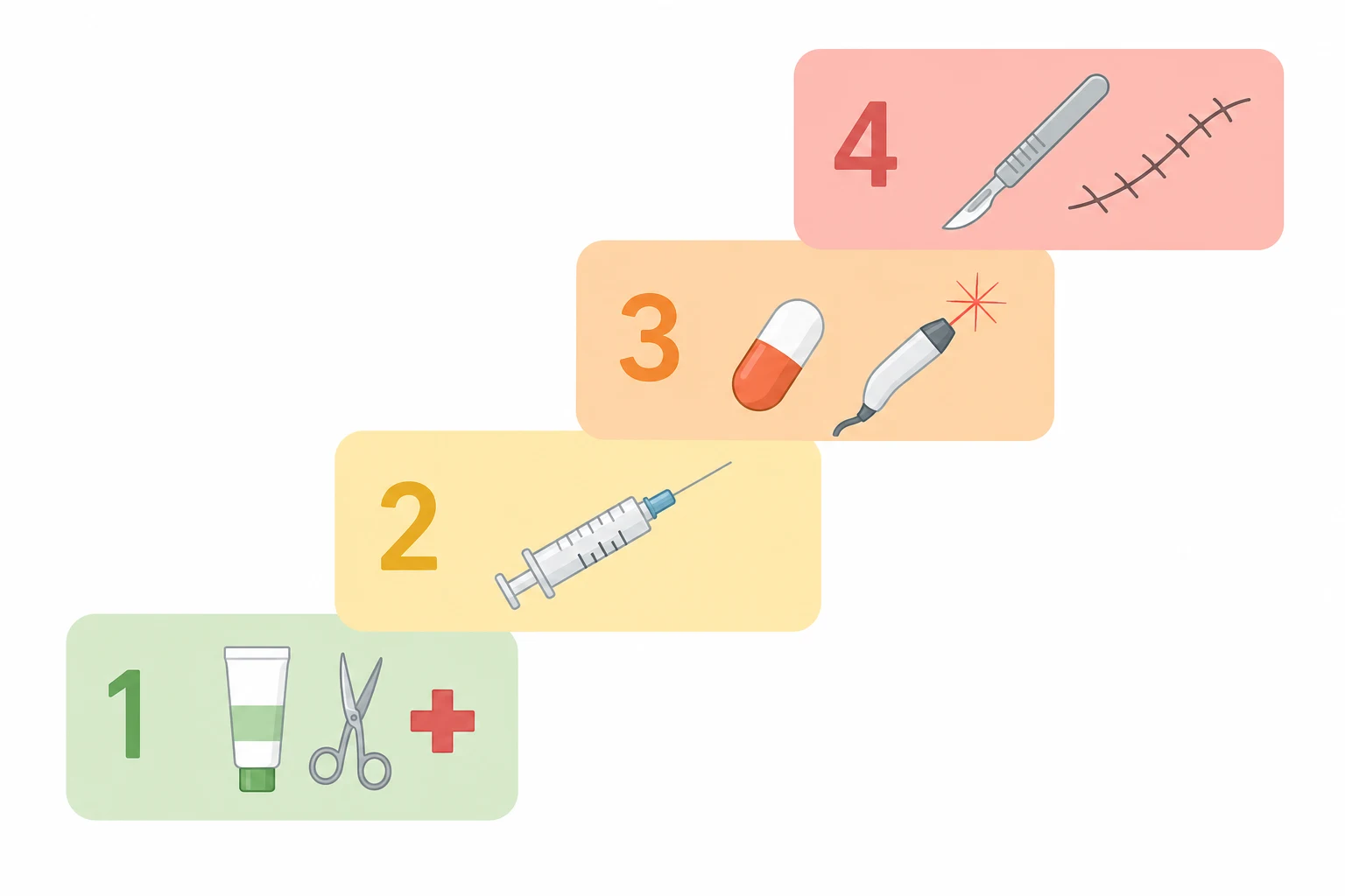
The therapeutic ladder is stage-based and combines trigger avoidance, medical therapy, physical modalities and surgery.[1][7][8]
Step 1 — Conservative measures and topical therapy (all stages; first-line for stage I)
First-line bundle for every AKN patient
Step 2 — Intralesional corticosteroid (stage II plaque disease; adjunct at all stages)
- Agent: triamcinolone acetonide, intralesional.
- Concentration: 10 mg/mL for softer/smaller papules, 40 mg/mL for firm keloidal plaques.
- Volume: 0.05–0.1 mL per papule, using a 30-gauge needle; total dose 20–30 mg per session (do not exceed 40 mg/session).
- Frequency: every 4–6 weeks until flattening (typically 3–6 sessions).
- Effect: flattens keloidal papules, reduces pruritus and pain.
- Adverse effects: skin atrophy, telangiectasia, hypopigmentation (especially relevant in skin of colour), and rare systemic absorption. Counsel the patient before injection.[1][2]
Adjuvant or alternative intralesional agents reported for refractory keloidal plaques include 5-fluorouracil (5-FU 50 mg/mL, alone or mixed with triamcinolone) and verapamil (2.5 mg/mL).[2]
Step 3 — Systemic therapy (moderate–severe, sinus/abscess, refractory to steps 1–2)
Anti-inflammatory tetracycline. Doxycycline 100 mg BD orally for 8–12 weeks (or minocycline 100 mg OD), continued long-term at the lowest effective dose if helpful. Warn about photosensitivity, gastrointestinal upset, oesophagitis (take with water, upright) and avoiding in pregnancy/children.[1]
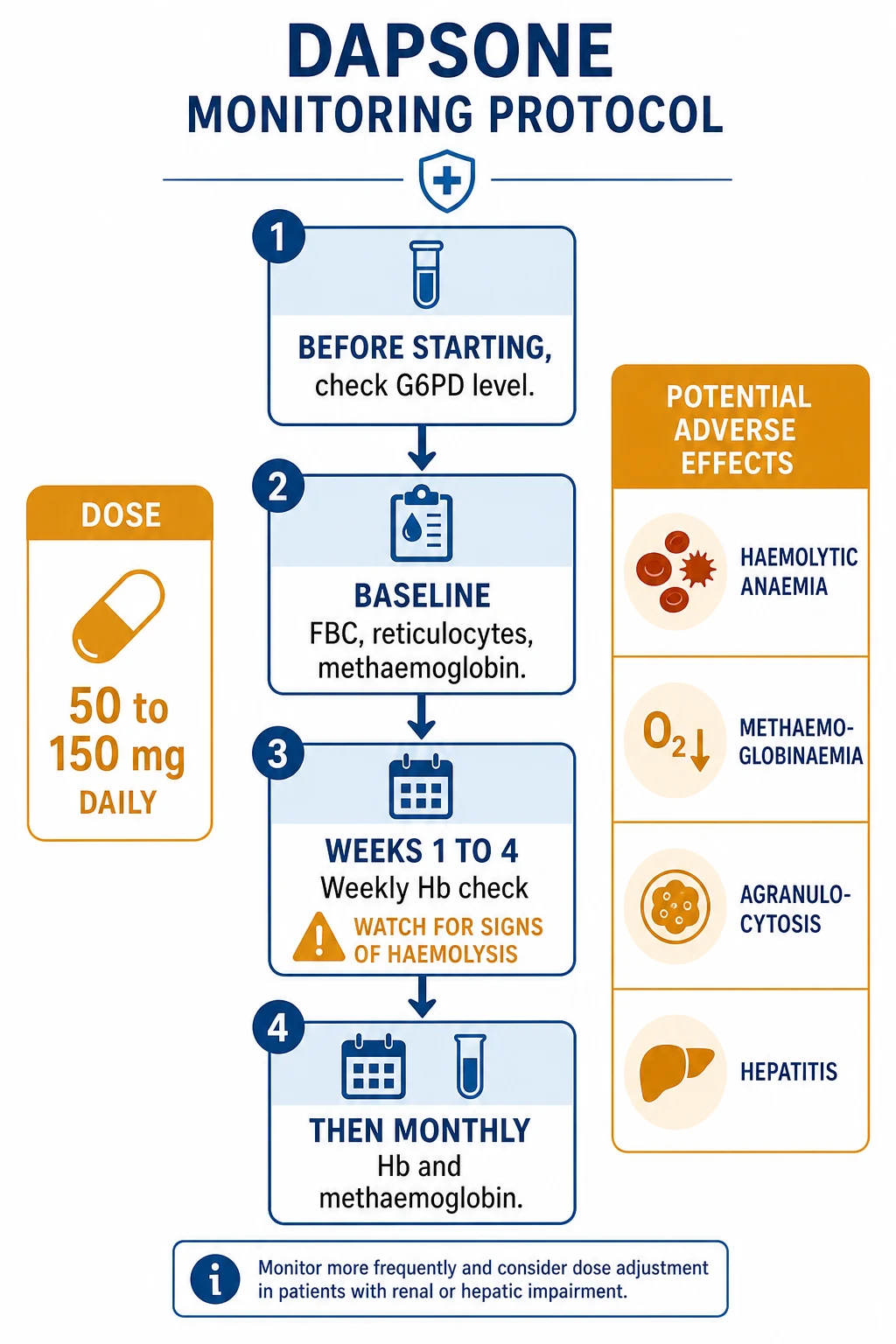
Combination antibiotic regimen — the follicular-occlusion combination. For refractory or sinus-stage disease, use the same regimen proven in folliculitis decalvans and other follicular occlusion disorders:[10]
[1]Other systemic agents used in resistant disease include isotretinoin (off-label; 0.5 mg/kg/day for 16–24 weeks; only in patients past childbearing potential; strictly contraceptive cover) for the follicular occlusion overlap, and a short course of systemic prednisolone (0.5 mg/kg/day tapering over 2–3 weeks) only for acute, severe inflammatory flares — avoid as routine (see pitfalls).[2]
Step 4 — Laser and physical modalities (adjuvant; key to recurrence prevention)
Laser hair removal is the single most important adjuvant because it destroys the culpable nuchal follicles and removes the trigger.[9]
- Laser of choice in skin of colour: long-pulsed Nd:YAG 1064 nm (deepest penetration, lowest melanin absorption → safest in Fitzpatrick IV–VI).
- Alternatives: diode 810 nm and long-pulsed alexandrite 755 nm (latter for lighter skin types only).
- Protocol: 4–8 sessions at 4–8 week intervals, after a test patch; combine with topical anaesthetic; counsel about transient erythema/oedema and the small risk of pigmentary change.
- Indication: any active disease (to reduce follicular load) and after any surgical excision to prevent new lesions at the periphery and in the surrounding nuchal skin.[9]
Cryotherapy (liquid nitrogen) has been used to flatten small keloidal papules but is unpredictable in skin of colour (risk of hypopigmentation) and has largely been superseded by laser. [1]
Electrodessication and radiofrequency ablation of individual papules are options for small-volume disease. [1]
Intralesional 5-FU and verapamil as above. [1]
Radiation therapy (superficial orthovoltage or electron beam) is rarely used as a post-excision adjuvant to prevent keloidal recurrence, mainly in patients with severe keloidal diathesis; the risk-benefit balance is unfavourable in young patients.[1]
Step 5 — Surgical excision (stage III tumoural/keloidal disease; refractory plaque/sinus disease)
Surgery is definitive for extensive or tumoural disease and for disease with sinus tracts or abscesses that has failed medical therapy.[7][8]
- Principle: complete excision of all involved skin and subcutaneous tissue, down to the deep cervical fascia / occipital periosteum, to remove every culpable follicle.
- Closure options (chosen by size, laxity and surgeon preference): primary closure for small defects; second-intention healing (excision and leave open — surprisingly well tolerated on the nape, low recurrence, slow healing over 8–12 weeks); split-thickness skin grafting; and local rotation or advancement flaps for large defects.
- Outcome: wide excision has the lowest recurrence rate of any single modality (under 10–15% in most series); recurrence is highest when excision is incomplete or when close clipping resumes.
- Post-operative adjuvants: intralesional triamcinolone to the healing scar at 4–6 weeks; laser hair removal to the surrounding nuchal skin to prevent new lesions; pressure garment (custom nuchal pressure pad) for 12–18 h/day for 4–6 months in keloidal-prone patients.[7][8]
Escalation triggers
- No response to step 1 after 8–12 weeks → add step 2 (intralesional triamcinolone).
- Active papular/plaque disease despite steps 1–2, or sinus/abscess → add step 3 (oral doxycycline or rifampicin+clindamycin) and arrange laser hair removal.
- Tumoural/keloidal plaque, persistent sinus tracts, or failure of medical therapy → step 5 (surgical excision).
- Any non-healing ulcer in a chronic plaque → biopsy to exclude Marjolin's ulcer before proceeding.[1]
Specific Subtypes & Scenarios
Early papular AKN (stage I). Trigger avoidance + topical retinoid + topical antibiotic/steroid is usually sufficient; add benzoyl peroxide wash. Counsel the patient that progression is preventable. [1]
Plaque-stage AKN (stage II). Combine first-line measures with intralesional triamcinolone every 4–6 weeks and an oral anti-inflammatory tetracycline; introduce laser hair removal early. [1]
Tumoural/keloidal AKN with sinus tracts (stage III). Medical therapy alone rarely resolves this stage. Plan surgical excision down to the fascia, swab and treat secondary infection first, and arrange post-operative laser hair removal to minimise recurrence.[7]
AKN in women. Rare; usually milder. Address chemical relaxers, traction and any overzealous nape trimming. Laser hair removal is particularly useful because shaving the nape is not part of most women's grooming. Avoid systemic retinoids in women of childbearing potential without reliable contraception. [1]
AKN with the follicular occlusion tetrad. Coordinated care with dermatology and surgery; treat the most symptomatic site first; consider adalimumab or other biologics approved for hidradenitis suppurativa in severe overlapping disease.[3]
Post-surgical AKN. Apply intralesional triamcinolone to the scar, laser to the surrounding skin, and an indefinite ban on close clipping. Recurrence is usually due to resumed clipping rather than failed surgery.[8]
AKN in the immunosuppressed (renal transplant on ciclosporin, HIV). May be more severe and atypical; reduce immunosuppression where possible ( ciclosporin has been implicated as a trigger); treat staphylococcal superinfection aggressively; avoid surgical excision in unstable immunosuppression without optimising the patient first.[1]
Complications & Pitfalls
Local complications. [1]
- Permanent scarring (cicatricial) alopecia — the dominant long-term complication; lost follicles do not regrow.
- Keloidal plaques — cosmetically significant, may itch and cause social embarrassment.
- Abscesses and sinus tracts — recurrent, malodorous, painful.
- Cosmetic disfigurement at the visible nape/hairline.[6]
Malignant transformation. A non-healing ulcer within a long-standing AKN plaque may be squamous cell carcinoma (Marjolin's ulcer) — a primary cicatricial alopecia is a recognised setting for scar-associated SCC. Biopsy any such ulcer.[1]
Psychological complications. Depression, anxiety, social withdrawal and impaired quality of life (high DLQI scores). Cosmetic concern is often the chief complaint.[6]
Complications of intralesional corticosteroid. Skin atrophy, telangiectasia, hypopigmentation (clinically important in skin of colour), and rare systemic absorption with repeated high-dose injection.[2]
Complications of surgery. Recurrence (highest with incomplete excision or resumed clipping), wound dehiscence, infection, hypertrophic scar/keloid of the scar itself, graft failure, and alopecia of the donor/recipient site.[7][8]
Pitfalls. [1]
- Mislabelling AKN as a true keloid and treating only with intralesional steroid, missing the follicular trigger and the role of trigger avoidance, retinoid, antibiotics and laser.
- Omitting laser hair removal after surgery — leading to avoidable recurrence.
- Continuing close clipping after treatment — guaranteeing relapse.
- Confusing AKN with pseudofolliculitis barbae (site: beard, not nape) or with folliculitis decalvans (site: vertex, not nape).[11]
- Failing to biopsy a chronic ulcer in a long-standing plaque — missing Marjolin's ulcer.[1]
- Prescribing tetracyclines or systemic retinoids in pregnancy — contraindicated.
- Using potent topical steroid on the face when treating associated acne or folliculitis — avoid.
- Under-treating early disease because it "looks mild" — early intervention prevents irreversible scarring alopecia.
Prognosis & Disposition
Natural history. AKN is chronic, relapsing and difficult to eradicate. Untreated, it progresses slowly over years to permanent scarring alopecia and disfiguring keloidal plaques. Spontaneous resolution is rare.[1]
Effect of treatment. Early disease (stage I) can be arrested and partially reversed with trigger avoidance and topical therapy. Plaque-stage disease (stage II) can be flattened and rendered asymptomatic with intralesional steroid, oral antibiotic and laser. Tumoural disease (stage III) requires surgical excision, after which recurrence rates are low (under 10–15% in most series) provided clipping is avoided and laser is used.[7][8]
Recurrence prevention. The single most effective measure to prevent recurrence at any stage is long-pulsed Nd:YAG laser hair removal to the nuchal follicles, combined with lifelong avoidance of close clipping.[9]
Disposition. Most patients are managed jointly by primary care and dermatology. Refer to a scalp surgery service for tumoural disease, sinus tracts or refractory plaque disease. Long-term dermatology follow-up is appropriate because of the chronic relapsing course and the psychological burden.[1]
Quality of life. DLQI scores are typically elevated; cosmetic concern and the visibility of the nape lesions are major drivers. Address this explicitly in the consultation.[6]
Special Populations
Pregnancy. Avoid tetracyclines (doxycycline, minocycline) and systemic retinoids (isotretinoin) — both teratogenic. Favour physical measures (trigger avoidance, laser hair removal — safe in pregnancy), topical antibiotic (clindamycin, erythromycin — both considered safe), and intralesional triamcinolone (single small doses — generally considered acceptable; avoid high cumulative doses). Defer surgery until after delivery if possible.[1]
Adolescents. Early-onset AKN in teenagers is increasingly recognised, often triggered by barbershop clipping and sports helmets. Management is the same as in adults, with particular attention to psychological and cosmetic impact and to avoiding systemic retinoids in adolescent girls of childbearing potential without contraception. Laser hair removal is acceptable and effective.[6]
Women. Rare; usually milder and often misdiagnosed as acne vulgaris or folliculitis. Address chemical relaxers, traction braids and overzealous nape trimming. Avoid systemic retinoids unless contraception is reliable. Laser hair removal is preferred over shaving.[12]
Immunosuppressed (renal transplant on ciclosporin, HIV, chemotherapy). AKN may be more severe, atypical and refractory. Reduce ciclosporin where possible (it has been implicated as a trigger). Treat staphylococcal superinfection aggressively. Avoid surgical excision in unstable immunosuppression without optimising the patient first.[1]
Lighter skin types (Asian, Hispanic, white men with curly hair). The keloidal quality is less pronounced and the diagnosis is more easily missed. The same management ladder applies; pigmentary adverse effects of intralesional steroid and laser are less of a concern.[1]
Evidence, Guidelines & Regional Differences
Guidelines. AKN lacks a single authoritative global guideline; recommendations are drawn from the American Academy of Dermatology clinical guidance and StatPearls/UpToDate syntheses (US), the British Association of Dermatologists and NICE Clinical Knowledge Summaries (UK), the European Academy of Dermatology and Venereology consensus statements (Europe), and the Indian Association of Dermatologists, Venereologists and Leprologists (IADVL) task-force guidance (India). All converge on the same stage-based ladder.[1][4]
Combination rifampicin + clindamycin. The regimen (300 mg BD each for 10–12 weeks) is best evidence-based for folliculitis decalvans (Miguel-Gómez et al., multicentre series, JAAD 2018) and is extrapolated to AKN as part of the same follicular occlusion spectrum. Reported response rates in folliculitis decalvans exceed 70%; AKN data are largely case series.[10]
Laser hair removal. Long-pulsed Nd:YAG 1064 nm is the preferred laser in skin of colour (Roberts, J Drugs Dermatol 2019), with the lowest risk of pigmentary adverse effects in Fitzpatrick IV–VI. Multiple case series show substantial reduction in new-lesion formation when laser is combined with surgical excision.[9]
Surgical excision. Multimodality surgical series (Madura et al., J Cutan Aesthet Surg 2021; Umar et al., JAAD Case Reports 2019) demonstrate that excision down to the fascia with second-intention healing or primary closure yields the lowest recurrence; lesion-type-tailored surgical selection improves cosmesis.[7][8]
AKN and folliculitis decalvans overlap. Doche et al. (Int J Dermatol 2019) argue that the two conditions share a final common pathway of follicular rupture and granulomatous scarring, supporting the use of overlapping therapies.[3]
Hairdensing and scalp disease in African adults. Khumalo et al. (Br J Dermatol 2007) documented a high prevalence of traction and traumatic scalp disorders in African adults associated with tight hairdensing practices — supporting the central role of mechanical triggers.[12]
Renaming controversy. Several authorities now prefer folliculitis keloidalis nuchae to "acne keloidalis nuchae" to remove the misleading "acne" and to emphasise the primary folliculitis. The term "dermatitis papillaris capillitii" is obsolete.[1]
Regional deltas. [1]
Exam Pearls
[1]NAPE — the four diagnostic anchors
TREAT — the management ladder
Red Flags
Exam application bank (NEET-PG / INICET)
One-line answer
Acne keloidalis nuchae (AKN) is a chronic scarring folliculitis of the nape of the neck and lower occipital scalp that produces firm keloidal papules, coalescent keloidal plaques, tufted hairs, sinus tracts and permanent scarring alopecia. It almost exclusively affects young men of African descent with tightly curled hair and is triggered by close clipping of the nape, friction from collars and helmets, and ingrown hairs. Despite the misleading name, AKN is neither true acne (no sebaceous gland or Cutibacterium acnes role) nor a true keloid pathologically — the keloidal appearance reflects a chronic foreign-body granulomatous fibrosing reaction to intra-dermal hair fragments. Management is a stepwise ladder: trigger avoidance and topical retinoid/antibiotic; intralesional triamcinolone 10–40 mg/mL; oral doxycycline or rifampicin 300 mg BD + clindamycin 300 mg BD for 10–12 weeks; long-pul [1]
Worked stems (answer without another resource)
Stem 1 — Classic presentation. Map symptoms to mechanism; name the first investigation and first treatment step with dose/route if drug therapy is standard. [1]
Stem 2 — Unstable / complicated. List red flags that force immediate resuscitation, theatre, ICU, antidote, or reperfusion — and what you do in the first 15 minutes. [1]
Stem 3 — Atypical group. Elderly, pregnancy, child, or immunocompromised: how presentation and thresholds change. [1]
Stem 4 — Differential trap. Name the three closest mimics and one discriminator for each. [1]
Stem 5 — Disposition. Who goes home with safety-netting, who is admitted, who needs HDU/ICU/theatre, and what follow-up is mandatory. [1]
Rapid viva checklist
- Definition + classification
- Pathophysiology chain
- Bedside signs / criteria
- Score with exact components (if any)
- Emergency bundle
- Definitive therapy with doses
- Complications of disease and of treatment
- Special populations
- Guideline/trial name if classic
- Three exam traps
Coverage self-check
If you cannot answer any stem above from this page alone, re-read the matching section — the page is intended to be self-sufficient for final-prof and NEET-PG/INICET questions on Acne keloidalis nuchae (folliculitis keloidalis nuchae).
References
- [1]Olagun-Samuel C, Alexis A, et al. Acne Keloidalis Nuchae JAMA Dermatol, 2025.PMID 40601307
- [2]Alves JP, Gozalo L, et al. Acne Keloidalis Nuchae J Cutan Med Surg, 2022.PMID 36124546
- [3]Doche I, Habinowa N, et al. Acne keloidalis nuchae and folliculitis decalvans: same process affecting the follicle or coexisting diseases? A retrospective study Int J Dermatol, 2019.PMID 31241169
- [4]Al Aboud DM, Piliang MP, et al. Acne Keloidalis Nuchae 2026.PMID 29083612
- [5]Gloster HM Jr. Acne keloidalis nuchae Dermatol Nurs, 2003.PMID 14735606
- [6]Patel A, Mirza S, et al. A Classic Presentation of Acne Keloidalis Nuchae in a Black Male Cureus, 2025.PMID 40656282
- [7]Madura C, Thappa BR, et al. Multimodality Surgical Treatment Approach to Acne Keloidalis Nuchae Based on Lesion Morphology J Cutan Aesthet Surg, 2021.PMID 34566374
- [8]Umar S, Etufugh CN, et al. Selection criteria and techniques for improved cosmesis and predictable outcomes in laser hair removal treatment of acne keloidalis nuchae JAAD Case Rep, 2019.PMID 31205997
- [9]Roberts WE, et al. Laser Treatment of Skin of Color for Medical and Aesthetic Uses With a New 650-Microsecond Nd:YAG 1064nm Laser J Drugs Dermatol, 2019.PMID 31017752
- [10]Miguel-Gomez L, et al. Folliculitis decalvans: Effectiveness of therapies and prognostic factors in a multicenter series of 60 patients with long-term follow-up J Am Acad Dermatol, 2018.PMID 29864465
- [11]Ogunbiyi A, George A, et al. Pseudofolliculitis barbae; current treatment options Clin Cosmet Investig Dermatol, 2019.PMID 31354326
- [12]Khumalo NP, Gumedze F, Lehloenya R, et al. Hairdressing and the prevalence of scalp disease in African adults Br J Dermatol, 2007.PMID 17725667