Dermatology · Medicine
Actinic keratosis
Also known as Actinic keratosis · AK · Solar keratosis · Senile keratosis
Actinic keratosis (AK, solar keratosis) is the commonest premalignant skin lesion — an intraepidermal keratinocyte dysplasia arising on chronically sun-damaged skin. It sits on a biological spectrum with SCC in situ (Bowen's disease) and invasive squamous cell carcinoma, driven by UV-induced p53 mutations and field cancerization. Clinically it presents as rough, scaly, erythematous macules or patches on sun-exposed sites, classically described as easier felt than seen. Management is stratified into lesion-directed therapy (cryotherapy, curettage, excision) for discrete lesions and field-directed therapy (topical 5-fluorouracil, imiquimod, photodynamic therapy, diclofenac) for multiple lesions or field cancerization. Any lesion that thickens, becomes indurated, ulcerates, or fails therapy must be biopsied to exclude invasive SCC.
On this page & tools
Your progress
Saved locally on this device.
Exam tags
Red flags
Overview & Definition
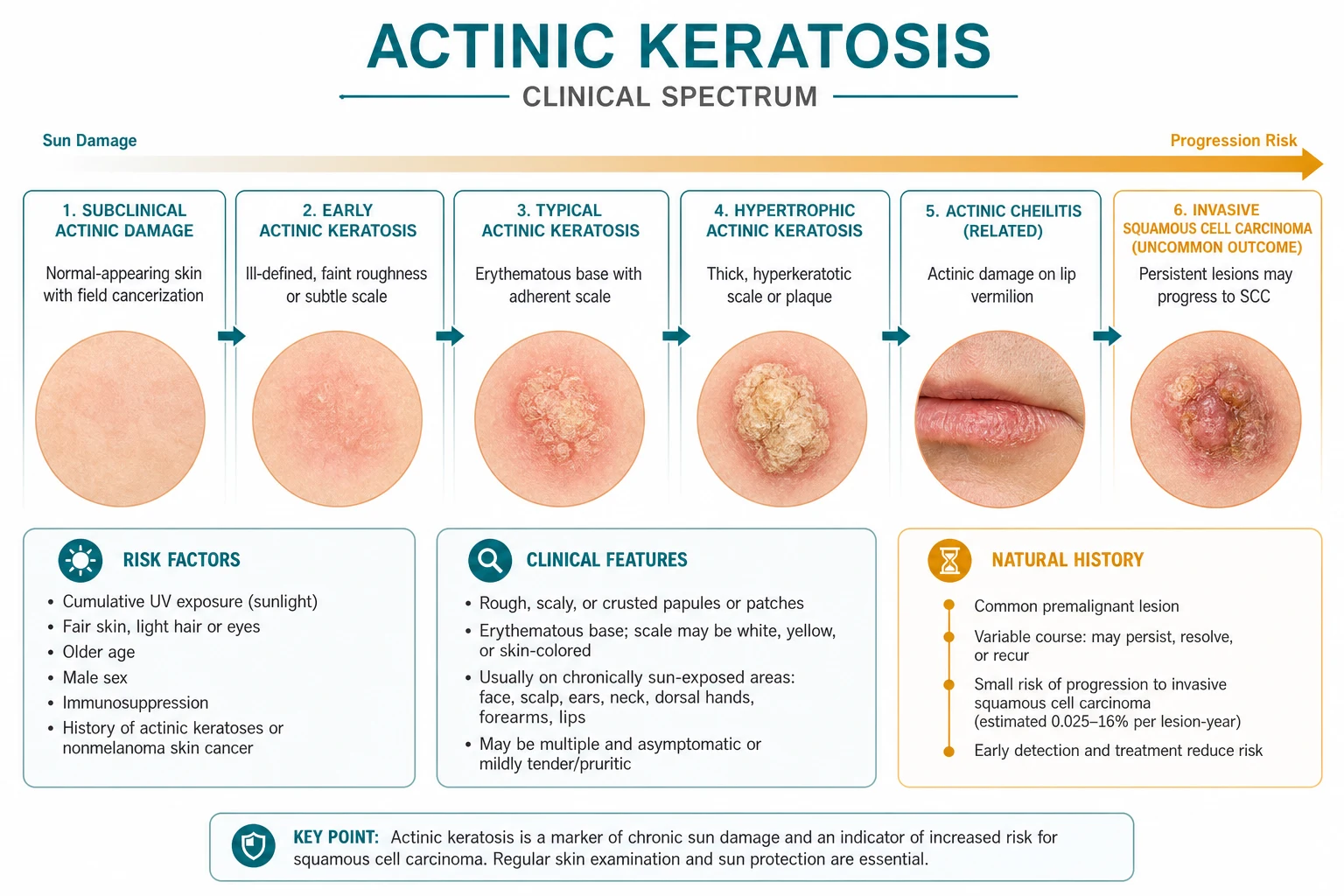
Actinic keratosis (AK), also called solar keratosis or senile keratosis, is a premalignant proliferation of atypical keratinocytes confined to the epidermis on skin that has sustained chronic ultraviolet (UV) damage. It is the commonest premalignant skin lesion in humans and represents the earliest clinically detectable stage on a continuum that extends through SCC in situ (Bowen's disease) to invasive cutaneous squamous cell carcinoma (cSCC).[6]
The lesion is not merely a local abnormality. Each visible AK arises within a broader field of cancerization — a sun-damaged skin field harbouring multiple independent premalignant clones that are not yet clinically apparent.[1] This concept explains why treating only the visible lesions often fails: new AKs continue to emerge from the surrounding damaged field unless the entire area is addressed. A patient presenting with one AK should therefore be examined and managed as someone with field damage, not merely as someone with a single spot.
The term "keratinocyte intraepidermal neoplasia" is sometimes used to emphasise the dysplastic nature of AK, but in clinical practice "actinic keratosis" remains the standard term. The distinction between AK and SCC in situ is histological — the extent of epidermal involvement — rather than molecular, and the two conditions share many driver mutations. This biological continuity is why any AK should be regarded as a potential precursor lesion and why surveillance is lifelong in high-risk patients. [1]
[1]Classification
AK can be classified clinically by morphology, anatomical site, and histological grade. The distinctions matter because thicker and more atypical lesions carry greater malignant potential and may require biopsy rather than simple destruction. [1]
Clinical variants
Classical / hypertrophic AK is the most common presentation. It appears as a rough, adherent yellow-brown scale on an erythematous base. Hypertrophic lesions are thicker and more palpable, making them harder to clear with brief cryotherapy; they also have a higher probability of harbouring invasive SCC, so biopsy should be considered when scale is markedly adherent or the lesion is indurated. [1]
Atrophic AK is a flat, pink-red macule or patch with minimal scale. It is easily missed on visual inspection and is often detected by palpation. Because the erythema is subtle, atrophic AK is commonly mistaken for rosacea, telangiectasia, or a scar. [1]
Pigmented AK presents as a brown to grey-brown macule with scale. It must be distinguished from solar lentigo, lentigo maligna, and seborrhoeic keratosis. Dermoscopy is particularly helpful: pigmented AK may show a pseudonetwork with brown-grey dots, globules, or annular-granular structures, whereas lentigo maligna shows more striking rhomboidal structures, asymmetrical pigmented follicles, and grey-blue dots. [1]
Actinic cheilitis is AK limited to the vermillion of the lower lip. It is generally considered to have a higher malignant potential than cutaneous AK because the lip mucosa is thin, chronically sun-exposed, and less protected by keratin. Presentations include scaling, fissuring, blurring of the vermillion border, atrophy, and leukoplakia. Any focal induration or ulceration warrants biopsy. [1]
Cutaneous horn is a conical column of compact keratin that can arise over an AK, SCC, keratoacanthoma, or verruca. The underlying pathology cannot be predicted from the horn itself; biopsy of the base is mandatory. [1]
Lichenoid AK is an uncommon variant with a violaceous or erythematous plaque and a surface that may resemble lichen planus. Histologically it shows a lichenoid band-like inflammatory infiltrate and interface changes in addition to the usual AK features. [1]
Histological grading
| Grade | Histological pattern | Malignant potential |
|---|---|---|
| Grade I (mild) | Atypical keratinocytes limited to the lower third of the epidermis | Lower |
| Grade II (moderate) | Atypical keratinocytes extending to the lower two-thirds | Intermediate |
| Grade III (severe) | Atypical keratinocytes involving most or all of the epidermis | Higher; approaches SCC in situ |
This grading overlaps conceptually with SCC in situ when atypia involves the full thickness of the epidermis without basement-membrane breach, and with invasive SCC once atypical cells cross the basement membrane into the dermis.[6]
Epidemiology & Risk Factors
AK is the most frequently encountered premalignant skin lesion in dermatology and primary care. Its prevalence rises steeply with age, cumulative UV exposure, and phenotypic susceptibility. Because the lesion is usually asymptomatic and often hidden within background sun damage, the true prevalence is almost certainly higher than the clinical detection rate. [1]
Geographic variation
Prevalence is strongly latitude-dependent. Australia and New Zealand report the highest rates, reflecting intense solar irradiation and a predominantly fair-skinned population of European ancestry. Northern European populations have lower prevalence but still substantial disease burden in elderly, fair-skinned individuals. In the United States, prevalence is higher in the sunbelt states than in the northern states. Globally, AK is uncommon in deeply pigmented populations but can occur in any phototype with sufficient cumulative UV exposure. [1]
Risk factors
The major risk factors can be grouped as intrinsic, environmental, and iatrogenic:[1][6]
Intrinsic
- Fair skin, light eyes, red or blonde hair (Fitzpatrick phototypes I–II).
- Poor tanning response and propensity to burn.
- Advanced age — cumulative UV dose and DNA-repair decline.
- Male sex — partly explained by greater occupational sun exposure and lower use of sun protection.
- Genetic disorders of DNA repair, especially xeroderma pigmentosum. [1]
Environmental
- Cumulative UV exposure is the dominant exogenous risk; both UVB (290–320 nm) and UVA contribute.
- Outdoor occupation or recreation at low latitude or high altitude.
- Use of tanning beds.
- Living closer to the equator or at higher altitude. [1]
Iatrogenic / comorbid
- Immunosuppression, especially solid-organ transplantation and chronic lymphocytic leukaemia.
- Chronic PUVA (psoralen plus UVA) therapy.
- Ionizing radiation or arsenic exposure.
- Previous cutaneous SCC or extensive AK burden. [1]
Pathophysiology
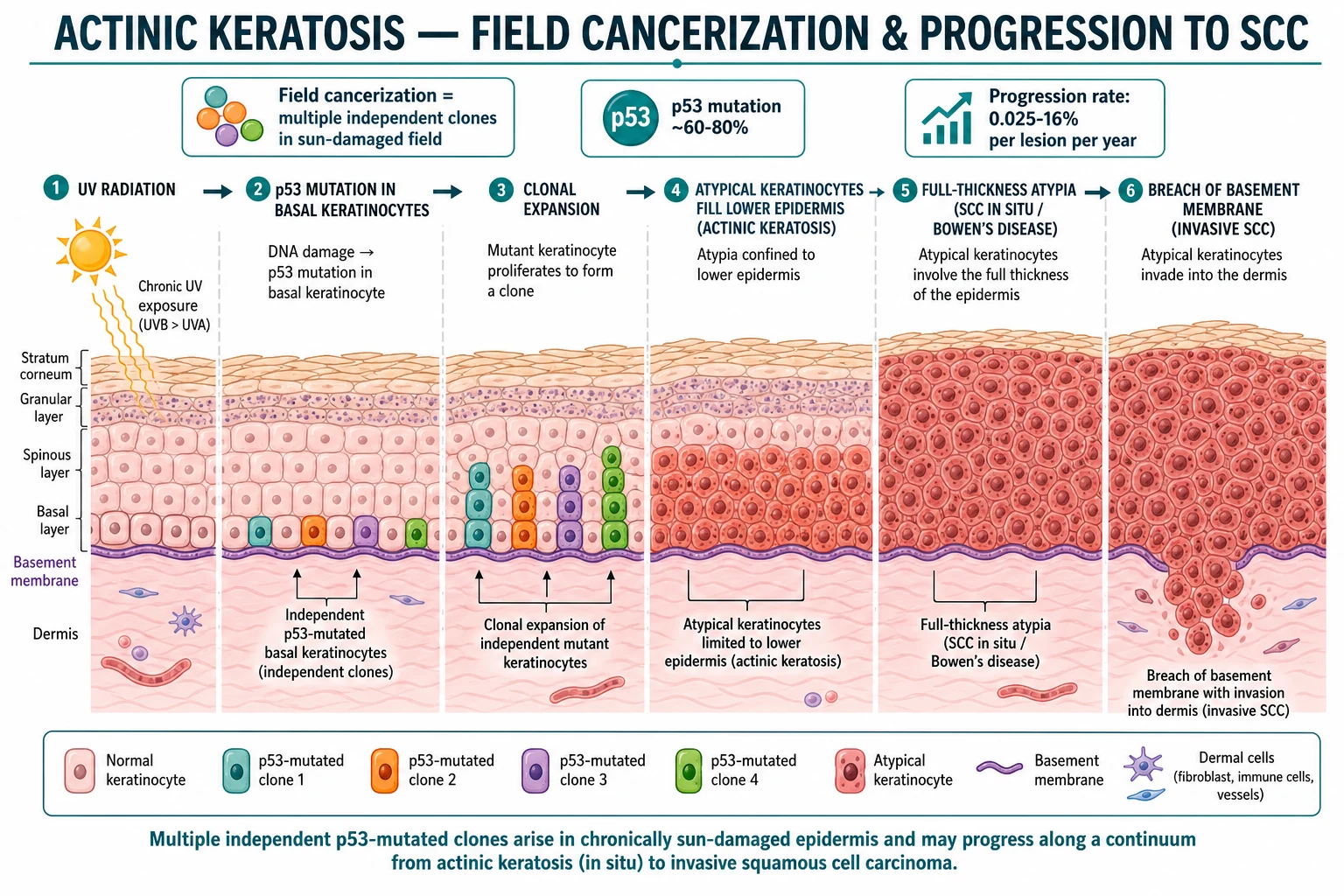
The molecular pathogenesis of AK is among the best understood of any human cancer. The initiating event is UV-induced DNA damage, predominantly cyclobutane pyrimidine dimers and 6-4 photoproducts, followed by characteristic UV signature mutations — most notably C→T and CC→TT transitions at dipyrimidine sites. UVB (290–320 nm) is the most carcinogenic waveband because it is directly absorbed by DNA, while UVA contributes indirectly through reactive oxygen species and by deepening the field of damage within the dermis. [1]
UV spectrum and DNA repair
After UV exposure, the nucleotide excision repair (NER) pathway normally removes cyclobutane pyrimidine dimers and restores genomic integrity. With cumulative UV exposure and advancing age, NER capacity declines. In patients with the rare autosomal recessive disorder xeroderma pigmentosum, defective NER produces an enormous lifetime burden of AK and NMSC at an early age, illustrating the central importance of DNA repair in AK prevention. [1]
p53 and clonal expansion
The TP53 tumour-suppressor gene is mutated in approximately 50–80% of AK lesions.[8] In normal keratinocytes, wild-type p53 arrests the cell cycle after DNA damage and directs damaged cells toward apoptosis. UV-induced p53 mutations abolish this checkpoint, allowing mutated keratinocytes to survive, proliferate, and form a clone of atypical cells within the lower epidermis.[8]
Other recurrent genomic alterations include NOTCH1, CDKN2A, and RAS family mutations — the same drivers seen in invasive SCC, supporting the view that AK and SCC share a common molecular lineage.[1]
Field cancerization
The term field cancerization describes a broad area of chronically sun-damaged skin containing multiple independent premalignant clones, each arising from separate UV-induced mutational events.[1] The clinical implication is profound: destroying one visible AK does not eliminate the field from which new lesions will emerge. Field-directed therapies are therefore designed to treat the entire damaged area rather than individual spots.
Progression to SCC
AK exists on a histological spectrum: [1]
| Stage | Histological hallmark | Clinical correlate |
|---|---|---|
| AK | Atypical keratinocytes in the lower epidermis | Rough scaly macule on sun-damaged skin |
| SCC in situ (Bowen's disease) | Full-thickness epidermal atypia; basement membrane intact | Persistent well-demarcated scaly red patch |
| Invasive SCC | Atypical keratinocytes breach the basement membrane into the dermis | Indurated, nodular, or ulcerated plaque |
Solar elastosis and dermal solar damage are almost always present on biopsy, confirming the UV aetiology.[6]
Clinical Presentation
AKs are classically described as rough, scaly, erythematous macules or patches that are "easier felt than seen." The gritty, sandpaper-like texture on palpation is often the first clue, even when erythema is subtle. Patients may not volunteer symptoms and may attribute the roughness to dry skin or age. [1]
Typical morphology and distribution
- Size: usually 2–10 mm, sometimes coalescing into larger patches or plaques when field cancerization is extensive.
- Colour: pink to red, sometimes with a yellow-brown scale; pigmented variants are brown-grey.
- Surface: dry, adherent scale that may feel rough or gritty; the scale may be picked off, leaving a slightly erythematous base.
- Symptoms: often asymptomatic; mild pruritus, tenderness, or burning may occur, especially after sun exposure.
- Distribution: sun-exposed sites — bald scalp, face (forehead, nose, cheeks), ears, dorsal hands, forearms, and the vermillion lower lip. [1]
Lesions usually occur multiply within a background of solar elastosis, telangiectases, dyspigmentation, and skin atrophy. This background field damage is itself a diagnostic clue. On the bald scalp, AKs may be numerous and confluent, producing a field of cancerization that is best treated with field-directed therapy. On the dorsal hands, lesions are often discrete and may coexist with solar lentigines and ageing-related changes. [1]
Actinic cheilitis
AK of the lower lip vermillion — actinic cheilitis — presents with blurring of the vermillion border, scaling, fissuring, atrophy, and pallor or erythema. The lower lip is more affected than the upper lip because it receives more direct sun exposure. It is generally regarded as having higher malignant potential than cutaneous AK and merits closer surveillance; biopsy is indicated if there is thickening, induration, ulceration, or failure of field therapy. [1]
Cutaneous horn
A cutaneous horn is a conical projection of compact keratin. When it overlies an AK, the base must be biopsied because the underlying pathology may be invasive SCC or keratoacanthoma rather than benign keratosis. The horn itself is not the disease; it is a morphological clue to an underlying disorder. [1]
Atypical presentations
- Patients with darker skin may show less erythema and more dyspigmentation or lichenification.
- Immunosuppressed patients often have more numerous, thicker, and less inflamed lesions because immune surveillance is impaired.
- Elderly patients may have extensive subclinical disease visible only after field therapy reveals the inflammatory response in previously inapparent areas. [1]
Dermoscopy
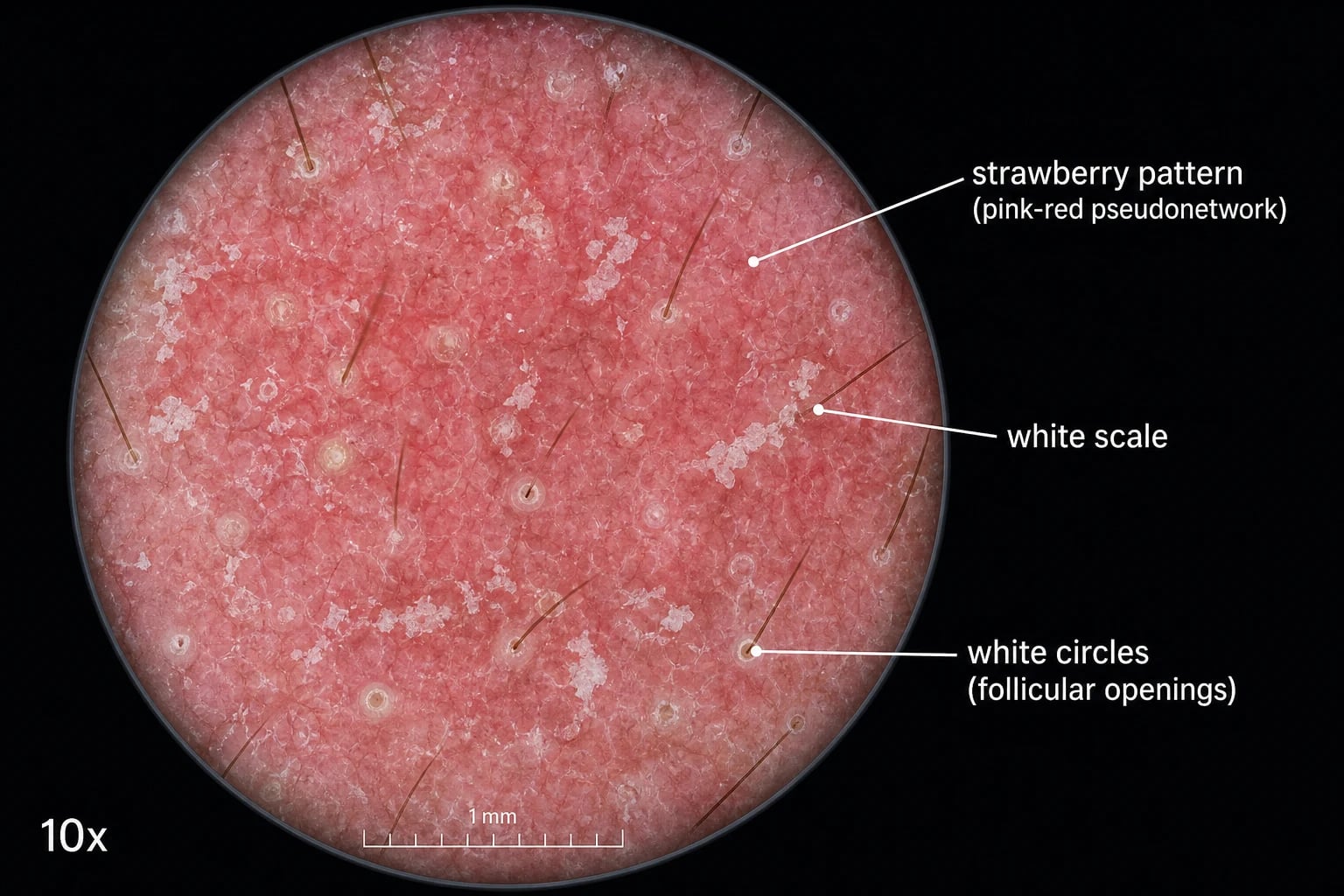
Dermoscopy improves diagnostic accuracy and helps distinguish AK from seborrhoeic keratosis, Bowen disease, and basal cell carcinoma. Different body sites produce different dermoscopic patterns, so the examination must be interpreted in context. [1]
Facial AK: the strawberry pattern
The classic dermoscopic pattern of facial, non-pigmented AK is the "strawberry pattern": [1]
- Erythematous pseudonetwork: red background created by unfocused vessels coursing between follicular openings.
- White-yellow scale: overlying keratotic scale that may be fine or coarse.
- White circles: halo-like white rings surrounding follicular openings (follicular plugs with perifollicular fibrosis). [1]
This pattern was originally described by Zalaudek and colleagues in facial non-pigmented AK and is highly characteristic, although not pathognomonic. [1]
Non-facial AK and other patterns
On the scalp, dorsal hands, and forearms, AK may show a less organised pattern with prominent white-yellow scale, dotted vessels, or a strawberry-like pseudonetwork without the well-defined follicular white circles. The background sun-damaged skin contributes solar elastosis and telangiectasia, which can obscure the pattern. [1]
Dermoscopic red flags for progression
In more advanced or hypertrophic lesions, dermoscopy may show: [1]
- Polymorphic vessels (a mixture of dotted, linear irregular, and hairpin vessels).
- Targetoid follicles or white-yellow opaque areas.
- Erosion or ulceration without preceding trauma.
- Blue-white veil or polymorphic vessels in pigmented lesions — these should prompt biopsy to exclude melanoma or pigmented SCC. [1]
The appearance of polymorphic vessels or erosion in a previously stable AK is a strong indicator that the lesion has progressed to SCC in situ or invasive SCC and must be biopsied. [1]
Differential Diagnosis
The differential diagnosis of AK includes benign keratoses, other premalignancies, and skin cancer. Distinguishing features are essential because the management differs markedly. [1]
| Condition | Key distinguishing features | Why it matters |
|---|---|---|
| SCC in situ (Bowen's disease) | Sharply demarcated, persistent, scaly red patch or plaque; histology shows full-thickness epidermal atypia without dermal invasion. | Often treated with topical 5-FU, imiquimod, PDT, or excision; biopsy confirms. |
| Invasive SCC | Indurated nodule or plaque that may ulcerate or bleed; firm on palpation; requires biopsy and staging. | Must not be missed; needs excisional management and possible staging. |
| Seborrhoeic keratosis | Waxy, "stuck-on" papule or plaque with milia-like cysts and comedo-like openings; not restricted to sun-exposed sites. | Benign; cryotherapy or curettage if symptomatic, but no malignant potential. |
| Porokeratosis | Annular plaque with a raised, grooved peripheral ridge (coronoid lamella); histology shows the characteristic lamella. | Premalignant; may progress to SCC; biopsy is diagnostic. |
| Viral wart | Hyperkeratotic papule with thrombosed capillaries; dermatoglyphic disruption; typically acral or periungual. | Infectious; cryotherapy, salicylic acid, or curettage; not premalignant. |
| Lichen simplex chronicus | Well-demarcated, lichenified, pruritic plaque on sites reachable for rubbing; not sun-damage restricted. | Benign; potent topical corticosteroids and behaviour modification. |
| Psoriasis | Well-demarcated erythematous plaques with silvery scale and symmetrical distribution; Auspitz sign and Koebner phenomenon. | Chronic inflammatory; topical vitamin D analogues, corticosteroids, or systemic therapy. |
| Discoid lupus erythematosus | Erythematous plaques with follicular plugging, atrophy, scarring, and dyspigmentation; photosensitive. | Autoimmune; requires biopsy and often systemic therapy; can scar. |
| Nummular eczema | Coin-shaped, pruritic, eczematous plaques; often on extremities; responds to topical corticosteroids. | Inflammatory; biopsy not needed if classic response. |
Clinical approach to the uncertain lesion
When a lesion is uncertain clinically, the safest approach is dermoscopy followed by biopsy if any red flags are present. A trial of topical corticosteroids or antifungals is not appropriate when malignancy is in the differential, because a transient partial response can delay diagnosis of SCC in situ or invasive SCC. [1]
Clinical & Bedside Assessment
The bedside assessment of suspected AK is primarily clinical and dermoscopic. The key aims are to confirm the diagnosis, assess the extent of field damage, and identify any lesion suspicious for invasive SCC. [1]
Focused history
- Cumulative sun exposure, outdoor occupation, sunburn history, phototype.
- Previous skin cancers or AKs.
- Immunosuppression, transplant status, or prior PUVA/radiation.
- Change in any specific lesion: thickening, growth, pain, bleeding, ulceration. [1]
Focused examination
- Inspect all sun-exposed sites, including scalp, face, ears, neck, dorsal hands, forearms, and lower lip.
- Palpate lesions — the gritty scale is often more obvious to touch than to sight.
- Assess background solar damage: elastosis, telangiectases, dyspigmentation.
- Look for red flags of malignancy: induration, nodularity, ulceration, rapid change. [1]
Dermoscopy
- Use a dermatoscope to look for the strawberry pattern and, importantly, for features of progression such as polymorphic vessels, erosion, or loss of the normal follicular pattern. [1]
Investigations
Most AKs are diagnosed clinically, with dermoscopy as a useful adjunct. However, biopsy is mandatory when malignancy is suspected or when the diagnosis is uncertain. [1]
Biopsy indications
Biopsy any AK that shows:[3][9]
- Thickening or induration
- Pain or tenderness
- Rapid growth
- Ulceration or bleeding
- Treatment failure after an adequate course
- Recurrence after apparent clearance
- Cutaneous horn at the base, to define the underlying pathology [1]
A shave, punch, or incisional biopsy should sample the most suspicious area, including enough dermis to assess for invasion. [1]
Histology
The histological hallmark of AK is dysplastic keratinocytes confined to the lower epidermis in a background of solar elastosis.[6]
Key features include: [1]
- Parakeratosis — retained nuclei in the stratum corneum.
- Atypical keratinocytes with enlarged, hyperchromatic nuclei and disordered maturation, primarily involving the lower third to lower two-thirds of the epidermis.
- Basal layer disorganization with budding of atypical cells.
- Solar elastosis in the upper dermis — basophilic degeneration of elastic fibres.
- Chronic inflammatory infiltrate in the superficial dermis. [1]
AK (mild to moderate)
SCC in situ (Bowen's disease)
Invasive SCC
In SCC in situ (Bowen's disease), atypia involves the full thickness of the epidermis. In invasive SCC, atypical keratinocytes breach the basement membrane and extend into the dermis. [1]
Management — Resuscitation
AK itself is not a medical emergency. The "resuscitation" equivalent in AK care is the rapid identification and expedited biopsy of a lesion suspicious for invasive SCC. Red-flag features — induration, rapid growth, ulceration, bleeding, or pain — should trigger urgent referral or same-week biopsy rather than watchful waiting or topical therapy.[9]
[1]Management — Definitive & Stepwise
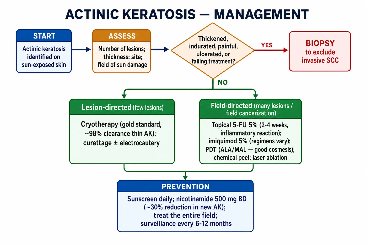
AK management is stratified by lesion number, thickness, location, patient preference, and comorbidities — especially immunosuppression.[9][10]
Lesion-directed therapy
For few, discrete, thin AKs, destructive or excisional treatments are preferred because they are quick, lesion-specific, and provide histology when needed. [1]
Cryotherapy (liquid nitrogen)
- The gold-standard lesion-directed therapy for isolated thin AKs.
- Technique: spray or cotton-tipped applicator; freeze the lesion and a narrow rim of normal skin, usually for 5–10 seconds, allowing a complete thaw before a second freeze-thaw cycle if used. A margin of 1–2 mm of clinically normal skin is included to catch subclinical lateral extension.
- Efficacy: clearance rates are high for thin lesions; thicker lesions may require repetition or alternative therapy. Studies report clearance rates approaching 98% for thin AKs after a single freeze-thaw cycle, but this falls with increasing lesion thickness.
- Adverse effects: pain, blistering, crusting, hypopigmentation (especially Fitzpatrick IV–VI), and rarely scarring or hypertrophic scarring. Hypopigmentation is the most common long-term cosmetic concern and is more pronounced after aggressive freeze-thaw cycles.
- Contraindications / cautions: areas with poor wound healing, very dark skin phototypes where dyspigmentation would be unacceptable, and lesions where invasive SCC is suspected (biopsy first). [1]
Curettage ± electrocautery
- Useful for thicker or hypertrophic AKs; provides tissue for histology.
- The lesion is scraped with a curette and the base is treated with electrocautery for haemostasis and destruction of residual atypia.
- May leave a hypopigmented or slightly atrophic scar; therefore it is used cautiously on cosmetically sensitive sites. [1]
Surgical excision
- Reserved for lesions suspicious for invasive SCC or for biopsy-proven SCC.
- Not indicated for straightforward, thin AK without diagnostic uncertainty, because the morbidity exceeds the benefit. [1]
Field-directed therapy
For multiple lesions, confluent AKs, or field cancerization, the therapeutic target is the entire sun-damaged field rather than individual lesions. Field-directed therapies reduce both visible AKs and the subclinical clones from which new lesions arise. [1]
| Agent | Dose and schedule | Typical response | Main adverse effects | Notes |
|---|---|---|---|---|
| 5-Fluorouracil 5% cream | Once or twice daily for 2–4 weeks to affected field | Brisk inflammatory reaction (erythema, erosion, crusting); clearance ~70–90% | Pain, erosion, crusting, post-inflammatory dyspigmentation | Inflammatory response correlates with efficacy; counsel patient before starting |
| Imiquimod 5% cream | 2–3 times/week for up to 16 weeks; or daily × 2 weeks on/2 weeks off × 2 cycles | Local inflammation, flulike symptoms, high clearance | Erythema, erosion, pruritus, headache, fatigue | Immune response modifier via TLR7 agonism |
| Photodynamic therapy (PDT) | ALA or MAL applied, incubated 1–3 hours, activated by red/blue light or daylight | Excellent for large facial/scalp fields; good cosmetic outcome | Pain during illumination, erythema, oedema, crusting | Single or few sessions; preferred when cosmetic outcome matters |
| Diclofenac 3% gel | Twice daily for 60–90 days | Milder inflammation; lower clearance than 5-FU or imiquimod | Erythema, pruritus, dryness | Useful for patients who cannot tolerate severe inflammation |
| Chemical peels / laser | Trichloroacetic acid, Jessner's solution, CO₂ or Er:YAG laser | Variable clearance | Scarring, dyspigmentation, infection risk | Operator-dependent; reserved for extensive or refractory disease |
The 2021 AAD guidelines give strong recommendations to cryosurgery, 5-FU, and imiquimod, and conditional recommendations to PDT and diclofenac as monotherapy or in combination regimens. [1]
Patient selection and practical tips
- Topical 5-FU is highly effective but requires patient buy-in because the inflammatory reaction can be alarming. Pre-treatment counselling should explain that erythema, erosion, and crusting are expected and indicate drug activity.
- Imiquimod is useful for patients who prefer a shorter intensive course or who have 5-FU intolerance. The 2-week on/2-week off regimen can improve adherence.
- PDT is ideal for patients with large fields, those who cannot apply creams reliably, or those who want fewer treatment visits. It is expensive and requires clinic-based light activation.
- Diclofenac is the gentlest option but has lower efficacy; it is best for patients with limited disease who cannot tolerate stronger agents. [1]
Combination and sequential therapy
For high-burden or refractory disease, combining lesion-directed and field-directed therapies can improve outcomes. Common approaches include: [1]
- Cryotherapy of thicker lesions followed by field 5-FU or imiquimod.
- PDT followed by topical maintenance for recurrent field disease.
- Sequential field therapy (e.g., 5-FU then imiquimod) for patients with extensive involvement. [1]
Follow-up and surveillance
After treatment, patients should be reviewed to document clearance and screen for new lesions. High-risk individuals — immunosuppressed patients, those with many AKs, or prior skin cancer — benefit from 6- to 12-monthly full-skin examinations.[1]
Specific Subtypes & Scenarios
Actinic cheilitis
Actinic cheilitis is AK limited to the lower lip vermillion. Because the lip has higher malignant potential than many cutaneous sites, management is more aggressive. Options include: [1]
- Topical 5-FU for diffuse involvement.
- Imiquimod for field disease.
- Photodynamic therapy for extensive disease.
- Vermillionectomy or laser ablation for severe, refractory, or suspicious cases.
- Biopsy any focal thickening, induration, or ulceration. [1]
Cutaneous horn overlying AK
A cutaneous horn is a morphological finding, not a diagnosis. When it overlies an AK, a biopsy of the base is required to exclude invasive SCC or keratoacanthoma. [1]
Immunosuppressed patients
Solid-organ transplant recipients have a dramatically increased AK burden and a substantially higher rate of progression to invasive SCC. Management principles include:[1]
- Aggressive field-directed therapy and regular surveillance.
- Lesion-directed destruction of all visible AKs.
- Consideration of mTOR inhibitor conversion (e.g., sirolimus) from calcineurin inhibitors where feasible, because mTOR inhibition may reduce NMSC burden.
- Nicotinamide was not effective for chemoprevention in transplant recipients in the ONTRANS trial, so it should not be relied upon in this group.[11]
Darker skin phototypes
AK is less common but not absent in Fitzpatrick IV–VI. It often presents with dyspigmentation and may be mistaken for seborrhoeic keratosis or lichen simplex chronicus. Cryotherapy carries a higher risk of hypo- and hyperpigmentation, so field-directed therapy and gentle cryotherapy technique are preferred. [1]
Complications & Pitfalls
Malignant progression
The risk of a single AK progressing to invasive SCC is low per lesion but clinically significant at the patient level because patients usually have multiple lesions and extensive field damage. Older literature cited a wide progression range; modern estimates suggest an annual per-lesion risk in the range of roughly 0.025–0.075%, with higher cumulative risk in patients with many lesions or immunosuppression.[1]
The greater danger is that an individual lesion that has already progressed to invasive SCC is mistaken for a benign AK and treated only with cryotherapy or topical 5-FU. This is why biopsy of thickened, indurated, or treatment-failing lesions is non-negotiable. [1]
Treatment pitfalls
- Undertreating thick lesions with cryotherapy: thick or hypertrophic AKs may need curettage or biopsy rather than a brief freeze. A single 5-second freeze is inadequate for a hyperkeratotic plaque and may leave residual atypia.
- Overtreating with field therapy for a single lesion: a single thin AK does not require a 4-week 5-FU course; cryotherapy is simpler, faster, and more cost-effective.
- Ignoring hypopigmentation risk: patients with darker skin require careful counselling and sometimes field-directed therapy to avoid dyspigmentation. The cosmetic impact of cryotherapy can be worse than the disease itself in some patients.
- Failing to treat the field: treating only visible AKs leaves subclinical clones behind, leading to recurrence and the appearance of new lesions within months.
- Neglecting prevention: even the best destructive or field therapy will be undermined if the patient continues intense unprotected sun exposure. Sun protection and chemoprevention are part of long-term disease control, not optional extras. [1]
Prevention & Surveillance
Primary prevention of AK aims to reduce cumulative UV exposure, while secondary prevention detects early lesions and prevents progression to invasive SCC. Both are integral to long-term management. [1]
Photoprotection
Sun protection is the cornerstone of AK prevention. Effective measures include: [1]
- Broad-spectrum sunscreen (SPF 30 or higher, UVB + UVA protection) applied generously to all sun-exposed skin and reapplied every 2 hours during outdoor activity or after swimming.
- Physical barriers: wide-brimmed hats, long-sleeved clothing, sunglasses, and seeking shade during peak UV hours (10 am to 4 pm).
- Avoidance of tanning beds and deliberate tanning.
- Vitamin D optimisation through diet or supplements rather than through sun exposure, especially in high-risk individuals. [1]
Sunscreen has been shown to reduce the development of new AKs and may allow partial regression of existing lesions. Regular use is particularly important in organ transplant recipients, although it is not sufficient on its own in this high-risk group. [1]
Chemoprevention
Nicotinamide (vitamin B3) 500 mg twice daily reduced the incidence of new AKs and NMSC in the ONTRAC trial of high-risk immunocompetent patients. It is safe, inexpensive, and well tolerated. It should be offered to patients with frequent new AKs, prior NMSC, or extensive field damage, but it does not replace sun protection or surveillance. [1]
In transplant recipients, the ONTRANS trial showed no significant benefit, so nicotinamide is not recommended as a chemopreventive agent in this population. [1]
Surveillance schedule
| Risk category | Definition | Follow-up interval |
|---|---|---|
| Low risk | Few thin AKs, immunocompetent, no prior SCC | Annual skin examination |
| Moderate risk | Multiple AKs, prior AK treatment, moderate field damage | 6–12 monthly review |
| High risk | Immunosuppressed, many AKs, prior SCC, extensive field damage | 3–6 monthly dermatology review |
Patient education should include self-examination for new or changing lesions, the ABCDE-like warning signs for AK progression (thickening, induration, bleeding, ulceration), and the importance of early review if these occur. [1]
Prognosis & Disposition
Most AKs remain stable for long periods, and a proportion regress spontaneously. However, the presence of AK marks a patient as having sufficient UV damage to be at increased risk of both further AKs and invasive SCC. The natural history is dynamic: individual lesions may regress, persist, or progress, and new lesions continue to arise from the surrounding field. [1]
Factors affecting prognosis
The prognosis of AK is determined more by the patient-level risk than by any single lesion. Key prognostic factors include: [1]
- Number of AKs: patients with many lesions have a higher cumulative risk of SCC.
- Extent of field cancerization: widespread sun damage predicts continued emergence of new lesions.
- Immunosuppression: transplant recipients have a markedly higher risk of progression and death from SCC.
- History of invasive SCC: previous SCC is the strongest predictor of future SCC.
- Anatomical site: actinic cheilitis and lesions on the scalp or ears may have higher malignant potential than truncal lesions. [1]
After successful treatment, disposition is determined by risk stratification: [1]
- Low-risk patients (few lesions, immunocompetent, no prior skin cancer): annual skin check and sun protection advice.
- High-risk patients (many lesions, immunosuppressed, prior SCC, extensive field damage): 6-monthly dermatology review, field maintenance therapy as needed, and patient education on self-examination. [1]
Special Populations
Elderly patients and frailty
Elderly patients have the highest prevalence of AK and often the greatest cumulative UV damage. Treatment choice should balance efficacy, frailty, wound-healing capacity, polypharmacy, and patient preference. Field therapy may be preferable to repeated cryotherapy visits for patients with limited mobility. For very frail patients with limited life expectancy, observation with sun protection may be reasonable if lesions are asymptomatic and not suspicious. However, any red flag for invasive SCC should still trigger biopsy. [1]
Pregnancy and lactation
Data on topical 5-FU, imiquimod, and PDT in pregnancy are limited. Systemic absorption of topicals is low, but many clinicians avoid 5-FU and imiquimod in pregnancy unless clearly necessary. Cryotherapy is generally considered safe in pregnancy because it is locally destructive and has minimal systemic absorption. PDT is usually deferred until after pregnancy because of limited safety data. Nicotinamide is generally regarded as safe in pregnancy at standard doses, but chemoprevention decisions should be individualised. [1]
Immunosuppressed patients
Solid-organ transplant recipients have a dramatically increased AK burden and a substantially higher rate of progression to invasive SCC. Management principles include: [1]
- Lower threshold for biopsy because the risk of occult invasive SCC is higher.
- Aggressive field-directed therapy with 5-FU, imiquimod, or PDT, often in combination with cryotherapy of discrete lesions.
- Regular surveillance every 3–6 months in high-risk transplant patients.
- Consideration of mTOR inhibitor conversion (e.g., from calcineurin inhibitors to sirolimus or everolimus) where feasible, because mTOR inhibition may reduce NMSC burden in some transplant recipients.
- Nicotinamide was not effective for chemoprevention in transplant recipients in the ONTRANS trial, so it should not be relied upon in this group.
- Patient education on rigorous sun protection and self-examination is essential. [1]
Patients with chronic lymphocytic leukaemia
CLL is associated with an increased risk of AK and aggressive SCC, even in the absence of exogenous immunosuppression. The disease-related immune dysfunction accelerates both AK development and malignant transformation. Management should be proactive, with frequent skin checks and early biopsy of suspicious lesions. [1]
Darker skin phototypes
AK is less common but not absent in Fitzpatrick IV–VI. It often presents with dyspigmentation rather than bright erythema and may be mistaken for seborrhoeic keratosis, lichen simplex chronicus, or post-inflammatory hyperpigmentation. Cryotherapy carries a higher risk of hypo- and hyperpigmentation, so field-directed therapy and gentle cryotherapy with narrow margins are preferred. Dermoscopy may be less dramatic because the erythematous pseudonetwork is less visible against darker skin; careful attention to scale, follicular patterns, and dyspigmentation is needed. [1]
Genetic disorders of DNA repair
Patients with xeroderma pigmentosum develop AK and NMSC at an extraordinarily young age due to defective nucleotide excision repair. Management requires aggressive photoprotection, frequent surveillance, and early treatment of all visible lesions. Other genodermatoses such as oculocutaneous albinism and dyskeratosis congenita also increase AK risk through reduced photoprotection or defective DNA maintenance. [1]
Patients on photosensitizing drugs
Medications such as voriconazole, amiodarone, thiazide diuretics, and some non-steroidal anti-inflammatory drugs can photosensitise the skin. In patients with AK, these agents may worsen photodamage and should be reviewed when possible. Voriconazole in particular is associated with aggressive SCC and AK in immunosuppressed patients. [1]
Evidence, Guidelines & Regional Differences
Landmark trials
ONTRAC (2015) — oral nicotinamide 500 mg twice daily reduced the rate of new non-melanoma skin cancers and actinic keratoses in high-risk immunocompetent patients over 12 months. The benefit was thought to relate to enhanced cellular energy metabolism, improved DNA repair, and reduced UV-induced immunosuppression. [1]
ONTRANS (2023) — in solid-organ transplant recipients, oral nicotinamide did not significantly reduce the incidence of new NMSC or AKs. This negative result is critical because transplant recipients have the highest AK burden and the highest malignant transformation risk. [1]
Systematic reviews (2023) — meta-analytic evidence supports high clearance rates with 5-FU, imiquimod, and PDT, with choice driven by lesion burden, patient preference, cost, and adverse-effect tolerance. [1]
AAD 2021 guidelines
The American Academy of Dermatology published updated guidelines in 2021 with 18 evidence-based recommendations for AK management: [1]
- Strong recommendations: cryosurgery, topical 5-FU, and imiquimod.
- Conditional recommendations: PDT, diclofenac, and combination or sequential regimens.
- Diagnostic recommendation: biopsy lesions suspicious for invasive SCC before destructive therapy.
- Prevention recommendation: photoprotection, patient education, and chemoprevention where appropriate. [1]
The guidelines emphasize that no single therapy is universally superior; treatment should be individualised based on number and thickness of lesions, field involvement, patient adherence, comorbidities, and cosmetic considerations. [1]
European and Australasian practice
European guidelines broadly align with the AAD: lesion-directed cryotherapy for discrete lesions, field-directed 5-FU, imiquimod, or PDT for multiple lesions. Access to daylight-PDT is more widespread in Europe and Australia than in many US centres. In Australia, where AK prevalence is among the highest in the world, PDT and field-directed topical therapy are standard of care, and chemoprevention with nicotinamide is commonly discussed in high-risk immunocompetent patients. [1]
Ingenol mebutate
Ingenol mebutate gel was previously used as a short-course field therapy for AK. It was withdrawn in the European Union due to safety concerns including severe local skin reactions and reports of skin necrosis. It remains available in some regions but is no longer a first-line option in many guidelines.[3]
Chemoprevention evidence
The ONTRAC trial demonstrated that oral nicotinamide 500 mg twice daily reduced the rate of new non-melanoma skin cancers and actinic keratoses in high-risk immunocompetent patients over 12 months.[4] However, the subsequent ONTRANS trial in solid-organ transplant recipients did not show a similar benefit, underscoring that chemoprevention efficacy is population-specific.[11]
Exam Pearls
AK-FIELD-CARE
Exam application bank (NEET-PG / INICET)
One-line answer
Actinic keratosis (AK, solar keratosis) is the commonest premalignant skin lesion — an intraepidermal keratinocyte dysplasia arising on chronically sun-damaged skin. It sits on a biological spectrum with SCC in situ (Bowen's disease) and invasive squamous cell carcinoma, driven by UV-induced p53 mutations and field cancerization. Clinically it presents as rough, scaly, erythematous macules or patches on sun-exposed sites, classically described as easier felt than seen. Management is stratified into lesion-directed therapy (cryotherapy, curettage, excision) for discrete lesions and field-directed therapy (topical 5-fluorouracil, imiquimod, photodynamic therapy, diclofenac) for multiple lesions or field cancerization. Any lesion that thickens, becomes indurated, ulcerates, or fails therapy must be biopsied to exclude invasive SCC.
Worked stems (answer without another resource)
Stem 1 — Classic presentation. Map symptoms to mechanism; name the first investigation and first treatment step with dose/route if drug therapy is standard. [1]
Stem 2 — Unstable / complicated. List red flags that force immediate resuscitation, theatre, ICU, antidote, or reperfusion — and what you do in the first 15 minutes. [1]
Stem 3 — Atypical group. Elderly, pregnancy, child, or immunocompromised: how presentation and thresholds change. [1]
Stem 4 — Differential trap. Name the three closest mimics and one discriminator for each. [1]
Stem 5 — Disposition. Who goes home with safety-netting, who is admitted, who needs HDU/ICU/theatre, and what follow-up is mandatory. [1]
Rapid viva checklist
- Definition + classification
- Pathophysiology chain
- Bedside signs / criteria
- Score with exact components (if any)
- Emergency bundle
- Definitive therapy with doses
- Complications of disease and of treatment
- Special populations
- Guideline/trial name if classic
- Three exam traps
Coverage self-check
If you cannot answer any stem above from this page alone, re-read the matching section — the page is intended to be self-sufficient for final-prof and NEET-PG/INICET questions on Actinic keratosis.
[1]References
- [1]Willenbrink TJ, Ruiz ES, Cornejo CM, et al. Field cancerization: Definition, epidemiology, risk factors, and outcomes J Am Acad Dermatol, 2020.PMID 32387665
- [2]Dianzani C, Conforti C, Giuffrida R, et al. Current therapies for actinic keratosis Int J Dermatol, 2020.PMID 32012240
- [3]Worley B, Harikumar V, Reynolds K, et al. Treatment of actinic keratosis: a systematic review Arch Dermatol Res, 2023.PMID 36454335
- [4]Chen AC, Martin AJ, Choy B, et al. A Phase 3 Randomized Trial of Nicotinamide for Skin-Cancer Chemoprevention N Engl J Med, 2015.PMID 26488693
- [5]Wang JY, Zeitouni N, Austin E, et al. Photodynamic therapy: Clinical applications in dermatology J Am Acad Dermatol, 2026.PMID 39986392
- [6]Ferrándiz C, Malvehy J, Guillén C, et al. Precancerous Skin Lesions Actas Dermosifiliogr, 2017.PMID 27658688
- [7]Zalaudek I, Ferrara G, Leinweber B, et al. Dermoscopy of facial nonpigmented actinic keratosis Br J Dermatol, 2006.PMID 17034524
- [8]Benjamin CL, Ullrich SE, Kripke ML, et al. p53 and the pathogenesis of skin cancer Toxicol Appl Pharmacol, 2007.PMID 17270229
- [9]Eisen DB, Asgari MM, Bennett DD, et al. Guidelines of care for the management of actinic keratosis: Executive summary J Am Acad Dermatol, 2021.PMID 34111497
- [10]Eisen DB, Asgari MM, Bennett DD, et al. Guidelines of care for the management of actinic keratosis J Am Acad Dermatol, 2021.PMID 33820677
- [11]Allen NC, Martin AJ, Murphy KE, et al. Nicotinamide for Skin-Cancer Chemoprevention in Transplant Recipients N Engl J Med, 2023.PMID 36856616