Dermatology · Medicine
Albinism
Also known as Albinism · Oculocutaneous albinism (OCA) · Ocular albinism (OA1) · OCA1 · OCA2 · Tyrosinase-negative albinism · Hermansky-Pudlak syndrome · Chédiak-Higashi syndrome
Albinism is a group of inherited disorders of melanin synthesis or melanosome biogenesis causing congenital hypopigmentation of skin, hair, and eyes (oculocutaneous albinism, OCA) or of the eyes alone (ocular albinism, OA1). The melanocytes are normal in NUMBER — the defect is in melanin PRODUCTION, which separates albinism from vitiligo (where melanocytes are lost). OCA1 (TYR, chr 11) is the severe tyrosinase-negative form; OCA2 (P gene, chr 15) is the most common worldwide and dominates in sub-Saharan Africa. All OCA types are autosomal recessive; OA1 (GPR143) is X-linked. Every type shares a characteristic OCULAR tetrad — nystagmus, foveal hypoplasia, iris transillumination, photophobia — plus abnormal chiasmal decussation. The dominant complication is SKIN CANCER (squamous cell carcinoma of the head and neck, up to 1000-fold risk), so lifelong sun protection (SPF 50+) and surveillance are the single most important interventions. Syndromic forms add systemic disease: Hermansky-Pudlak (platelet dense-granule deficiency, pulmonary fibrosis, colitis) and Chédiak-Higashi (neutrophil dysfunction, giant lysosomal granules, haemophagocytic lymphohistiocytosis).
On this page & tools
Your progress
Saved locally on this device.
Exam tags
Red flags
Albinism is one of the oldest recognised genetic conditions and one of the most common inherited disorders of pigmentation worldwide. It is best understood not as a single disease but as a shared phenotype — congenital hypopigmentation — produced by mutations in any of a dozen genes along the melanin-synthesis pathway or in the biogenesis of the melanosome, the specialised organelle in which melanin is made and stored. What unites every type is that the melanocyte is present in normal numbers; what fails is the chemistry of pigment production. This single distinction is the hinge on which the differential diagnosis turns, separating albinism (a factory that makes a defective product) from vitiligo and piebaldism (a factory that has lost its workers or relocated them).[2][5]
Because melanin protects the skin from ultraviolet radiation and is central to the development of the fovea and the normal wiring of the optic nerves, its absence produces two parallel clinical stories: a cutaneous one dominated by sun damage and skin cancer, and an ophthalmic one defined by poor vision, involuntary eye movements, and light sensitivity. In syndromic forms — Hermansky-Pudlak and Chédiak-Higashi — the same intracellular trafficking machinery that builds the melanosome also builds platelet dense granules and lysosomes, so the phenotype extends to bleeding, lung fibrosis, immune failure, and a risk of fatal haemophagocytic lymphohistiocytosis.[3][7]
Classification
Albinism divides first by distribution (oculocutaneous vs ocular) and then by gene. The non-syndromic oculocutaneous albinisms (OCA1 through OCA8) are all autosomal recessive and share the same ocular features; they differ chiefly in severity, residual pigment, and ancestry. Ocular albinism type 1 (OA1) is X-linked recessive and spares the skin and hair. Two syndromic forms — Hermansky-Pudlak syndrome (HPS) and Chédiak-Higashi syndrome (CHS) — combine partial albinism with systemic disease because the mutated genes govern lysosome-related organelles more broadly.[2][5]
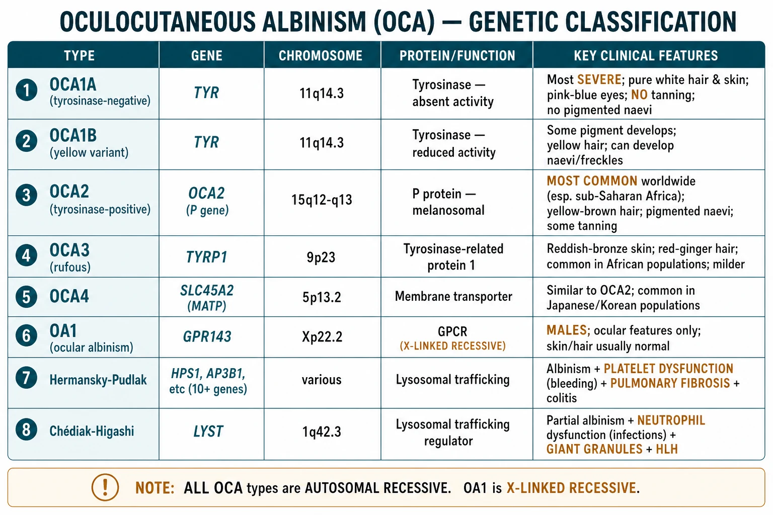
The principal non-syndromic albinisms at a glance
OCA1A (tyrosinase-negative)
OCA1B (yellow variant)
OCA2 (tyrosinase-positive)
OCA3 (rufous)
OCA4
OA1 (ocular albinism)
The remaining non-syndromic types — OCA5 (mapped to 4p15.2), OCA6 (SLC24A5), OCA7 (LRMDA / C10orf11) and OCA8 (DCT) — are rare and behave clinically like OCA2, distinguished only by molecular testing. The syndromic forms are clinically unmistakable and are dealt with under Specific Subtypes below.[5]
Epidemiology & Risk Factors
Albinism is rare globally but strikingly common in selected populations, a pattern that reflects both founder effects and the recessive inheritance that allows carriers to be silent. [1]
Worldwide, OCA1 is the most common type in European/Caucasian and Japanese populations, while OCA2 dominates in sub-Saharan Africa, where its carrier frequency reaches 1 in 25 to 1 in 50 and affected individuals may number 1 in 1,000 in Tanzania and Zimbabwe.[6] OCA4 is enriched in Japanese and Korean cohorts, and OCA3 (rufous albinism) is seen almost exclusively in African populations. OA1 affects males (X-linked) at roughly 1 in 60,000.[2]
Sex incidence is equal for the autosomal recessive OCA types. The dominant risk factors for adverse outcome are not genetic but environmental: consanguinity (which raises the chance that two carriers meet), equatorial residence with intense year-round ultraviolet exposure, and poor access to sunscreen, protective clothing and dermatology services. In much of sub-Saharan Africa these factors converge catastrophically, and skin cancer — not the albinism itself — is the leading cause of premature death, with some series reporting a median age at skin-cancer death below 40 years.[6][9]
Pathophysiology
Understanding albinism requires the melanin biosynthetic pathway, because each OCA type corresponds to a block at a defined point along it. [1]
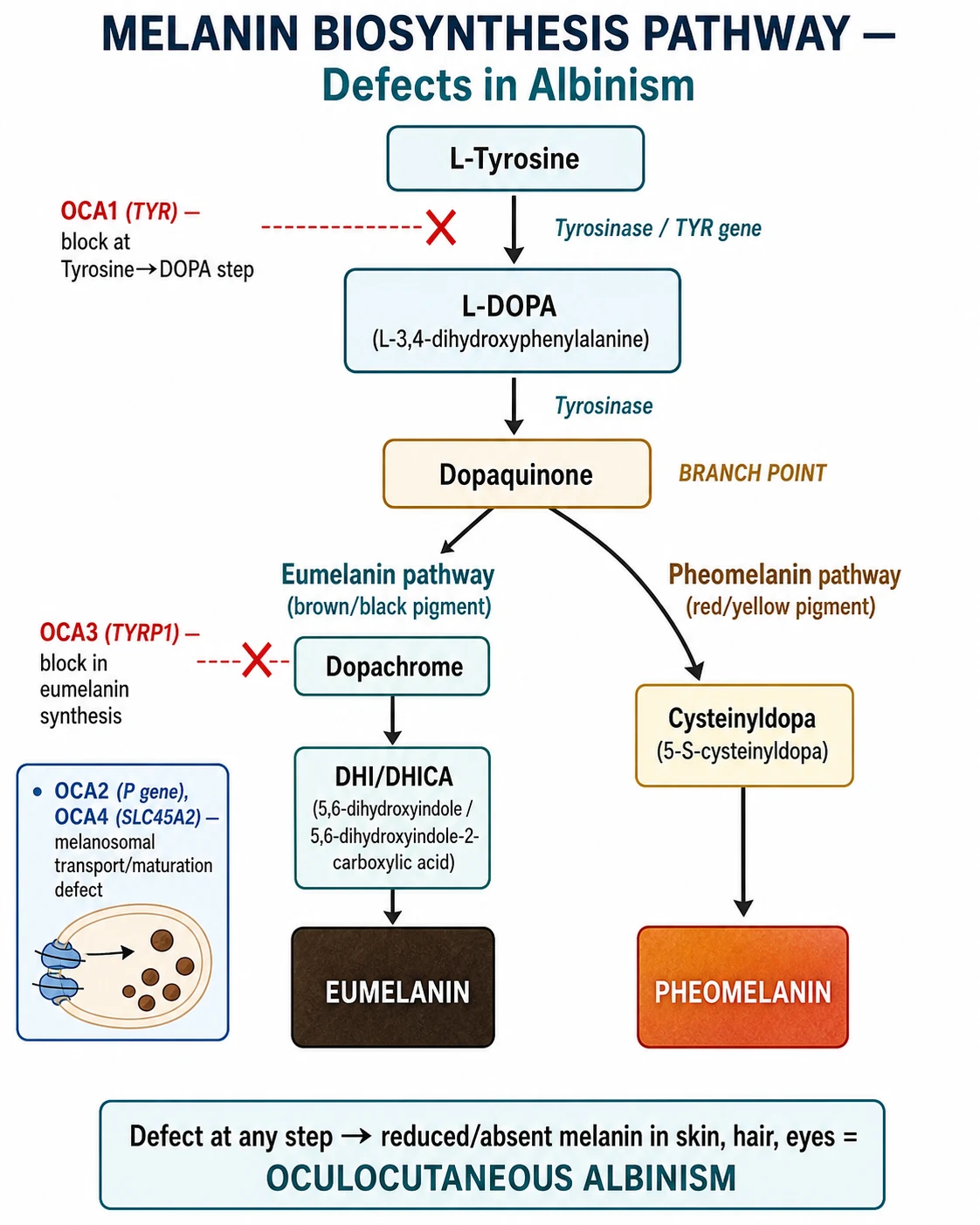
Melanin is synthesised inside the melanosome, a lysosome-related organelle within the melanocyte. The pathway begins with the amino acid L-tyrosine. The enzyme tyrosinase (encoded by TYR on chromosome 11q14) catalyses the first two and rate-limiting steps: hydroxylation of tyrosine to L-DOPA, and oxidation of L-DOPA to dopaquinone. Dopaquinone is the branch point. In the presence of cysteine it is diverted into the pheomelanin pathway (red-yellow pigment); otherwise it proceeds via dopachrome to eumelanin (brown-black pigment). Both pigments are then packaged into mature stage-IV melanosomes and transferred to neighbouring keratinocytes, where they sit over the nucleus as a parasol against ultraviolet damage.[1]
From tyrosine to pigment — and where albinism blocks it
L-Tyrosine enters the melanosome.
TYROSINASE (TYR, 11q14) converts tyrosine → L-DOPA → dopaquinone. LOSS of tyrosinase = OCA1A (no melanin at all).
DOPAQUINONE branches: + cysteine → pheomelanin (red-yellow); otherwise → eumelanin (brown-black).
TYRP1 (TYRP1, 9p23) stabilises tyrosinase and drives eumelanin synthesis. Loss = OCA3 (rufous/red phenotype).
Melanosome pH and maturation governed by the P protein (OCA2) and SLC45A2 transporter (OCA4). Defect → poorly melanised melanosomes.
Mature melanosomes transferred to keratinocytes and positioned over nuclei as UV shielding. Absent in all OCA → UV damage accumulates → skin cancer.
Two downstream consequences define the disease. In the skin, the absence of melanin removes the natural ultraviolet filter, so photons reach and mutate keratinocyte and melanocyte DNA directly; cumulative actinic damage produces the characteristic skin-cancer burden. In the eye, melanin is required in the retinal pigment epithelium and iris during development, and its absence causes foveal hypoplasia (the fovea never differentiates its pit or cone mosaic), iris transillumination (the depigmented iris lets light pass through, visible as a pink-red glow with the slit-lamp), and a striking developmental error of the optic nerves: abnormal decussation at the chiasm, in which an excess of fibres from each temporal retina cross to the contralateral hemisphere. This misrouting is detectable on visual evoked potentials (VEP) as an asymmetrical cortical response and is one of the most sensitive objective confirmations of albinism.[8]
The chiasmal misrouting deserves a closer look because it is the single most objective laboratory confirmation of albinism available. In normal development the proportion of retinal ganglion-cell axons that cross at the optic chiasm is set by the molecular identity of the retina, and melanin signalling from the retinal pigment epithelium is one of the cues that marks ganglion cells for an uncrossed (ipsilateral) fate. When melanin is absent, this cue is lost, and an excess of fibres — including those that should have remained ipsilateral — cross to the contralateral lateral geniculate nucleus. The result is a markedly asymmetrical cortical representation of each visual hemifield that is detectable on pattern-reversal VEP as a larger contralateral-than-ipsilateral response over the occipital cortex when each eye is stimulated in turn. This monocular VEP asymmetry is found in essentially all forms of albinism, including the clinically subtle autosomal recessive ocular albinism, and is the basis for VEP's role as a sensitive confirmatory test when the cutaneous signs are equivocal.[8]
The syndromic forms share a deeper cellular problem. Hermansky-Pudlak and Chédiak-Higashi arise from mutations in genes governing lysosome-related organelle biogenesis — the machinery that builds not only melanosomes but also platelet dense granules (HPS — absent dense bodies, defective secondary-wave aggregation) and the lysosomes of leucocytes and cytotoxic T cells (CHS — giant peroxidase-positive granules, defective phagocyte killing and degranulation, impaired cytotoxicity that predisposes to HLH).[3][7]
Clinical Presentation
The presentation is cutaneous, ocular, and — in syndromic cases — systemic. The cutaneous and ocular features are present from birth; the systemic complications of HPS and CHS declare themselves across infancy, childhood and early adult life. [1]
Cutaneous features
The skin and hair phenotype tracks the residual melanin-producing capacity of each genotype. OCA1A is the most striking: pure white hair that never darkens, porcelain-white skin that never tans, and light blue to pink irides, with no pigmented naevi or freckles throughout life — a useful bedside discriminator, because the presence of naevi argues against OCA1A.[5] OCA1B (yellow variant) is born similarly pale but develops yellow-tan pigment over the first years, accumulates naevi and freckles, and includes the striking temperature-sensitive sub-variant in which pigment develops only in the cooler extremities (the Siamese-cat pattern). OCA2 shows the most pigment of the common types: yellow-brown hair, pigmented naevi, freckling in sun-exposed areas, and a limited ability to tan; the characteristic pigmented pink macules (naevi) scattered on exposed skin are a recognised clue. OCA3 (rufous) produces a reddish-bronze complexion with red-ginger hair and is confined almost entirely to African kindreds. OCA4 cannot be distinguished from OCA2 clinically.[4]
Ocular features (present in ALL types — the defining morbidity)
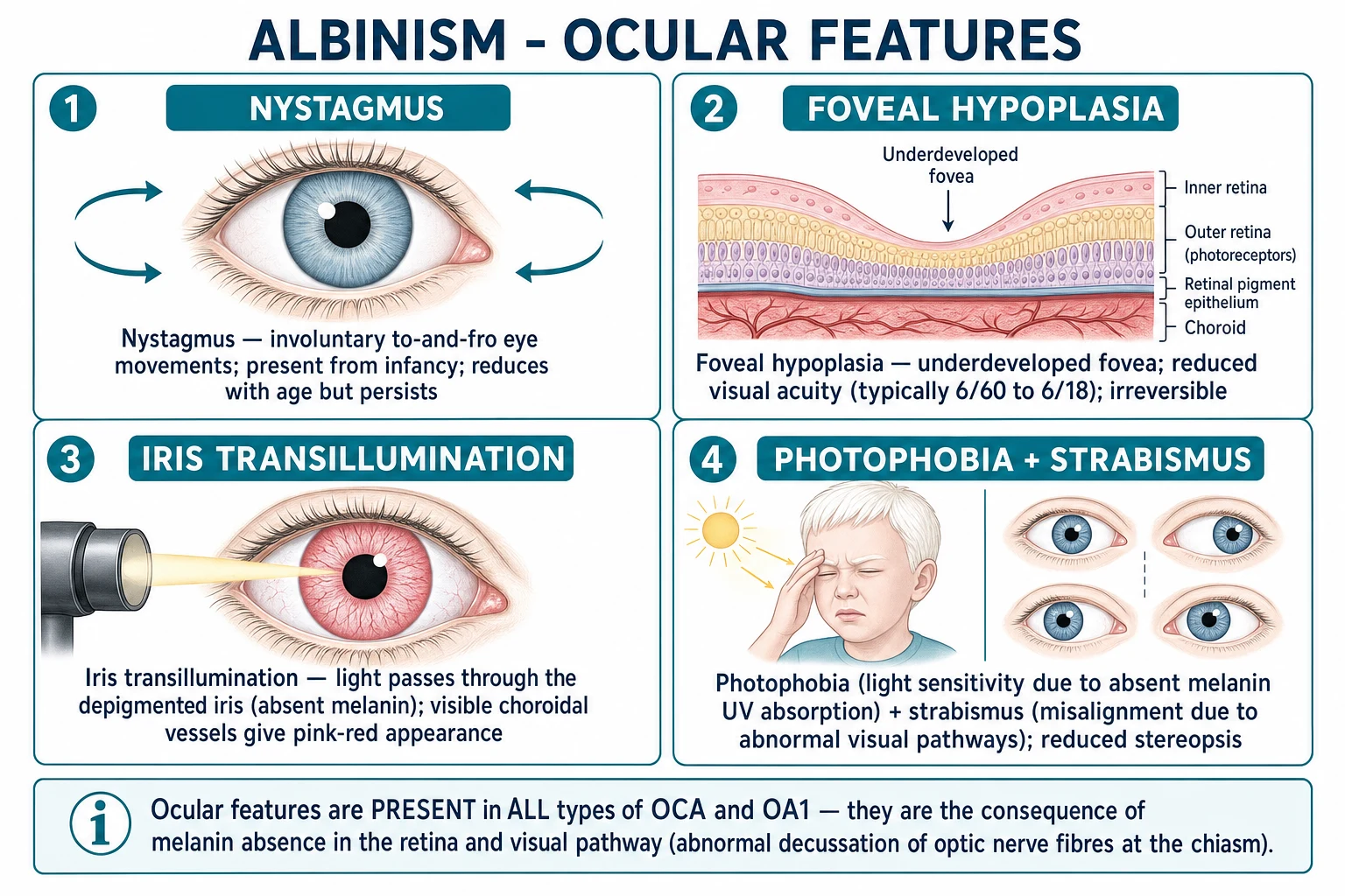
The ocular features are the same across OCA types because the developing eye requires melanin regardless of skin type: [1]
- Nystagmus — involuntary to-and-fro eye movements, usually pendular and horizontal, present from infancy; it often dampens with age and the child adopts a compensatory null point (an anomalous head posture that minimises the movements).[4]
- Foveal hypoplasia — the fovea fails to develop its pit and cone mosaic; this is the chief cause of the permanently reduced visual acuity (typically 6/18 to 6/60, occasionally worse) and is visible on OCT and fundoscopy as an absent foveal reflex with persistence of retinal vessels through the macula.[4]
- Iris transillumination — depigmented iris stroma transmits light; slit-lamp examination shows a pink-red glow and visible radial iris vessels. The degree of transillumination parallels residual pigment (marked in OCA1A, subtle in OCA2).[2]
- Photophobia and strabismus (usually esotropia or exotropia) are near-universal.
- Reduced stereopsis and an anomalous head posture complete the picture.[8]
Visual acuity is the single best functional measure: OCA1A is typically 6/60, OCA2 and OCA4 cluster around 6/24–6/36, and OA1 is similar to OCA1. Crucially, the visual impairment is stable — it does not progressively worsen through life once the developmental window closes. [1]
Atypical and easily missed presentations
Examiners test the corners deliberately. Several presentations are routinely missed: [1]
- Autosomal recessive ocular albinism — OCA (most often OCA1B or OCA2) with minimal, easily overlooked cutaneous signs, presenting as isolated infantile nystagmus or strabismus. The skin looks normal; the diagnosis rests on the ocular findings plus molecular testing.[4]
- Prader-Willi and Angelman syndromes unmasking OCA2 — the OCA2 gene sits within the 15q11-q13 deletion region; a child with hypotonia, feeding difficulty and developmental delay who is also strikingly fair may carry a contiguous gene deletion. Recognising this changes the genetic counselling entirely.[5]
- OA1 in a male infant with normal-appearing skin and only nystagmus and photophobia — easily attributed to congenital motor nystagmus until transillumination and VEP are checked.
- CHS in early infancy presenting as recurrent infection rather than as a pigmentary disorder — the silvery hair and partial albinism may be subtle, but the giant granules on the blood film are pathognomonic.[3]
Syndromic presentations
When albinism is accompanied by systemic symptoms, a syndromic diagnosis must be actively sought: [1]
- Hermansky-Pudlak syndrome adds a bleeding diathesis (easy bruising, epistaxis, gum bleeding, menorrhagia, surgical bleeding), progressive pulmonary fibrosis presenting as exertional dyspnoea and a restrictive defect on lung function (typically onset in the third to fifth decade), and a granulomatous colitis that mimics Crohn's disease.[7]
- Chédiak-Higashi syndrome presents with recurrent pyogenic infections (skin, respiratory, perianal), partial albinism with characteristic silvery metallic hair, hepatosplenomegaly, and the risk of a sudden accelerated phase indistinguishable from haemophagocytic lymphohistiocytosis (HLH): fever, hepatosplenomegaly, pancytopenia, hyperferritinaemia and hypertriglyceridaemia.[3]
Differential Diagnosis
The differential separates the congenital hypomelanoses (in which melanocytes are present but under-producing) from the acquired and patterned leukodermas (in which the defect is different). The single most discriminating feature is the eye: every true albinism has the ocular tetrad; the mimics do not. [1]
Hypopigmentation that is NOT albinism
Vitiligo
Piebaldism
Waardenburg syndrome
Phenylketonuria (PKU)
Tietz syndrome
Nevus depigmentosus / ash-leaf macules (tuberous sclerosis)
A practical rule for the viva: a child with white skin, white hair, and nystagmus and photophobia has an albinism until proven otherwise; a child with a white forelock, patchy depigmentation and normal eyes and hearing has piebaldism; a child with a white forelock and deafness or heterochromia has Waardenburg syndrome; a fair child with seizures and developmental delay has PKU until screened.[2]
Clinical & Bedside Assessment
The focused assessment is built around three questions: Is this an albinism?, Which type?, and Is there a syndromic component that changes management? [1]
Cutaneous examination documents skin and hair colour, the presence and distribution of pigmented naevi, freckles and lentigines in sun-exposed areas (their presence favours OCA2/OCA1B over OCA1A), signs of chronic actinic damage (actinic keratoses, scaling, ulceration), and any lesion suspicious for skin cancer. A Wood's lamp can help delineate the extent of depigmentation. A pinch of hair examined against a dark background reveals the degree of residual pigment. [1]
Ophthalmic examination is the cornerstone. Document visual acuity (distance and near, with and without correction), the nystagmus (type, amplitude, and the null point that the patient adopts to dampen it), and any strabismus. On slit-lamp, look for iris transillumination (the pink-red glow and visible radial vessels are pathognomonic). On fundoscopy, grade foveal hypoplasia: the foveal reflex is absent or dull, the macula lacks its yellow xanthophyll pit, and retinal vessels course uninterrupted through the macular area; the fundus is blond with readily visible choroidal vessels.[4]
Systemic examination hunts for syndromic features: bruising, epistaxis, gum hypertrophy and signs of blood loss (HPS); hepatosplenomegaly, lymphadenopathy, recurrent skin or respiratory infection, silvery hair, and focal neurological signs (CHS); and dyspnoea, fine basal crackles and finger clubbing (HPS pulmonary fibrosis). Examine the parents and siblings where possible, and review the developmental history (CHS causes progressive neurologic decline).[3][7]
Investigations
Albinism is diagnosed clinically and molecularly, not by biopsy. The investigation sequence is: confirm the clinical phenotype, define the ocular anatomy, sequence the genes, and screen for systemic disease in suspected syndromic cases. [1]
Genetic testing
A multi-gene panel sequencing TYR, OCA2, TYRP1, SLC45A2, SLC24A5, LRMDA, DCT, GPR143 and the HPS genes confirms the subtype, distinguishes OCA from OA1, identifies carriers for counselling, and enables prenatal or preimplantation diagnosis in future pregnancies. A historical and still-taught bedside test is the hair-bulb incubation with tyrosine and DOPA: bulbs that fail to darken are tyrosinase-negative (OCA1A), while those that darken are tyrosinase-positive (OCA2 and most others).[5]
Ophthalmic investigations
The full ophthalmic work-up includes cycloplegic refraction (high ametropia is common), slit-lamp for iris transillumination, fundus photography, optical coherence tomography (OCT) to grade foveal hypoplasia objectively, visual evoked potentials (VEP) — which demonstrate the characteristic asymmetry of chiasmal misrouting and are among the most sensitive confirmatory tests — and electroretinography (ERG), which is typically normal because retinal function is preserved (the defect is structural, not degenerative).[8]
Syndromic investigations
When a syndromic form is suspected, targeted testing is mandatory: [1]
- Hermansky-Pudlak — the platelet count is normal but the bleeding time is prolonged; platelet aggregation studies show absent second-wave aggregation with ADP and epinephrine; platelet electron microscopy (the gold standard) shows absent dense bodies. Add pulmonary function tests and high-resolution CT to stage pulmonary fibrosis, and screen for colitis if bowel symptoms are present.[7]
- Chédiak-Higashi — the peripheral blood film shows giant peroxidase-positive cytoplasmic granules in neutrophils, eosinophils and lymphocytes (pathognomonic). Natural-killer-cell cytotoxicity is reduced. If the accelerated phase is suspected, check ferritin, triglycerides, fibrinogen, soluble IL-2 receptor and bone-marrow haemophagocytosis against HLH criteria.[3]
Skin biopsy (rarely needed)
A biopsy is occasionally requested to settle a difficult differential. In albinism the melanocytes are present in normal numbers but contain only early (stage I–II), poorly melanised melanosomes — the opposite of vitiligo, where melanocytes are absent altogether. Biopsy does not usually change management. [1]
Management — Resuscitation and the Immediate Priorities
Albinism is not, in the newborn, an acute emergency — but three situations are time-critical and examiners expect them recognised: [1]
- Chédiak-Higashi accelerated phase (HLH). A febrile CHS child with hepatosplenomegaly and falling blood counts is in the accelerated phase and will die without treatment. Manage as HLH: urgent haematology referral, the HLH-2004 protocol (dexamethasone, etoposide, ciclosporin), supportive care for cytopenias and organ failure, and planning for curative haematopoietic stem cell transplantation.[3]
- Hermansky-Pudlak with major bleeding. Withhold all antiplatelet drugs and NSAIDs. Treat with desmopressin (DDAVP) 0.3 micrograms/kg intravenously, antifibrinolytics (tranexamic acid), and platelet transfusion for severe or surgical bleeding; recombinant activated factor VII is reserved for refractory cases. Avoid nasal packing that traumatises mucosa unnecessarily.[7]
- The newly diagnosed newborn. Confirm the diagnosis, institute sun protection from day one, refer to ophthalmology and clinical genetics, and check vitamin D status — strict sun avoidance can lower it. Provide the family with written, age-appropriate information and put them in touch with a patient organisation.
Management — Definitive and Stepwise
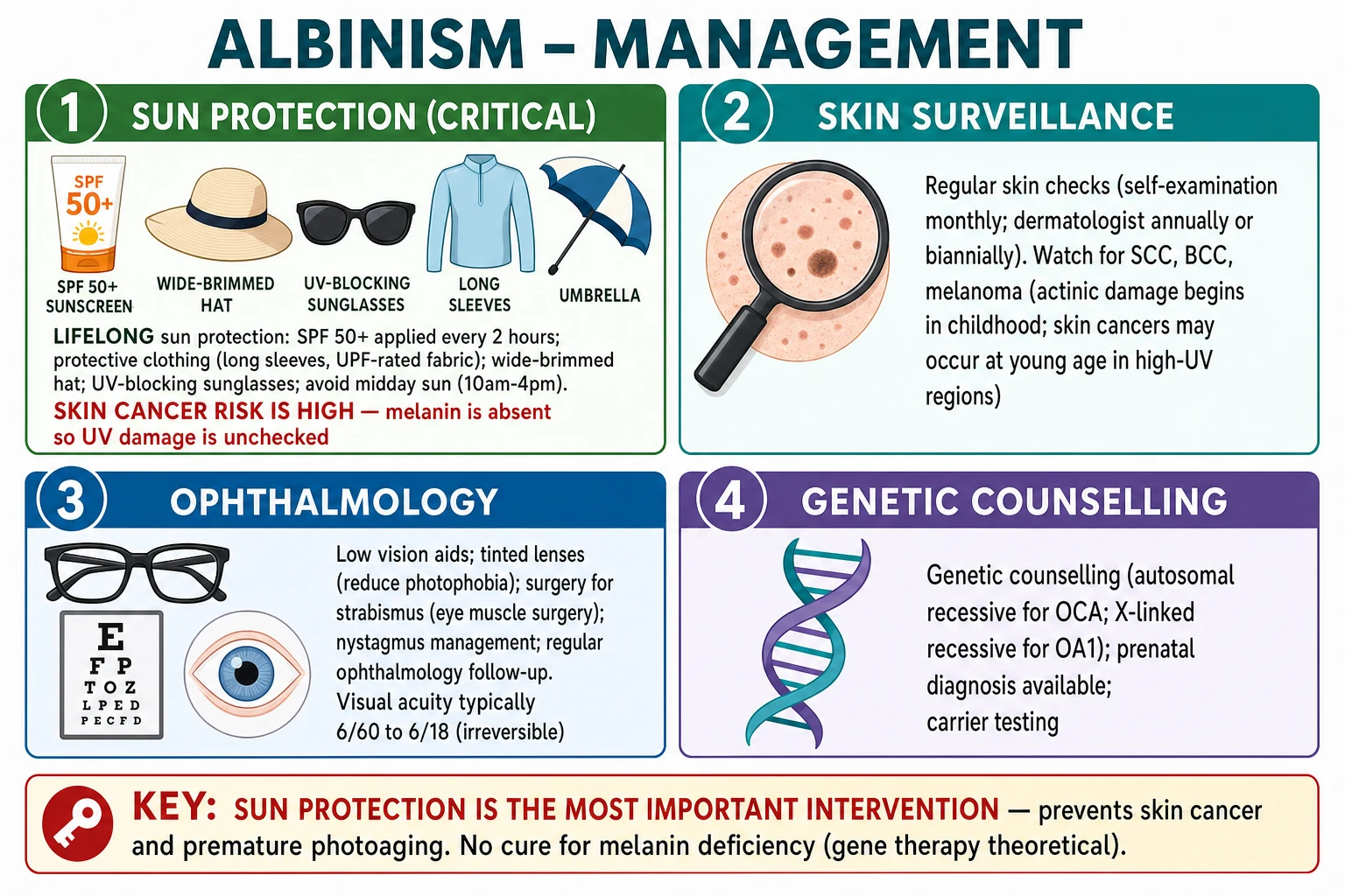
There is no cure for albinism. Management is lifelong prevention and support, organised around four pillars: photoprotection, skin surveillance, ophthalmology, and genetic and psychosocial support.[2][9]
A life managed with albinism
Pillar 1 — Photoprotection (the single most important intervention)
Photoprotection from birth is the only effective prevention for the skin-cancer burden, and its rigor determines long-term survival in high-ultraviolet environments.[9]
- Broad-spectrum sunscreen SPF 50+ applied to all exposed skin every two hours and after swimming or sweating.
- UPF 50+ clothing (long sleeves, long trousers, tightly woven or specially rated fabric).
- A wide-brimmed hat (brim at least 7.5 cm) and, where possible, a neck flap.
- UV-blocking, wrap-around sunglasses that also protect the adnexa and reduce photophobia.
- Avoid midday sun (10 am to 4 pm); seek shade; rearrange school or work schedules where possible.
- Vitamin D adequacy: strict sun avoidance lowers vitamin D; supplement 400–800 IU/day (more if deficient) and monitor. [1]
Pillar 2 — Skin surveillance
- Monthly self-examination using the ABCDE criteria (Asymmetry, Border irregularity, Colour variation, Diameter over 6 mm, Evolution).
- Full-body dermatologist examination at least annually, and every six months in tropical or high-UV regions and after any skin cancer.[6]
- Biopsy any non-healing, growing, ulcerated or bleeding lesion — and remember that melanoma in albinism may be amelanotic (pink) and easily dismissed.
When a skin cancer is confirmed, treatment follows standard oncologic principles but with attention to the field-change burden that characterises chronically photodamaged albino skin: squamous cell carcinoma in situ (Bowen disease, actinic keratoses) is treated with 5-fluorouracil 5% cream twice daily for three to six weeks, imiquimod 5% three times weekly, cryotherapy, or photodynamic therapy; invasive SCC is managed with wide local excision (4–6 mm margins for low-risk lesions, 6 mm or more for high-risk) or Mohs micrographic surgery for head-and-neck lesions where tissue conservation matters, with radiotherapy reserved for patients unfit for surgery; basal cell carcinoma is treated with surgical excision, Mohs surgery, imiquimod for superficial lesions, or radiotherapy; and melanoma — which is frequently amelanotic in albinism and so easily missed — is excised with margins dictated by Breslow thickness, with sentinel-lymph-node biopsy for lesions over 0.8 mm or ulcerated, exactly as in pigmented melanoma. Field-directed therapy for extensive actinic damage (5-fluorouracil plus calcipotriol, or acitretin 25–30 mg daily for chemoprevention in high-risk patients) reduces the rate of new SCC formation. The key examiner point is that amelanotic melanoma is the great mimic in albino skin: any non-healing pink nodule or ulcer must be biopsied, never assumed benign.[6][9]
Pillar 3 — Ophthalmology
There is no treatment that restores foveal development, so the goal is to maximise available vision and reduce symptoms: [1]
- Low-vision aids — magnifiers, large-print materials, electronic reading devices, smartphone accessibility features.
- Refractive correction and dark-tinted, polarised wrap-around lenses or contact lenses to reduce photophobia.
- Prisms to improve an anomalous head posture, and strabismus surgery (typically bimedial recession) for cosmesis and function.
- Nystagmus surgery (the Anderson or Kestenbaum-Anderson head-rotating procedure) shifts the null point towards the primary position to widen the field of best vision and improve the head posture.[4]
Pillar 4 — Genetic and psychosocial support
- Genetic counselling — autosomal recessive OCA carries a 25 percent recurrence risk; X-linked OA1 means carrier females and affected sons. Cascade testing of relatives, and prenatal diagnosis (chorionic-villus sampling or amniocentesis) or preimplantation genetic testing (PGT-M) once the familial mutation is known.[5]
- Psychosocial support — address bullying, stigma and self-esteem; vocational guidance; educational accommodations; and connection with patient organisations (for example Under the Same Sun and the Africa Albinism Network in sub-Saharan Africa, or national albinism societies in Europe and North America).
Specific Subtypes and Syndromic Scenarios
Hermansky-Pudlak syndrome (HPS)
HPS is the most common syndromic albinism in many populations and is enriched in Puerto Rico (HPS1 founder mutation). It is defined by the triad of oculocutaneous albinism, a platelet dense-body deficiency causing a bleeding diathesis, and — in the most common subtypes — pulmonary fibrosis. At least eleven HPS genes (HPS1 through HPS11) have been identified, all encoding components of the lysosome-related-organelle biogenesis complexes (BLOC-1, BLOC-2, BLOC-3 and AP-3).[7]
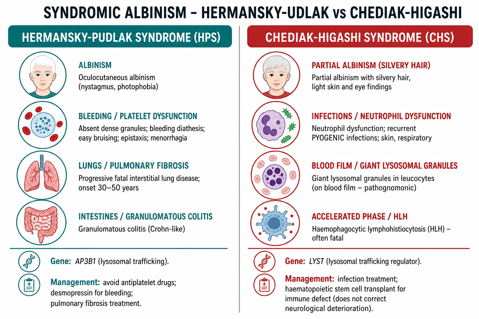
Clinically the albinism resembles OCA2 (moderate hypopigmentation with the full ocular tetrad). The bleeding history (easy bruising, epistaxis, menorrhagia, prolonged bleeding after dental work or surgery) reflects absent dense granules: the platelet count is normal, the bleeding time prolonged, and aggregation studies show absent second-wave aggregation with ADP and epinephrine. Pulmonary fibrosis is the life-limiting complication in HPS1 and HPS2: a progressive interstitial lung disease resembling idiopathic pulmonary fibrosis, typically presenting between 30 and 50 years of age with exertional dyspnoea and a restrictive defect. Granulomatous colitis (Crohn-like) and renal failure (glomerulonephritis) add further morbidity.[7]
Management combines the general albinism care above with: avoidance of antiplatelet drugs and NSAIDs; desmopressin, antifibrinolytics and platelet transfusion for bleeding; pirfenidone for HPS1 pulmonary fibrosis (slows decline); and lung transplantation for end-stage disease. Colitis is treated with corticosteroids and anti-TNF agents. [1]
Chédiak-Higashi syndrome (CHS)
CHS is rarer and more lethal. Mutations in LYST (lysosomal trafficking regulator, chromosome 1q42) produce abnormally large lysosomes and lysosome-related organelles in every cell: the result is partial oculocutaneous albinism with characteristic silvery metallic hair, recurrent pyogenic infections from defective neutrophil degranulation and chemotaxis, giant peroxidase-positive granules in leucocytes (pathognomonic on the blood film), mild bleeding from platelet dense-body deficiency, and a risk of the accelerated phase (HLH) that is often fatal in the first decade.[3]
The accelerated phase presents with fever, hepatosplenomegaly, lymphadenopathy, pancytopenia, hyperferritinaemia, hypertriglyceridaemia, hypofibrinogenaemia and haemophagocytosis on marrow. It is treated with the HLH-2004 protocol and curative haematopoietic stem cell transplantation, which corrects the immune defect and prevents the accelerated phase — but does not correct the albinism, and does not prevent a late-onset neurodegenerative syndrome (cerebellar ataxia, peripheral neuropathy, parkinsonism and cognitive decline) that emerges in survivors in the third and fourth decades.[3]
Ocular albinism type 1 (OA1)
OA1 (Nettleship-Falls) is X-linked recessive (GPR143, Xp22.2), affecting males with the full ocular tetrad while the skin and hair are usually normal or only minimally hypopigmented. Carrier females are clinically normal but show a characteristic mosaic retinal pigment pattern (irregular radially arranged pigmented and depigmented lacunae) on fundoscopy, reflecting X-inactivation lyonisation. The misrouting of optic fibres at the chiasm is as marked as in OCA. Management is the ocular and low-vision support above; photoprotection is still advisable but the skin-cancer risk is far lower than in OCA.[2]
Rarer non-syndromic and syndromic forms
The remaining non-syndromic OCAs are molecular diagnoses that resemble OCA2 clinically and are distinguished only by gene-panel sequencing: OCA5 (mapped to chromosome 4p15.2), OCA6 (SLC24A5, a calcium/potassium-dependent sodium-calcium exchanger also famous for its role in human skin-colour evolution), OCA7 (LRMDA / C10orf11) and OCA8 (DCT, dopachrome tautomerase). They are rare and confer no special management beyond the standard photoprotection and surveillance.[5]
Two rarer syndromic entries complete the differential. Griscelli syndrome (myosin Va, RAB27A or MLPH) shares silvery hair and immune dysregulation with CHS but shows normal melanosome number with clumped pigment rather than giant granules, and differentiates clinically by the absence of the leucocyte inclusions — RAB27A-positive type 2 carries the same HLH risk. Cross-McKusick-Breen syndrome (also called Elejalde or neuroectodermal melanolysosomal disease) combines albinism with profound psychomotor retardation and a silvery hair shaft, and is now recognised to overlap with the syndromic albinisms. The examiner point is that whenever silvery hair accompanies albinism, a blood film for giant granules (CHS) and a careful neurodevelopmental assessment are mandatory, because the prognosis and the transplant decision depend entirely on the underlying gene.[3]
Complications and Pitfalls
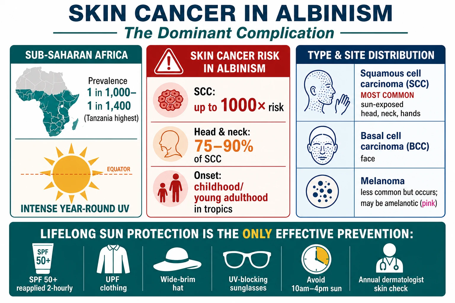
The complications divide into cutaneous, ocular, and psychosocial, with the syndromic forms adding their own lethal trajectories. [1]
Cutaneous. Skin cancer is the dominant complication of non-syndromic albinism and the leading cause of premature death where UV exposure is intense. Squamous cell carcinoma is the most common (three-quarters or more occur on the sun-exposed head, neck and hands), followed by basal cell carcinoma on the face and melanoma — which in albinism is frequently amelanotic (pink) and easily mistaken for a benign lesion. The lifetime risk is many hundreds-fold higher than in the general population.[6][9]
Ocular. The visual impairment is permanent (typically 6/18–6/60) and non-progressive after childhood, but it limits education, employment, driving and independence. Photophobia and strabismus reduce quality of life. [1]
Psychosocial. Albinism carries a heavy social burden everywhere, but it is most severe in parts of sub-Saharan Africa, where deeply rooted misconceptions associate albinism with magical properties and have driven attacks, mutilation and killings of people with albinism. Stigma, bullying, reduced marriage and employment prospects, and low self-esteem are near-universal and demand active psychosocial support.[6]
The common pitfalls examiners probe: [1]
- Missing a syndromic diagnosis. A bleeding history or recurrent infections in a "fair" child must trigger platelet studies (HPS) or a blood film (CHS). Treating only the albinism misses the lethal systemic disease.[3][7]
- Dismissing a pink lesion. Amelanotic melanoma is easily mistaken for a benign nodule in lightly pigmented skin — biopsy any non-healing, growing or bleeding lesion.
- Confusing OCA2 with normal pigmented skin in darker-skinned patients, who show more residual pigment; the skin-cancer risk is nonetheless very high.
- Over-operating on nystagmus. Surgery improves the head posture and widens the null point; it does not cure the nystagmus or the visual acuity.
- Neglecting vitamin D in patients practising strict photoprotection.
- Forgetting late neurodegeneration in CHS survivors after successful HSCT.[3]
Prognosis and Disposition
For non-syndromic OCA and OA1, life expectancy is normal provided skin cancer is prevented through lifelong photoprotection and surveillance. The visual impairment is permanent and stable; it does not worsen with age. Tropical or equatorial residence dramatically worsens the prognosis because of skin cancer, and patients in these environments need the most aggressive surveillance and support.[6][9]
For Hermansky-Pudlak syndrome, prognosis is governed by pulmonary fibrosis (the leading cause of death in HPS1), bleeding, and colitis; survival has improved with pirfenidone and transplantation but remains reduced.[7] For Chédiak-Higashi syndrome, prognosis is poor without HSCT — most untreated children die of infection or the accelerated phase in the first decade; even successfully transplanted survivors face late neurodegeneration in adulthood.[3]
Disposition is shared care: a dermatologist leads skin surveillance, an ophthalmologist/optometrist leads vision, a clinical geneticist counsels the family, and a haematologist/respiratory physician co-manages HPS and CHS. The primary care physician coordinates vitamin D monitoring, psychosocial support, school and workplace liaison, and ensures the sun-protection habit is sustained across a lifetime. [1]
Special Populations
Newborns and infants are usually diagnosed at birth on the cutaneous and ocular findings; photoprotection and ophthalmology begin immediately, with developmental surveillance for CHS. [1]
Children need educational accommodations — front-of-class seating, large-print materials, electronic magnification, and mobility training — alongside active management of bullying and self-esteem. Sun-safety habits built in childhood are the foundation of lifelong skin-cancer prevention. [1]
Pregnancy. OCA does not affect pregnancy itself. Affected parents or known carriers are offered genetic counselling: an autosomal recessive couple has a 25 percent recurrence risk; an OA1 carrier mother has a 50 percent chance each pregnancy of passing the X chromosome to a son (who would be affected) or a daughter (who would be a carrier like her). Prenatal diagnosis (CVS at 11–13 weeks or amniocentesis at 15–20 weeks) and preimplantation genetic testing (PGT-M) are available once the familial mutation is known.[5]
Immunocompromised context (CHS). Aggressive treatment of every infection, prophylactic antibiotics as guided by immunology, vigilance for the accelerated phase, and early referral for HSCT define management.[3]
Darker-skinned patients. OCA2 and OCA3 in African and Asian patients retain more residual pigment and may be labelled "mild," yet the skin-cancer risk remains very high and surveillance must not be relaxed.[6]
Evidence, Guidelines and Regional Differences
The classification and ocular phenotyping rest on several landmark reviews. Grønskov and colleagues (Orphanet Journal of Rare Diseases, 2007) set out the modern OCA classification by gene and chromosome that still anchors teaching.[5] Kruijt and colleagues (Ophthalmology, 2018) defined the phenotypic spectrum of albinism and standardised the grading of foveal hypoplasia that now underpins ophthalmic assessment.[4] Ather and colleagues (Human Brain Mapping, 2019) detailed the aberrant visual pathway development — the excess chiasmal crossing — that explains the VEP asymmetry and the stereoscopic deficit.[8] The syndromic forms are covered by De Jesus Rojas and Young (Seminars in Respiratory and Critical Care Medicine, 2020) for HPS, and by Talbert and colleagues (Current Opinion in Hematology, 2023) for CHS.[3][7]
The skin-cancer and photoprotection literature is dominated by the African experience. Nakkazi (Lancet, 2019) documented the devastating skin-cancer burden in people with albinism in Africa,[6] and Wright and Norval (Photochemistry and Photobiology, 2023) reviewed UV exposure and photoprotective strategies in South Africa.[9] Thawabteh and colleagues (Molecules, 2023) reviewed the biochemistry of skin pigmentation and its therapeutic targets.[1]
Across regions the disease is the same but the threats differ. In sub-Saharan Africa, community health-worker-led skin surveillance, distribution of sunscreen and protective clothing, and advocacy (Under the Same Sun, the Africa Albinism Network) are the cornerstones, because access to dermatology and surgery is the chief barrier and the UV burden is extreme. In the United Kingdom and Europe, specialist genetics, dermatology and low-vision services are generally accessible, and the focus is on coordinated multidisciplinary care following GeneReviews and national photoprotection guidance. In the United States, the American Academy of Dermatology sun-protection guidance and GeneReviews management framework apply. In India and much of South Asia, consanguinity and founder effects raise OCA prevalence; genetic counselling and access to sunscreen are the principal gaps.
Exam Pearls
ALBINISM
The examiner-rewarded one-liners: [1]
- Definition: albinism = inherited defect of melanin synthesis with normally numbered melanocytes; OCA affects skin+hair+eye, OA affects eye only.
- Genes: OCA1 = TYR (11q14); OCA2 = P gene (15q12, most common worldwide); OCA3 = TYRP1 (9p23); OCA4 = SLC45A2 (5p13); OA1 = GPR143 (Xp22, X-linked).
- OCA1A = tyrosinase-negative (most severe, no pigment); OCA2 = tyrosinase-positive (most common, some pigment).
- Ocular tetrad: nystagmus + foveal hypoplasia + iris transillumination + photophobia (plus strabismus, reduced acuity).
- Chiasmal misrouting: excess optic fibres cross at the chiasm → asymmetrical VEP — a sensitive objective confirmation.
- Piebaldism vs albinism: piebaldism is autosomal dominant, white forelock, patchy, normal eyes.
- HPS: albinism + platelet dense-granule deficiency (bleeding) + pulmonary fibrosis + colitis.
- CHS: partial albinism + neutrophil dysfunction + giant granules (blood film, pathognomonic) + HLH; gene LYST.
- Skin cancer: SCC is number one, head and neck; risk up to 1000-fold; sun protection (SPF 50+) is the intervention.
- All OCA autosomal recessive (25 percent recurrence); OA1 X-linked (males); genetic counselling and prenatal diagnosis once the familial mutation is known. [1]
Exam application bank (NEET-PG / INICET)
One-line answer
Albinism is a group of inherited disorders of melanin synthesis or melanosome biogenesis causing congenital hypopigmentation of skin, hair, and eyes (oculocutaneous albinism, OCA) or of the eyes alone (ocular albinism, OA1). The melanocytes are normal in NUMBER — the defect is in melanin PRODUCTION, which separates albinism from vitiligo (where melanocytes are lost). OCA1 (TYR, chr 11) is the severe tyrosinase-negative form; OCA2 (P gene, chr 15) is the most common worldwide and dominates in sub-Saharan Africa. All OCA types are autosomal recessive; OA1 (GPR143) is X-linked. Every type shares a characteristic OCULAR tetrad — nystagmus, foveal hypoplasia, iris transillumination, photophobia — plus abnormal chiasmal decussation. The dominant complication is SKIN CANCER (squamous cell carcinoma of the head and neck, up to 1000-fold risk), so lifelong sun protection (SPF 50+) and surveillanc
Worked stems (answer without another resource)
Stem 1 — Classic presentation. Map symptoms to mechanism; name the first investigation and first treatment step with dose/route if drug therapy is standard. [1]
Stem 2 — Unstable / complicated. List red flags that force immediate resuscitation, theatre, ICU, antidote, or reperfusion — and what you do in the first 15 minutes. [1]
Stem 3 — Atypical group. Elderly, pregnancy, child, or immunocompromised: how presentation and thresholds change. [1]
Stem 4 — Differential trap. Name the three closest mimics and one discriminator for each. [1]
Stem 5 — Disposition. Who goes home with safety-netting, who is admitted, who needs HDU/ICU/theatre, and what follow-up is mandatory. [1]
Rapid viva checklist
- Definition + classification
- Pathophysiology chain
- Bedside signs / criteria
- Score with exact components (if any)
- Emergency bundle
- Definitive therapy with doses
- Complications of disease and of treatment
- Special populations
- Guideline/trial name if classic
- Three exam traps
Coverage self-check
If you cannot answer any stem above from this page alone, re-read the matching section — the page is intended to be self-sufficient for final-prof and NEET-PG/INICET questions on Albinism.
Self-test: which gene, and why does it matter?
A Tanzanian child has yellow-brown hair, pigmented naevi, nystagmus and visual acuity of 6/36. The parents are unaffected. What is the most likely subtype, what is the gene and chromosome, and what single intervention most protects this child's life? [1]
Answer: This is OCA2 — the most common OCA subtype worldwide and the dominant type in sub-Saharan Africa, characterised by residual pigment, pigmented naevi and a moderate ocular phenotype. The gene is the OCA2 (P) gene on chromosome 15q12-q13, autosomal recessive. The single most life-protecting intervention is lifelong photoprotection (SPF 50+, UPF clothing, hat, sunglasses, sun avoidance) with annual dermatological skin surveillance, because SCC of the head and neck is the leading cause of premature death in this population.[6][9]
The companion eight single-best-answer questions, the short-answer question, and the cross-table viva for this topic are linked below and rehearse the high-yield discriminators: the gene–chromosome pairs, the ocular tetrad, the chiasmal-misrouting VEP, the HPS and CHS syndromic cores, and the skin-cancer rationale for lifelong photoprotection. [1]
References
- [1]Thawabteh AM, Jibreen A, Karaman D, et al. Skin Pigmentation Types, Causes and Treatment-A Review Molecules, 2023.PMID 37375394
- [2]Adam MP, Bick S, Mirzaa GM, et al. Oculocutaneous Albinism and Ocular Albinism Overview 1993.PMID 37053367
- [3]Talbert ML, Malicdan MCV, Introne WJ. Chediak-Higashi syndrome Curr Opin Hematol, 2023.PMID 37254856
- [4]Kruijt CC, de Wit GC, Bergen AA, et al. The Phenotypic Spectrum of Albinism Ophthalmology, 2018.PMID 30098354
- [5]Grønskov K, Ek J, Brondum-Nielsen K. Oculocutaneous albinism Orphanet J Rare Dis, 2007.PMID 17980020
- [6]Nakkazi E. People with albinism in Africa: contending with skin cancer Lancet, 2019.PMID 31423986
- [7]De Jesus Rojas W, Young LR. Hermansky-Pudlak Syndrome Semin Respir Crit Care Med, 2020.PMID 32279294
- [8]Ather S, Proudlock FA, Welton T, et al. Aberrant visual pathway development in albinism: From retina to cortex Hum Brain Mapp, 2019.PMID 30511784
- [9]Wright CY, Norval M. Solar Ultraviolet Radiation, Skin Cancer and Photoprotective Strategies in South Africa(†) Photochem Photobiol, 2023.PMID 35841370