Dermatology · General Medicine
Alopecia & Hair Disorders
Also known as Alopecia · Hair loss · Alopecia areata · Androgenetic alopecia · Male/female pattern baldness · Telogen effluvium · Cicatricial alopecia
Hair loss (alopecia) is the single most psychologically charged dermatological presentation. The pivotal skill is the scarring vs non-scarring distinction: non-scarring (reversible — androgenetic, alopecia areata, telogen effluvium, anagen effluvium, traction, trichotillomania, tinea capitis) preserves the follicle and is potentially recoverable, whereas scarring (cicatricial) (lichen planopilaris, frontal fibrosing alopecia, central centrifugal cicatricial alopecia, discoid lupus, folliculitis decalvans, pseudopelade) destroys follicular stem cells and is permanent and irreversible. Androgenetic alopecia (miniaturisation by dihydrotestosterone via 5-alpha-reductase type II) is graded by the Hamilton-Norwood scale in men and the Ludwig scale in women and treated with topical minoxidil 5% and oral finasteride 1 mg daily (men only). Alopecia areata (autoimmune T-cell attack on anagen follicles) presents as a smooth patch with exclamation-mark hairs and is now treated with JAK inhibitors (baricitinib, ritlecitinib) for severe disease — FDA-approved 2022-2023. Telogen effluvium is diffuse shedding two to three months after a trigger and is self-limiting.
On this page & tools
Your progress
Saved locally on this device.
Exam tags
Red flags
Overview & Definition
Hair loss (alopecia) is one of the most psychologically devastating dermatological presentations — the patient's fear of disfigurement frequently outweighs the clinical severity of the disease. The clinician's central skill rests on a single binary branch-point, made at the bedside before any test: is the hair loss scarring or non-scarring?[1]
- Non-scarring alopecia preserves the hair follicle. The follicle is structurally intact, the follicular ostia (openings) remain visible, and — if the underlying cause is removed or treated — hair can regrow. Members: androgenetic alopecia, alopecia areata, telogen effluvium, anagen effluvium, traction alopecia, trichotillomania, tinea capitis, loose anagen syndrome.
- Scarring (cicatricial) alopecia destroys the follicle. Follicular stem cells at the bulge region and the sebaceous gland are obliterated and replaced by fibrosis, the follicular openings vanish, the scalp becomes smooth and shiny, and the loss is permanent and irreversible. Members: lichen planopilaris (LPP), frontal fibrosing alopecia (FFA), central centrifugal cicatricial alopecia (CCCA), discoid lupus erythematosus (DLE), folliculitis decalvans, dissecting cellulitis, pseudopelade of Brocq. [1]
The clinical implication is direct: non-scarring disease can be reversed; scarring disease can only be halted. Every day of untreated active scarring inflammation destroys more follicles that will never return — hence the dictum "biopsy early in any scarring-looking alopecia".[1][7]
After the binary split, the second task is pattern recognition: which non-scarring alopecia is this (androgenetic vs areata vs effluvium)? And which scarring alopecia (lymphocytic vs neutrophilic)? The answers dictate therapy. Two therapeutic revolutions define modern practice: 5-alpha-reductase inhibitors and topical minoxidil for androgenetic alopecia, and oral JAK inhibitors (baricitinib, ritlecitinib) — FDA-approved 2022-2023 — for severe alopecia areata.[3][5]
Classification
Alopecia is classified along two axes: scarring vs non-scarring (the bedside branch-point), and — within the scarring group — the dominant inflammatory infiltrate on biopsy (lymphocytic, neutrophilic, or mixed), which determines therapy.[1][7]
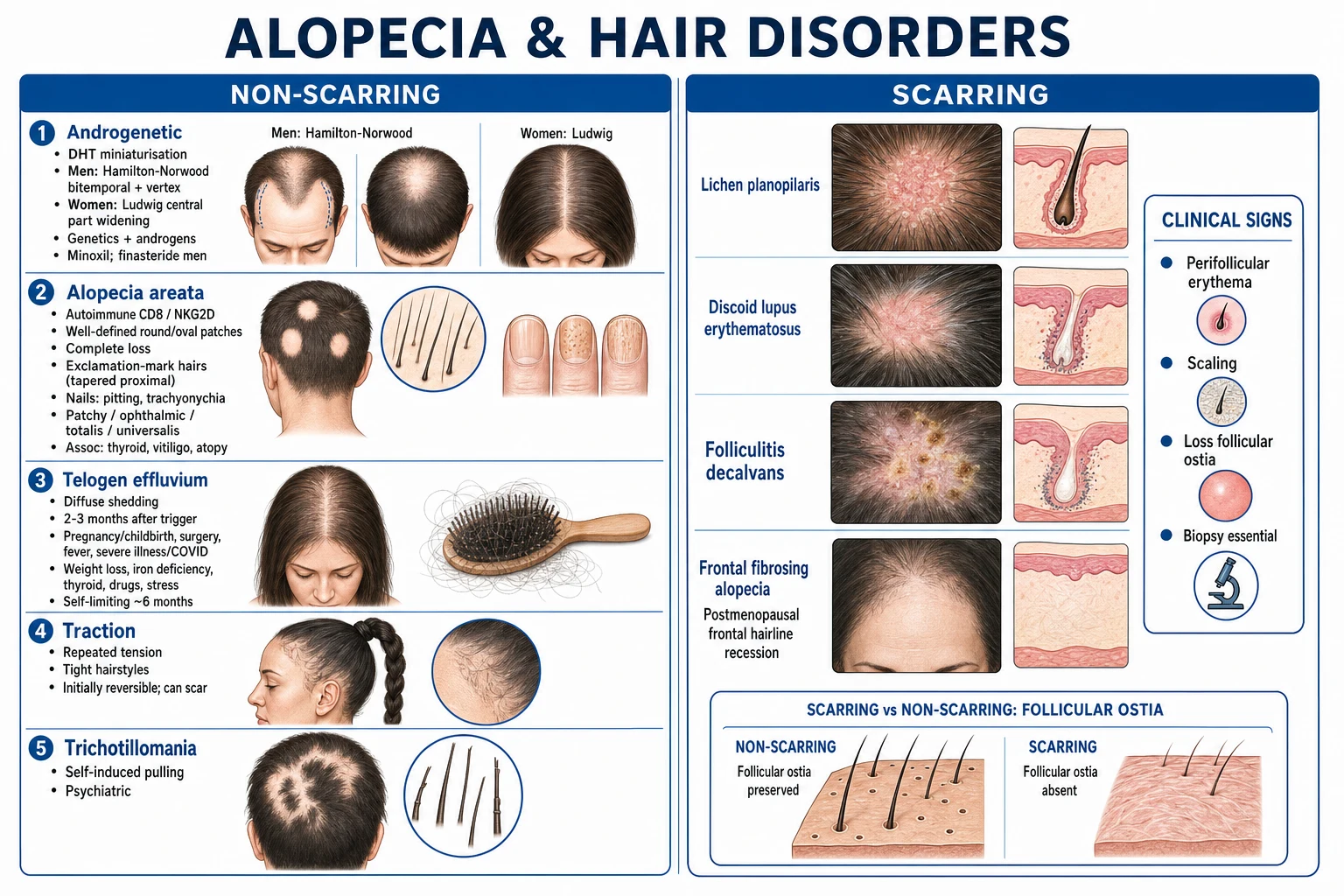
A second, mechanistic classification is by which phase of the hair cycle is disturbed: [1]
- Anagen disorders — androgenetic (anagen progressively shortens → miniaturisation), alopecia areata (T cells attack anagen follicles), anagen effluvium (chemotherapy poisons the anagen matrix).
- Catagen/telogen shift — telogen effluvium (a trigger throws follicles prematurely into telogen). [1]
Epidemiology & Risk Factors
Androgenetic alopecia (AGA) is the commonest cause of hair loss in both sexes. Approximately 50% of Caucasian men show some degree by age 50, rising to 80% by age 70; in women the prevalence climbs more slowly — roughly 50% of women by age 80 — and presents with diffuse central thinning rather than receding hairline. Prevalence is lower in Asian and African populations. Risk factors are polygenic family history (paternal and maternal), androgen exposure, age, smoking, insulin resistance and metabolic syndrome, and coronary risk. The androgen receptor gene lies on the X chromosome, which explains the strong maternal-line influence but not the whole picture; AGA is genuinely polygenic.[1][11]
Alopecia areata (AA) has a lifetime prevalence of approximately 2% — i.e. roughly 1 in 50 people will experience at least one episode. Peak incidence is in childhood and young adulthood (most cases begin before age 40). Sex incidence is roughly equal. Severe variants (totalis, universalis, ophiasis) account for fewer than 10% of cases but carry the heaviest burden. AA is strongly associated with other autoimmune diseases: autoimmune thyroid disease (Hashimoto, Graves) — screen TSH in every patient — vitiligo, atopy/eczema/asthma, psoriasis, inflammatory bowel disease, and Down syndrome (in which AA is more severe and refractory).[12]
Telogen effluvium (TE) is the commonest cause of diffuse hair loss in adult women. It follows any physiological stressor two to three months earlier: pregnancy (post-partum is the classic), severe illness, surgery, febrile illness (including dengue, malaria, COVID-19), crash dieting or rapid weight loss, iron deficiency, thyroid dysfunction (hypo- or hyper-), severe emotional stress, and drugs (retinoids, anticoagulants, anticonvulsants, beta-blockers, ACE inhibitors).[8]
Anagen effluvium is overwhelmingly iatrogenic — cytotoxic chemotherapy (doxorubicin, cyclophosphamide, paclitaxel, etoposide; also radiation). It is dose-dependent and affects virtually all patients on combination regimens. [1]
Scarring alopecias segregate by demographic: LPP and FFA in postmenopausal white women (FFA mean age early 60s); CCCA in Black women (related to traction, heat, chemical relaxers, and likely a genetic susceptibility); folliculitis decalvans and dissecting cellulitis in young adult Black men (follicular occlusion spectrum); DLE in young-middle-aged adults with a female preponderance, particularly in sun-exposed phototypes.[6][9][10]
Pathophysiology
The normal hair cycle
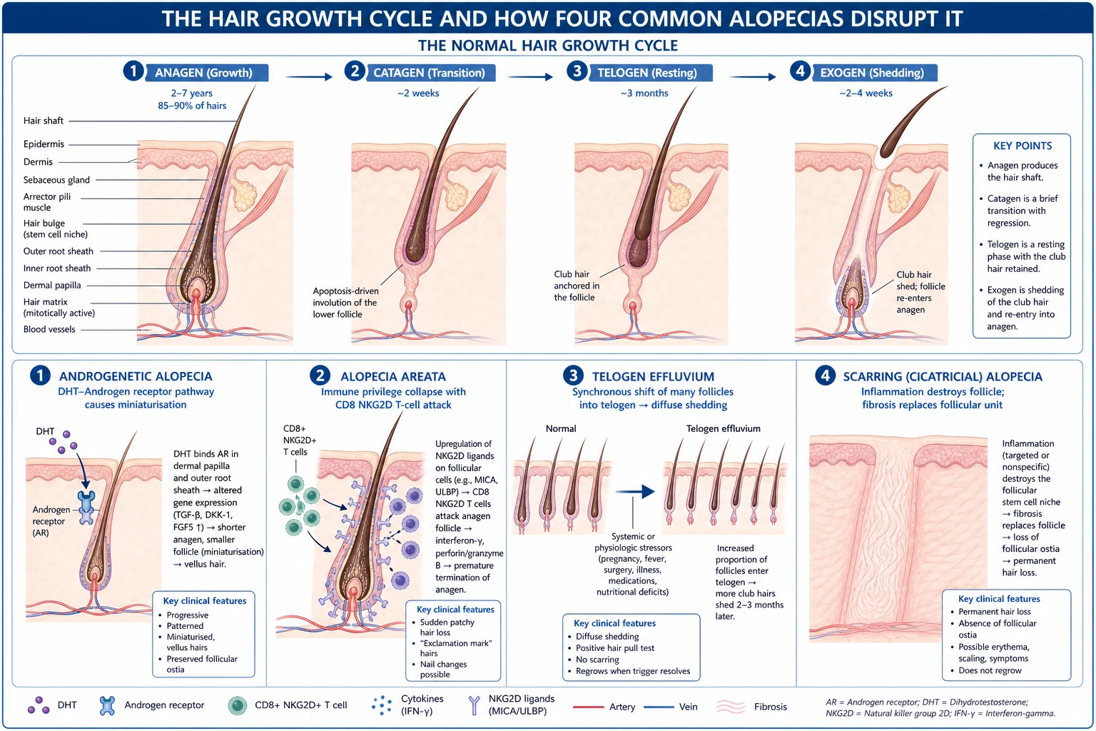
Every follicle cycles continuously: anagen (active growth, 2-7 years, 85-90% of scalp hairs) → catagen (regression, 2 weeks) → telogen (resting, about 3 months, 10-15% of hairs) → exogen (shedding). Normal daily loss is 50-100 hairs. Each alopecia disturbs this cycle in a characteristic way.[1]
Androgenetic alopecia — DHT and miniaturisation
Testosterone is converted to dihydrotestosterone (DHT) by 5-alpha-reductase type II within the dermal papilla. DHT binds the intracellular androgen receptor, the complex translocates to the nucleus, and the net effect on genetically susceptible scalp follicles is a shortening of anagen and a progressive decrease in hair-shaft diameter — the miniaturisation that converts thick terminal hairs into fine vellus hairs. Follicles over the bitemporal and vertex scalp are most androgen-sensitive, while the occipital scalp is androgen-independent — which is why the occiput is the donor site for hair transplantation.[1][11]
This immediately explains the two pharmacological anchors of AGA therapy: finasteride 1 mg daily inhibits 5-alpha-reductase type II (lowering scalp and serum DHT), and topical minoxidil prolongs anagen and increases follicle size (mechanism via sulphotransferase-converted minoxidil sulphate opening ATP-sensitive potassium channels). Both work only while taken; stopping reverses gains within months. [1]
Alopecia areata — collapse of immune privilege
The anagen hair follicle normally enjoys immune privilege: the hair matrix expresses low MHC class I/II and is shielded from autoimmune attack. In AA, immune privilege collapses — follicles upregulate MHC and present autoantigens (notably melanocyte-derived peptides, which is why AA targets pigmented hairs and "sparing" of grey hairs is sometimes seen). Autoreactive CD8+ NKG2D+ T cells infiltrate the peribulbar region and attack the anagen follicle. The follicle is shocked into catagen but not destroyed, which is why regrowth is always possible — and why JAK inhibitors, which block the cytokine signalling (IFN-γ/IL-15 axis) driving these T cells, work so dramatically.[3][12]
Telogen effluvium — premature catagen entry
A systemic stressor (fever, surgery, blood loss, crash diet, iron deficiency, thyroid disease, drugs) shifts an abnormally large cohort of follicles simultaneously from anagen into telogen. Two to three months later — the duration of telogen — these follicles synchronously shed. The mechanism is therefore one of synchronisation of what is normally a staggered, asynchronous cycle.[8]
Anagen effluvium — matrix cell poisoning
Cytotoxic chemotherapy arrests the rapidly dividing matrix cells at the base of the anagen follicle. The hair shaft narrows to a fragile point (Pohl-Pinkus constriction) and fractures within days to two weeks of the dose — hence the rapid, near-total loss seen on combination chemotherapy. [1]
Scarring alopecia — stem-cell destruction and fibrosis
The shared end-point of all scarring alopecias is destruction of follicular stem cells (which reside at the bulge region of the outer root sheath, where the arrector pili muscle inserts) and the sebaceous gland, replaced by fibrosis. Because the stem cells are gone, the follicle cannot regenerate — hence permanent loss. The inciting inflammation differs by subtype (lymphocytic in LPP/FFA/CCCA/DLE; neutrophilic in folliculitis decalvans) but the outcome is shared.[7]
Clinical Presentation
Androgenetic alopecia
Men — the Hamilton-Norwood scale. Male AGA follows the Hamilton-Norwood classification (types I-VII). The earliest definite change is type II (small bitemporal recession). Type III is the clinical threshold of baldness; type III vertex adds a balding vertex. Types IV-V show progressive recession anteriorly and a growing vertex bald spot separated by a band of hair. Type VI merges the two areas, leaving only a band-and-a-half of hair across the sides and back. Type VII is the most severe — a narrow horseshoe of hair remains over the occiput and temporal scalp. The occipital donor area is preserved in every type.[1]
Women — the Ludwig (Savin) scale. Female AGA follows the Ludwig classification (grades I-III). There is diffuse thinning over the crown with widening of the central hair part, preservation of the frontal hairline, and a characteristic "Christmas-tree" pattern of anterior hairline tapering. Grade I is mild (visible widening of the part), grade II moderate (increased thinning over the crown), grade III severe (near-complete thinning of the crown). Recession of the frontal hairline does NOT occur in classical Ludwig — if a woman has a receding hairline, suspect frontal fibrosing alopecia, traction, or virilisation.[1][6]
[1]Alopecia areata
AA presents as one or more well-circumscribed, perfectly smooth, completely hairless patches on the scalp (or beard, eyebrows, eyelashes, body). The skin within the patch is normal in colour, with preserved follicular ostia (non-scarring). The pathognomonic finding at the advancing margin is the exclamation-mark (cadaverised) hair: a hair broken 3-4 mm above the scalp with a tapered proximal end and a broader distal end, easily plucked. Nail involvement is common (pitting, ridging, trachyonychia — "twenty-nail dystrophy" — and onycholysis).[12]
Severity spectrum. Most patients have patchy AA (one or a few coins of loss). The ophiasis pattern is a band-like band of loss around the occipital and temporal margins — prognostically worse and more refractory. Alopecia totalis is loss of the entire scalp; alopecia universalis is loss of all body hair. The rare Marie Antoinette (sudden whitening) phenomenon — overnight greying — reflects selective loss of pigmented anagen hairs with sparing of grey ones.[12]
Telogen effluvium
TE presents as diffuse shedding over the whole scalp, beginning two to three months after a clearly identifiable trigger (post-partum, febrile illness, surgery, crash diet, blood loss, thyroid dysfunction, drug). The patient describes hair "coming out in handfuls" — in the shower, on the pillow, in the brush — but there are no patches and the hairline is preserved. The pull test is positive in active shedding. The frontotemporal recession of female AGA is absent.[8]
Anagen effluvium
Sudden, rapid loss of large amounts of hair within one to two weeks of starting chemotherapy (or radiation), producing anagen bulbs (no telogen club) at the shed end. May progress to near-complete baldness; recovers one to three months after cessation of the offending agent. [1]
Traction and trichotillomania
Traction alopecia produces frontotemporal and vertex-margin hair loss along the line of tension of tight braids, ponytails, buns, extensions, weaves, hair rollers. There may be perifollicular erythema and scaling along the hairline ("traction signs"). Early traction is reversible; chronic traction produces scarring that is permanent. [1]
Trichotillomania (an obsessive-compulsive-spectrum disorder) produces bizarre, irregular patches of hair loss with hairs broken at varying lengths on an otherwise normal scalp — the patient pulls, twists, or rubs the hair. Perifollicular pitting and haemorrhage may be seen. There is often a coexistent psychiatric comorbidity (anxiety, depression, OCD). [1]
Tinea capitis
In children, the commonest cause of patchy hair loss. Black-dot tinea (broken hair shafts flush with the scalp), grey-patch scaling, kerion (a boggy, tender, pustular, inflammatory mass — a hypersensitivity reaction to the dermatophyte, NOT a bacterial abscess and NOT treated with antibiotics alone), and cervical lymphadenopathy. KOH-positive; culture-positive.[1]
Scarring alopecias — the unifying clinical signs
Across all scarring alopecias, look for the trio of (1) perifollicular erythema and hyperkeratosis (the activity marker), (2) loss of follicular ostia (smooth, shiny scalp devoid of follicular openings — the irreversibility marker), and (3) patches of complete baldness. Subtype-specific features:[6][7]
- Lichen planopilaris (LPP) — perifollicular violaceous erythema and perifollicular hyperkeratosis at the periphery of central patchy scarring on the vertex scalp; mucosal/skin lichen planus may coexist.
- Frontal fibrosing alopecia (FFA) — a variant of LPP in postmenopausal women: slowly progressive frontotemporal hairline recession (often several centimetres), perifollicular erythema, loss of eyebrows (and body hair), and — strongly linked — use of facial sunscreens/moisturisers on the face/forehead.
- Central centrifugal cicatricial alopecia (CCCA) — Black women, centrifugal scarring spreading outward from the vertex, attributed to traction, heat, chemical relaxers and likely a genetic susceptibility (PADI3 mutation described).
- Discoid lupus (DLE) — well-demarcated erythematous plaques with follicular plugging, adherent scale ("carpet-tack" sign) and scarring, often in sun-exposed scalp; systemic lupus in a minority.
- Folliculitis decalvans — recurrent pustular folliculitis with tufted folliculitis (multiple hairs emerging from a single ostium), crusting and scarring; Staphylococcus aureus commonly cultured.
- Pseudopelade of Brocq — burnt-out, inactive scarring — small, flesh-coloured, atrophic patches likened to "footprints in the snow", with no active inflammation. [1]
Differential Diagnosis
The differential diagnosis depends on the pattern of loss.[1]
[1]The single most important distinction is scarring from non-scarring. If follicular ostia are lost and the scalp is smooth and shiny, treat as scarring and biopsy.[7]
Clinical & Bedside Assessment
Diagnosis is overwhelmingly clinical, supported by trichoscopy. The bedside examination establishes the scarring/non-scarring split, the pattern, and the activity of shedding.[1]
History. Take a meticulous history of: onset and rate (sudden TE vs gradual AGA), pattern, recent triggers in the last three months (illness, surgery, pregnancy, weight loss, drugs, stress), hairstyle practices (traction, relaxers, heat), family history of baldness, autoimmune and thyroid history, drug history (chemotherapy, retinoids, anticoagulants, beta-blockers), and the psychological impact (use a DLQI or HADS). [1]
Three bedside tests: [1]
[1]Trichoscopy (dermoscopy of the scalp) is now first-line and often diagnostic:[1]
- Androgenetic — hair diameter diversity >20% (a key diagnostic threshold), more than four yellow dots per cm², empty follicles, and a predominance of thin (vellus) hairs.
- Alopecia areata — yellow dots (dilated empty follicles infundibula), black dots (cadaverised hairs), exclamation-mark hairs, dystrophic hairs, and short regrowing vellus hairs.
- Telogen effluvium — upright regrowing hairs, empty follicles, but uniform hair diameter (no diversity) — the key contrast with AGA.
- Tinea capitis — comma hairs, corkscrew hairs, broken hairs, black dots (M-shaped comma is pathognomonic).
- Scarring alopecia — loss of follicular openings, perifollicular scaling and erythema, white structureless areas of fibrosis, and tufted folliculitis (multiple hairs from one ostium) in the neutrophilic types. [1]
Investigations
Most non-scarring alopecia needs no investigation beyond clinical + trichoscopy. Tests are reserved for diffuse / uncertain / scarring presentations.[1]
Blood tests for diffuse hair loss (especially in women and TE): [1]
- Ferritin — target at least 70 ng/mL for optimal hair cycling; iron-deficiency is a reversible driver of TE.
- TSH (± free T4) — both hypo- and hyperthyroidism cause TE.
- Vitamin D (25-OH), zinc, vitamin B12 / folate.
- CBC, CRP — anaemia of chronic disease, inflammation.
- In women with AGA + hirsutism / acne / oligomenorrhoea — total and free testosterone, DHEAS, LH, FSH, prolactin, 17-OH-progesterone to exclude PCOS or other hyperandrogenism; consider pelvic ultrasound.
- ANA if lupus (DLE) is suspected. [1]
Scalp biopsy (4 mm punch, ideally two samples from the active edge) is mandatory for any scarring alopecia, any uncertain diagnosis, and any non-response to therapy. Horizontal sectioning allows follicular counts, the anagen:telogen ratio (normal at least 8:1), and assessment of miniaturisation — the most informative single investigation for diffuse disease.[7]
KOH preparation and fungal culture is indicated for any child with patchy hair loss, black-dot, scaling, or kerion. [1]
The SALT (Severity of Alopecia Tool) score — the percentage scalp hair loss, graded 0-100 — is the standard outcome measure in AA clinical trials (SALT of 50 = 50% loss; the SALT at most 20 response is the conventional treatment target in JAK inhibitor trials).[3][4]
Management — Resuscitation
Alopecia is rarely a physiological emergency, but several situations demand prompt action:[1]
Kerion in a child. A boggy, pustular, tender inflammatory tinea mass is not a bacterial abscess. Do not treat with antibiotics alone, and do not incise and drain. Treat with oral antifungal (griseofulvin or terbinafine) plus a short course of systemic corticosteroid (prednisolone 1-2 mg/kg/day for 1-2 weeks) to suppress the hypersensitivity inflammation. Mismanagement produces scarring. [1]
Active scarring alopecia. Every day of untreated inflammation destroys more follicles permanently. Start high-potency topical or intralesional corticosteroid immediately and arrange an urgent biopsy and dermatology referral. Do not wait weeks for clinic. [1]
Severe psychological distress or suicidal ideation (particularly in young women with severe AA) — acknowledge the impact, screen with PHQ-9, offer psychological support, and expedite systemic therapy (JAK inhibitor pathway).[5]
Uncovered systemic disease in TE (severe iron-deficiency anaemia, hypothyroidism) — treat the cause. [1]
Management — Definitive & Stepwise
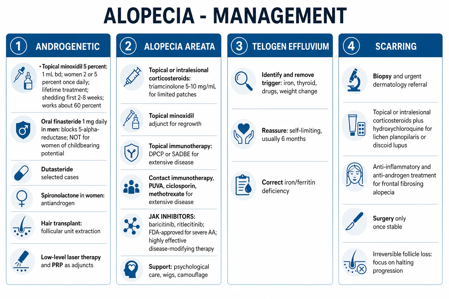
Therapy is entirely subtype-specific. There is no role for systemic corticosteroids as long-term therapy in most alopecias (chronic side-effects; relapse on taper).[1]
Androgenetic alopecia — men
First-line bundle for male androgenetic alopecia
Androgenetic alopecia — women
Minoxidil 2% solution (1 mL BD) or 5% foam once daily is first-line. Finasteride is CONTRAINDICATED in women of childbearing potential (teratogenic — causes hypospadias and feminisation of the male fetus). In postmenopausal women finasteride 1-1.25 mg daily may be used off-label. Spironolactone 100-200 mg daily (antiandrogen) is the oral agent of choice for premenopausal women with female pattern loss and signs of hyperandrogenism, always with reliable contraception (teratogenic). Combined oral contraceptive pill is added for the same. Always rule out PCOS if hirsutism, acne, or oligomenorrhoea coexist.[1][11]
Alopecia areata — stepwise
AA-LADDER — the stepwise treatment of alopecia areata
Intralesional triamcinolone — 5-10 mg/mL into patches on the scalp (lower concentration 2.5-5 mg/mL for the face, eyebrows, beard); maximum 20 mg per session and 40 mg per month; repeat every 4-6 weeks. Avoid skin atrophy by superficial injection.[1]
Contact immunotherapy — DPCP (diphenylcyclopropenone) or SADBE (squaric acid dibutylester) applied weekly to induce a mild allergic contact dermatitis. Mechanism: diversion of the immune response. Useful for extensive patchy AA; slow (response at 6-12 months); not suitable for totalis/universalis. [1]
JAK inhibitors (the modern revolution in severe AA):[3][4][5]
Telogen effluvium
Identify and remove the trigger. Correct iron deficiency (oral ferrous sulphate 200 mg BD until ferritin is at least 70 ng/mL), treat hypothyroidism (levothyroxine), review drugs. Reassure — TE is self-limiting, regrows within six months of removing the trigger. Chronic telogen effluvium lasts more than six months without an identified trigger (middle-aged women, described by Whiting); treatment is supportive plus a trial of minoxidil.[8]
Anagen effluvium
Scalp cooling (cold cap) during chemotherapy reduces the cytotoxic effect on the follicle by vasoconstriction; begun before and continued through and after infusion. Reassure — recovers within one to three months of stopping chemotherapy. Provide wig/prosthesis and psychological support.[1]
Traction and trichotillomania
Traction — stop the offending hairstyle immediately; topical minoxidil to stimulate regrowth; intralesional corticosteroid for any active perifollicular inflammation; for established scarring, hair transplant is the only option once disease is burnt-out. Trichotillomania — cognitive-behavioural therapy (habit-reversal training) is first-line; SSRI (fluoxetine, sertraline) or clomipramine for the underlying OCD-spectrum disorder; topical corticosteroid for any follicular inflammation.[1]
Tinea capitis — ALWAYS oral
Griseofulvin 20-25 mg/kg/day for 6-8 weeks is the gold standard (best evidence for Microsporum). Terbinafine (weight-based dosing) is preferred for Trichophyton species and is now first-line in many guidelines. Adjunctive ketoconazole 2% or selenium sulphide 2.5% shampoo twice weekly reduces shedding and contagion. Kerion adds a short course of systemic corticosteroid. Treat household contacts.[1]
Scarring alopecias — the goal is to HALT, not reverse
Therapy aims to suppress active inflammation and prevent further scarring — lost follicles will not regrow.[6][7][9][10]
Subtype-specific management of scarring alopecia
Escalation triggers
- AGA: no response to minoxidil at six months → add finasteride (men); no response to combination at 12 months → consider hair transplant.
- AA: failure of intralesional steroid in patchy disease → contact immunotherapy → JAK inhibitor for moderate-to-severe (SALT at least 50).
- Scarring: active inflammation despite topical steroid at three months → add hydroxychloroquine/doxycycline → systemic immunosuppressant.
- Folliculitis decalvans: recurrent pustular flares despite doxycycline → rifampicin + clindamycin combination. [1]
Specific Subtypes & Scenarios
Ophiasis AA. Band-like occipitotemporal loss is more refractory — start systemic therapy (JAK inhibitor) earlier; topical and intralesional alone usually fail.[12]
Paediatric alopecia. Tinea capitis is the commonest cause of patchy hair loss in children (always oral therapy; rule out kerion). AA is second. Traction (tight braids) and trichotillomania are common in school-age children. Loose anagen syndrome (anagen hairs that pull out painlessly with no bulb — in young blonde children) is benign and self-resolves. Aplasia cutis congenita (a congenital vertex defect) is a structural mimic. Minoxidil is generally avoided in prepubertal children.[1]
Pregnancy and post-partum. Post-partum telogen effluvium is classic — diffuse shedding two to three months after delivery, resolving spontaneously within six months. Avoid finasteride, dutasteride, and systemic retinoids in pregnancy and breastfeeding. Topical minoxidil is not recommended in pregnancy. Topical and intralesional corticosteroids are safe.[1]
Chemotherapy-induced anagen effluvium. Scalp cooling pre-, intra-, and post-infusion. Reassure: regrows within one to three months, sometimes with a change in texture/colour ("chemo curl").[1]
Frontal fibrosing alopecia. Treat early — once the hairline has receded it will not return. Stop facial sunscreens/moisturisers; 5-alpha-reductase inhibitor + intralesional corticosteroid.[6]
CCCA in Black women. Stop traction, heat, and chemical relaxers. A trial of a 5-alpha-reductase inhibitor is increasingly supported.[9]
Complications & Pitfalls
Psychological complications. Hair loss carries a substantial psychological burden across all types — depression, anxiety, low self-esteem, social withdrawal, and, in severe AA, suicidal ideation. Always screen and offer psychological support. DLQI scores in AA parallel those of severe psoriasis.[5][12]
Permanent irreversible loss — the dominant complication of scarring alopecia; also of late-stage traction and late-diagnosed CCCA. Squamous cell carcinoma may arise in chronic discoid lupus scars (Marjolin-type) — biopsy any non-healing ulcer.[7]
Adverse effects of minoxidil. Initial shedding in the first 2-8 weeks (reassure; do not stop), facial hypertrichosis (especially women on 5%), contact dermatitis (especially the propylene glycol vehicle — switch to foam), palpitations; systemic absorption is low. Not recommended in pregnancy.[2]
Adverse effects of finasteride. Decreased libido, erectile dysfunction, reduced ejaculate volume, gynaecomastia, and a post-finasteride syndrome of persistent sexual and mood symptoms after cessation (rare, debated). PSA falls by approximately 50% — adjust the prostate-cancer threshold (multiply measured PSA by 2). Strictly contraindicated in pregnancy (male fetus).[11]
Adverse effects of JAK inhibitors. Herpes zoster reactivation (consider Shingrix before starting), serious infection (TB screen), venous thromboembolism, major adverse cardiovascular events, malignancy (long-term), cytopenia, lipid changes. The FDA class warning cautions use in patients over 50 with at least one cardiovascular risk factor. Counsel women of childbearing potential.[3][5]
Diagnostic and therapeutic pitfalls. [1]
- Mislabelling tinea capitis as AA — a child with scaling, black-dot hairs, or lymphadenopathy needs KOH/culture and oral antifungal, not steroid.
- Missing scarring — any smooth, shiny, ostia-free patch must be biopsied; treating scarring as non-scarring loses follicles irreversibly.
- Forgetting iron/thyroid in TE — the reversible causes.
- Giving finasteride to a pregnant or possibly pregnant woman — catastrophic teratogenicity.
- Under-treating scarring until irreversible — early, aggressive anti-inflammatory therapy is the only way to halt progression.
- Using long-term systemic corticosteroid for AA — works short-term, relapses on taper, and accumulates steroid toxicity; JAK inhibitors have replaced chronic prednisolone.[5]
- Treating kerion with antibiotics alone — the boggy mass is a hypersensitivity reaction to a dermatophyte, not a bacterial abscess.
Prognosis & Disposition
Androgenetic alopecia is chronic and progressive without treatment; treatment stabilises the loss and partially regrows hair, but relapses within months of stopping. Lifelong therapy is the expectation. Hair transplant is permanent for the transplanted follicles (occipital donor is androgen-independent), but does not stop loss of non-transplanted hair — medical therapy continues.[1]
Alopecia areata has an unpredictable course. Patchy AA in an adult with no atopy, no nail involvement, short duration has a high rate of spontaneous regrowth within one year (often full). Prognostically worse features: ophiasis pattern, totalis/universalis, childhood onset, atopy, nail involvement, long duration (more than one year), and a positive family history. JAK inhibitors produce robust regrowth but relapse occurs in approximately 60% within months of stopping.[3][12]
Telogen effluvium is self-limiting within six months of removing the trigger. Chronic TE lasts more than six months; treat any underlying cause and consider a minoxidil trial.[8]
Anagen effluvium recovers within one to three months of stopping the offending drug.[1]
Scarring alopecia is permanent. The realistic goal is to halt progression; regrowth of follicles is impossible. Burnt-out, stable disease (more than two years inactive) may be amenable to hair transplant for cosmesis.[7]
Disposition. Most AGA and TE are managed in primary care. Refer to dermatology for any scarring alopecia, severe/refractory AA (for JAK inhibitor pathway), diagnostic uncertainty, and consideration of hair transplant.[1]
Special Populations
Children. Weight-based dosing for all systemic therapy. Oral antifungal is mandatory for tinea capitis (topical alone is inadequate). Minoxidil is generally avoided in prepubertal children. Intralesional corticosteroid is safe for AA in small doses. Address the psychological impact in school-age children (trichotillomania, visible AA).[1]
Pregnancy and breastfeeding. AVOID finasteride, dutasteride, and systemic retinoids (teratogenic). Topical minoxidil is not recommended. Topical and intralesional corticosteroids are safe. Post-partum TE is common and self-limiting — reassure.[1]
Menopause. FFA and female pattern loss predominate. 5-alpha-reductase inhibitors (finasteride, dutasteride) are now widely used in postmenopausal FFA and CCCA. Consider bone density with long-term systemic corticosteroid.[6]
Immunosuppressed (HIV, transplant, chemotherapy). Rule out infection (tinea, atypical mycobacteria, syphilis) — biopsy early. JAK inhibitors relatively contraindicated in significant immunosuppression.[1]
Black patients. CCCA, traction alopecia, and acne keloidalis nuchae predominate; trichoscopy of curved/curled hair has pitfalls (false "black dots"). Take a culturally sensitive history of hair practices (braids, weaves, extensions, relaxers, heat).[9]
Evidence, Guidelines & Regional Differences
JAK inhibitors — the modern landmark. The BRAVE-AA1 and BRAVE-AA2 phase 3 trials (King et al., NEJM 2022) established baricitinib as the first FDA-approved systemic therapy for severe AA: roughly 35-40% of patients on baricitinib 4 mg reached a SALT score of 20 or less at 36 weeks (versus 3-5% on placebo).[3] The ALLEGRO phase 2b/3 trial (King et al., Lancet 2023) showed ritlecitinib 50 mg achieved a SALT score of 20 or less in around 23-31% at 24 weeks and is the first JAK inhibitor approved for adolescents (aged 12 and over).[4] A 2023 JAAD review (King) summarises the JAK inhibitor class for AA.[5]
AGA. Cochrane and comprehensive reviews confirm modest efficacy of finasteride and minoxidil, with combination superior to either alone; dutasteride is more potent than finasteride.[2][11]
Scarring alopecia. The EADV 2026 task force (Seyed Jafari et al.) published a stepwise consensus for LPP management. FFA and CCCA reviews support 5-alpha-reductase inhibitors.[6][7][9]
Folliculitis decalvans. The Miguel-Gómez multicentre series (JAAD 2018) underpins the rifampicin + clindamycin regimen.[10]
Telogen effluvium. Whiting's classic descriptions of chronic TE in middle-aged women remain definitive.[8]
Exam Pearls
[1]THE-HAIR — quick pattern diagnosis
BOLD — the four first-line drugs
Red Flags
Exam application bank (NEET-PG / INICET)
One-line answer
Hair loss (alopecia) is the single most psychologically charged dermatological presentation. The pivotal skill is the scarring vs non-scarring distinction: non-scarring (reversible — androgenetic, alopecia areata, telogen effluvium, anagen effluvium, traction, trichotillomania, tinea capitis) preserves the follicle and is potentially recoverable, whereas scarring (cicatricial) (lichen planopilaris, frontal fibrosing alopecia, central centrifugal cicatricial alopecia, discoid lupus, folliculitis decalvans, pseudopelade) destroys follicular stem cells and is permanent and irreversible. Androgenetic alopecia (miniaturisation by dihydrotestosterone via 5-alpha-reductase type II) is graded by the Hamilton-Norwood scale in men and the Ludwig scale in women and treated with topical minoxidil 5% and oral finasteride 1 mg daily (men only). Alopecia areata (autoimmune T-cell attack on anagen folli [1]
Worked stems (answer without another resource)
Stem 1 — Classic presentation. Map symptoms to mechanism; name the first investigation and first treatment step with dose/route if drug therapy is standard. [1]
Stem 2 — Unstable / complicated. List red flags that force immediate resuscitation, theatre, ICU, antidote, or reperfusion — and what you do in the first 15 minutes. [1]
Stem 3 — Atypical group. Elderly, pregnancy, child, or immunocompromised: how presentation and thresholds change. [1]
Stem 4 — Differential trap. Name the three closest mimics and one discriminator for each. [1]
Stem 5 — Disposition. Who goes home with safety-netting, who is admitted, who needs HDU/ICU/theatre, and what follow-up is mandatory. [1]
Rapid viva checklist
- Definition + classification
- Pathophysiology chain
- Bedside signs / criteria
- Score with exact components (if any)
- Emergency bundle
- Definitive therapy with doses
- Complications of disease and of treatment
- Special populations
- Guideline/trial name if classic
- Three exam traps
Coverage self-check
If you cannot answer any stem above from this page alone, re-read the matching section — the page is intended to be self-sufficient for final-prof and NEET-PG/INICET questions on Alopecia & Hair Disorders.
References
- [1]Dakkak M, Forde KM, Lavery H. Hair Loss: Diagnosis and Treatment Am Fam Physician, 2024.PMID 39283847
- [2]Gupta AK, Talukder M, Venkataraman M, Bamimore MA. Minoxidil: a comprehensive review J Dermatolog Treat, 2022.PMID 34159872
- [3]King B, Sikirica V, Sinclair R, et al. Two Phase 3 Trials of Baricitinib for Alopecia Areata N Engl J Med, 2022.PMID 35334197
- [4]King B, Mesinkovska NA, Craiglow B, et al. Efficacy and safety of ritlecitinib in adults and adolescents with alopecia areata: a randomised, double-blind, multicentre, phase 2b-3 trial Lancet, 2023.PMID 37062298
- [5]King BA, Freilich E, Ohyama M, et al. Janus kinase inhibitors for alopecia areata J Am Acad Dermatol, 2023.PMID 37591562
- [6]Landau M, Fitch Valicenti C, Latham P. Frontal Fibrosing Alopecia: A Comprehensive Guide for Cosmetic Dermatologists Dermatol Ther (Heidelb), 2025.PMID 39607666
- [7]Seyed Jafari SM, Fricke AC, Carvalho L, et al. Management of classic lichen planopilaris: The EADV task force on hair diseases position statement J Eur Acad Dermatol Venereol, 2026.PMID 41848299
- [8]Whiting DA. Chronic telogen effluvium: increased scalp hair shedding in middle-aged women J Am Acad Dermatol, 1996.PMID 8959948
- [9]Choi J, DiChiacchio L, Tangpricha V, et al. Central Centrifugal Cicatricial Alopecia: A Survey of Treatment Practices Among Dermatology Residents and Attending Physicians Cureus, 2025.PMID 41122576
- [10]Miguel-Gómez L, Rodrigues-Barata AR, Pérez-González Y, et al. Folliculitis decalvans: Effectiveness of therapies and prognostic factors in a multicenter series of 60 patients with long-term follow-up J Am Acad Dermatol, 2018.PMID 29864465
- [11]Valdez-Zertuche JA, Ponce-Rodríguez M, Vargas-Mora P, et al. Efficacy, safety and tolerability of drugs for alopecia: a comprehensive review Expert Opin Drug Metab Toxicol, 2025.PMID 39893632
- [12]Terrany A, Bergman R, Ramot Y. Alopecia areata: current concepts Wien Klin Wochenschr, 2026.PMID 42307740