Dermatology · Medicine
Angular cheilitis (perleche)
Also known as Angular cheilitis · Perleche · Angular stomatitis · Cheilosis
Angular cheilitis is an inflammatory condition characterised by painful, erythematous, fissured lesions at the angles (commissures) of the mouth. It is multifactorial, caused by saliva pooling in skin folds created by decreased vertical dimension (ill-fitting dentures, edentulism), providing a moist environment for Candida albicans (most common) and Staphylococcus aureus colonisation. Risk factors include nutritional deficiencies (iron, B12, folate), immunosuppression (HIV, diabetes), and dry mouth. Treatment: topical antifungal (miconazole/clotrimazole) ± topical antibiotic (mupirocin for S. aureus); correct predisposing factors (denture adjustment, nutritional supplementation).
On this page & tools
Your progress
Saved locally on this device.
Exam tags
Red flags
Overview & Definition
Angular cheilitis — also called perleche (from the French pourlécher, "to lick one's lips"), angular stomatitis, or cheilosis — is a common inflammatory disorder of the labial commissures (the angles or corners of the mouth). It is characterised clinically by erythema, maceration, fissuring, crusting, and pain at one or, more typically, both oral commissures. The condition straddles the boundary between the lip vermilion (the dry, red-coloured portion of the lip) and the perioral skin, and it often extends a few millimetres onto both surfaces. [1]
The term cheilitis broadly refers to inflammation of the lip; angular cheilitis specifically localises this inflammation to the commissural angles. It is distinct from other forms of cheilitis — actinic cheilitis (sun-damage damage to the lower lip vermilion), contact/allergic cheilitis (eczematous reaction across the entire lip surface), exfoliative cheilitis (chronic peeling), glandular cheilitis (inflammation of minor salivary glands), and granulomatous cheilitis (a component of orofacial granulomatosis / Melkersson-Rosenthal syndrome). These distinctions matter because the aetiology, management, and prognostic implications differ fundamentally. [1]
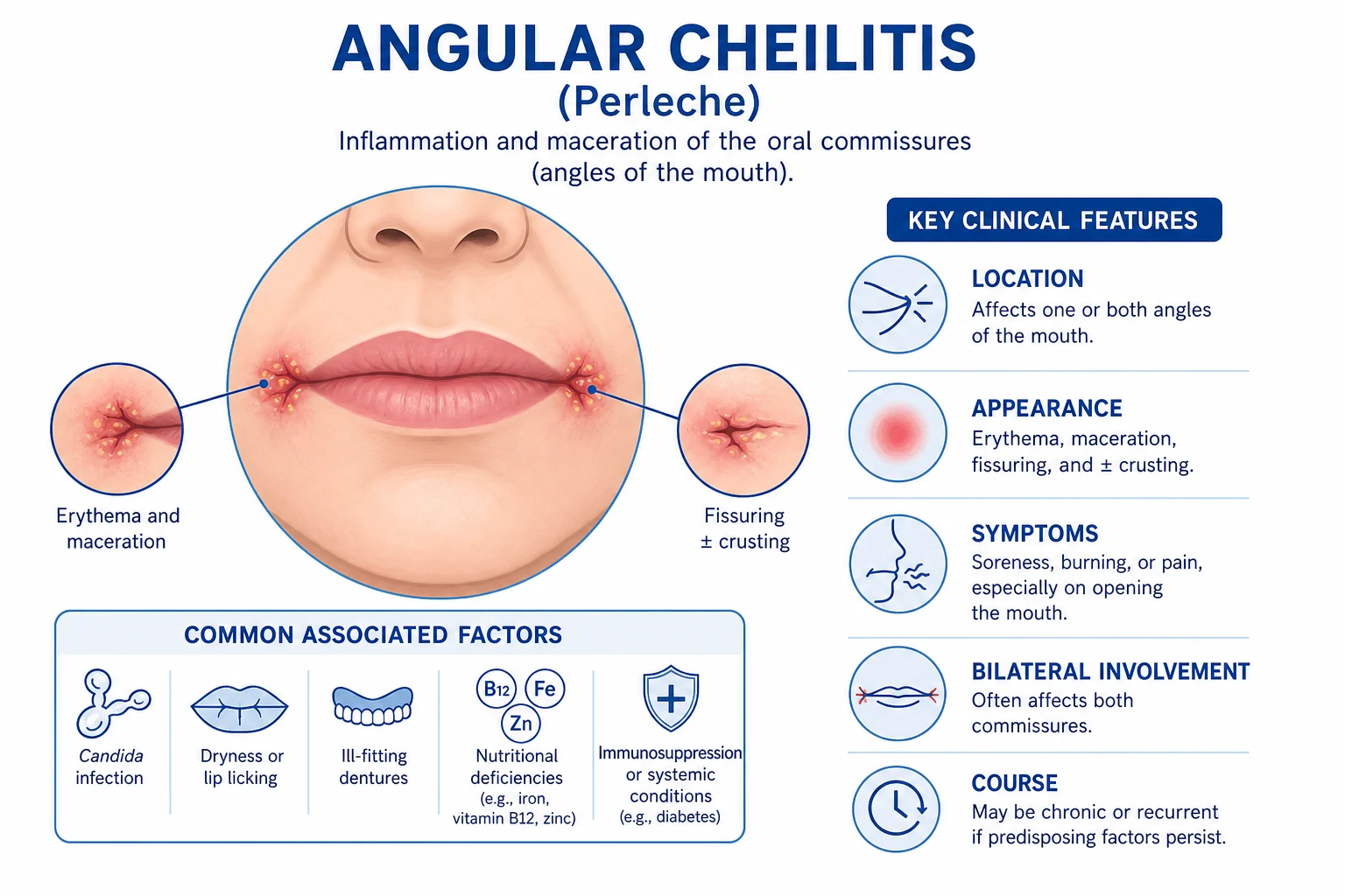
Angular cheilitis is one of the most common oral mucosal disorders encountered in primary care, dental practice, and dermatology clinics. It affects all age groups but is over-represented among the elderly (especially the edentulous and denture wearers), infants (from lip-licking and pacifier use), and patients with nutritional deficiency, diabetes mellitus, HIV/AIDS, and xerostomia. While usually benign and self-limited when treated, it is a sentinel condition: persistent or recurrent disease demands a systematic search for an underlying systemic predisposition — iron, vitamin B12, or folate deficiency, diabetes, immunodeficiency, or haematological malignancy.[1][3]
[1]Classification
Angular cheilitis can be classified along two axes — duration and aetiology. Both axes are clinically useful because they guide investigation and management. [1]
By duration
| Type | Duration | Characteristics |
|---|---|---|
| Acute | Less than two weeks | Sudden onset; often related to a trigger (new denture, intercurrent illness, lip-licking); responds rapidly to topical therapy |
| Chronic | More than two weeks, recurrent | Persistent or relapsing; strongly suggests an unaddressed predisposing factor — nutritional deficiency, poorly controlled diabetes, ill-fitting dentures, immunodeficiency |
By aetiology
This classification is the more useful one clinically, as it directly determines the treatment strategy: [1]
Causes of angular cheilitis — ANGLES
The aetiological categories are: [1]
- Infectious — Candida-associated (the single largest category, approximately 70 percent; Candida albicans is the predominant species), bacterial (Staphylococcus aureus in approximately 20 percent, often from anterior nares carriage; beta-haemolytic streptococci less commonly), and mixed (Candida plus Staphylococcus co-colonisation, very common).
- Non-infectious / nutritional — iron deficiency anaemia (including Plummer-Vinson syndrome), vitamin B12 deficiency (pernicious anaemia), folate deficiency, and riboflavin deficiency. In the Indian subcontinent and other regions where malnutrition is prevalent, nutritional deficiency is the leading predisposing factor.
- Mechanical / anatomical — overclosure of the mandible from decreased vertical dimension (edentulism, ill-fitting or worn dentures), prominent commissural folds (ageing, obesity), orthodontic appliances, and habitual lip-licking.
- Systemic / immunological — diabetes mellitus, HIV/AIDS, xerostomia (Sjogren syndrome, drug-induced), inflammatory bowel disease (Crohn disease with orofacial granulomatosis), corticosteroid therapy, and haematological malignancy. [1]
In practice, most cases are multifactorial: an elderly edentulous patient with iron deficiency and a Candida infection has mechanical, nutritional, and infectious components all contributing simultaneously.[1]
Epidemiology & Risk Factors
Angular cheilitis is common worldwide, but precise prevalence figures vary widely because study populations differ (community vs dental clinic vs institutionalised elderly). Key epidemiological points:[1][2]
Age and demographics
- Elderly — the highest prevalence group. The combination of edentulism (decreased vertical dimension → commissural folds), denture wear, xerostomia (medication-related), and nutritional deficiency creates a perfect storm. Prevalence in institutionalised elderly may reach 30 to 60 percent.
- Infants and young children — common due to lip-licking habits, pacifier/dummy use (saliva pooling), and drooling during teething.
- Adults of any age with predisposing factors (nutritional deficiency, diabetes, HIV, orthodontic appliances). [1]
Systemic risk factors
| Risk factor | Mechanism | Clinical clue |
|---|---|---|
| Iron deficiency | Impaired epithelial integrity, reduced cell-mediated immunity, Plummer-Vinson syndrome | Pallor, fatigue, koilonychia, glossitis, dysphagia |
| Vitamin B12 deficiency | Mucosal vulnerability, megaloblastic changes in epithelium | Glossitis (smooth beefy tongue), neurological symptoms, macrocytic anaemia |
| Folate deficiency | Impaired epithelial turnover | Macrocytic anaemia, glossitis |
| Diabetes mellitus | Hyperglycaemia → impaired neutrophil function, increased Candida adherence and virulence | Polyuria, polydipsia, recurrent candidiasis, neuropathy |
| HIV/AIDS | CD4 depletion → mucocandidiasis; angular cheilitis may be an early presenting feature | Oral thrush, weight loss, opportunistic infections; angular cheilitis with CD4 under 200 |
| Xerostomia | Reduced salivary antimicrobial function (lysozyme, histatins), altered saliva composition | Medications (anticholinergics, antidepressants, antihistamines), Sjogren syndrome, radiotherapy |
| Down syndrome | Immune dysregulation, anatomical factors; prevalence significantly elevated | Characteristic facies, intellectual disability |
Mechanical and local factors
- Edentulism / ill-fitting dentures — the single most common predisposing factor in developed countries. Loss of teeth decreases the vertical dimension (lower face height), causing the mandible to overclose and creating deep commissural skin folds that trap saliva.[2]
- Orthodontic appliances (braces, retainers) — trap moisture, irritate the commissure.
- Habitual lip-licking — chronic saliva exposure → maceration and irritant dermatitis; common in children and anxious adults.
- Anatomical variants — prominent perioral folds, macroglossia.
- Crohn disease — orofacial granulomatosis may present with angular cheilitis, facial swelling, and mucosal tags.[5]
Medications
- Isotretinoin — severe xerostomia and cheilitis.
- Anticholinergics, tricyclic antidepressants, antihistamines — xerostomia.
- Systemic corticosteroids and immunosuppressants — immune suppression.
- Broad-spectrum antibiotics — disrupt oral flora, predispose to Candida overgrowth. [1]
Pathophysiology
The pathogenesis of angular cheilitis follows a characteristic three-step cascade: (1) anatomical predisposition to saliva pooling, (2) maceration of the stratum corneum, and (3) microbial colonisation with ensuing inflammation. [1]
Step 1: Anatomical predisposition and saliva pooling
The labial commissures are the point where the upper and lower lips meet laterally. In health, this is a flat or shallow angle. When the vertical dimension of the face is reduced — most commonly from loss of teeth (edentulism) or a poorly fitting denture that does not restore facial height — the mandible overcloses (the chin moves upward and forward). This overclosure creates deep, overlapping skin folds at the commissures, particularly in the ageing face where skin elasticity is already reduced. [1]
Saliva naturally collects in these folds. The commissural angle acts as a dependent recess — saliva pools by gravity, especially in patients with reduced lower facial height. Additionally, patients with xerostomia paradoxically may develop angular cheilitis because their thickened, reduced-quantity saliva loses its normal antimicrobial properties (lysozyme, lactoferrin, histatins, secretory IgA), while sialorrhoea (excess saliva, as in children or neurological conditions) directly increases the moisture burden.[1][2]
Step 2: Maceration
Chronic exposure to saliva macerates the stratum corneum at the commissure. Saliva is alkaline relative to skin (pH approximately 6.5 to 7.0 versus skin pH approximately 5.0), and it contains digestive enzymes (amylase, maltase) that further break down the protective barrier. The macerated skin appears white, softened, and fragile — the stratum corneum lifts and fissures with even minor mechanical stress (opening the mouth, chewing). [1]
This macerated, moist, warm, nutrient-rich environment is an ideal growth medium for fungi and bacteria that are normally present only as commensals on skin and oral mucosa.[3]
Step 3: Microbial colonisation and inflammation
Candida albicans is the single most common colonising organism, present in approximately 70 percent of cases. C. albicans is a normal oral commensal in 30 to 60 percent of healthy adults. In the macerated commissure, it transitions from a harmless yeast form to an invasive pseudohyphal form — this dimorphic switch is triggered by temperature (37 degrees C), pH above 6.5, and nutrient availability. The pseudohyphae invade the macerated epithelium, secreting secreted aspartyl proteinases (SAPs) and phospholipases that degrade host tissues. This drives an inflammatory response: vasodilation (erythema), neutrophil infiltration (pus in more severe cases), epidermal hyperproliferation (scaling/crusting), and fissure formation.[3]
Staphylococcus aureus is the second most common pathogen, found in approximately 20 percent of cases. The anterior nares are the usual reservoir — nasal carriage seeds the commissure, especially in patients who nose-pick or rub their nose and mouth. Beta-haemolytic streptococci (particularly Streptococcus pyogenes) are less common but well documented, especially in children. [1]
Mixed infection (Candida plus Staphylococcus) is extremely common — up to half of all culture-positive cases — which is why a single topical agent with both antifungal and antibacterial activity (miconazole) is rational first-line therapy. [1]
The role of nutritional deficiency
Nutritional deficiency does not directly cause angular cheilitis but creates mucosal vulnerability and immune dysfunction that allow the saliva-pooling/maceration/colonisation cascade to proceed unchecked: [1]
- Iron deficiency reduces epithelial cell turnover and impairs cell-mediated immunity (particularly T-cell function). In Plummer-Vinson syndrome (iron deficiency, dysphagia, oesophageal webs, post-cricoid cancer risk), angular cheilitis and glossitis are classical features.
- Vitamin B12 deficiency impairs DNA synthesis in rapidly dividing epithelial cells, producing megaloblastic changes and a thin, vulnerable mucosa.
- Folate deficiency has a similar megaloblastic mechanism.
- Riboflavin (B2) deficiency classically causes angular stomatitis, glossitis, and seborrhoeic dermatitis — though it is rare in isolation in developed countries. [1]
Clinical Presentation
Morphology
The classical lesion of angular cheilitis is an erythematous, moist, macerated, fissured plaque at one or both labial commissures. The morphology evolves through characteristic stages: [1]
- Early: mild erythema and a subtle moist sheen at the commissural angle; the patient may notice only mild discomfort or burning.
- Established: well-defined erythematous maceration with a greyish-white surface (the macerated stratum corneum), extending several millimetres from the commissure onto both the vermilion and perioral skin. A linear fissure (rhagade) develops in the crease — this is the source of the characteristic pain on mouth opening.
- Advanced: deep fissuring with yellowish crusting (serous exudate drying to crust), surrounding erythema and scaling, and sometimes secondary haemorrhage from the fissure base. The commissure feels thickened and tender.
- Chronic: with persistence, the fissures may deepen, and the surrounding skin develops lichenification and post-inflammatory hyperpigmentation. Recurrent episodes can leave subtle scarring and commissural widening. [1]
Distribution
- Bilateral is the rule — both commissures are affected, though severity may be asymmetric.
- Unilateral involvement is less common and should prompt consideration of local factors (trauma, a dental appliance rubbing on one side) or an alternative diagnosis (herpes simplex labialis, squamous cell carcinoma, traumatic ulcer).[1]
Symptoms
- Pain — the cardinal symptom, typically sharp and tearing, worsened by opening the mouth wide (stretching the fissure), eating (especially acidic, spicy, or salty foods), and yawning.
- Burning and stinging sensation.
- Cosmetic concern — visible crusted lesions at the mouth corners.
- Difficulty eating — pain limits mouth opening and food intake, which may exacerbate underlying nutritional deficiency (a vicious cycle).
- Bleeding — from deep fissures, especially with trauma. [1]
Atypical presentations
Associated findings suggesting aetiology
- Oral candidiasis (thrush) — white, curdy, wipeable plaques on buccal mucosa, palate, or tongue → check HIV, diabetes, immunosuppression.
- Atrophic glossitis (smooth, beefy-red, depapillated tongue) → iron, B12, or folate deficiency; niacin deficiency (pellagra); riboflavin deficiency.
- Denture stomatitis — chronic erythema under the denture-bearing palatal mucosa → denture hygiene issue, Candida colonisation of the denture.
- Facial skin changes — pallor (anaemia), hyperpigmentation of palmar creases (Addison disease), butterfly rash (SLE), orofacial swelling and fissured tongue (Melkersson-Rosenthal).
- Koilonychia (spoon-shaped nails) → iron deficiency. [1]
Differential Diagnosis
The differential diagnosis of lesions at the oral commissure encompasses a range of dermatological, infectious, and neoplastic conditions. A systematic approach based on distribution, morphology, and associated features is essential. [1]
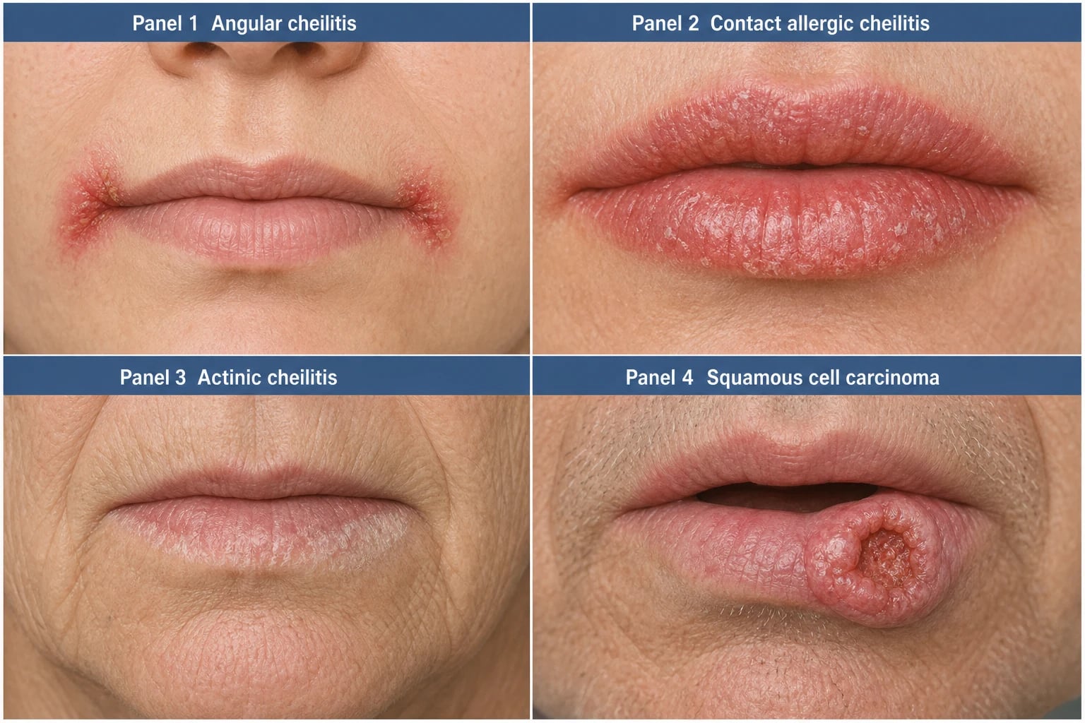
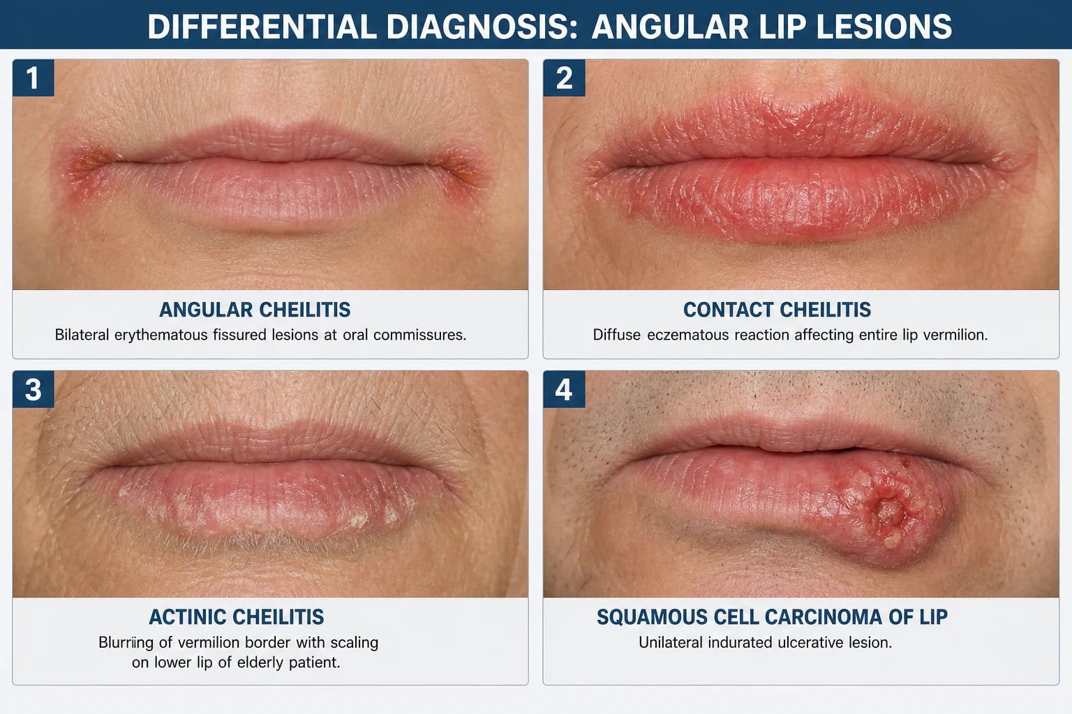
Contact / allergic cheilitis
Allergic contact cheilitis produces a diffuse eczematous reaction affecting the entire vermilion border (not just the commissures), with associated dryness, fissuring, scaling, and lichenification. Common allergens include lip balms (lanolin, fragrances, propolis/beeswax), toothpaste, dental materials, nail varnish (transferred by lip-licking), and foods (mango, citrus). Irritant contact cheilitis from lip-licking (lip-licker's dermatitis) produces a well-demarcated perioral erythematous patch encircling the lips — the so-called lip-licker's ring. The key distinguishing feature from angular cheilitis is the distribution: contact cheilitis involves the whole lip or perioral skin, while angular cheilitis is confined to the commissures. [1]
Lip-licker's dermatitis
This is a form of irritant contact dermatitis caused by chronic lip-licking. Saliva contains digestive enzymes that irritate the skin; evaporation dries and fissures it further. It produces a circumoral (encircling the lips) erythematous, scaly, sharply demarcated patch that may include the commissures. Common in children (especially in winter) and anxious adults. The history of habitual lip-licking and the circumoral (not commissural) distribution distinguish it. Management is barrier protection (petrolatum) and behavioural counselling to stop the habit. [1]
Actinic cheilitis (solar cheilosis)
Actinic cheilitis is a premalignant keratinocyte dysplasia of the lower lip vermilion caused by chronic UV exposure. It affects elderly, fair-skinned, outdoor workers (farmers, sailors). Clinical features: blurring of the vermilion border, dry scaling, atrophy (thin, pale vermilion), white plaques (leucoplakia), and occasionally ulceration or focal erythroplakia (red plaques — high risk of malignant transformation). It is confined to the lower lip (sun-exposed), not the commissures. Biopsy is mandatory to exclude squamous cell carcinoma. Management: sun protection, topical 5-fluorouracil or imiquimod, laser ablation, vermilionectomy (lip shave) for severe dysplasia. [1]
Plasma cell cheilitis
A rare condition characterised by a dense plasma cell infiltrate in the lamina propria, producing a well-demarcated, flat, erythematous, and sometimes eroded plaque, usually on the lower lip. It is the mucosal analogue of plasma cell balanitis (Zoon's). Diagnosis is by biopsy (showing a band-like plasma cell infiltrate). Management: topical corticosteroids (clobetasol). [1]
Squamous cell carcinoma of the lip
SCC of the lip is the most serious condition to exclude. It typically presents as a unilateral, indurated, non-healing ulcer or nodule on the lower lip vermilion, often with a rolled, everted edge and a granular, friable base that bleeds on minor trauma. Risk factors: chronic sun exposure (actinic damage), tobacco (especially smokeless tobacco/betel quid in South Asia), alcohol, and immunosuppression. Any unilateral commissural or lip lesion that does not respond to appropriate treatment within two to four weeks must be biopsied to exclude SCC. SCC of the lip metastasises to submental and submandibular lymph nodes.[1]
Herpes simplex labialis
Recurrent herpes simplex (cold sore, fever blister) is caused by HSV-1 (occasionally HSV-2). It presents as a cluster of vesicles on an erythematous base, usually on the lip vermilion border (unilateral). The vesicles rupture to form painful, crusted erosions that heal over 7 to 10 days. Key distinguishing features from angular cheilitis: vesicular onset (angular cheilitis does not have vesicles), unilateral, confined to the vermilion (not the commissure), and self-limited (resolves without treatment). Triggered by UV light, fever, stress, menstruation. Diagnosis is clinical; Tzanck smear or PCR if doubt. [1]
Orofacial granulomatosis / Melkersson-Rosenthal syndrome
Orofacial granulomatosis is a non-caseating granulomatous inflammation that can cause angular cheilitis, lip/facial swelling, fissured tongue (scrotal tongue), and facial nerve palsy. The full triad of recurrent facial swelling, fissured tongue, and recurrent facial palsy constitutes Melkersson-Rosenthal syndrome. It is associated with Crohn disease and sarcoidosis. The swelling is persistent, non-pitting, and firm (unlike the soft, transient oedema of acute cheilitis). Biopsy shows non-caseating granulomas. Management: intralesional corticosteroids, exclusion diets (cinnamon/benzoate-free), and in severe cases, systemic immunosuppression.[5]
Clinical & Bedside Assessment
A focused but systematic clinical assessment is the cornerstone of managing angular cheilitis. The goal is to identify the predisposing factor — treatment of the infection alone, without correcting the cause, leads to near-certain recurrence. [1]
History
A structured history should cover: [1]
- Onset and duration: acute (new denture, intercurrent illness) vs chronic/recurrent (systemic predisposition).
- Unilateral or bilateral: bilateral is typical; unilateral may suggest local trauma, HSV, or neoplasia.
- Dental/denture history: Does the patient wear dentures? When were they last fitted/adjusted? Do they remove them at night? How do they clean them?
- Dietary history: adequacy of intake, vegetarian/vegan diet (B12, iron risk), food insecurity.
- Systemic symptoms: fatigue, pallor (anaemia), weight loss, polyuria/polydipsia (diabetes), recurrent infections, night sweats (immunodeficiency/malignancy), dysphagia (Plummer-Vinson), diarrhoea (malabsorption, IBD).
- Medications: xerostomia-inducing drugs, broad-spectrum antibiotics, corticosteroids, immunosuppressants.
- Habits: lip-licking, thumb-sucking (children), smoking, betel quid.
- Sexual history / HIV risk: if recurrent/refractory.
- Sun exposure history: for actinic damage assessment. [1]
Examination
Bedside assessment of denture fit and vertical dimension
Assess the patient with and without the denture in place. Key observations: [1]
- Facial proportions: the lower third of the face should be approximately equal to the middle third. Loss of vertical dimension (overclosure) makes the lower face appear shortened, the chin prominent, and the nasolabial folds deepened.
- Commissural folding: with the mouth slightly open, look for deep skin folds at the commissures — the "drooping angle" sign. These folds trap saliva.
- Lip competence: can the patient close the lips at rest without strain?
- Denture adaptation: does the denture rock, click, or drop? Is there friable, erythematous palatal mucosa underneath (denture stomatitis)? [1]
Signs of nutritional deficiency
| Sign | Deficiency |
|---|---|
| Pallor of conjunctiva/palmar creases | Iron deficiency anaemia |
| Koilonychia (spoon nails) | Iron deficiency |
| Atrophic glossitis (smooth, beefy-red tongue) | Iron, B12, folate, niacin, riboflavin |
| Angular stomatitis | Iron, B12, folate, riboflavin |
| Oral aphthous ulcers | B12, folate, iron |
| Generalised hyperpigmentation | B12 deficiency (rare), Addison disease |
| Dermatitis (seborrhoeic, periorificial) | Zinc, riboflavin, niacin, biotin |
Investigations
Angular cheilitis is fundamentally a clinical diagnosis. Investigations are not required in every case but are essential when: [1]
- The condition is persistent (more than two weeks despite topical therapy).
- It is recurrent (two or more episodes).
- There are clinical clues to systemic disease (glossitis, pallor, oral thrush, polyuria).
- The lesion is unilateral, indurated, or non-healing (exclude SCC — biopsy). [1]
First-line investigation panel (persistent/recurrent cases)
- Full blood count (FBC) — anaemia: microcytic hypochromic (low MCV, low MCH) suggests iron deficiency; macrocytic (high MCV) suggests B12 or folate deficiency. Also check for leucopenia/lymphopenia (HIV, marrow infiltration) or thrombocytopenia.
- Serum ferritin — the most sensitive indicator of iron stores. A level under 15 micrograms per litre confirms iron deficiency (in the absence of inflammation; ferritin is an acute-phase reactant and may be falsely normal/elevated in chronic disease — check C-reactive protein concurrently).[1]
- Vitamin B12 — a level under 150 ng per litre (approximately 110 pmol per litre) confirms deficiency. If borderline (150 to 250), check methylmalonic acid and homocysteine (elevated in true B12 deficiency).
- Red cell folate (more reliable than serum folate) — under 140 micrograms per litre confirms deficiency.
- HbA1c / fasting glucose — screen for diabetes. HbA1c of 48 mmol per mol (6.5 percent) or above on two occasions establishes the diagnosis. This is mandatory in recurrent candidal angular cheilitis.
Microbiological studies
- Oral/commissural swab for bacterial and fungal culture — if resistant to first-line therapy, to identify the organism (Candida species, including non-albicans Candida like C. glabrata or C. krusei which may be azole-resistant) and check Staphylococcus aureus including MRSA. Send for culture and sensitivity, not just Gram stain.
- If nasal carriage of S. aureus is suspected (recurrent staphylococcal angular cheilitis), swab the anterior nares and treat with mupirocin nasal ointment (decolonisation). [1]
Further investigations (selected cases)
- HIV test — if risk factors present or if angular cheilitis is recurrent and associated with oral candidiasis. A fourth-generation HIV antigen/antibody combination test is the standard.
- Iron studies (transferrin saturation, TIBC) if ferritin equivocal.
- Intrinsic factor antibodies — if pernicious anaemia suspected (B12 deficiency).
- Coeliac screen (tissue transglutaminase IgA) — if malabsorption suspected.
- Patch testing — if allergic/contact cheilitis suspected.
- Biopsy — if the lesion is unilateral, indurated, non-healing, or if SCC is suspected. [1]
Management — Resuscitation
Angular cheilitis is not a dermatological emergency in the classic sense, but several scenarios require prompt or urgent attention: [1]
Acute pain management
- Petrolatum-based lip balm or zinc oxide paste applied frequently as a barrier to reduce pain from salivary irritation and promote fissure healing.
- Topical anaesthetic (lidocaine gel 2 percent) applied sparingly before meals may help severe pain, but avoid in young children (risk of methaemoglobinaemia and systemic toxicity with overuse).
- Soft diet and avoidance of acidic, spicy, and salty foods during the acute phase. [1]
Secondary bacterial cellulitis
If the angular cheilitis has surrounding spreading erythema, warmth, swelling, and systemic symptoms (fever, malaise), treat as perioral cellulitis: [1]
- Oral antibiotics: flucloxacillin 500 mg four times daily (or cephalexin 500 mg four times daily) for 5 to 7 days — covers Staphylococcus aureus and streptococci.
- If MRSA suspected or known: doxycycline 100 mg twice daily or clindamycin 300 mg four times daily (adjust based on sensitivities).
- Assess for systemic signs — admit for IV antibiotics if cellulitis is rapidly spreading, the patient is systemically unwell, or immunocompromised. [1]
Immunocompromised patients
In patients with HIV/AIDS, poorly controlled diabetes, or neutropenia, angular cheilitis may be the tip of the iceberg of invasive mucocandidiasis. These patients need: [1]
- Prompt systemic antifungal therapy (oral fluconazole) in addition to topical therapy.
- Urgent glycaemic optimisation (diabetes) or immune reconstitution (HIV — antiretroviral therapy).
- Investigation for oesophageal candidiasis (dysphagia, odynophagia) which may require systemic therapy. [1]
Management — Definitive & Stepwise
The management of angular cheilitis rests on two pillars applied simultaneously: (1) treat the infection (topical antimicrobial) and (2) correct the predisposing factor (denture, nutrition, diabetes, habit). Treatment of the infection alone without addressing the cause guarantees recurrence. [1]
Step 1: Treat the infection (first-line topical therapy)
Topical antifungal is the first-line agent because Candida is the most common organism: [1]
| Agent | Concentration | Frequency | Duration | Notes |
|---|---|---|---|---|
| Miconazole 2 percent gel (Daktarin) | 2 percent | Four times daily | Two weeks | First-line — has BOTH antifungal AND weak antibacterial (anti-staphylococcal) activity; preferred for this reason |
| Clotrimazole 1 percent cream | 1 percent | Two to three times daily | Two to four weeks | Alternative; purely antifungal (no antibacterial activity) |
| Nystatin 100,000 units per gram cream | 100,000 units/g | Two to three times daily | Two to four weeks | Alternative; not absorbed; useful if azole resistance suspected |
| Ketoconazole 2 percent cream | 2 percent | Once to twice daily | Two weeks | Effective but rarely used topically (hepatotoxicity concern with oral form) |
First-line topical antifungal: MICONAZOLE is the answer
Why miconazole is preferred: Miconazole has dual activity — it inhibits fungal ergosterol synthesis (azole mechanism) AND has weak activity against Gram-positive bacteria, including Staphylococcus aureus. Since mixed Candida-Staphylococcus infection is common, miconazole covers both. Clotrimazole, in contrast, is purely antifungal. [1]
IMPORTANT drug interaction: Miconazole oral gel (even when applied to the lips, some is swallowed) inhibits CYP2C9 and can potentiate warfarin (dangerous INR elevation and bleeding), sulfonylureas (hypoglycaemia), and statins (myopathy). Check the patient's medication list. In anticoagulated patients, use nystatin or clotrimazole instead, or use miconazole cream (less systemic absorption than gel) with close INR monitoring.[1]
Step 2: Add topical antibacterial if Staphylococcus confirmed
If a swab confirms Staphylococcus aureus (or empirically, if there is yellow crusting suggesting bacterial superinfection and the patient does not respond to antifungal alone): [1]
| Agent | Concentration | Frequency | Duration | Notes |
|---|---|---|---|---|
| Fusidic acid 2 percent cream | 2 percent | Three times daily | Seven to ten days | Active against S. aureus including some MRSA; available in UK/India |
| Mupirocin 2 percent cream/ointment | 2 percent | Three times daily | Seven to ten days | Active against S. aureus including MRSA; gold standard in US; reserve for confirmed staph |
If nasal carriage of S. aureus is the source (recurrent staphylococcal angular cheilitis): treat with mupirocin 2 percent nasal ointment applied to the anterior nares twice daily for 5 to 7 days (decolonisation), plus chlorhexidine body wash. [1]
Step 3: Correct the predisposing factor
This is mandatory — without it, recurrence is near-certain. [1]
Denture management:[2]
- Denture adjustment or replacement — if ill-fitting or worn (loss of vertical dimension). Refer to a prosthodontist or dentist for relining, rebasing, or remaking to restore the correct vertical dimension and eliminate commissural folds.
- Denture hygiene regimen:
- Remove dentures at night — never sleep in them (allows oral tissues to rest, reduces Candida colonisation of the denture).
- Brush and rinse dentures daily with a soft brush and non-abrasive denture cleaner (do not use regular toothpaste — it scratches the acrylic).
- Soak dentures overnight in a denture-cleansing solution: either chlorhexidine gluconate 0.2 percent (30 minutes to overnight), sodium hypochlorite (dilute Milton-type soak, 10 to 15 minutes), or alkaline peroxide tablets (overnight). This kills Candida biofilm on the denture surface.
- Clean the palate under the denture — if denture stomatitis is present, apply miconazole oral gel to the fitting surface of the denture and the palatal mucosa.
Nutritional replacement: [1]
[1]Glycaemic control (diabetic patients):
- Optimise diabetes management — the candidal infection will not resolve with persistent hyperglycaemia. Review oral hypoglycaemics/insulin, diet, and HbA1c target (under 53 mmol/mol / 7 percent for most). [1]
Habit modification:
- Stop lip-licking — behavioural counselling, especially in children. Use petrolatum-based lip balm as a saliva barrier and to break the dry-lick cycle.
- Discourage thumb-sucking and pacifier use in affected children. [1]
Barrier measures:
- Petrolatum (Vaseline) or zinc oxide paste applied to the commissures and lips — protects the skin from saliva, reduces pain, and promotes fissure healing. Apply before meals, at night, and between medication doses. [1]
Step 4: Systemic therapy (refractory or extensive disease)
If the angular cheilitis is refractory to two to four weeks of topical therapy, or in immunocompromised patients (HIV, malignancy) with extensive disease: [1]
- Oral fluconazole 50 to 100 mg once daily for 7 to 14 days — systemic azole; well absorbed; covers Candida albicans. Check LFTs and drug interactions (fluconazole inhibits CYP2C9/3A4 — interacts with warfarin, statins, sulfonylureas, phenytoin).
- Oral itraconazole — alternative if fluconazole resistance suspected (non-albicans Candida).
- In HIV-positive patients with fluconazole-resistant Candida (e.g., C. krusei, C. glabrata): oral voriconazole or posaconazole, or IV echinocandin (caspofungin, micafungin) — specialist input. [1]
Step 5: Referral
Refer to dermatology or oral medicine if:
- Diagnosis is uncertain (consider biopsy for SCC, plasma cell cheilitis, granulomatous cheilitis).
- Disease is refractory to topical and systemic therapy.
- There is an underlying condition requiring specialist input (orofacial granulomatosis, severe nutritional deficiency, immunodeficiency). [1]
Refer to maxillofacial/prosthodontics for:
- Denture adjustment, relining, or remaking.
- Vermilionectomy or biopsy if SCC suspected. [1]
Specific Subtypes & Scenarios
Candida-associated angular cheilitis (the commonest subtype)
Clinical features: bilateral, moist, macerated, erythematous lesions with greyish-white surface; often associated with oral candidiasis (thrush) and denture stomatitis (erythematous palate under denture). Burning sensation is prominent. The diagnosis is essentially clinical but can be confirmed by fungal swab. [1]
Management: miconazole 2 percent gel applied to the commissures four times daily for two weeks; if the patient wears dentures, also apply miconazole to the denture fitting surface and palatal mucosa. Soak dentures overnight in chlorhexidine. Correct any nutritional deficiency. If the patient is immunocompromised (HIV), add oral fluconazole 50 to 100 mg daily for 7 to 14 days.[3]
Bacterial (Staphylococcal) angular cheilitis
Clinical features: more likely to have yellow crusting, honey-coloured exudate (impetiginous appearance), and to be associated with nasal vestibulitis or Staphylococcus aureus nasal carriage. May be unilateral. More common in children (nose-picking) and atopic patients. [1]
Management: fusidic acid 2 percent cream or mupirocin 2 percent cream applied three times daily for 7 to 10 days. If recurrent, check for nasal carriage and decolonise with mupirocin nasal ointment twice daily for 5 to 7 days plus chlorhexidine body wash. Take a swab to check for MRSA. Often co-exists with Candida — use combined or dual therapy (miconazole plus fusidic acid, alternating applications). [1]
Nutritional deficiency-associated angular cheilitis
Clinical features: associated with glossitis (atrophic, smooth, beefy-red tongue), pallor, fatigue, koilonychia (iron), neurological symptoms (B12 — peripheral neuropathy, subacute combined degeneration), macrocytic anaemia (B12/folate). May be the presenting feature of iron deficiency, pernicious anaemia, or coeliac disease. In Plummer-Vinson syndrome (sideropenic dysphagia), angular cheilitis accompanies dysphagia, glossitis, and oesophageal webs (with an increased risk of post-cricoid SCC). [1]
Management: treat the deficiency AND the infection simultaneously. Iron replacement (oral ferrous sulfate 200 mg TDS), B12 replacement (IM hydroxocobalamin), folate replacement (oral folic acid 5 mg daily). Topical miconazole for the commissural infection. Investigate the cause of the deficiency (coeliac screen, upper GI endoscopy if Plummer-Vinson suspected, intrinsic factor antibodies for pernicious anaemia).[1]
Mechanical / overclosure angular cheilitis
Clinical features: elderly edentulous patients, or patients with worn or ill-fitting dentures. Deep commissural folds, "drooping angle" sign, overclosure of the mandible (shortened lower facial third, deepened nasolabial folds). No systemic symptoms. Often bilateral, chronic, recurrent. [1]
Management: the definitive treatment is restoring the vertical dimension — denture relining, rebasing, or replacement by a prosthodontist. Topical miconazole controls the infection while the denture is being corrected. Denture hygiene regimen (overnight soak in chlorhexidine). Without denture correction, topical therapy alone will fail and recurrence is guaranteed. Dental referral is essential.[2]
Angular cheilitis in HIV/AIDS
Clinical features: may be the first manifestation of HIV. Often severe, bilateral, rapidly progressive, with concurrent oral candidiasis, oral hairy leukoplakia (white, vertical corrugated streaks on lateral tongue border, EBV-associated), and periodontal disease. Resistant to topical therapy alone. High CD4 count correlation — angular cheilitis typically appears when CD4 count is under 200 cells per microlitre. [1]
Management: oral fluconazole 100 mg daily for 14 days (topical therapy alone usually insufficient). Antiretroviral therapy (immune reconstitution) is the definitive treatment — recurrent candidiasis resolves with viral suppression and CD4 recovery. Check for azole resistance (non-albicans Candida) and use voriconazole or echinocandins if resistant. Screen for other opportunistic infections.[3]
Angular cheilitis in infants and children
Clinical features: common from lip-licking, pacifier/dummy use, and drooling during teething. Often associated with perioral irritant dermatitis (lip-licker's ring). Streptococcal impetiginous crusting is more common in this age group. Nutritional deficiency should be considered (especially in the developing world). [1]
Management: stop the habit (pacifier, lip-licating counselling), apply petrolatum barrier, and use topical miconazole if Candida is suspected or topical fusidic acid/mupirocin if streptococcal/staphylococal crusting. Weight-based dosing if systemic therapy is needed (fluconazole 3 to 6 mg per kg daily). Screen for nutritional deficiency if recurrent. [1]
Complications & Pitfalls
Complications
- Secondary bacterial infection / cellulitis — the fissured, macerated skin is a portal of entry for Staphylococcus aureus and streptococci. Watch for spreading erythema, warmth, and systemic symptoms. Treat with oral antibiotics (flucloxacillin).[1]
- Chronic fissuring and scarring — deep, recurrent fissures heal with fibrosis and commissural widening, producing a permanently altered commissural contour (cosmetic concern).
- Post-inflammatory hyperpigmentation — common in darker skin types; may persist for months after resolution.
- Bleeding — from deep fissures, especially with trauma (eating, yawning).
- Pain and nutritional compromise — pain on eating may reduce intake, worsening any underlying nutritional deficiency (vicious cycle).
- Recurrence — near-certain if the predisposing factor is not addressed. Each recurrence deepens the fissure and worsens scarring.
Pitfalls
Pitfalls in angular cheilitis — DEFECT
Prognosis & Disposition
Prognosis
The prognosis of angular cheilitis is excellent when the underlying cause is identified and treated: [1]
- With appropriate topical therapy (miconazole for 2 weeks) and correction of the predisposing factor (denture, nutrition, diabetes, habit), most cases resolve within 1 to 3 weeks.
- Recurrence rate without cause correction: high (over 50 percent within months).
- Recurrence rate with cause correction: low (under 10 percent).[1]
- In immunocompromised patients (HIV, malignancy), prognosis depends on immune reconstitution — recurrent disease persists until the underlying immunodeficiency is addressed.
Factors predicting recurrence
- Unaddressed predisposing factor (denture, nutritional deficiency, diabetes).
- Continued lip-licking habit.
- Poor denture hygiene (not removing at night).
- Immunosuppression (HIV, corticosteroids).
- Xerostomia from medications. [1]
Disposition
- Primary care / dental practice: most cases are managed here with topical therapy and denture/habit correction.
- Dermatology referral: refractory disease, diagnostic uncertainty, suspected SCC, or complex underlying dermatosis (plasma cell cheilitis, granulomatous cheilitis).
- Oral medicine referral: recurrent/refractory disease, orofacial granulomatosis, complex immunocompromised patients.
- Maxillofacial/prosthodontics referral: denture adjustment, relining, or remaking; biopsy or excision of suspected SCC.
- Haematology/gastroenterology referral: if severe nutritional deficiency, pernicious anaemia, or malabsorption is identified. [1]
Special Populations
[1]Evidence, Guidelines & Regional Differences
Guidelines
- NICE Clinical Knowledge Summaries (UK) — recommends topical miconazole or clotrimazole as first-line for angular cheilitis, with topical fusidic acid if S. aureus is confirmed. Advises investigating for nutritional deficiency (iron, B12, folate) and diabetes in recurrent/persistent cases. Denture hygiene advice (soak overnight).[1]
- American Academy of Oral Medicine — supports topical antifungals (miconazole, clotrimazole) as first-line; emphasises correction of predisposing factors. Recommends mupirocin (US) for staphylococcal disease (fusidic acid less commonly used in the US due to limited availability and resistance concerns).
- Primary Care Dermatology Society (UK) — miconazole 2 percent gel first-line; fusidic acid 2 percent cream if bacterial.
Evidence base
The evidence base for angular cheilitis treatment is limited by the absence of large randomised controlled trials — most evidence is from narrative reviews, case series, and expert opinion:[1]
- Ophaswongse and Maibach (2020) — a comprehensive narrative review that synthesised available evidence and expert opinion. Key conclusions: miconazole is the most-studied and preferred topical agent (dual antifungal/antibacterial); treatment must address the underlying cause; nutritional deficiency is common and under-diagnosed. There is no high-quality RCT comparing individual antifungal agents head-to-head for angular cheilitis specifically.[1]
- Akpan and Morgan (2002) — a widely cited review of oral candidiasis (including angular cheilitis as a form) establishing the role of Candida albicans as the predominant organism and the importance of topical azoles. This remains a foundational reference.[3]
- Viljanto et al. (2023) — an interdisciplinary review of denture stomatitis (which co-exists with angular cheilitis in denture wearers), emphasising the role of denture hygiene (overnight soaking, Candida biofilm removal from acrylic) and interdisciplinary collaboration (dental and medical).[2]
- Cataldi et al. (2022) — a case series of angular cheilitis in COVID-19 patients, possibly related to mask-wearing (moist environment, friction), immunosuppression, and steroid therapy. This introduced a new epidemiological context — "mask mouth" and COVID-era angular cheilitis.[4]
- Greuter et al. (2021) — a review of dermatological manifestations in inflammatory bowel disease, including orofacial granulomatosis presenting with angular cheilitis, facial swelling, and fissured tongue in Crohn disease. This reminds clinicians that angular cheilitis can be a cutaneous marker of IBD.[5]
Controversies
- Systemic vs topical therapy in immunocompetent patients — topical therapy is sufficient for the vast majority of immunocompetent patients. Oral fluconazole is reserved for refractory, extensive, or immunocompromised cases. Routine systemic therapy is not recommended for uncomplicated angular cheilitis.
- Combined antifungal-antibacterial preparations — some advocate a single combined cream (e.g., miconazole plus fusidic acid) to cover both Candida and Staphylococcus; others prefer sequential or alternating application with separate agents. There is no clear evidence that combined preparations are superior.
- Role of topical corticosteroids — generally not recommended (may worsen Candida infection, mask SCC, and cause skin atrophy). A short course may be justified in a granulomatous cheilitis (Melkersson-Rosenthal) component, but this is specialist territory.
- Prophylactic antifungals in recurrent disease — there is no evidence to support routine prophylactic topical antifungals; the key is correcting the predisposing factor. [1]
Exam Pearls
[1]Red flags
Exam application bank (NEET-PG / INICET)
One-line answer
Angular cheilitis is an inflammatory condition characterised by painful, erythematous, fissured lesions at the angles (commissures) of the mouth. It is multifactorial, caused by saliva pooling in skin folds created by decreased vertical dimension (ill-fitting dentures, edentulism), providing a moist environment for Candida albicans (most common) and Staphylococcus aureus colonisation. Risk factors include nutritional deficiencies (iron, B12, folate), immunosuppression (HIV, diabetes), and dry mouth. Treatment: topical antifungal (miconazole/clotrimazole) ± topical antibiotic (mupirocin for S. aureus); correct predisposing factors (denture adjustment, nutritional supplementation).
Worked stems (answer without another resource)
Stem 1 — Classic presentation. Map symptoms to mechanism; name the first investigation and first treatment step with dose/route if drug therapy is standard. [1]
Stem 2 — Unstable / complicated. List red flags that force immediate resuscitation, theatre, ICU, antidote, or reperfusion — and what you do in the first 15 minutes. [1]
Stem 3 — Atypical group. Elderly, pregnancy, child, or immunocompromised: how presentation and thresholds change. [1]
Stem 4 — Differential trap. Name the three closest mimics and one discriminator for each. [1]
Stem 5 — Disposition. Who goes home with safety-netting, who is admitted, who needs HDU/ICU/theatre, and what follow-up is mandatory. [1]
Rapid viva checklist
- Definition + classification
- Pathophysiology chain
- Bedside signs / criteria
- Score with exact components (if any)
- Emergency bundle
- Definitive therapy with doses
- Complications of disease and of treatment
- Special populations
- Guideline/trial name if classic
- Three exam traps
Coverage self-check
If you cannot answer any stem above from this page alone, re-read the matching section — the page is intended to be self-sufficient for final-prof and NEET-PG/INICET questions on Angular cheilitis (perleche).
[1]References
- [1]Ophaswongse S, Maibach HI. Treatment of angular cheilitis: A narrative review and authors' clinical experience Oral Dis, 2020.PMID 31464357
- [2]Viljanto A, et al. Denture stomatitis-An interdisciplinary clinical review J Prosthodont, 2023.PMID 36988151
- [3]Akpan A, Morgan R. Oral candidiasis Postgrad Med J, 2002.PMID 12185216
- [4]Cataldi A, et al. Angular cheilitis of COVID-19 patients: A case-series and literature review Oral Dis, 2022.PMID 33043573
- [5]Greuter T, et al. Dermatological Manifestations in Inflammatory Bowel Diseases J Clin Med, 2021.PMID 33477990