Dermatology · Medicine
Aphthous ulcers
Also known as Aphthous ulcers · Canker sores · Recurrent aphthous stomatitis · RAS · Aphthae · Complex aphthosis
Recurrent aphthous stomatitis is a clinical diagnosis of painful recurrent round or oval ulcers on non-keratinized oral mucosa, classified as minor, major, or herpetiform aphthae. The examiner expects candidates to distinguish aphthae from HSV, hand-foot-mouth disease, trauma, oral SCC, lichen planus and pemphigus; screen for Behçet disease, IBD, coeliac disease, HIV, cyclic neutropenia and haematinic deficiency when disease is severe or atypical; and manage stepwise with analgesia, chlorhexidine, topical corticosteroids, tetracycline mouthwash, deficiency replacement and specialist systemic therapy for refractory disease.
On this page & tools
Your progress
Saved locally on this device.
Exam tags
Red flags
Overview & Definition
Aphthous ulcers, commonly called canker sores, are painful round or oval ulcers of the oral mucosa with a yellow-white fibrinous base and an erythematous halo. The recurrent syndrome is recurrent aphthous stomatitis (RAS). It is not herpes, not candidiasis, not a manifestation of poor hygiene, and not a diagnosis to apply to every oral ulcer. It is a clinicopathologic pattern in which a susceptible mucosal barrier develops episodic immune-mediated ulceration, usually on non-keratinized movable mucosa such as the buccal mucosa, labial mucosa, floor of mouth, ventral tongue and soft palate.[1][3]
The key examination definition is deliberately narrow: recurrent, painful, shallow, well-demarcated oral ulcers; no preceding vesicles; no fixed indurated edge; no adherent white plaque; and no primary systemic illness unless features point beyond simple RAS. A single traumatic ulcer after biting the cheek is not RAS. A grouped vesicular eruption on the hard palate is not RAS. A solitary ulcer persisting for weeks in a smoker is oral cancer until proven otherwise. The diagnosis is safe only after the morphology, site, tempo and recurrence pattern have been matched to aphthae and the red flags have been actively sought.[3][5]
[1]Classification
The classical clinical classification is minor aphthous ulcers, major aphthous ulcers and herpetiform aphthous ulcers. The categories are not academic trivia: they determine expected healing time, scarring risk, severity of pain, likelihood of systemic association, and threshold for investigation or referral. Minor aphthae are the common everyday pattern; major aphthae and complex aphthosis are the patterns that make examiners ask about HIV, Behçet disease, inflammatory bowel disease, coeliac disease, neutropenia and haematinic deficiency.[1][2]
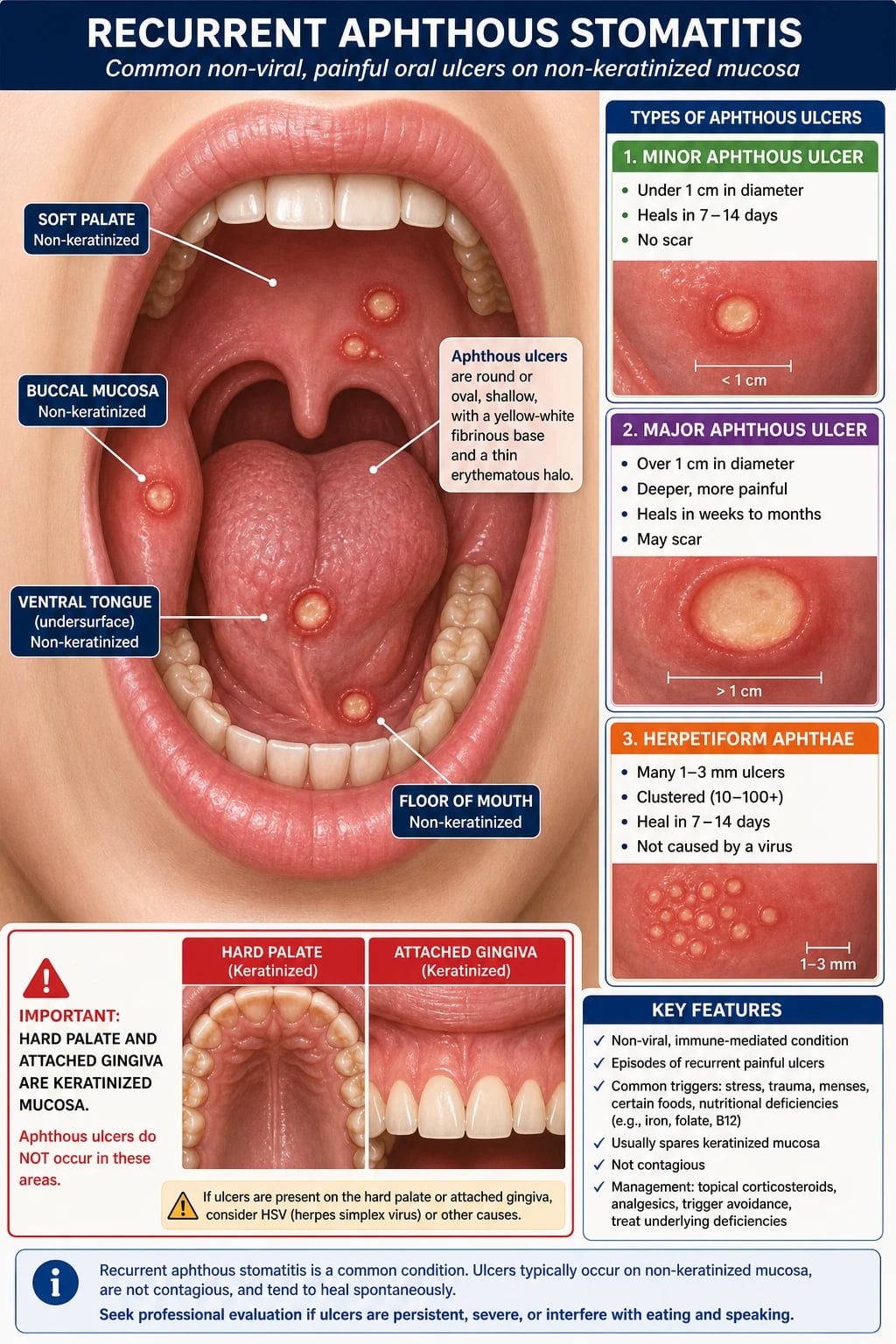
Minor aphthae
Most common
- Usually under 1 cm in diameter.
- One to five ulcers per episode are typical.
- Heal in 7-14 days.
- No scarring.
- Usually on labial or buccal mucosa, ventral tongue, floor of mouth or soft palate.
Major aphthae
Sutton disease / periadenitis mucosa necrotica recurrens
- Usually greater than 1 cm and deeper than minor aphthae.
- Can persist for weeks to months.
- May scar and may cause severe odynophagia or poor intake.
- Raises the threshold for haematinic, HIV, neutropenia, IBD and Behçet assessment.
- Often needs specialist therapy when recurrent or disabling.
Herpetiform aphthae
Not HSV
- Dozens of 1-3 mm ulcers in crops.
- May coalesce into larger irregular erosions.
- No vesicular stage and HSV PCR is negative unless coincidental infection is present.
- More frequent in adults than children.
- The name describes the clustered appearance, not the cause.
A practical fourth label, complex aphthosis, describes almost constant or very frequent oral aphthae, recurrent genital aphthae, or major and herpetiform patterns that behave more severely than simple minor RAS but do not yet meet diagnostic criteria for Behçet disease. Complex aphthosis is a management and referral category rather than a separate histological disease. It is useful because it warns the clinician not to reassure too quickly: repeated genital ulcers, ocular symptoms, skin lesions, gastrointestinal symptoms, fever cycles or immunosuppression may declare an underlying systemic disorder over time.[2][6]
Epidemiology & Risk Factors
RAS is common, with population estimates often around one fifth of people affected at some stage, although frequency varies by definition, age group and ascertainment. Onset is typically in childhood, adolescence or young adulthood; many patients describe a tendency that gradually becomes less frequent with age. A family history is common, supporting genetic susceptibility, but a positive family history does not remove the need to examine for systemic disease when the pattern is severe or atypical.[1][5]
High-yield numbers
Risk factors and triggers should be elicited as modifiable contributors rather than assumed causes. Local trauma from cheek biting, a sharp tooth, orthodontic appliance, vigorous toothbrushing or recent dental work can initiate an ulcer in susceptible mucosa. Emotional stress, sleep deprivation and intercurrent illness are common patient-reported triggers. Sodium lauryl sulfate toothpaste can worsen ulcer frequency in some patients, probably by irritating or altering mucosal barrier function. Acidic or spicy foods, nuts, chocolate, cheese, tomatoes and citrus may aggravate pain and sometimes act as reproducible triggers, but blanket dietary restriction is less useful than a symptom diary.[1][2]
Haematinic deficiency is an examiner favourite because it is common enough to matter and treatable enough to miss at one's peril. Iron deficiency, low ferritin, vitamin B12 deficiency, folate deficiency and sometimes zinc deficiency are associated with recurrent oral ulceration. The presence of angular cheilitis, glossitis, fatigue, menorrhagia, gastrointestinal symptoms, a restricted diet, bariatric surgery, vegan diet or malabsorption symptoms should lower the threshold for testing. Correcting a true deficiency is disease-modifying; supplementing a normal patient is not the same thing.[2][8]
The systemic associations are not rare zebras in examinations. Behçet disease is the classic systemic vasculitis association; inflammatory bowel disease can produce aphthous-like oral ulcers and other oral inflammatory changes; coeliac disease may present with oral aphthae and iron deficiency; HIV and other immunodeficiency states may produce large, persistent, treatment-resistant ulcers; and cyclic neutropenia produces periodic fever, infections and oral ulcers. Medication-related ulceration, especially with nicorandil, NSAIDs, bisphosphonates or cytotoxic drugs, should be considered when the temporal history fits.[2][6][9]
Pathophysiology
RAS is best understood as a final common pathway of mucosal barrier vulnerability plus dysregulated immune injury. The oral epithelium normally tolerates constant mechanical trauma, microbial exposure and dietary irritants. In RAS, a local trigger appears to expose epithelial antigens, stress signals or microbial products to a primed immune system. T-cell dominated inflammation, particularly Th1-skewed cytokine activity, recruits macrophages and neutrophils, producing epithelial necrosis, fibrin deposition and the familiar yellow-white pseudomembrane. Healing occurs by re-epithelialisation once the inflammatory episode resolves.[1][4]
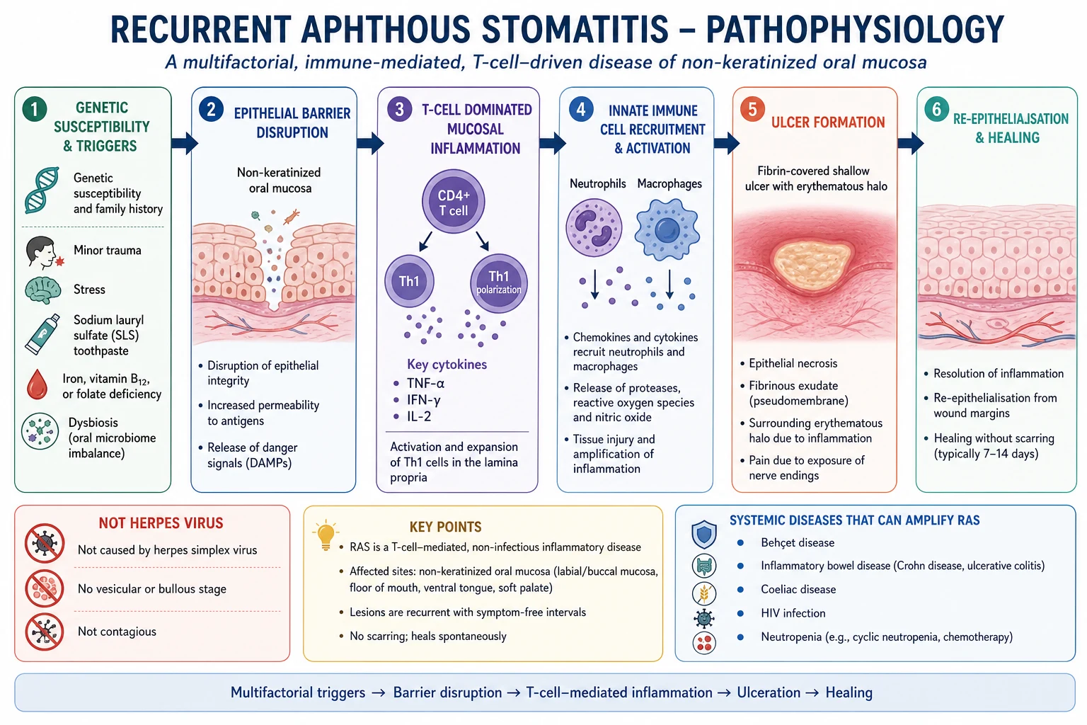
The yellow-white centre is not pus in the usual bacterial abscess sense. It is fibrinous exudate and necrotic debris over an epithelial defect. The red halo reflects adjacent inflammatory hyperaemia. This explains why chlorhexidine may reduce secondary colonisation and discomfort but does not treat the primary immune driver; why topical corticosteroids started early can shorten attacks; and why antivirals do not work for true aphthae even when the herpetiform subtype resembles herpes clinically.[1][3]
Aphthous ulcers are also a good example of why oral mucosa is not merely skin in a wet environment. The typical distribution on non-keratinized mucosa reflects differences in epithelial thickness, keratinisation, mechanical exposure and immune-microbial interface. Keratinized attached gingiva and hard palate are relatively spared in idiopathic RAS. When a patient has recurrent lesions on keratinized mucosa, especially if lesions begin as vesicles, the diagnostic centre of gravity moves toward HSV, varicella-zoster, erythema multiforme, trauma or autoimmune blistering disease rather than simple aphthae.[3][5]
The microbial story is nuanced. RAS is not a disease caused by one bacterium or one virus, but the oral microbiome, epithelial barrier and mucosal immune system communicate continuously. Dysbiosis may lower the threshold for inflammation, and systemic inflammatory diseases may alter mucosal immunity. This is why the examination answer should say "noninfectious and immune-mediated" rather than "sterile" in a simplistic way. It is also why routine bacterial swabs are usually unhelpful unless secondary infection, candidiasis or immunocompromise is suspected.[4]
Clinical Presentation
The typical patient describes a prodrome of burning, tingling or soreness for one to two days followed by a painful round or oval ulcer. Minor aphthae usually appear on the inner lip, buccal mucosa, ventral tongue, floor of mouth or soft palate. Pain is often disproportionate to size because oral movement, speaking, eating, toothpaste and acidic foods repeatedly stimulate the exposed nerve endings. The ulcer is shallow, has a soft base, and lacks the rolled indurated edge that makes malignancy dangerous.[1][3]
The history must establish recurrence, duration, site, number, size and healing. Ask whether lesions heal completely between episodes, whether the same site is repeatedly traumatised, whether there were vesicles first, whether ulcers occur on the hard palate or gingiva, whether there are genital ulcers, eye symptoms, skin lesions, fever cycles, diarrhoea, weight loss, arthralgia, immunosuppression, medication changes, nutritional risk or family history. A patient with two minor ulcers per year needs a different workup from a patient with large ulcers every month, weight loss and diarrhoea.[2][5]
Major aphthae are larger, deeper and more disabling. They may involve the soft palate, fauces or oropharynx and cause odynophagia, poor oral intake, dehydration risk and weight loss. They may heal with scarring. A major ulcer persisting for several weeks can look alarming, but the clinician must not simply label it "major aphtha" without reconsidering oral squamous cell carcinoma, deep fungal infection, TB, syphilis, Crohn disease, HIV-associated ulceration or drug-induced ulceration when the appearance or history is atypical.[2][3]
Herpetiform aphthae present as crops of many tiny ulcers. The name is a trap: they are not caused by herpes simplex virus. Unlike HSV, true herpetiform aphthae do not start as vesicles and do not preferentially involve keratinized mucosa. They may coalesce and become irregular, which can make them look like erosive inflammatory disease. HSV PCR from an early lesion is useful when the history is vesicular, when keratinized mucosa is involved, when the patient is immunocompromised, or when antiviral treatment would change management.[1][3]
Special presentations deserve explicit mention. Children with mouth ulcers and fever may have primary HSV gingivostomatitis, hand-foot-mouth disease, herpangina, PFAPA or cyclic neutropenia rather than simple RAS. Pregnant patients may report fluctuating disease, but systemic therapy choices are constrained. Immunocompromised patients, including advanced HIV, transplant recipients and patients on chemotherapy, can develop large aphthous-like ulcers and opportunistic ulcer mimics such as HSV, CMV, Candida-associated erosions and neutropenic ulceration. Older adults with new-onset recurrent oral ulceration require a medication review and a lower threshold for malignancy evaluation because "new aphthae" is less reassuring late in life.[2][5]
Differential Diagnosis
The differential diagnosis is the section that separates a safe clinician from a candidate who has memorised a single line. Examiners ask it because the mouth has few ways to show many diseases: infection, trauma, autoimmune blistering disease, inflammatory dermatoses, cancer, nutritional deficiency and systemic inflammatory disease can all produce painful erosions or ulcers. The discriminators are site, onset, vesicles, number, recurrence, systemic symptoms, induration, healing time and mucosal background.[3][5]
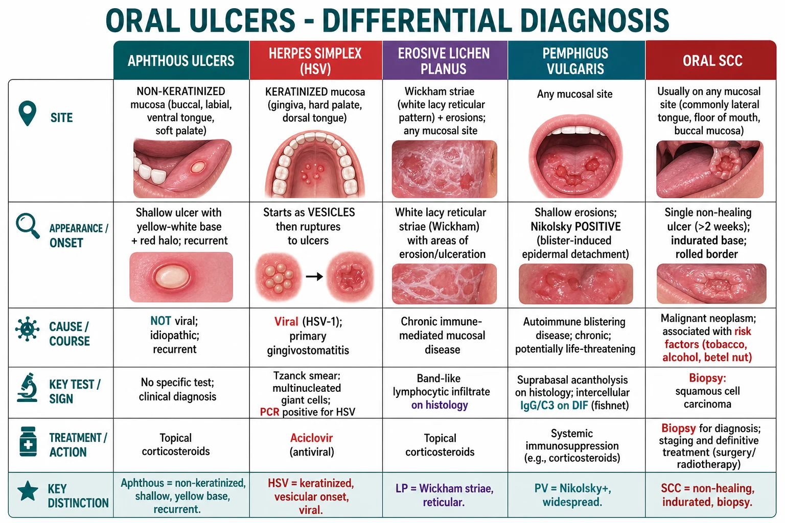
| Mimic | Clue against simple RAS | How to confirm or act |
|---|---|---|
| Herpes simplex virus | Vesicles precede ulcers; keratinized gingiva or hard palate; systemic primary gingivostomatitis; recurrent same-site clusters | HSV PCR from fresh lesion if uncertain; antivirals for confirmed HSV |
| Hand-foot-mouth disease | Child or outbreak setting; fever; vesicles/ulcers in mouth plus palms, soles or buttocks | Clinical diagnosis; supportive care and infection-control advice |
| Traumatic ulcer | Single lesion at bite line or near sharp tooth, denture or orthodontic appliance | Remove trauma; review healing; biopsy if persistent |
| Oral squamous cell carcinoma | Persistent solitary ulcer, induration, rolled edge, bleeding, fixation, cervical node, tobacco, alcohol or betel risk | Urgent oral medicine, ENT or maxillofacial referral and biopsy |
| Erosive lichen planus | Bilateral white lacy Wickham striae with erythema or erosions; chronic soreness | Biopsy with histology and direct immunofluorescence if atypical |
| Pemphigus vulgaris | Widespread fragile erosions, desquamative gingivitis, positive Nikolsky sign, skin blisters | Biopsy for histology and perilesional direct immunofluorescence |
| Mucous membrane pemphigoid | Desquamative gingivitis, tense blisters, ocular symptoms or scarring | Biopsy with direct immunofluorescence; eye assessment if suspected |
| Erythema multiforme | Acute mucosal erosions with target lesions; HSV, Mycoplasma or drug trigger | Clinical pattern; manage trigger and severity |
| Fixed drug eruption or drug ulcer | Same site after medication exposure; nicorandil can cause deep oral ulceration | Medication review and withdrawal if safe |
| Crohn disease or coeliac disease | GI symptoms, weight loss, anaemia, perianal disease, growth failure in children | Directed bloods, coeliac serology and gastroenterology referral |
A common unsafe answer is "HSV is painful and aphthae are painful too, so they are hard to distinguish." They can be hard in real life, but the high-yield discriminator is the vesicular phase and mucosal site. HSV classically produces vesicles that rupture into ulcers, and recurrent intraoral HSV favours keratinized mucosa. Aphthae are ulcers from the outset and favour movable non-keratinized mucosa. When the patient is immunocompromised, when lesions are atypical, or when the distinction changes treatment, swab early for HSV PCR rather than guessing.[3][5]
Oral squamous cell carcinoma is the can-not-miss mimic. Pain is not reassuring, because cancers can be painful once ulcerated or infected. A non-healing ulcer for more than 2-3 weeks, particularly with induration, rolled border, spontaneous bleeding, erythroplakia, leukoplakia, fixation, dysphagia, odynophagia, referred otalgia, neck mass, or tobacco, alcohol or betel exposure, needs urgent specialist assessment. Reassuring the patient with "it is probably aphthous" without safety-net review is a classic pitfall.[3][5]
Clinical & Bedside Assessment
Bedside assessment begins before investigations. Inspect the entire oral cavity with good light, gloves and a tongue depressor: lips, labial mucosa, buccal mucosa, gingiva, floor of mouth, ventral and dorsal tongue, lateral tongue, hard palate, soft palate, tonsillar pillars and oropharynx. Note size, depth, border, base, number, site, symmetry, surrounding mucosa, dental trauma and whether lesions are ulcers, erosions, vesicles, plaques or bullae. Palpate any persistent ulcer gently for induration and palpate the neck for lymphadenopathy.[3][5]
[1]Ask targeted systemic questions. Behçet disease screening requires oral ulcer recurrence, genital ulcers, eye pain, photophobia, blurred vision, floaters, erythema nodosum, acneiform lesions, pathergy-like reactions, arthralgia, neurological symptoms and vascular events. Gastrointestinal screening covers chronic diarrhoea, abdominal pain, rectal bleeding, weight loss, perianal symptoms and growth faltering. Haematinic screening covers fatigue, pallor, pica, heavy menstrual bleeding, vegan diet, bariatric surgery, malabsorption and glossitis. Neutropenia screening covers recurrent fevers, bacterial infections and a striking periodicity.[2][6][8]
The pathergy test is relevant to Behçet disease but should not be oversold. A sterile needle prick causing a papule or pustule after 24-48 hours supports Behçet disease in the right clinical setting, but sensitivity varies by ethnicity and technique, and a negative test does not exclude the disease. The International Study Group style of exam question often centres on recurrent oral ulcers plus two of genital ulcers, eye lesions, skin lesions or positive pathergy; modern classification systems are more nuanced, but the bedside principle remains that ocular symptoms require urgent ophthalmology.[6]
Investigations
Simple, infrequent minor RAS in a well child or young adult with a long stable pattern and no red flags can be diagnosed clinically without a large test panel. Investigation is appropriate when ulcers are frequent, severe, major, herpetiform, adult-onset, persistent, treatment-resistant, associated with systemic symptoms, associated with abnormal examination, or occurring in an immunocompromised patient. The workup should be targeted; a scattergun panel is less impressive than a clear rationale.[1][2]
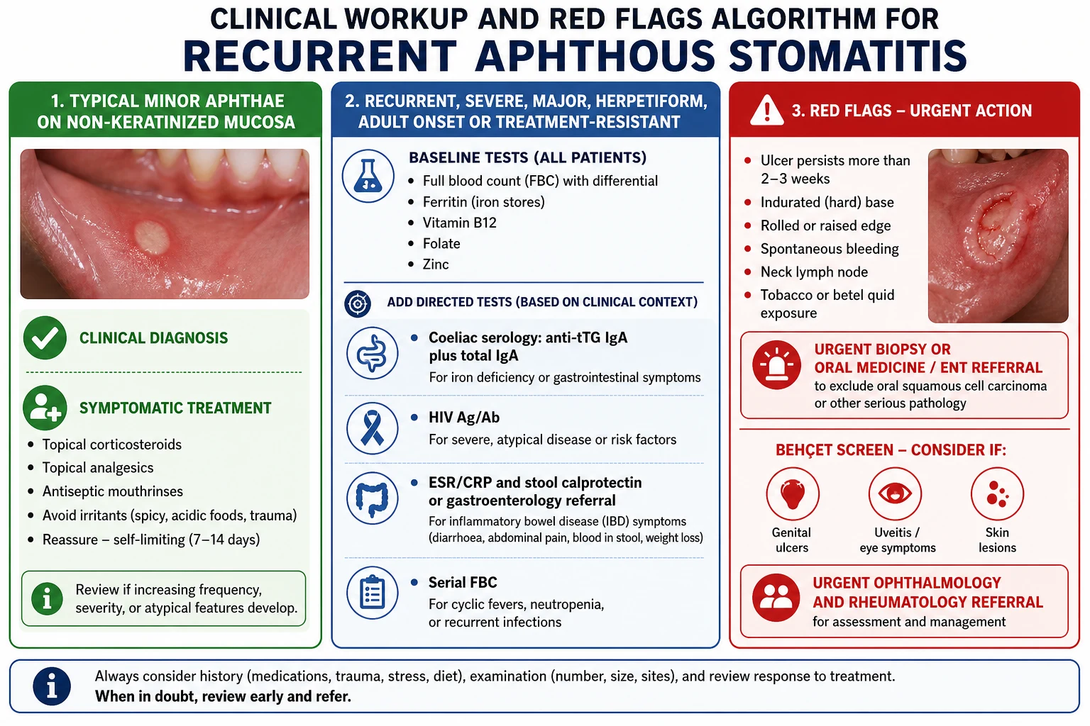
| Test | When to order | What it answers |
|---|---|---|
| FBC with differential | Frequent, severe, major, systemic symptoms, paediatric fever cycles, immunocompromise | Anaemia, neutropenia, lymphopenia or haematological disease |
| Ferritin and iron studies | Recurrent disease, glossitis, fatigue, heavy menses, GI symptoms, restricted diet | Iron deficiency and possible occult blood loss or malabsorption |
| Vitamin B12 and folate | Recurrent disease, glossitis, neuropathy, vegan diet, malabsorption, macrocytosis | Treatable haematinic deficiency |
| Zinc | Severe or recurrent disease when nutritional risk exists | Less common but potentially correctable deficiency |
| Coeliac serology | Iron deficiency, GI symptoms, growth issues, family history or recurrent unexplained ulcers | Usually anti-tTG IgA with total IgA; adjust for IgA deficiency |
| HIV Ag/Ab | Severe, major, atypical, persistent, recurrent infection, risk factors or immunosuppression | HIV-associated aphthous-like disease and opportunistic mimics |
| ESR, CRP, faecal calprotectin or gastroenterology referral | Diarrhoea, rectal bleeding, abdominal pain, weight loss, perianal disease | IBD screen and referral justification |
| Serial FBC | Periodic fever, infections and ulcers every few weeks | Cyclic neutropenia pattern |
| HSV PCR | Vesicular onset, keratinized mucosal lesions, immunocompromise or diagnostic uncertainty | Confirms HSV and avoids inappropriate steroid-only treatment |
| Biopsy | Persistent ulcer, induration, rolled edge, unexplained solitary lesion, atypical mucosa | Excludes SCC, dysplasia, autoimmune blistering disease or granulomatous disease |
Histopathology is not required for classic minor RAS. When biopsied, an aphthous ulcer is nonspecific: ulceration with fibrinopurulent surface exudate, mixed inflammatory infiltrate and granulation tissue. That is why biopsy is performed not to "prove RAS" but to exclude malignancy, autoimmune blistering disease, lichen planus, infection or granulomatous disease. Direct immunofluorescence is essential if pemphigus, pemphigoid or another immunobullous disorder is suspected; the sample should include perilesional mucosa, not only necrotic ulcer bed.[3][5]
Management — Resuscitation
RAS is rarely a resuscitation problem, but the examiner map still asks what is immediately dangerous. Severe oral pain can prevent drinking, particularly in children, older adults and immunocompromised patients. Immediate priorities are hydration status, ability to swallow, airway warning signs, sepsis signs, neutropenic fever and ocular symptoms suggesting Behçet uveitis. A child who is drooling, unable to drink, febrile and toxic should not be managed as routine aphthae. A patient with chemotherapy-associated neutropenia and oral ulceration needs urgent infection assessment.[3][5]
[1]Initial supportive care includes oral fluids, soft diet, avoidance of acidic and spicy foods, regular simple analgesia when appropriate, topical anaesthetic before meals, and safety-net review. If the patient is immunocompromised or lesions have a vesicular or necrotic appearance, avoid escalating topical or systemic corticosteroids until HSV, CMV, Candida-associated erosions, neutropenic ulceration or other infection has been considered. The safest resuscitation answer is therefore: assess intake and systemic illness first, then treat pain, then investigate red flags.[3][5]
Management — Definitive & Stepwise
Management has five aims: reduce pain, shorten ulcer duration, reduce recurrence, identify treatable triggers, and avoid missing systemic disease or cancer. Most patients need explanation and topical therapy rather than systemic immunosuppression. The patient should be told that RAS is common, noncontagious, not due to poor hygiene and not HSV, but they should also be given clear reasons to return: ulcer persistence, increasing size, induration, systemic symptoms, genital ulcers, eye symptoms or weight loss.[1][2]
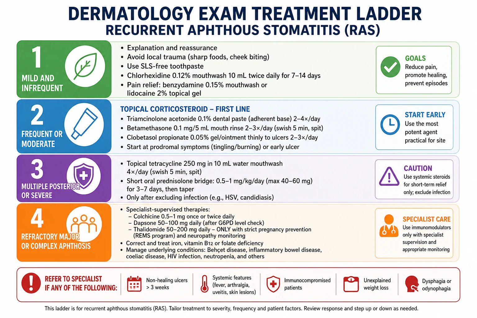
For mild infrequent disease, advise a soft toothbrush, correction of dental trauma, avoidance of reproducible food triggers, SLS-free toothpaste trial, and topical pain control. Chlorhexidine mouthwash can reduce secondary bacterial colonisation and may reduce severity, but it stains teeth and alters taste, so it should be used as a short course rather than a permanent habit. Benzydamine mouthwash, lidocaine gel or other topical anaesthetics can help eating, but anaesthetised mucosa can be bitten, and children require careful dosing and supervision.[1][2][3]
Topical corticosteroids are first-line pharmacological therapy for frequent, moderate or painful RAS. The principle is early local anti-inflammatory treatment, ideally during prodrome or at ulcer onset. Common regimens include triamcinolone acetonide 0.1% dental paste applied thinly to dried ulcer two to four times daily after meals and at bedtime; betamethasone sodium phosphate 0.5 mg dissolved in 10-15 mL water as a rinse and spit two to four times daily for multiple ulcers; clobetasol gel or ointment applied carefully for severe local disease under specialist advice; or fluticasone spray directed to difficult posterior sites. Patients should spit, not swallow, steroid rinses unless specifically instructed, and prolonged courses increase candidiasis risk.[1][2]
| Treatment | Typical use | Practical caution |
|---|---|---|
| Triamcinolone acetonide 0.1% dental paste | Accessible labial or buccal ulcers; apply thinly after meals and at bedtime | Dry mucosa first; best started early |
| Betamethasone sodium phosphate 0.5 mg rinse | Multiple or posterior ulcers; dissolve in 10-15 mL water, rinse and spit | Do not swallow routinely; watch for candidiasis with repeated courses |
| Chlorhexidine 0.12-0.2% mouthwash | Short adjunct course to reduce secondary colonisation and discomfort | Tooth staining and taste disturbance limit prolonged use |
| Topical lidocaine or benzydamine | Pain control before meals | Analgesic only; avoid excess and supervise children |
Tetracycline mouthwash is sometimes used for recurrent or herpetiform disease because of anti-inflammatory and antimicrobial effects. A common specialist pattern is doxycycline or tetracycline capsule contents dispersed in water, used as a rinse and spit for several minutes, usually for a short course. It is not appropriate for young children or pregnancy, can irritate mucosa, and should not become a reflex prescription when topical corticosteroids and trigger correction have not been optimised.[2]
Systemic corticosteroids are rescue therapy rather than chronic management. A short oral prednisolone course may be used for severe major aphthae causing major pain or poor intake, but infection and malignancy must be excluded when the presentation is atypical. Repeated steroid bursts without a diagnosis are a warning sign that the patient needs oral medicine, dermatology or rheumatology review. Long-term systemic steroids are not a good chronic strategy for idiopathic RAS.[2][5]
Refractory major aphthae, complex aphthosis or Behçet-associated mucocutaneous disease may require systemic steroid-sparing therapy. Colchicine is often used for mucocutaneous Behçet-like disease and recurrent aphthosis under specialist supervision; gastrointestinal intolerance, renal impairment, hepatic impairment and drug interactions must be considered. Dapsone can help neutrophil-mediated ulceration but requires G6PD testing, FBC and liver monitoring because haemolysis, methaemoglobinaemia and agranulocytosis are serious risks. Thalidomide is highly effective in HIV-associated severe aphthous ulcers and refractory major aphthae, but teratogenicity, neuropathy, sedation and thrombosis risk make it specialist-only with strict pregnancy prevention and monitoring. Apremilast is used in Behçet oral ulcers in some regions and is a specialist decision rather than an MBBS first-line answer.[2][6][7]
Treat deficiencies and associated disease. Iron replacement, vitamin B12 replacement, folate replacement or zinc replacement should be given when deficiency is demonstrated, and the cause of deficiency should be sought. Coeliac disease requires a gluten-free diet and gastroenterology follow-up, not simply repeated oral gels. HIV-associated ulcers require antiretroviral optimisation and exclusion of HSV, CMV and fungal disease. Crohn disease oral ulcers often improve with control of intestinal inflammation. Behçet disease requires organ-based management, especially urgent eye care when uveitis is suspected.[2][6][8][9]
Specific Subtypes & Scenarios
Minor RAS is managed with reassurance, topical analgesia, trigger control and topical corticosteroid for frequent episodes. The key counselling point is expectation: ulcers should heal in 7-14 days without scarring. If the patient returns with a lesion still present after 3 weeks, the diagnosis must be revisited rather than continuing to call it minor RAS. Recurrence prevention is pragmatic: identify trauma, SLS toothpaste, stress/sleep triggers, nutritional deficiency and medication associations; do not impose broad restrictive diets without a reproducible trigger.[1][2]
Major RAS requires a lower threshold for investigation because it causes morbidity and mimics cancer. Check hydration, nutrition, dysphagia, weight change and pain control. Examine for genital ulcers, ocular symptoms, skin lesions, GI disease and immunosuppression. If a major ulcer is solitary, indurated, persistent or in a high-risk patient, biopsy pathway referral is more important than trying another mouthwash. For recurrent confirmed major aphthae, oral medicine or dermatology may use high-potency topical corticosteroids, systemic bridge therapy and steroid-sparing agents.[2][3][5]
Herpetiform aphthae create the HSV trap. They are multiple tiny ulcers without a vesicular stage and are not improved by aciclovir unless there is true coincident HSV. Because numerous ulcers can be extremely painful, mouth rinses are often more practical than dental paste. Consider topical steroid rinses, chlorhexidine, pain control and sometimes tetracycline rinse in non-pregnant adults under appropriate guidance. If lesions are vesicular, on keratinized mucosa or recurrent at the same site, test for HSV rather than relying on the name "herpetiform".[1][2][3]
Behçet disease should be suspected when recurrent oral ulcers occur with genital ulcers, ocular inflammation, erythema nodosum, acneiform lesions, pathergy, arthritis, neurological symptoms or vascular disease. Oral ulcers are usually the earliest and most frequent manifestation, so dermatology and oral medicine clinicians often see the patient before the systemic diagnosis is obvious. Eye symptoms are time-critical: uveitis and retinal vasculitis can threaten vision. Mucocutaneous disease may be treated with topical corticosteroids, colchicine, apremilast or other systemic agents depending on region and severity; organ-threatening disease requires specialist immunosuppression.[6]
Inflammatory bowel disease and coeliac disease should be considered when aphthae accompany chronic diarrhoea, abdominal pain, rectal bleeding, weight loss, iron deficiency, growth faltering, perianal symptoms or a family history. Aphthous-like ulcers can precede gastrointestinal diagnosis. Coeliac disease can present with oral aphthae and nutritional deficiencies; anti-tTG IgA with total IgA is the usual screening framework, adjusted for IgA deficiency and local pathways. Do not diagnose coeliac disease from mouth ulcers alone, and do not start a gluten-free diet before testing if coeliac diagnosis is being pursued.[2][8][9]
HIV-associated aphthous ulcers are often large, deep, persistent and painful, especially with advanced immunosuppression. Opportunistic infections can mimic aphthae, so HSV, CMV, candidiasis and malignancy must be considered. Thalidomide showed efficacy for oral aphthous ulcers in HIV infection in a randomized trial, but current use is constrained by teratogenicity and toxicity and sits firmly in specialist care. In modern practice, antiretroviral therapy optimisation and careful infectious differential diagnosis are part of the management plan.[2][7]
Cyclic neutropenia presents with recurrent fever, malaise, bacterial infections, gingivitis or oral ulcers at roughly three-week intervals. A single normal neutrophil count does not exclude it if the timing is wrong. Serial FBC with differential across several weeks may be needed when the history is convincing. This scenario is a favourite viva corner because the mouth ulcer is the visible clue to a haematological rhythm.[2][5]
Complications & Pitfalls
Most simple aphthae heal without sequelae, but the complications that matter are pain, impaired oral intake, sleep disturbance, school or work absence, scarring from major aphthae, secondary infection, candidiasis from repeated topical steroids, and psychological burden from unpredictable recurrence. The bigger danger is diagnostic: missed oral SCC, missed Behçet uveitis, missed IBD, missed HIV, missed neutropenia, or unnecessary antivirals and antibiotics for a noninfectious condition.[2][3][5]
[1]A subtle pitfall is over-investigation of straightforward disease and under-investigation of atypical disease. The patient with a 10-year history of two minor ulcers per year, classic non-keratinized sites and complete healing does not need repeated autoimmune panels. The patient with new adult-onset major ulcers, weight loss and anaemia does. Exam answers should show that the candidate can calibrate investigation to risk rather than memorising a fixed battery for every mouth ulcer.[2][5]
Prognosis & Disposition
Minor RAS is benign and self-limiting, although it may be painful and recurrent for years. Many patients experience decreasing frequency with age. Major RAS has a more prolonged course, greater pain, possible scarring and greater association with systemic disease or immunosuppression. Herpetiform aphthae can recur frequently and be disabling despite small individual ulcer size. Prognosis improves when triggers, deficiencies and systemic associations are identified rather than simply suppressing each episode.[1][2]
Disposition depends on pattern. Manage classic minor RAS in primary care or general dermatology with safety-net review. Refer to oral medicine, dermatology, ENT or maxillofacial surgery for non-healing ulcers, suspicious morphology, uncertain diagnosis, major or complex aphthosis, severe pain, scarring, dysphagia, recurrent treatment failure, need for biopsy, or consideration of systemic therapy. Refer to ophthalmology urgently for suspected Behçet eye involvement. Refer to gastroenterology for IBD or coeliac features, infectious diseases or HIV services for suspected or known HIV with severe ulcers, and haematology or paediatrics for suspected cyclic neutropenia.[2][5][6]
Safety-netting is part of the treatment, not an administrative afterthought. Tell the patient to return if an ulcer persists beyond 2-3 weeks, becomes firm or raised, bleeds, enlarges, is associated with a neck lump, causes weight loss or dysphagia, or if genital ulcers, eye pain, photophobia, blurred vision, fever cycles, diarrhoea or new skin lesions develop. Give a review date for atypical lesions rather than relying on the patient to interpret progression alone.[3][5]
Special Populations
Children commonly present with mouth ulcers, but RAS is only one possibility. Primary HSV gingivostomatitis causes fever, diffuse gingivitis, vesicles and ulcers and may cause dehydration. Hand-foot-mouth disease causes oral lesions plus acral or buttock rash in an outbreak context. PFAPA causes periodic fever, aphthae, pharyngitis and cervical adenitis in young children. Cyclic neutropenia causes periodic fevers, infections and ulcers. In children, always assess hydration, fever, systemic toxicity, immunisation context, exposure history and ability to drink.[3][5]
Pregnancy requires conservative treatment whenever possible. Trigger avoidance, chlorhexidine, simple analgesic choices compatible with pregnancy, and carefully selected topical corticosteroids are usually preferred. Tetracycline mouthwash is generally avoided in pregnancy. Thalidomide is absolutely contraindicated because of severe teratogenicity. Dapsone and colchicine decisions in pregnancy require specialist indication and risk-benefit assessment. The exam answer should not list systemic agents without saying pregnancy status must be checked before prescribing.[2][7]
Older adults deserve a medication and malignancy lens. New-onset oral ulceration in later life is less typical for idiopathic RAS. Review nicorandil, NSAIDs, bisphosphonates, cytotoxic drugs, immunosuppressants and oral appliances. Examine for denture trauma and xerostomia. Maintain a low threshold for biopsy of persistent or atypical ulcers, especially with tobacco, alcohol or betel exposure, neck nodes, dysphagia or induration.[3][5]
Immunocompromised patients are high risk for both severe aphthous-like disease and mimics. HIV, transplant immunosuppression, chemotherapy, neutropenia and biologic therapy change the differential. HSV, CMV, deep fungal infection, candidiasis-associated erosions, EBV-associated lesions, drug ulcers and malignancy should be considered. If topical steroids are used, follow-up must be closer; if systemic steroids are contemplated, infection exclusion and specialist review are prudent.[2][5][7]
Evidence, Guidelines & Regional Differences
The evidence base for RAS is a mixture of clinical reviews, small randomized trials, specialist experience and extrapolation from Behçet disease or HIV-associated aphthous ulcer trials. Topical corticosteroids are widely accepted as first-line therapy for frequent or painful disease because they target the inflammatory phase with low systemic exposure. Chlorhexidine and topical anaesthetics are symptomatic adjuncts. Systemic agents have more toxicity and less generalisable evidence, so they should be reserved for severe, refractory or systemic-associated disease under specialist care.[1][2][5]
Thalidomide for HIV-associated oral aphthae
New England Journal of Medicine
Randomized controlled trial in patients with HIV infection and oral aphthous ulcers.
Key finding
Thalidomide was effective for severe oral aphthous ulcers in this immunocompromised population.
Practice change
Established thalidomide as an effective but specialist-only option because teratogenicity and neuropathy make safety controls mandatory.
Regional differences are mainly about drug availability, referral pathways and pregnancy-prevention regulation rather than the diagnostic concept. In Australia and New Zealand, the practical pathway is often general practice or dentistry to oral medicine, dermatology, ENT or oral and maxillofacial surgery for persistent or suspicious ulcers; Behçet eye symptoms require urgent ophthalmology. In the United Kingdom, suspected oral cancer follows urgent cancer referral pathways, and oral medicine or maxillofacial services often coordinate biopsy. In the United States, oral medicine, ENT, dermatology, dentistry and oral-maxillofacial surgery pathways vary by insurance and local access; thalidomide access is governed by strict risk-management programs. The safe universal rule is biopsy pathway for persistent suspicious ulcers and specialist supervision for systemic immunomodulators.[5][6][7]
In ANZ practice, document the ulcer duration and cancer red flags clearly, arrange oral medicine, oral and maxillofacial, ENT or dermatology referral according to local access, and send same-day ophthalmology referral for suspected Behçet uveitis or retinal symptoms.
Exam Pearls
[1]Screen severe aphthae with MOUTH
MOUTH
Malignancy red flags: persistent, indurated, bleeding, neck node.
Ocular and genital symptoms: Behçet disease until assessed.
Underlying gut disease: IBD or coeliac, especially with anaemia or GI symptoms.
Tests for deficiencies: FBC, ferritin, B12, folate, zinc.
HIV or haematology: severe ulcers, immunosuppression, neutropenia or periodic fevers.
Viva trap: how do you distinguish aphthous ulcers from HSV?
Aphthous ulcers are non-vesicular ulcers from the outset and classically affect non-keratinized movable mucosa. HSV usually has grouped vesicles before ulceration and recurrent intraoral HSV favours keratinized mucosa such as hard palate or attached gingiva. If uncertain, especially in immunocompromise, send HSV PCR from a fresh lesion before escalating steroids.
Exam application bank (NEET-PG / INICET)
One-line answer
Recurrent aphthous stomatitis is a clinical diagnosis of painful recurrent round or oval ulcers on non-keratinized oral mucosa, classified as minor, major, or herpetiform aphthae. The examiner expects candidates to distinguish aphthae from HSV, hand-foot-mouth disease, trauma, oral SCC, lichen planus and pemphigus; screen for Behçet disease, IBD, coeliac disease, HIV, cyclic neutropenia and haematinic deficiency when disease is severe or atypical; and manage stepwise with analgesia, chlorhexidine, topical corticosteroids, tetracycline mouthwash, deficiency replacement and specialist systemic therapy for refractory disease.
Worked stems (answer without another resource)
Stem 1 — Classic presentation. Map symptoms to mechanism; name the first investigation and first treatment step with dose/route if drug therapy is standard. [1]
Stem 2 — Unstable / complicated. List red flags that force immediate resuscitation, theatre, ICU, antidote, or reperfusion — and what you do in the first 15 minutes. [1]
Stem 3 — Atypical group. Elderly, pregnancy, child, or immunocompromised: how presentation and thresholds change. [1]
Stem 4 — Differential trap. Name the three closest mimics and one discriminator for each. [1]
Stem 5 — Disposition. Who goes home with safety-netting, who is admitted, who needs HDU/ICU/theatre, and what follow-up is mandatory. [1]
Rapid viva checklist
- Definition + classification
- Pathophysiology chain
- Bedside signs / criteria
- Score with exact components (if any)
- Emergency bundle
- Definitive therapy with doses
- Complications of disease and of treatment
- Special populations
- Guideline/trial name if classic
- Three exam traps
Coverage self-check
If you cannot answer any stem above from this page alone, re-read the matching section — the page is intended to be self-sufficient for final-prof and NEET-PG/INICET questions on Aphthous ulcers.
[1]One-page clinical workflow
Definition
Painful recurrent shallow oral ulcers with yellow-white fibrin base and red halo, usually on non-keratinized movable mucosa.
Classification
Minor heals in 7-14 days without scarring; major is larger, deeper and may scar; herpetiform is clustered pinpoint aphthae and is not HSV.
Key Investigations
For recurrent, severe, major or atypical disease: FBC with differential, ferritin, B12, folate and zinc; add coeliac, HIV, IBD and serial neutrophil testing when indicated.
Management
Education, trauma reduction, SLS-free toothpaste, chlorhexidine and topical analgesia; topical corticosteroids first-line; systemic colchicine, dapsone or thalidomide only under specialist supervision.
Prognosis
Minor RAS is benign and self-limiting; major or complex aphthosis needs referral, systemic screen and safety-netting for non-healing suspicious ulcers.
Key References
References
- [1]Gasmi Benahmed A, Noor S, Menzel A, et al. Oral Aphthous: Pathophysiology, Clinical Aspects and Medical Treatment Arch Razi Inst, 2021.PMID 35355774
- [2]Saikaly SK, Saikaly TS, Saikaly LE. Recurrent aphthous ulceration: a review of potential causes and novel treatments J Dermatolog Treat, 2018.PMID 29278022
- [3]Hargitai IA. Painful Oral Lesions Dent Clin North Am, 2018.PMID 30189985
- [4]Lin D, Yang L, Wen L, et al. Crosstalk between the oral microbiota, mucosal immunity, and the epithelial barrier regulates oral mucosal disease pathogenesis Mucosal Immunol, 2021.PMID 34040155
- [5]Stoopler ET, Villa A, Bindakhil M, et al. Common Oral Conditions: A Review JAMA, 2024.PMID 38530258
- [6]Alibaz-Oner F, Direskeneli H. Update on the Diagnosis of Behçet's Disease Diagnostics (Basel), 2022.PMID 36611332
- [7]Jacobson JM, Greenspan JS, Spritzler J, et al. Thalidomide for the treatment of oral aphthous ulcers in patients with human immunodeficiency virus infection. National Institute of Allergy and Infectious Diseases AIDS Clinical Trials Group N Engl J Med, 1997.PMID 9154767
- [8]Koparal M, Ege B, Dogan EI, et al. Evaluation of biochemical variables in patients with recurrent aphthous stomatitis J Stomatol Oral Maxillofac Surg, 2023.PMID 36162803
- [9]Sahin Y. Celiac disease in children: A review of the literature World J Clin Pediatr, 2021.PMID 34316439