Dermatology · Medicine
Atopic dermatitis
Also known as Atopic eczema · Eczema · Dermatitis · Endogenous eczema
Atopic dermatitis is a chronic, relapsing, inflammatory skin disease driven by skin-barrier dysfunction and type 2 inflammation. Fellowship-level assessment requires understanding of filaggrin biology, the IL-4/IL-13/IL-31 axis, the atopic march, severity scoring (EASI, IGA, DLQI), trigger avoidance, site-specific topical therapy, phototherapy, conventional systemic immunosuppressants, and the mechanisms and landmark trial evidence for biologics and JAK inhibitors.
On this page & tools
Your progress
Saved locally on this device.
Exam tags
Red flags
Definition & Classification
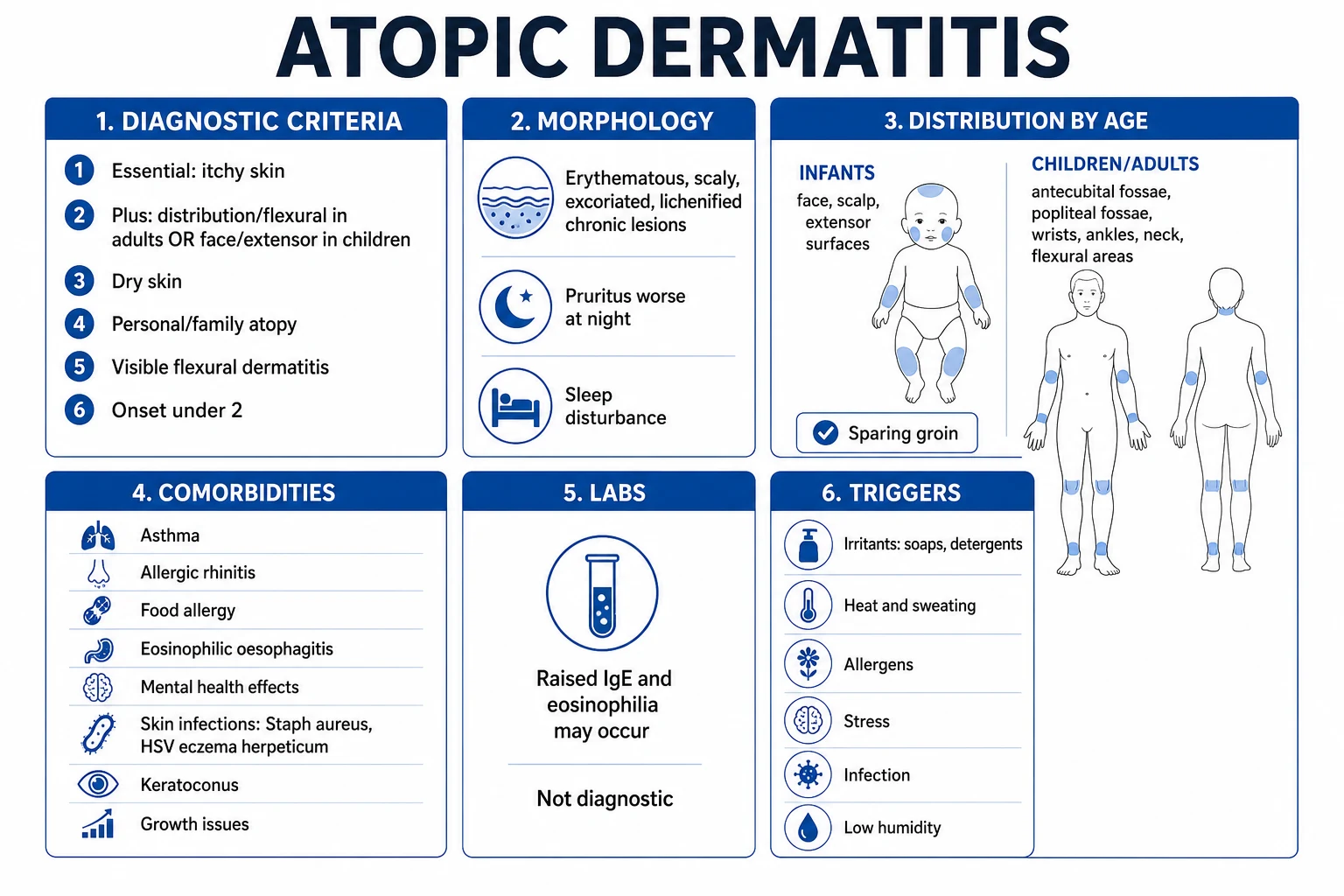
Atopic dermatitis (AD) is a chronic, relapsing, inflammatory skin disease characterised by pruritus, eczematous morphology, and a typical age-dependent distribution. It is the cutaneous component of the atopic march, with strong associations with asthma, allergic rhinitis and food allergy.[1]
| Term | Meaning |
|---|---|
| Acute eczema | Erythematous papules and vesicles with oedema, exudate and crusting |
| Subacute eczema | Erythematous, scaly, slightly lichenified plaques |
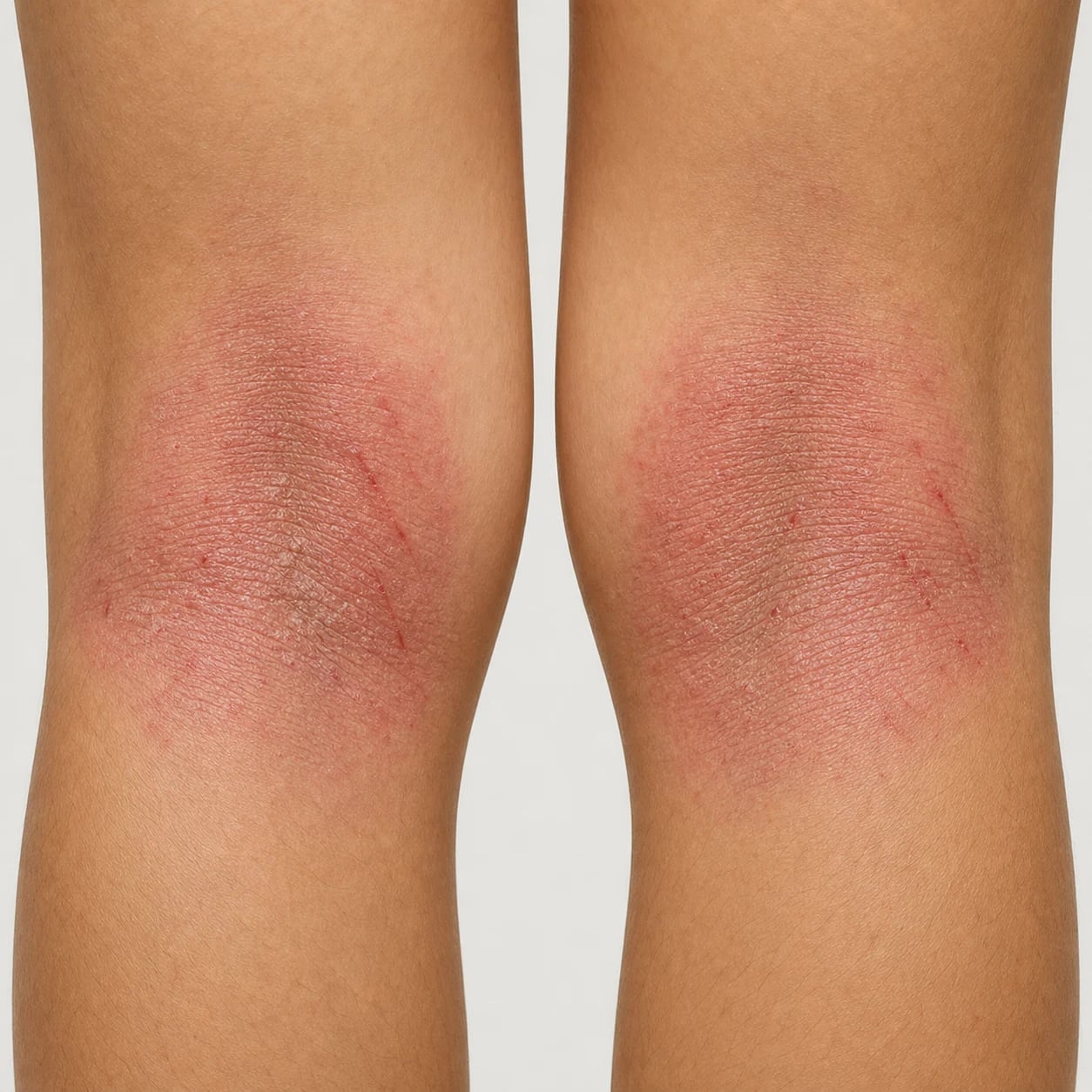
| Chronic eczema | Lichenified, thickened plaques with prominent skin lines and excoriations |
| Lichenified eczema | Long-standing plaques with lichenification from the itch–scratch cycle |
Epidemiology
AD affects up to 15–20% of children and 2–10% of adults worldwide, with rising prevalence in industrialised settings and wide geographic variation.[1] Onset is usually in infancy; about 60% of cases begin in the first year of life. A substantial minority persists into adulthood or recurs after quiescence.
Risk factors [1]
- Genetic: family history of atopy (atopic dermatitis, asthma, allergic rhinitis); filaggrin (FLG) loss-of-function mutations are the strongest known genetic risk factor.[6]
- Environmental: urbanisation, hard water, low humidity, exposure to allergens, tobacco smoke, and certain occupational irritants.
- Barrier defects: reduced ceramides, altered pH, and tight-junction abnormalities increase transepidermal water loss and allergen penetration.[7]
- Microbiome: colonisation with Staphylococcus aureus is common during flares and correlates with disease severity.[2]
Atopic dermatitis at a glance — the high-yield numbers
Pathophysiology
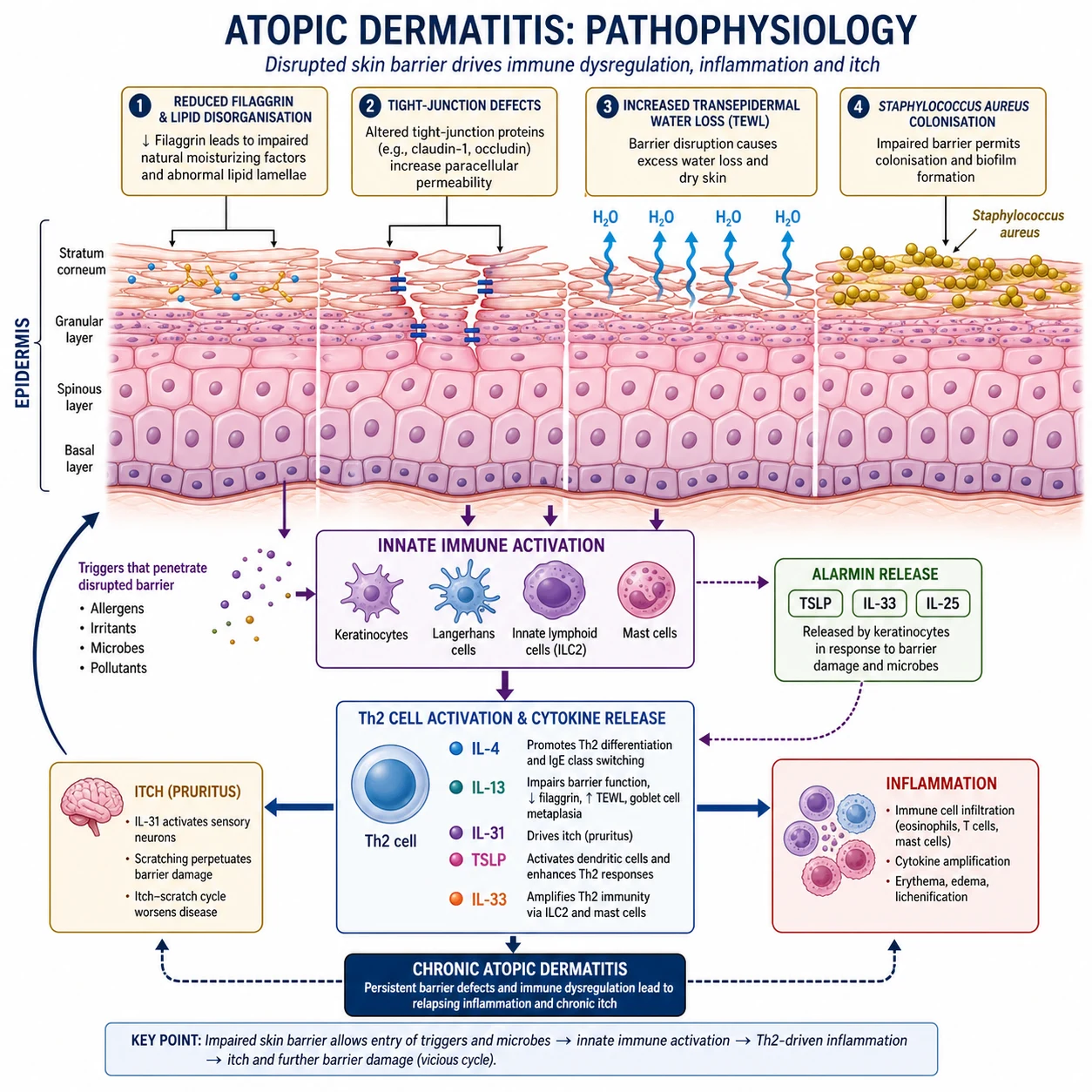
AD is a barrier-driven, type 2 inflammatory disease. [1]
Skin-barrier dysfunction
Filaggrin aggregates keratin filaments and is proteolysed into natural moisturising factors. FLG loss-of-function mutations, or Th2-driven downregulation of filaggrin and tight-junction proteins, increase transepidermal water loss and allow allergen and microbial entry.[6][7]
Type 2 inflammation
Keratinocytes release alarmins such as thymic stromal lymphopoietin (TSLP), IL-25 and IL-33, which promote Th2 responses. Key cytokines include: [1]
- IL-4 and IL-13 — central drivers of barrier dysfunction, IgE class switching, eosinophil recruitment and fibrosis.
- IL-31 — the "itch cytokine", acting on peripheral nerves to drive pruritus.
- IL-5 and IL-9 — eosinophil and mast-cell survival.
- IL-22 and IL-17 — contribute to epidermal hyperplasia in chronic lesions. [1]
Clinical Features
History
- Intense pruritus is the hallmark; scratching triggers the itch–scratch cycle.
- Personal or family history of atopy.
- Flares with stress, infection, allergens, irritants, temperature change, or hormonal factors.
- Sleep disturbance and reduced quality of life. [1]
Morphology
- Acute: erythematous papules, vesicles, oedema, exudate, crusting.
- Subacute/chronic: poorly demarcated erythematous plaques with fine scale, lichenification, and excoriations.
- Common secondary changes: excoriations, lichen simplex chronicus, prurigo nodules, dyspigmentation (post-inflammatory hypopigmentation or hyperpigmentation). [1]
Distribution by age
| Age | Typical sites |
|---|---|
| Infants | Face (cheeks), scalp, extensor surfaces, trunk |
| Children | Flexural sites (antecubital and popliteal fossae), wrists, ankles, neck |
| Adolescents/adults | Flexures, hands, feet, eyelids, nipples; head-and-neck dermatitis may predominate |
| Older adults | Lichenified, localised plaques; hand and nummular patterns more common |
Severity and quality-of-life measures
| Tool | What it measures | Notes |
|---|---|---|
| EASI | Extent and severity of erythema, oedema/papulation, excoriations, lichenification | 0–72; EASI-75 = ≥75% improvement |
| IGA | Investigator Global Assessment | 0 (clear) to 4 (severe); IGA 0/1 is a common trial endpoint |
| DLQI | Dermatology Life Quality Index | 0–30; ≥11 indicates large effect on life |
| POEM | Patient-Oriented Eczema Measure | Patient-reported symptoms, 0–28 |
| SCORAD | Severity scoring of atopic dermatitis | Combines signs, extent and symptoms |
EASI severity bands — atopic dermatitis trial and biologic-eligibility benchmark
Mild (EASI 1.1-7.0)
2 — Localised or limited-body-surface-area disease. Topical therapy mainstay; phototherapy if widespread.
How each score is calculated
| Score | Range | Calculation | What counts as response |
|---|---|---|---|
| EASI (Eczema Area and Severity Index) | 0 to 72 | Sum of four body-region subscores (head/neck, trunk, upper limbs, lower limbs). Each subscore = body surface area % times severity sum (erythema, oedema/papulation, excoriations, lichenification, each 0-3). Multiplied by an age-adjusted weighting. | EASI-50 = at-least-50% improvement; EASI-75 = at-least-75% (the modern benchmark for biologic/JAK response); EASI-90 = at-least-90% (high bar). |
| SCORAD (Scoring Atopic Dermatitis) | 0 to 103 | Extent (0-100% BSA via rule-of-nines, A) + 6 intensity items (0-18, B) + subjective itch and sleep (0-20, C). Formula: A/5 + 7B/2 + C. | Mild less than 25; moderate 25-50; severe greater than 50. SCORAD-50 (50% improvement) is a common endpoint. |
| IGA (Investigator Global Assessment) | 0 to 4 | Single 5-point clinician rating of overall lesion severity. Validated 0-4 scales (e.g. vIGA-AD) require no residual induration/papulation for 0 or 1. | IGA 0/1 with at-least-2-grade improvement is the FDA-recognised primary endpoint for many recent AD trials. |
| DLQI (Dermatology Life Quality Index) | 0 to 30 | 10-item patient questionnaire covering symptoms, daily activities, leisure, work/school, personal relationships, treatment. | at-least-11 = "very large effect on life"; 6-10 = "moderate"; less-than-6 = "small/none". DLQI at-least-11 is a common biologic-eligibility threshold. |
| POEM (Patient-Oriented Eczema Measure) | 0 to 28 | 7-item patient-reported symptom frequency over the prior week (itch, sleep, dryness, etc.). | POEM at-least-17 = severe; 8-16 = moderate; 3-7 = mild; 0-2 = clear/almost clear. |
| NRS itch (Numerical Rating Scale for worst itch) | 0 to 10 | Single-item 0-10 patient scale for worst itch over 24 hours. | at-least-4-point reduction is a clinically meaningful itch improvement; JAK inhibitors often achieve at-least-4-point reduction within days. |
| BSA (Body Surface Area) | 0 to 100% | Clinician estimate using rule-of-nines or handprint method (patient palm + fingers ≈ 1% BSA). | BSA >10% is a common biologic-eligibility threshold. |
Severity-guided thresholds for systemic therapy
The AAD 2024 systemic guideline and the UK NICE TA814 framework converge on broadly comparable entry thresholds for systemic immunosuppressants, biologics, or JAK inhibitors in moderate-to-severe AD: EASI at-least-16, IGA at-least-3, DLQI at-least-11 (or POEM at-least-16), and BSA >10%, after failure of optimised topical therapy for at-least-4-8 weeks. Trials of dupilumab, upadacitinib and abrocitinib enrolled on EASI at-least-16, IGA at-least-3 and at-least-10% BSA, with DLQI/POEM corroborating the patient-burden side. Tralokinumab ECZTRA 1/2 used IGA at-least-3 and EASI at-least-16 with BSA at-least-10%. Treat-to-target endpoints are EASI-75 or IGA 0/1 by week 16 — if not achieved, escalate or switch.[2][5]
Drug Doses — Specific Protocols
The following protocols reflect standard induction and maintenance dosing used in pivotal trials and current AAD 2024-2025 guidelines. Local formularies and product labels may differ. [1]
Clobetasol propionate 0.05% ointment/cream
Tacrolimus 0.1% ointment
Dupilumab (IL-4Rα antagonist)
Upadacitinib (oral JAK1-selective inhibitor)
Abrocitinib (oral JAK1-selective inhibitor)
Baricitinib (oral JAK1/JAK2 inhibitor)
Tralokinumab (anti-IL-13 monoclonal antibody)
Lebrikizumab (anti-IL-13 monoclonal antibody)
Nemolizumab (anti-IL-31 receptor A monoclonal antibody)
Methotrexate
Mycophenolate mofetil (MMF)
Azathioprine (AZA)
Ciclosporin (cyclosporine A)
Landmark Trials — Quick Reference
SOLO 1 and SOLO 2 — Dupilumab monotherapy in moderate-to-severe AD
Two replicate 16-week, double-blind, randomised, placebo-controlled phase 3 trials; 671 and 708 adults with moderate-to-severe AD inadequately controlled by topical therapy
Key finding
EASI-50 at week 16 in ~85%; EASI-75 in 44-51% with dupilumab vs 12-15% placebo. IGA 0/1 achieved in 36-38% vs 8-10%. Pruritus NRS fell by at-least-4 points in ~40% of dupilumab patients vs ~10% placebo. Safety profile favourable, with injection-site reactions and conjunctivitis as the most consistent adverse signals. Established dupilumab as the first targeted systemic biologic approved for AD.
Measure Up 1 and Measure Up 2 — Upadacitinib in moderate-to-severe AD
Two replicate 16-week, double-blind, randomised, placebo-controlled phase 3 trials; 847 and 836 adults and adolescents with moderate-to-severe AD
Key finding
EASI-75 at week 16 in 70% (15 mg) and 80% (30 mg) vs 16% placebo in Measure Up 1; 60% and 73% vs 13% in Measure Up 2. Itch NRS reduction at-least-4 points within days — significantly faster than biologic comparators. Acne in ~10%, mild lab abnormalities. Established upadacitinib as a rapid-onset oral option for moderate-to-severe AD.
ECZTRA 1 and ECZTRA 2 — Tralokinumab in moderate-to-severe AD
Two 52-week, double-blind, randomised, placebo-controlled phase 3 trials; 802 and 794 adults with moderate-to-severe AD
Key finding
EASI-75 at week 16 in 25-33% (monotherapy) vs 11-13% placebo; rose to ~56% with concomitant TCS in ECZTRA 3. IGA 0/1 in ~15-22% monotherapy. Conjunctivitis rate low (~2-5%) — favourable safety profile. Established selective IL-13 blockade as a viable alternative to IL-4Rα blockade with lower ocular signal.
ADvocate 1 and ADvocate 2 — Lebrikizumab in moderate-to-severe AD
Two 16-week, double-blind, randomised, placebo-controlled phase 3 trials; 424 and 427 adults with moderate-to-severe AD
Key finding
EASI-75 at week 16 in 43-51% vs 13-18% placebo; IGA 0/1 in 33-38% vs 9-11%. Very low conjunctivitis rate (~2-5%); injection-site reactions and URTI most common adverse events. q4w maintenance preserved response in those achieving clear/almost-clear at week 16. Confirmed selective IL-13 blockade as a high-efficacy, low-ocular-toxicity option.
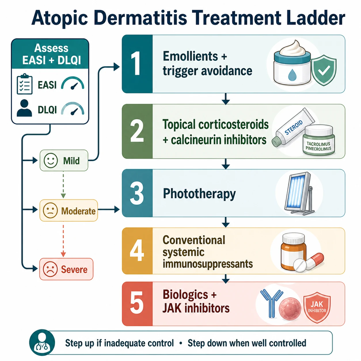
Stepwise Treatment Ladder
Stepwise treatment ladder — emollients to JAK inhibitors
Foundation for every patient
Emollients liberally and frequently (250-500 g/week for an adult), soap substitutes, trigger avoidance (irritants, allergens, heat, sweat, stress), bleach baths 0.005% twice weekly for recurrent S. aureus superinfection, and psychosocial support.
Mild-to-moderate AD — topical anti-inflammatory
Topical corticosteroids by site and severity: mild (hydrocortisone 1%) for face/flexures; moderate (betamethasone valerate 0.025%, clobetasone butyrate 0.05%) for trunk/limbs; potent/ultra-potent (clobetasol propionate 0.05%, mometasone furoate 0.1%) for thick plaques on palms/soles. Twice-daily during flare, then step-down. Topical calcineurin inhibitors (tacrolimus 0.1% ointment BD, pimecrolimus 1% cream BD) for face/flexures and steroid-sparing maintenance.
Refractory moderate-to-severe — phototherapy
Narrowband UVB (311 nm) 2-3 sessions/week for 8-12 weeks; PUVA as second-line for refractory disease. Phototherapy may be combined with topical therapy and is often used as a bridge to slower systemic agents.
Refractory moderate-to-severe — conventional systemics
Ciclosporin 3-5 mg/kg/day for rapid control (max 1 year); methotrexate 10-25 mg weekly with folic acid 5 mg the next day; azathioprine 1-3 mg/kg/day dosed by TPMT; mycophenolate mofetil 1-1.5 g twice daily. Choose by patient profile (comorbidities, pregnancy intent, TPMT activity, speed of onset required).
Biologic — first-line systemic (AAD 2024 strong recommendation)
Dupilumab 600 mg loading then 300 mg every other week (first-line for most patients). Tralokinumab 600 mg loading then 300 mg every other week, or lebrikizumab 500 mg loading then 250 mg every other week, as narrower IL-13 alternatives with lower conjunctivitis risk. Nemolizumab 60 mg loading then 30 mg every 4 weeks for itch-dominant disease (AAD 2025 update, with concomitant topical therapy).
JAK inhibitor — rapid onset or biologic-experienced
Upadacitinib 15 mg or 30 mg daily; abrocitinib 100 mg or 200 mg daily; baricitinib 2-4 mg daily. Choose for rapid itch and clearance, biologic-experienced patients, or where speed of onset is paramount. Class boxed warning for VTE/MACE/malignancy/infection — baseline labs (FBC, LFT, lipids), ongoing monitoring, pregnancy contraindicated.
Treat-to-target and step-down
Aim for EASI-75 or IGA 0/1 by week 16 — if achieved, continue to maintenance with the lowest effective dose; if not, switch or escalate. Consider topical ruxolitinib or tapinarof for steroid-sparing maintenance in selected patients. Treat comorbidities — sleep, mental health, infection.
Differential Diagnosis
[1] [1]Investigations
Diagnosis is usually clinical. Investigations are used to exclude differential diagnoses, assess severity/comorbidity, or before systemic therapy. [1]
- Skin swab/culture if secondary bacterial (S. aureus) or viral (HSV) infection suspected.
- KOH preparation to exclude tinea corporis.
- Patch testing if contact dermatitis is suspected, particularly in adults with hand or refractory disease.
- Total IgE and specific IgE / radioallergosorbent testing may support atopic status but are not diagnostic.
- Biopsy if diagnosis uncertain, atypical features, or treatment resistance.
- Baseline bloods before systemic immunosuppression: FBC, U&E, creatinine, LFT, hepatitis B/C, HIV, TB screening (IGRA or Mantoux), pregnancy test where relevant. [1]
Histopathology
Histology is not usually required. Findings include spongiosis (intercellular oedema) with epidermal thickening in chronic lesions, parakeratosis, a superficial perivascular inflammatory infiltrate with lymphocytes, and eosinophils. Unlike psoriasis, there are no regular acanthosis, Munro microabscesses, or neutrophilic collections.[1]
Management — General Measures
- Emollients — apply liberally and frequently (multiple times daily) to all skin, including unaffected areas; use as soap substitute and bath additive.
- Trigger avoidance — identify and reduce irritants (fragrances, detergents, wool, overheating), allergens and stressors.
- Psychosocial support — address sleep disturbance, anxiety, depression, school/work impact.
- Infection control — bleach baths or antiseptic washes may reduce S. aureus burden in selected patients; treat secondary infection promptly. [1]
Management — Topical Therapy
Topical anti-inflammatory therapy is the cornerstone for most patients. [1]
| Agent | Typical use | Notes |
|---|---|---|
| Topical corticosteroids | First-line anti-inflammatory for flares | Match potency to site and severity; use once or twice daily; apply to active lesions and adjacent skin that was active within 48 hours.[3] |
| Topical calcineurin inhibitors (tacrolimus, pimecrolimus) | Second-line for sensitive sites (face, eyelids, skin folds) and steroid-sparing maintenance | Use twice daily; no skin atrophy; transient stinging is common.[3] |
| Crisaborole (topical PDE4 inhibitor) | Mild-to-moderate disease, including face and skin folds | Apply twice daily; stinging may occur. |
| Ruxolitinib cream (topical JAK inhibitor) | Short-term, non-continuous use for mild-to-moderate disease in patients 12 years and older | Apply to limited body surface area twice daily. |
| Tapinarof cream / roflumilast cream | Steroid-free topical options for plaque areas | Recently endorsed in the AAD 2025 focused update.[5] |
Steroid potency and site
- Face, eyelids, genitals, skin folds: mild potency (e.g., hydrocortisone).
- Trunk and limbs: moderate potency for moderate disease.
- Thick, lichenified plaques on palms/soles or limbs: potent/very potent for short courses.
- Fingertip-unit guidance helps quantify quantity. [1]
Management — Phototherapy
Narrowband UVB (NB-UVB) is the most commonly used phototherapy for moderate-to-severe or widespread AD that has not responded to topical therapy. Typical regimens are two to three times weekly for 8–12 weeks, with dose titration based on skin type/minimal erythema dose. PUVA is an alternative for refractory disease but has greater long-term carcinogenic risk.[1]
Management — Systemic Therapy
Systemic therapy is indicated for moderate-to-severe disease that is refractory to optimised topical therapy and phototherapy, or when there is major quality-of-life impairment. [1]
Conventional systemic immunosuppressants
The AAD 2024 systemic guideline gives conditional recommendations for phototherapy, azathioprine, ciclosporin, methotrexate and mycophenolate, and recommends against systemic corticosteroids because of rebound and adverse effects.[1]
- Ciclosporin — often used for rapid control of severe flares; monitoring of blood pressure and renal function is essential.
- Methotrexate, azathioprine, mycophenolate mofetil — steroid-sparing options; dosing and monitoring per local formulary and specialist guidance.
- Systemic corticosteroids — avoid for chronic disease; short courses only for exceptional acute flares with a clear plan. [1]
Biologics and small molecules
The AAD 2024 systemic guideline gives strong recommendations for dupilumab, tralokinumab, abrocitinib, baricitinib and upadacitinib; the 2025 focused update adds strong recommendations for lebrikizumab and nemolizumab (with concomitant topical therapy), and for tapinarof and roflumilast creams.[5]
| Class | Drug | Target/mechanism | Key trial |
|---|---|---|---|
| IL-4Rα antagonist | Dupilumab | Blocks IL-4 and IL-13 signalling | SOLO 1 and SOLO 2 (NEJM 2016)[8] |
| Anti-IL-13 | Tralokinumab | Neutralises IL-13 | ECZTRA 1 and 2 (BJD 2021)[10] |
| Anti-IL-13 | Lebrikizumab | Neutralises IL-13 | ADvocate 1 and 2 (NEJM 2023)[11] |
| JAK1 inhibitor | Upadacitinib | Blocks JAK1-dependent cytokine signalling | Measure Up 1 and 2 (Lancet 2021)[9] |
| JAK1 inhibitor | Abrocitinib | Blocks JAK1-dependent cytokine signalling | JADE trials |
| JAK1/2 inhibitor | Baricitinib | Blocks JAK1/2-dependent cytokine signalling | BREEZE-AD trials |
| Anti-IL-31Rα | Nemolizumab | Blocks IL-31 signalling | ARCADIA trials (with topical therapy) |
AD systemic-therapy decision aid — the 'CLEAR-IT' ladder
CLEAR-IT
EASI ≥16, IGA ≥3, DLQI ≥11, or BSA >10% — and confirm failure of optimised topical therapy and trigger avoidance for ≥4–8 weeks
Bring vaccinations up to date BEFORE immunosuppression — live vaccines are contraindicated on biologics and JAK inhibitors
Hepatitis B sAg/core, hepatitis C, HIV, TB (IGRA), Strongyloides if endemic — document and treat latent TB before biologic/JAK
Baseline ocular review; conjunctivitis occurs in up to 10% of dupilumab-treated patients and may need ophthalmology input
Ciclosporin or upadacitinib for rapid severe-disease control; dupilumab or tralokinumab for durable, long-term steroid-sparing therapy
FBC, U&E, creatinine, LFT, lipids (JAK inhibitors), pregnancy test; repeat at 8–16 weeks then every 3–6 months
Aim for EASI-75 / IGA 0–1 by week 16; reassess at week 16 — switch or escalate if not achieved
Dupilumab (IL-4Rα antagonist)
First-line biologic for moderate-to-severe AD
- Blocks IL-4 and IL-13 signalling via the IL-4Rα subunit — the broadest type-2 blockade of any current biologic
- SOLO 1 and SOLO 2 (NEJM 2016) — EASI-50 at week 16 in ~85%, EASI-75 in ~44–51%; long-term safety now to 5 years
- Loading 600 mg SC, then 300 mg every other week; adolescent weight-based regimens
- Conjunctivitis in 8–10% (and blepharitis/keratitis); injection-site reactions; transient blood eosinophilia in some patients
- Avoid co-administration with live vaccines; minimal to no need for routine laboratory monitoring
Upadacitinib (oral JAK1 inhibitor)
Rapid itch and skin clearance
- Once-daily oral JAK1-selective inhibitor — Measure Up 1 and 2 (Lancet 2021) and Heads Up vs dupilumab
- EASI-75 at week 16 in ~70–80% — numerically higher than dupilumab; itch reduction within days
- Dose 15 mg or 30 mg daily; adolescent ≥12 years weight-based 15 mg
- Boxed warning (VTE, MACE, malignancy, infection, mortality); lipid rise, neutropenia, anaemia, acne, nausea
- Pregnancy CONTRAINDICATED; live vaccines contraindicated; baseline and ongoing labs required (FBC, LFT, lipids)
Tralokinumab and Lebrikizumab (anti-IL-13)
Targeted IL-13 neutralisation
- Both neutralise IL-13 only — narrower than dupilumab but with very favourable safety profile
- Tralokinumab ECZTRA 1 and 2 — EASI-75 at week 16 in ~25–33% (monotherapy); ~56% with concomitant TCS; q2w then q4w maintenance
- Lebrikizumab ADvocate 1 and 2 — EASI-75 in ~43–51% at week 16; induction q2w then q4w maintenance
- Lower conjunctivitis rate than dupilumab (~2–5%); injection-site reactions and upper-respiratory infections most common adverse events
- Sensible choices when IL-4Rα blockade is contraindicated, not tolerated, or in patients preferring monotherapy with a narrower mechanism
Pre-treatment screening for systemic immunosuppression/biologics
- Infection screen: hepatitis B surface antigen/core antibody, hepatitis C antibody, HIV, TB (IGRA/Mantoux) as per local guideline.
- Vaccination status: ensure inactivated vaccines up to date before starting immunosuppression; live vaccines contraindicated during therapy.
- Baseline labs: FBC, U&E, creatinine, LFT, lipids (for JAK inhibitors), pregnancy test. [1]
Special Sites and Populations
Children
- Emollients and mild-to-moderate topical corticosteroids are first-line.
- Topical calcineurin inhibitors are useful for facial/flexural maintenance.
- Avoid potent steroids on face and skin folds.
- Phototherapy and systemic therapy only under specialist care.
Pregnancy and breastfeeding
- Emollients and mild topical corticosteroids are generally safe.
- Avoid most systemic immunosuppressants and biologics unless specialist-directed.
- Ciclosporin may be used in selected cases under specialist care.
Hand and foot eczema
- Potent topical steroids under occlusion for thick plaques; patch test if occupational/contact aetiology suspected.
Eyelid eczema
- Low-potency steroids or topical calcineurin inhibitors; avoid chronic potent steroid use because of glaucoma/cataract risk.
Complications and Emergencies
Secondary infection
- Bacterial: S. aureus superinfection presents with weeping, crusting, pustules and rapid flare; treat with antiseptics or antibiotics as appropriate.
- Viral: eczema herpeticum is a dermatology emergency caused by herpes simplex virus, producing monomorphic punched-out erosions with haemorrhagic crusting and systemic symptoms. Treat promptly with systemic aciclovir; consider admission.
- Molluscum contagiosum may be more extensive and persistent in AD.
Erythroderma
- Generalised erythema and scale covering most of the body surface area; requires admission for fluid, electrolyte and temperature management.
Ocular complications
- Conjunctivitis, keratitis and blepharitis are more common in AD; conjunctivitis has been reported with dupilumab and requires ophthalmology review if severe.
Comorbidities
The AAD 2022 comorbidity guideline summarised associations between adult AD and allergic, atopic, immune-mediated, mental-health and bone-health disorders, as well as skin infections. Clinicians should maintain awareness of asthma, allergic rhinitis, food allergy, anxiety, depression, and sleep disturbance.[4]
Prognosis and Follow-Up
AD is chronic and relapsing; the goal is control, not cure. Modern targeted therapies can achieve high rates of EASI-75 or IGA 0/1, but relapse is common after stopping treatment. Follow-up intervals depend on therapy and severity: [1]
- Topical therapy: review at 4–8 weeks.
- Phototherapy: review every 2–4 weeks during induction.
- Systemic/biologic therapy: review at 8–16 weeks initially, then every 3–6 months, with laboratory monitoring as appropriate. [1]
Evidence, Guidelines and Regional Differences
- AAD 2023 topical guideline — updated recommendations on emollients, topical corticosteroids, calcineurin inhibitors, crisaborole and ruxolitinib.[3]
- AAD 2024 systemic/phototherapy guideline — strong recommendations for dupilumab, tralokinumab, abrocitinib, baricitinib and upadacitinib; conditional for phototherapy, ciclosporin, methotrexate, azathioprine and mycophenolate; recommends against systemic corticosteroids.[1]
- AAD 2025 focused update — adds strong recommendations for tapinarof cream, roflumilast cream, lebrikizumab and nemolizumab with topical therapy.[5]
- NICE CG57 — UK guidance for atopic eczema in children under 12 years emphasises emollients, stepped topical corticosteroids, and calcineurin inhibitors as second-line.
- European (EuroGuiDerm) and IADVL guidelines provide regional variations in phototherapy access, biologic eligibility and monitoring.
Exam Pearls
[1]Red Flags
Exam application bank (NEET-PG / INICET)
One-line answer
Atopic dermatitis is a chronic, relapsing, inflammatory skin disease driven by skin-barrier dysfunction and type 2 inflammation. Fellowship-level assessment requires understanding of filaggrin biology, the IL-4/IL-13/IL-31 axis, the atopic march, severity scoring (EASI, IGA, DLQI), trigger avoidance, site-specific topical therapy, phototherapy, conventional systemic immunosuppressants, and the mechanisms and landmark trial evidence for biologics and JAK inhibitors.
Worked stems (answer without another resource)
Stem 1 — Classic presentation. Map symptoms to mechanism; name the first investigation and first treatment step with dose/route if drug therapy is standard. [1]
Stem 2 — Unstable / complicated. List red flags that force immediate resuscitation, theatre, ICU, antidote, or reperfusion — and what you do in the first 15 minutes. [1]
Stem 3 — Atypical group. Elderly, pregnancy, child, or immunocompromised: how presentation and thresholds change. [1]
Stem 4 — Differential trap. Name the three closest mimics and one discriminator for each. [1]
Stem 5 — Disposition. Who goes home with safety-netting, who is admitted, who needs HDU/ICU/theatre, and what follow-up is mandatory. [1]
Rapid viva checklist
- Definition + classification
- Pathophysiology chain
- Bedside signs / criteria
- Score with exact components (if any)
- Emergency bundle
- Definitive therapy with doses
- Complications of disease and of treatment
- Special populations
- Guideline/trial name if classic
- Three exam traps
Coverage self-check
If you cannot answer any stem above from this page alone, re-read the matching section — the page is intended to be self-sufficient for final-prof and NEET-PG/INICET questions on Atopic dermatitis.
[1]References
- [1]Langan SM, Irvine AD, Weidinger S. Atopic dermatitis Lancet, 2020.PMID 32738956
- [2]Guttman-Yassky E, Brunner PM, Neumann AU, et al. Atopic dermatitis Lancet, 2025.PMID 39955121
- [3]Sidbury R, Davis DMR, Cohen DE, et al. Guidelines of care for the management of atopic dermatitis in adults with topical therapies J Am Acad Dermatol, 2023.PMID 36641009
- [4]Davis DMR, Drucker AM, Alikhan A, et al. American Academy of Dermatology Guidelines: Awareness of comorbidities associated with atopic dermatitis in adults J Am Acad Dermatol, 2022.PMID 35085682
- [5]Davis DMR, Narla S, Drucker AM, et al. Focused update: Guidelines of care for the management of atopic dermatitis in adults J Am Acad Dermatol, 2025.PMID 40531067
- [6]Drislane C, Irvine AD. The role of filaggrin in atopic dermatitis and allergic disease Ann Allergy Asthma Immunol, 2020.PMID 31622670
- [7]Margolis DJ, Gandhi NA, Hoffstad OJ, et al. Atopic dermatitis: filaggrin and skin barrier dysfunction Br J Dermatol, 2022.PMID 35128630
- [8]Simpson EL, Bieber T, Guttman-Yassky E, et al. Two Phase 3 Trials of Dupilumab versus Placebo in Atopic Dermatitis N Engl J Med, 2016.PMID 27690741
- [9]Guttman-Yassky E, Teixeira HD, Simpson EL, et al. Once-daily upadacitinib versus placebo in adolescents and adults with moderate-to-severe atopic dermatitis (Measure Up 1 and Measure Up 2): results from two replicate double-blind, randomised controlled phase 3 trials Lancet, 2021.PMID 34023008
- [10]Wollenberg A, Blauvelt A, Guttman-Yassky E, et al. Tralokinumab for moderate-to-severe atopic dermatitis: results from two 52-week, randomized, double-blind, multicentre, placebo-controlled phase III trials (ECZTRA 1 and ECZTRA 2) Br J Dermatol, 2021.PMID 33000465
- [11]Silverberg JI, Guttman-Yassky E, Thaçi D, et al. Two Phase 3 Trials of Lebrikizumab for Moderate-to-Severe Atopic Dermatitis N Engl J Med, 2023.PMID 36920778