Dermatology · Medicine
Atopic dermatitis in infancy
Also known as Infantile atopic eczema · Infantile eczema · Atopic eczema of infancy
Atopic dermatitis in infancy is a chronic itchy inflammatory dermatosis with facial, scalp and extensor predominance driven by barrier failure (including filaggrin) and type 2 inflammation. Fellowship exams test age-related distribution, differentiation from infantile seborrhoeic and nappy dermatitis, emollient-first care, infant-safe topical corticosteroid/TCI use, eczema herpeticum recognition, and specialist pathways including early-life dupilumab evidence where licensed.
On this page & tools
Your progress
Saved locally on this device.
Exam tags
Red flags
Definition & Classification
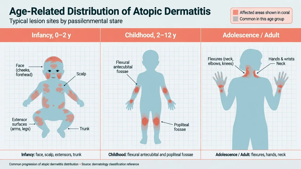
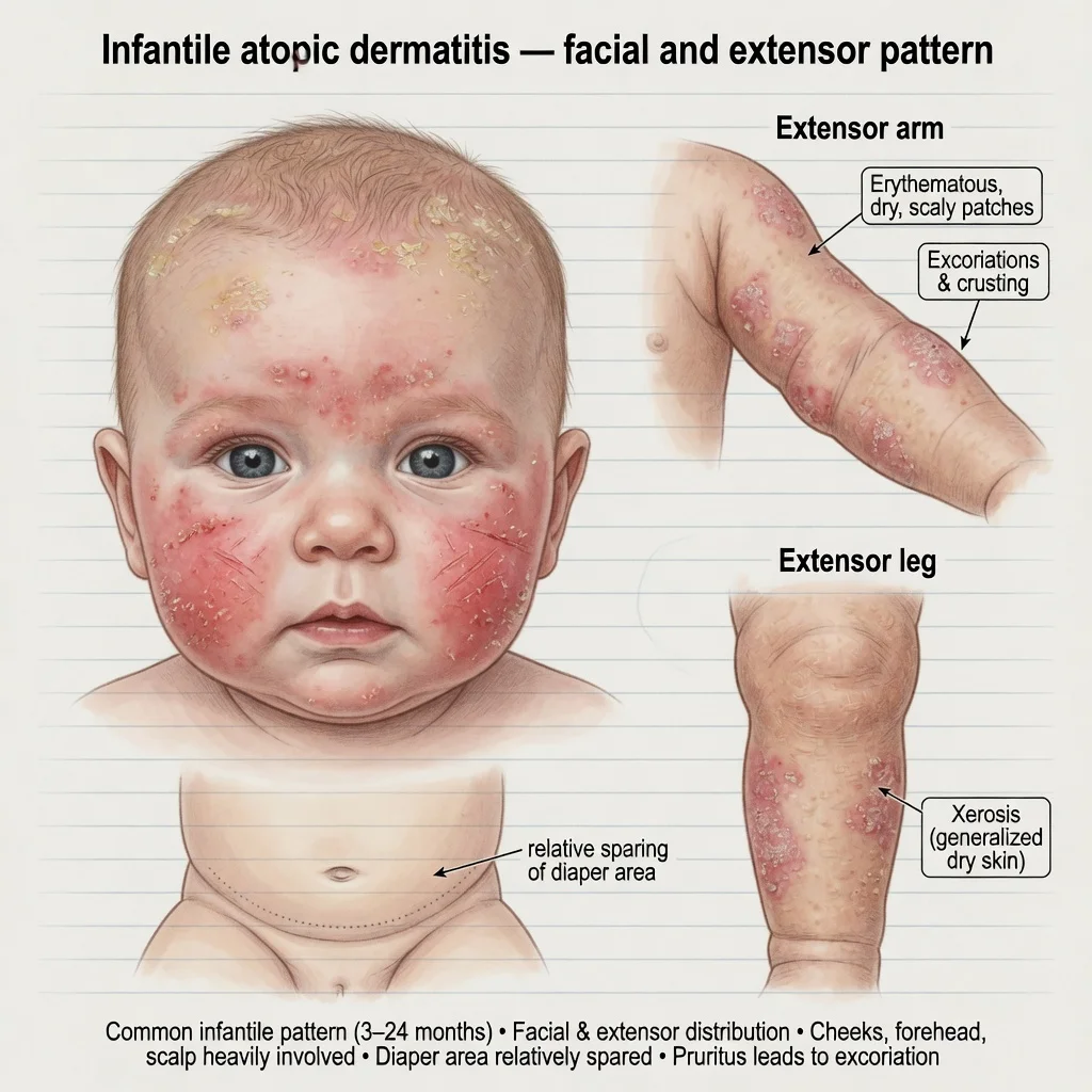
Atopic dermatitis (AD; atopic eczema) is a chronic relapsing inflammatory skin disease defined clinically by pruritus, typical morphology and age-related distribution, and a chronic or chronically relapsing course, often with personal or family atopic history.[1][2][4] In infancy (commonly onset in the first year), lesions favour the face, scalp, trunk and extensor surfaces; later childhood shifts toward flexures.[1][2]
Diagnosis is clinical. AAD guidelines summarise diagnosis and assessment principles without mandatory laboratory confirmation for typical disease.[2]
Epidemiology & Risk Factors
AD affects a substantial minority of children worldwide (often cited around 15–20% in high-prevalence settings), with many cases beginning in infancy.[1][4] Risk factors include family history of atopy, FLG loss-of-function variants, urban environments, and skin barrier stressors.[5][6] Not every infant with FLG mutation develops AD, and not every AD infant carries FLG variants — barrier failure is multifactorial.[5][6]
Pathophysiology
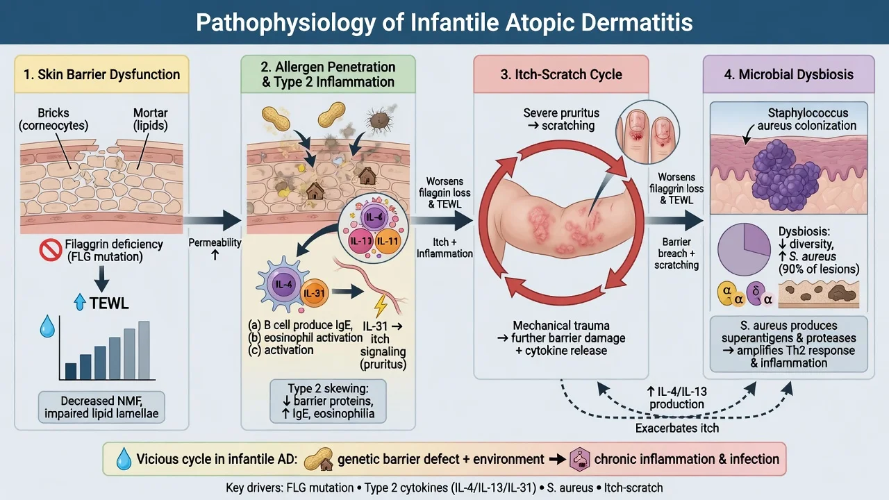
Three interlocking pillars: [1]
- Epidermal barrier dysfunction — reduced filaggrin and natural moisturising factor, lipid abnormalities, increased transepidermal water loss (TEWL).[5][6]
- Type 2 inflammation — IL-4, IL-13 (barrier and inflammation), IL-31 (itch signalling).[1][4][7]
- Microbial dysbiosis — Staphylococcus aureus colonisation amplifies inflammation and flares.[1][4]
Infant AD skin has age-specific molecular features compared with adult AD, relevant to therapeutic targeting and natural history research.[7] The itch–scratch cycle mechanically worsens barrier injury.
Clinical Presentation
- Intense pruritus (scratching, irritability, sleep loss) is mandatory for the diagnosis framing.[2]
- Erythematous, dry, scaly, excoriated patches on cheeks, forehead, scalp; extensors of arms/legs; trunk.
- Xerosis generalised; Dennie–Morgan lines and other atopic stigmata may appear with time.
- Napkin area often relatively spared compared with seborrhoeic or candidal napkin disease — a classic teaching contrast.
- Secondary crusting, oozing, pustules suggest bacterial superinfection.
- Severe disease may approach erythroderma.
Age-Related Distribution
| Age | Typical pattern |
|---|---|
| Infancy | Face, scalp, trunk, extensors |
| Childhood | Antecubital and popliteal fossae, wrists, ankles |
| Adolescent/adult | Flexures, hands, head and neck |
Differential Diagnosis
| Condition | Distinguishing clue |
|---|---|
| Infantile seborrhoeic dermatitis | Greasy yellow scale; little itch; scalp/flexures/napkin folds; early months |
| Irritant nappy dermatitis | Convex surfaces; fold sparing |
| Candidal napkin rash | Folds involved; satellite pustules |
| Scabies | Burrows, acral/genital, family itch |
| Psoriasis | Sharper plaques; less itch sometimes; family history |
| Immunodeficiency / Netherton / nutritional | FTT, erythroderma, recurrent infection, atypical persistence |
Bedside Assessment & Severity
Assess itch/sleep, extent, infection signs, growth, caregiver stress, and prior steroid/antibiotic exposure.[2] Severity language (mild/moderate/severe) and scores (EASI/SCORAD concepts) support communication and trial literacy even when formal scoring is not done every visit.[2][12]
Investigations
- Usually none for typical mild–moderate disease.[2]
- Skin swab if recurrent crusting/pustules.
- Allergy evaluation only when history suggests IgE-mediated food reactions or refractory disease — avoid shotgun panels that drive harmful diets.[2][8]
- Biopsy/work-up for atypical, purpuric, ulcerative, or systemically unwell infants (LCH, immunodeficiency, other genodermatoses).
Management — Foundation (all infants)
- Education — chronic relapsing course; written flare plan.
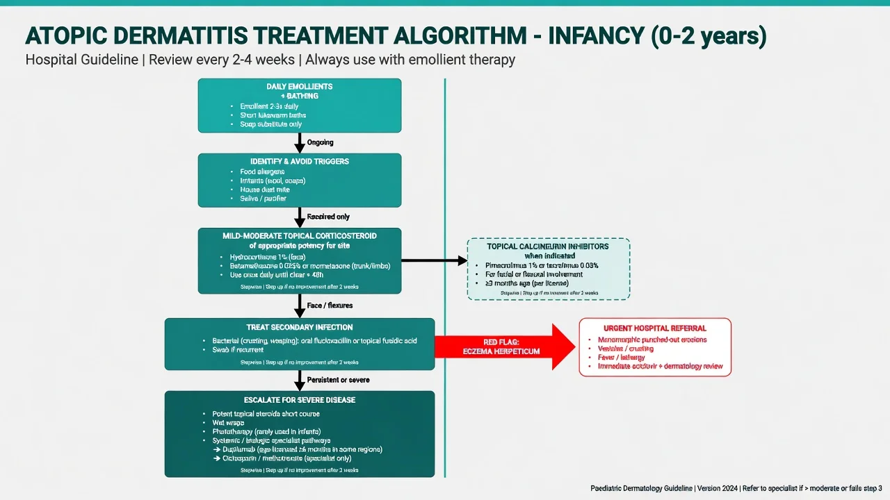
- Emollients liberally and frequently — cornerstone of barrier repair.[3][9][12]
- Bathing — short lukewarm baths; gentle cleansers; emollient after pat-dry.
- Trigger minimisation — wool, harsh soaps, heat, saliva; individualise food only with clear history.[3][8]
Management — Topical Anti-Inflammatory Therapy
Use the least potent agent that controls disease for the shortest effective time, then step down, while continuing emollients.[3][9]
- Face/neck/flexures: low-potency topical corticosteroid (e.g. hydrocortisone class teaching) or topical calcineurin inhibitors when age-licensed and indicated.[3][12]
- Trunk/limbs: mild-to-moderate potency as needed for flares.[3]
- Wet wraps for severe flares under experienced supervision.[3][12]
- Network meta-analysis supports multiple effective topical classes; choice depends on site, age, cost and access.[9]
European and AAD-aligned pathways emphasise proactive maintenance in frequently relapsing sites after induction of control.[3][12]
Management — Infection & Emergencies
- Treat clinically infected AD with appropriate topical/systemic antimicrobials plus anti-inflammatory and barrier care.[3]
- Eczema herpeticum: monomorphic punched-out erosions ± fever, malaise — urgent systemic aciclovir/valaciclovir pathway and specialist care; do not escalate steroids alone.[10]
Management — Phototherapy & Systemic Therapy
Phototherapy is rarely used in young infants (practical and safety constraints). Severe, refractory disease needs paediatric dermatology for systemic options. Dupilumab has demonstrated efficacy and acceptable safety signals in young children (including open-label extension data in ages 6 months to 5 years) and is a landmark targeted option where regulatory labelling and access allow.[11] JAK inhibitors and conventional immunosuppressants remain specialist tools with age-specific labelling — do not invent doses outside licensed specialist practice.[8][12]
Special Populations & Feeding
- Breastfeeding encouraged per general paediatric guidance; maternal diet restriction is not routine without specialist indication.
- Elimination diets without proven allergy risk failure to thrive — a major pitfall.
- Routine immunisations are not withheld solely because of AD.
- Screen for primary immunodeficiency if severe refractory AD plus infections, diarrhoea, or poor growth. [1]
Comorbidities & Atopic March
Infant AD increases risk trajectories for food allergy, asthma and allergic rhinitis, though not all children “march.” Sleep loss and parental mental health are part of disease burden and counselling.[1][4]
Prognosis & Follow-Up
Many improve through childhood; a substantial minority persist or relapse. FLG-related disease may be more persistent.[5][6] Provide follow-up proportional to severity, with clear escalation instructions for infection or herpeticum.
Evidence, Guidelines & Regional Differences
Anchor documents for examiners: Lancet overviews,[1][4] AAD diagnosis and topical sections,[2][3] European consensus,[12] AAAAI/ACAAI JTF 2023 GRADE-informed guidance,[8] topical network meta-analysis,[9] and early-life dupilumab extension data.[11] NICE/UK, IADVL and ANZ pathways share emollient-first logic with regional formulary differences for TCIs and biologics access.
Exam Pearls
Red Flags
Exam application bank (NEET-PG / INICET)
One-line answer
Atopic dermatitis in infancy is a chronic itchy inflammatory dermatosis with facial, scalp and extensor predominance driven by barrier failure (including filaggrin) and type 2 inflammation. Fellowship exams test age-related distribution, differentiation from infantile seborrhoeic and nappy dermatitis, emollient-first care, infant-safe topical corticosteroid/TCI use, eczema herpeticum recognition, and specialist pathways including early-life dupilumab evidence where licensed.
Worked stems (answer without another resource)
Stem 1 — Classic presentation. Map symptoms to mechanism; name the first investigation and first treatment step with dose/route if drug therapy is standard. [1]
Stem 2 — Unstable / complicated. List red flags that force immediate resuscitation, theatre, ICU, antidote, or reperfusion — and what you do in the first 15 minutes. [1]
Stem 3 — Atypical group. Elderly, pregnancy, child, or immunocompromised: how presentation and thresholds change. [1]
Stem 4 — Differential trap. Name the three closest mimics and one discriminator for each. [1]
Stem 5 — Disposition. Who goes home with safety-netting, who is admitted, who needs HDU/ICU/theatre, and what follow-up is mandatory. [1]
Rapid viva checklist
- Definition + classification
- Pathophysiology chain
- Bedside signs / criteria
- Score with exact components (if any)
- Emergency bundle
- Definitive therapy with doses
- Complications of disease and of treatment
- Special populations
- Guideline/trial name if classic
- Three exam traps
Coverage self-check
If you cannot answer any stem above from this page alone, re-read the matching section — the page is intended to be self-sufficient for final-prof and NEET-PG/INICET questions on Atopic dermatitis in infancy.
Expanded exam teaching (depth pass)
Clinical reasoning
For Atopic dermatitis in infancy, examiners test whether you can prioritise life threats, choose the right first test, and give specific therapy (agent, dose, route, timing). Generic phrases without numbers score poorly.
Mechanism → feature map
Build a short chain: cause → pathophysiologic intermediate → clinical feature → complication. Every major symptom in the classic vignette should sit on that chain.
Investigation strategy
- Bedside/first-line tests that change immediate management
- Confirmatory or staging tests
- What a normal result does not exclude
- When not to delay treatment for imaging (unstable patient)
Management ladder
- Resuscitation / ABC / sepsis or haemorrhage bundle as relevant
- Specific antidote / procedure / antimicrobial / reperfusion / surgery
- Supportive care and monitoring targets
- Definitive long-term therapy and secondary prevention
- Disposition and safety-net advice
Special populations
Always prepare one line each for children, pregnancy, elderly, renal/hepatic impairment, and immunocompromised patients when the topic allows.
Pitfalls that fail candidates
- Treating the number not the patient
- Missing pregnancy status when relevant
- Imaging before stabilisation
- Wrong empiric cover or wrong antidote timing
- Incomplete counselling on recurrence, adherence, or red-flag return
Atopic dermatitis in infancy is a chronic itchy inflammatory dermatosis with facial, scalp and extensor predominance driven by barrier failure (including filaggrin) and type 2 inflammation. Fellowship exams test age-related distribution, differentiation from infantile seborrhoeic and nappy dermatitis, emollient-first care, infant-safe topical corticosteroid/TCI use, eczema herpeticum recognition, and specialist pathways including early-life dupilumab evidence where licensed. [1]
Structured revision sheet
Must-know numbers and names
List every score, size threshold, dose, and time window from this topic on a blank page from memory, then check against the sections above.
Three classic MCQ angles
- Most likely diagnosis given a vignette
- Next best step in management
- Most appropriate investigation
Three classic SAQ angles
- Pathophysiology in five steps
- Management algorithm with doses
- Complications and prevention
Clinical station flow
Greet → focused history → targeted exam → investigations → explain diagnosis → emergency care → definitive plan → safety-net / follow-up → answer examiner questions on mechanism and pitfalls.
Exam anchors
References
- [1]Langan SM, Irvine AD, Weidinger S. Atopic dermatitis Lancet, 2020.PMID 32738956
- [2]Eichenfield LF, Tom WL, Chamlin SL, et al. Guidelines of care for the management of atopic dermatitis: section 1. Diagnosis and assessment of atopic dermatitis J Am Acad Dermatol, 2014.PMID 24290431
- [3]Eichenfield LF, Tom WL, Berger TG, et al. Guidelines of care for the management of atopic dermatitis: section 2. Management and treatment of atopic dermatitis with topical therapies J Am Acad Dermatol, 2014.PMID 24813302
- [4]Weidinger S, Novak N. Atopic dermatitis Lancet, 2016.PMID 26377142
- [5]Palmer CN, Irvine AD, Terron-Kwiatkowski A, et al. Common loss-of-function variants of the epidermal barrier protein filaggrin are a major predisposing factor for atopic dermatitis Nat Genet, 2006.PMID 16550169
- [6]Drislane C, Irvine AD. The role of filaggrin in atopic dermatitis and allergic disease Ann Allergy Asthma Immunol, 2020.PMID 31622670
- [7]Renert-Yuval Y, Del Duca E, Pavel AB, et al. The molecular features of normal and atopic dermatitis skin in infants, children, adolescents, and adults J Allergy Clin Immunol, 2021.PMID 33453290
- [8]AAAAI/ACAAI JTF Atopic Dermatitis Guideline Panel, Chu DK, Schneider L, et al. Atopic dermatitis (eczema) guidelines: 2023 American Academy of Allergy, Asthma and Immunology/American College of Allergy, Asthma and Immunology Joint Task Force on Practice Parameters GRADE- and Institute of Medicine-based recommendations Ann Allergy Asthma Immunol, 2024.PMID 38108679
- [9]Chu DK, Chu AWL, Rayner DG, et al. Topical treatments for atopic dermatitis (eczema): Systematic review and network meta-analysis of randomized trials J Allergy Clin Immunol, 2023.PMID 37678572
- [10]Traidl S, Roesner L, Zeitvogel J, et al. Eczema herpeticum in atopic dermatitis Allergy, 2021.PMID 33844308
- [11]Paller AS, Siegfried EC, Simpson EL, et al. Dupilumab Safety and Efficacy up to 1 Year in Children Aged 6 Months to 5 Years with Atopic Dermatitis: Results from a Phase 3 Open-Label Extension Study Am J Clin Dermatol, 2024.PMID 38743155
- [12]Wollenberg A, Barbarot S, Bieber T, et al. Consensus-based European guidelines for treatment of atopic eczema (atopic dermatitis) in adults and children: part I J Eur Acad Dermatol Venereol, 2018.PMID 29676534