Dermatology · Medicine
Atypical / Dysplastic Naevus
Also known as Atypical naevus · Dysplastic naevus · Clark naevus · Atypical mole · B-K mole
An atypical (dysplastic) naevus is a benign melanocytic naevus with clinical features of irregularity (asymmetry, border irregularity, colour variegation, diameter 5 mm) and/or histological features of architectural disorder and cytological atypia. It is primarily a melanoma risk marker rather than a direct precursor, and management is risk-stratified: observation and surveillance for most, with excision of changing or suspicious lesions.
On this page & tools
Your progress
Saved locally on this device.
Exam tags
Red flags
Overview & Definition
An atypical naevus (also called a dysplastic naevus, Clark naevus, or atypical mole) is a benign melanocytic naevus that shows either clinical features of irregularity or histological features of architectural disorder and cytological atypia, or both.[2][4] The term "atypical" is usually applied to the clinical appearance, whereas "dysplastic" is used when the histology shows the characteristic architectural and cytological changes. The two terms are often used interchangeably in clinical practice, but they are not synonymous: a clinically atypical naevus may have a histologically banal appearance, and a histologically dysplastic naevus may look clinically normal.[4]
Clark naevus is named after Wallace Clark, who described lesions with irregular borders and variegated pigmentation, and who first linked such naevi to an increased risk of melanoma. The historical term B-K mole (from the initials of two families with familial melanoma) is now largely replaced by the term FAMMM syndrome (familial atypical multiple mole melanoma syndrome). [1]
A key clinical distinction is that atypical usually refers to the clinical appearance, while dysplastic refers to the histological findings. The clinical-histological correlation is imperfect: a lesion that looks clinically atypical may have a histologically banal appearance, and vice versa. Therefore, management must integrate both clinical and histological information, not rely on one alone.[4]
For undergraduate and postgraduate examinations, the high-yield concepts are the distinction between clinical atypia and histological dysplasia, the ABCDE and ugly duckling screening tools, the risk-stratified classification (sporadic, AMS, FAMMM), the CDKN2A genetics of FAMMM, and the evidence-based management that favours observation over prophylactic excision for most lesions. [1]
[1]Classification
Atypical naevi can be classified by clinical context, which is the main driver of melanoma risk and management.[1][2]
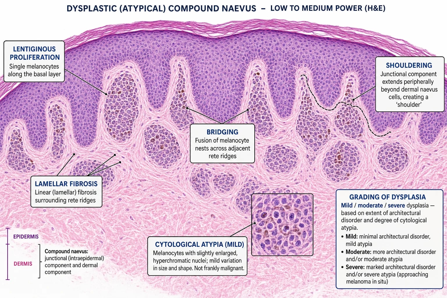
Histologically, dysplastic naevi are graded by the degree of architectural disorder and cytological atypia into mild, moderate, or severe.[5] This grading has practical management implications, because severely dysplastic naevi with positive margins are usually re-excised, whereas mildly or moderately dysplastic naevi with positive margins are often observed.[8][9]
Epidemiology & Risk Factors
Approximately 2–8% of the general population have at least one clinically atypical naevus.[2] The prevalence is higher in fair-skinned individuals, in those with a tendency to sunburn rather than tan, and in populations with high cumulative sun exposure. Atypical naevi usually begin to appear in childhood and adolescence, and their number may increase through early adulthood. They are uncommon in darker skin phototypes.
The most important risk factors are: [1]
- Fair skin and light eye/hair colour — associated with a greater naevus count and sun sensitivity.
- Intermittent high-dose UV exposure and sunburns, especially in childhood — drive the acquisition of new naevi and melanocytic atypia.[1]
- Family history of melanoma — strongly suggests FAMMM syndrome when combined with multiple atypical naevi.
- High total naevus count — more than 50 common naevi is itself a melanoma risk factor.
- Immunosuppression — organ transplant recipients and patients on chronic immunosuppression develop more naevi and have a higher melanoma risk.[1]
- Photosensitising medications — drugs such as voriconazole, hydrochlorothiazide, and amiodarone can increase photosensitivity and the risk of UV-induced skin changes, although their direct effect on atypical naevus formation is less clear.
The number of naevi and the number of atypical naevi are both independent risk factors for melanoma. A patient with more than 100 common naevi has a substantially higher risk than a patient with fewer than 15, and the presence of even one atypical naevus approximately doubles the risk. [1]
FAMMM syndrome is rare, autosomal dominant, and most often associated with germline mutations in CDKN2A (encoding the tumour suppressors p16^INK4a and p14^ARF). Penetrance is incomplete and variable, but carriers have a markedly elevated lifetime melanoma risk, as well as an increased risk of pancreatic cancer.[3]
Pathophysiology
The histological diagnosis of a dysplastic naevus requires both architectural disorder and cytological atypia.[4][5] The combination of these features distinguishes a dysplastic naevus from a benign compound naevus on one side and from melanoma on the other. However, the boundary between a severely dysplastic naevus and a thin melanoma is not always sharp, and even experienced dermatopathologists may disagree on the same section.
Architectural disorder includes: [1]
- Lentiginous or junctional proliferation of melanocytes along the basal layer, often as single cells or small nests rather than the regular nests of a benign naevus.
- Bridging of rete ridges by melanocytic nests.
- Shouldering — extension of the junctional melanocyte component beyond the dermal component at the periphery.
- Lamellar fibrosis or concentric fibrosis in the papillary dermis surrounding the nests.
- Chronic mild inflammatory infiltrate.
- Variable degrees of pigmentation. [1]
Cytological atypia refers to melanocytes with enlarged, hyperchromatic, pleomorphic nuclei, sometimes with prominent nucleoli. The degree of atypia is graded as mild, moderate, or severe depending on the nuclear size, chromatin pattern, and degree of pleomorphism.[5]
At the molecular level, sporadic atypical naevi are thought to arise from UV-induced or stochastic genetic alterations in melanocytes, most commonly activating BRAF mutations (particularly BRAF V600E), but without the additional driver events needed for malignant transformation. NRAS and NF1 mutations are less common. The initial benign proliferation is constrained by normal senescence and DNA repair mechanisms, and the lesion remains on the benign side of the melanoma spectrum unless further oncogenic hits accumulate, such as inactivation of CDKN2A, PTEN loss, or TERT promoter activation.[1][3]
Senescence and the melanoma barrier: Ordinary and dysplastic naevi commonly show BRAF mutations, yet most do not become melanoma because oncogene-induced senescence arrests further growth. This is thought to be mediated by p16INK4a, which is encoded by CDKN2A. In FAMMM syndrome, one copy of CDKN2A is already defective from birth, lowering the threshold for malignant progression when a second hit or additional driver mutation occurs.[3]
Microenvironment and inflammation: Dysplastic naevi often show a mild chronic inflammatory infiltrate and altered dermal collagen (lamellar fibrosis). The interaction between melanocytes, keratinocytes, fibroblasts, and immune cells in the tumour microenvironment may influence whether a lesion remains stable or progresses. Immune surveillance, particularly by T-cells and natural killer cells, is believed to suppress early malignant transformation; immunosuppression can therefore permit progression.[1]
The dysplastic naevus controversy
For decades, pathologists and clinicians debated whether dysplastic naevi are true melanoma precursors or merely risk markers. The modern consensus is that they are primarily risk markers, but severely dysplastic lesions may occasionally represent melanoma in situ or early invasive melanoma that is difficult to distinguish histologically. This uncertainty explains why severely dysplastic naevi with positive margins are usually re-excised, while mild-to-moderate dysplasia is observed. The controversy also underlies the variable terminology and grading systems used in different laboratories.[2][4]
Clinical Presentation
The classic clinical features of an atypical naevus are captured by the ABCDE criteria, originally described for melanoma screening but applicable to atypical naevi:[7]
- A — Asymmetry: one half does not match the other half when an imaginary line is drawn through the centre.
- B — Border irregularity: notched, scalloped, indistinct, or poorly defined edges.
- C — Colour variegation: two or more colours (tan, brown, black, red, white, blue) within the same lesion.
- D — Diameter: usually larger than 5 mm (some older sources use 6 mm). Diameter alone is a poor discriminator because many benign naevi exceed 5 mm, and some melanomas are smaller.
- E — Evolution: any change in size, shape, colour, elevation, surface, or symptoms such as bleeding, itching, or crusting. Evolution is often the most important clue. [1]
Atypical naevi are often larger than common naevi, measuring 5–12 mm or more. They may be macular, papular, or have a target-like or "fried-egg" appearance — a raised pink or tan centre with a darker, flat peripheral rim. This morphology is common in younger patients and in those with atypical mole syndrome. The lesion may also have a pink or flesh-coloured centre if the dermal component predominates, while the periphery remains pigmented. [1]
Other morphological variants include: [1]
- Flat, macular lesions with irregular borders — often the earliest clinical sign of atypia.
- Papular or dome-shaped lesions with irregular pigmentation — may be mistaken for melanoma or Spitz naevus.
- Lesions with peripheral darkening — a darker rim with a lighter centre, which can mimic regression.
- Multiple lesions in the same patient that share some irregularity but differ in size and colour — typical of atypical mole syndrome. [1]
Their distribution tends to be trunk and extremities, often sparing the face and acral sites. Multiple lesions are common and may show considerable variation in size, shape, and colour.[2]
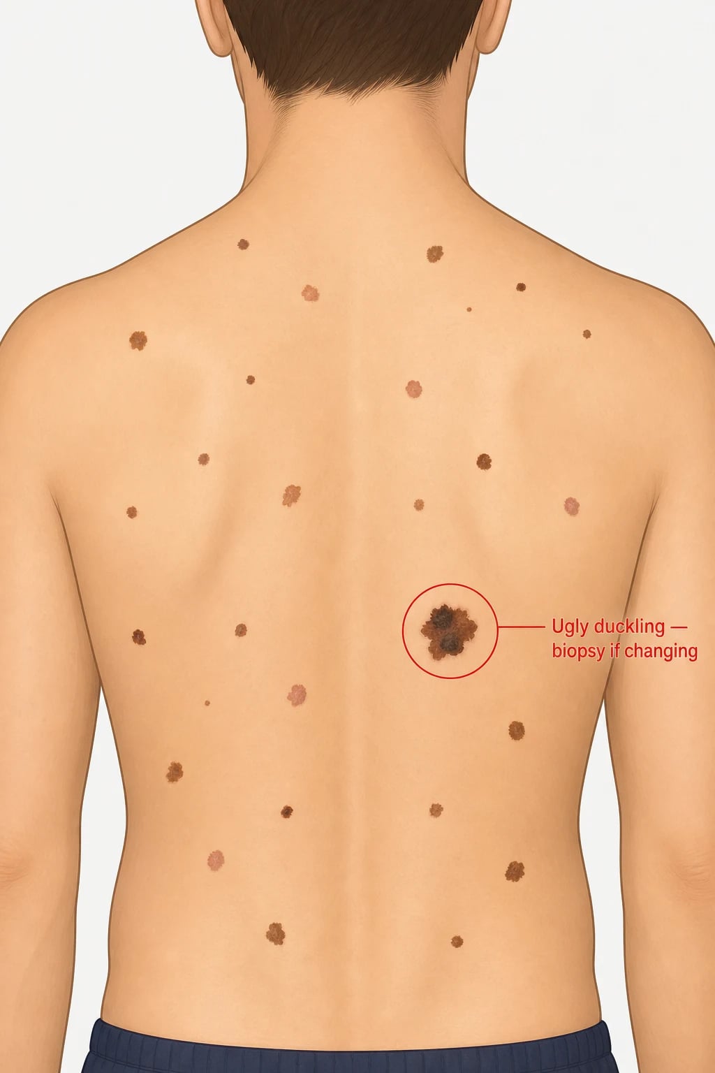
The "ugly duckling" sign is one of the most useful clinical concepts in patients with many naevi. Each person has a characteristic pattern of naevi in terms of size, shape, and colour. The naevus that looks different from the patient's other naevi — the outlier — is the one most likely to be melanoma. This sign is particularly valuable in atypical mole syndrome and FAMMM syndrome, where applying the ABCDE criteria to every lesion can be overwhelming and insensitive.[1]
ABCDE
Atypical presentations
- Children: Atypical naevi may first appear around puberty. Childhood melanoma is rare, but when it occurs it is often associated with FAMMM syndrome or congenital naevi. Any rapidly changing or unusually large lesion in a child warrants referral and biopsy.[6]
- Pregnancy: Existing naevi may darken or enlarge due to hormonal changes; this is usually benign. Biopsy is reserved for lesions that show suspicious clinical or dermoscopic change.[1]
- Immunosuppression: Solid-organ transplant recipients and patients on chronic immunosuppression develop more new naevi and have a higher risk of melanoma; surveillance should be more frequent.[1]
- Elderly: New or changing pigmented lesions in older adults have a higher pre-test probability of melanoma; a low threshold for biopsy is appropriate.
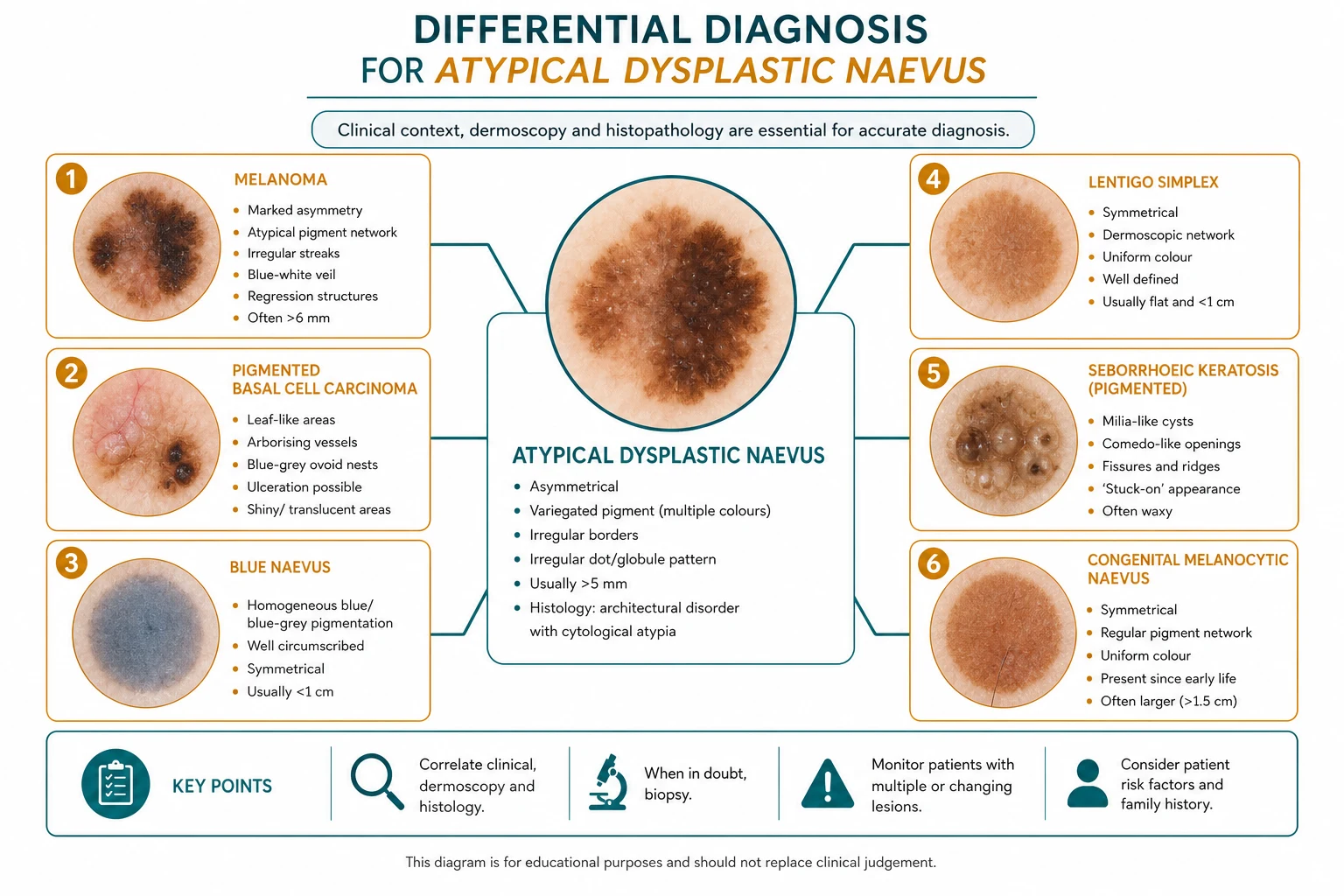
Differential Diagnosis
The main diagnostic challenge is distinguishing an atypical naevus from melanoma, because both can show asymmetry, border irregularity, colour variegation, and diameter >5 mm. The key discriminating features are listed below. [1]
Other important mimics include: [1]
| Mimic | Clinical features | Dermoscopic clues |
|---|---|---|
| Melanoma | Evolving, asymmetrical, irregular border, multiple colours, diameter >5 mm, may ulcerate or bleed. | Blue-white veil, polymorphous vessels, irregular streaks/pseudopods, regression structures, marked asymmetry. |
| Seborrhoeic keratosis | "Stuck-on" waxy plaque with sharp demarcation; surface may be greasy or verrucous. | Milia-like cysts, comedo-like openings, pseudofollicular openings, fingerprint vessels, absence of pigment network. |
| Solar lentigo | Flat, uniformly pigmented macule on sun-damaged skin; often on face or dorsal hands. | Regular faint network, moth-eaten borders, uniform light-brown colour. |
| Pigmented basal cell carcinoma | Pearly papule or plaque with telangiectasia; may ulcerate centrally. | Arborising vessels, blue-grey ovoid nests, ulceration, multiple small erosions. |
| Congenital melanocytic naevus | Present at birth or early infancy; usually larger and more uniform; hair may grow from it. | Regular or slightly irregular network, globules, homogeneous areas; risk depends on size. |
| Spitz naevus | Pink-red dome-shaped papule or nodule, often in children; may be pigmented or non-pigmented. | Starburst pattern, dotted vessels, homogeneous blue-white veil in blue Spitz; rapid growth can mimic melanoma. |
| Halo naevus | Central pigmented naevus with surrounding depigmented halo; indicates immune regression. | Central network or globules with peripheral white halo; vessels may be visible in the halo. |
| Dermatofibroma | Firm dermal nodule, often on legs; may be pigmented. | Peripheral pigment ring, central white scar-like area, delicate vessels; positive dimple sign on pinching. |
Clinical & Bedside Assessment
The focused examination for atypical naevi should be performed in good light, preferably with a dermatoscope. The examination should include: [1]
- Full skin examination — including scalp, nails, palms, soles, genitalia, and mucous membranes. This is essential because melanoma risk is a field effect; a new primary can arise anywhere.
- Total body photography (TBP) — a set of standardised clinical photographs used as a baseline for future comparison. TBP improves the detection of new and changing lesions, particularly in patients with many naevi.
- Dermoscopy — non-invasive examination of subsurface structures using polarised or non-polarised light and immersion fluid. It increases diagnostic accuracy for melanoma compared with naked-eye examination alone.
- Sequential digital dermoscopy (SDD) — serial dermoscopic images of individual lesions over months to years to detect subtle change. Change is a strong indicator of malignancy.
- The ugly duckling sign — identify the lesion that looks different from the patient's other naevi. This is more sensitive than applying ABCDE to every lesion in a patient with many naevi.
- Documentation — record number of naevi, number of atypical naevi, and any suspicious lesions. Photograph or dermoscopically image any lesion that is not clearly benign. [1]
At the bedside, ask about change in any lesion (evolution), symptoms (itching, bleeding, tenderness), personal or family history of melanoma, sun exposure history, tanning or burning tendency, and immunosuppression. [1]
Investigations
Dermoscopy
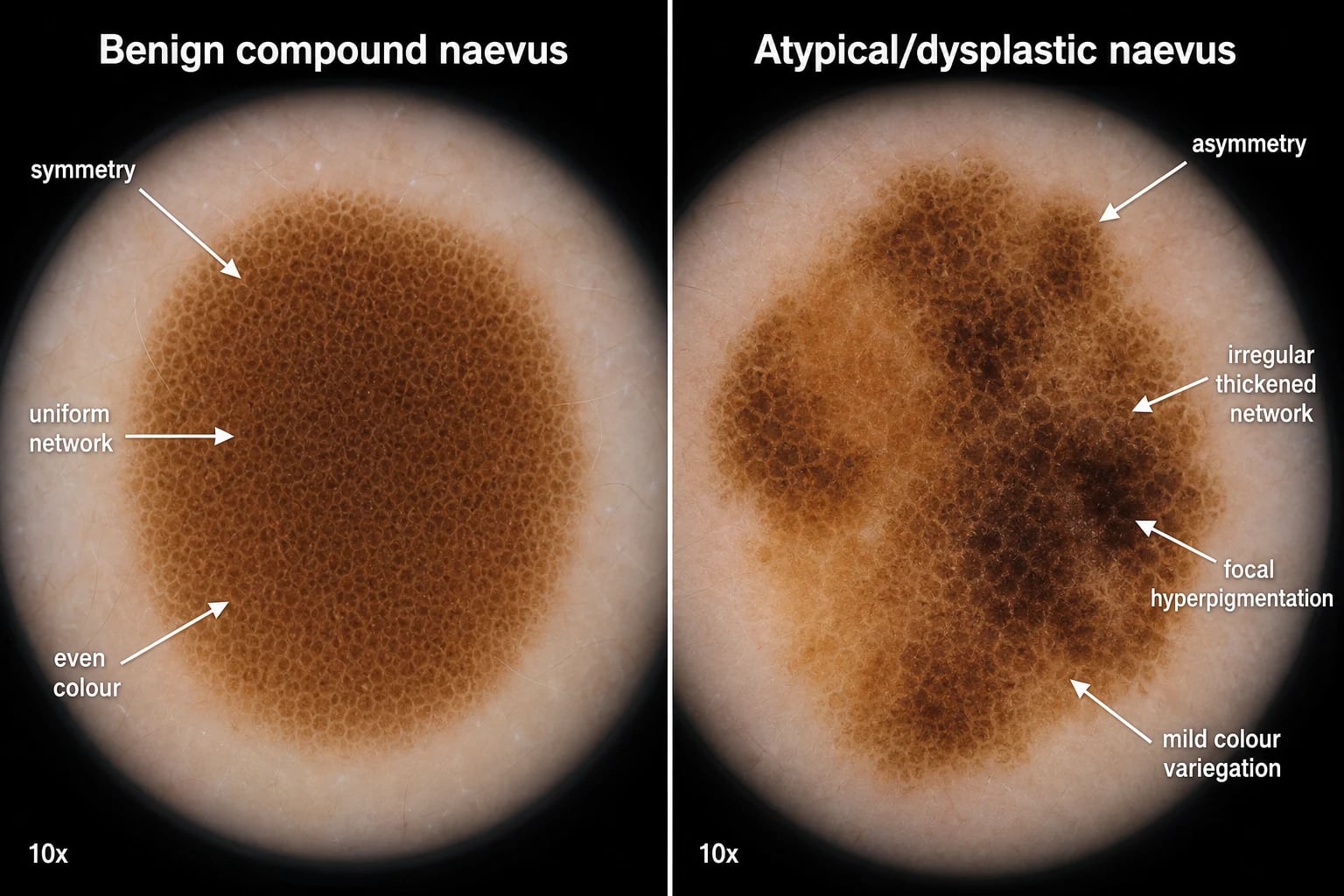
Dermoscopy is the most important office-based investigation for evaluating atypical naevi. The dermoscopic features of an atypical naevus include:[1]
- Asymmetry of colour and structure: the lesion does not have a uniform pattern.
- Atypical or irregular pigment network: network lines that are thickened, widened, abruptly ending, or unevenly distributed. This is the hallmark feature of an atypical naevus.
- Irregular globules or dots: brown-black dots or globules of varying size and distribution.
- Hyperpigmented areas or structureless zones: areas without a clear network or globular pattern.
- Peripheral faint network: a faded network at the periphery, sometimes called a negative pigment network or reticular depigmentation when the network is reversed.
- Regression structures: subtle white scar-like areas or peppering, though prominent regression should raise concern for melanoma. [1]
Pattern analysis vs algorithmic approaches: Experienced dermoscopists often use pattern analysis, which assesses the overall symmetry, colours, and structures. For learners, several algorithms help distinguish benign from malignant lesions: [1]
- ABCD rule of dermoscopy (not to be confused with the clinical ABCDE): assigns scores to asymmetry, border, colour, and differential structures; a higher score suggests melanoma.
- Menzies method: identifies 11 criteria, none of which should be present in a benign lesion. Positive features include blue-white veil, multiple brown dots, pseudopods, and broad radial streaming.
- Seven-point checklist: assesses atypical pigment network, blue-white veil, atypical vascular pattern, irregular peripheral streaks, irregular pigmentation, regression structures, and irregular dots/globules. [1]
These algorithms are useful aids, but they do not replace clinical judgement or histology when a lesion is suspicious. [1]
Melanoma-specific features that should prompt excision include blue-white veil (blue colour with overlying white haze), atypical vessels (polymorphous, dotted, or milky-red), regression structures (white scar-like areas or peppering), irregular streaks or pseudopods at the periphery, atypical blotches, ulceration, and marked asymmetry with multiple colours. The presence of any of these features shifts the lesion from "atypical naevus" to "suspicious for melanoma" and mandates excisional biopsy.[7]
Reflectance confocal microscopy (RCM) is an advanced non-invasive imaging technique that visualises the epidermis and superficial dermis at near-histological resolution. It can be used as an adjunct when dermoscopy is inconclusive. Features such as pagetoid cells, non-edged papillae, and atypical melanocytes in the dermis favour melanoma. However, RCM is not universally available and does not replace histology when melanoma is suspected.[1]
Biopsy
An excisional biopsy with a narrow margin (usually 1–3 mm) is preferred for any lesion suspicious for melanoma. This allows full histological assessment, including Breslow thickness if melanoma is found. Shave or punch biopsy should be avoided when melanoma is suspected, because partial sampling can miss the invasive component and preclude accurate Breslow measurement.[8]
Histopathology
The histological diagnosis requires both architectural disorder and cytological atypia, as described above. The pathologist should report:[5]
Grading of dysplasia
The degree of dysplasia is usually reported as mild, moderate, or severe, although the criteria are not perfectly standardised and interobserver agreement is moderate.[5] A practical approach considers both architectural disorder and cytological atypia:
- Mild dysplasia: Focal architectural disorder with minimal bridging or shouldering. Cytological atypia is mild, with only slightly enlarged nuclei and minimal pleomorphism. Mitotic figures are rare or absent.
- Moderate dysplasia: More pronounced architectural disorder with bridging between several rete ridges and more prominent shouldering. Cytological atypia is moderate, with enlarged, hyperchromatic nuclei and some pleomorphism.
- Severe dysplasia: Marked architectural disorder with extensive bridging, shouldering, and lamellar fibrosis. Cytological atypia is severe, with large, hyperchromatic, pleomorphic nuclei and prominent nucleoli. The distinction from melanoma in situ can be difficult, and the presence of pagetoid spread or dermal mitoses would tip the diagnosis toward melanoma. [1]
Some centres use a Duke system that assigns numerical scores to architectural and cytological features to improve reproducibility. Others have moved to a binary low-grade vs high-grade classification because of the poor reproducibility of the three-tier system. Regardless of the grading system used, the margin status and clinical appearance are equally important in management decisions. [1]
The histological report should also comment on any concerning features for melanoma, such as pagetoid spread of melanocytes above the basal layer, dermal mitoses, asymmetrical dermal nests, ulceration, or lymphovascular invasion. These features should prompt a diagnosis of melanoma rather than dysplastic naevus. [1]
Genetic testing
Genetic counselling and testing for CDKN2A germline mutations should be considered in families with multiple cases of melanoma, especially if pancreatic cancer is also present. Testing should be preceded by formal genetic counselling, and it is usually offered to adults rather than children.[3]
Management — Resuscitation
Atypical naevi are not a life-threatening emergency, but the diagnosis of melanoma must not be missed. The "resuscitation" equivalent here is urgent recognition and referral. Any lesion with the following features should be biopsied promptly or referred urgently to dermatology: [1]
- Rapid evolution (weeks to months).
- New nodularity or ulceration.
- Bleeding without trauma.
- Dermoscopy showing blue-white veil, polymorphous vessels, or marked asymmetry.
- An ugly duckling lesion that is changing. [1]
Management — Definitive & Stepwise
The cornerstone of management is risk stratification. The majority of atypical naevi do not need treatment; they need surveillance. Management decisions integrate the clinical phenotype, patient risk factors, dermoscopic findings, and histological grade. [1]
Why observation, not prophylaxis? A landmark concept is that the presence of atypical naevi reflects an increased field cancerisation risk across the entire skin, not a high per-lesion risk. Removing every atypical naevus would not eliminate the risk of melanoma arising de novo from uninvolved skin, and would cause substantial morbidity. Therefore, the goal is to detect melanoma early while minimising unnecessary surgery.[1][4]
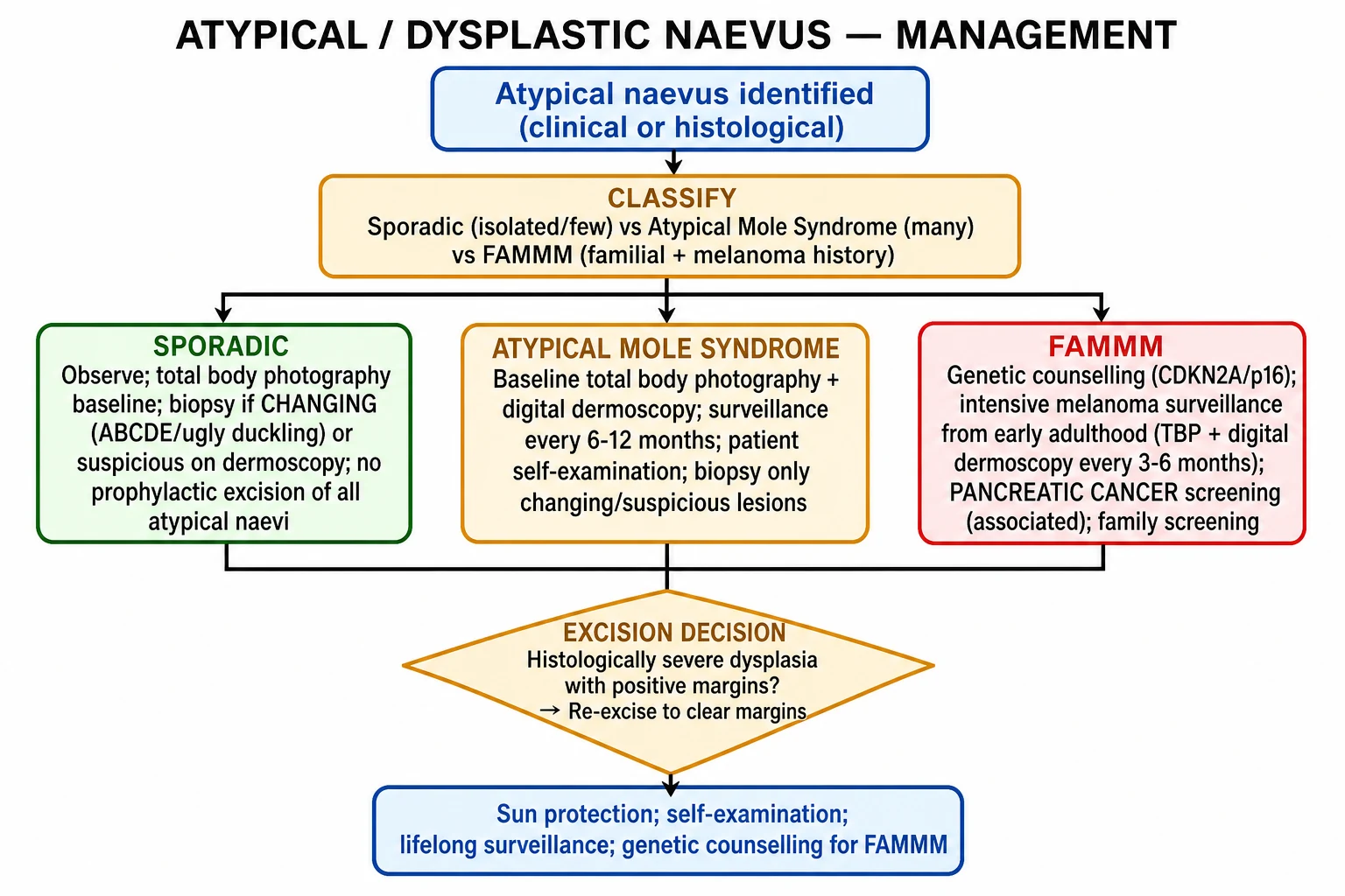
General principles
- Do not prophylactically excise all atypical naevi. Most melanomas arise de novo, not from pre-existing naevi. Mass excision does not reduce melanoma mortality and causes scarring, cost, and anxiety.[1][4]
- Biopsy only changing, suspicious, or ugly-duckling lesions. A lesion that is stable over time, with no suspicious dermoscopic features, can be observed even if clinically atypical.
- Use total body photography and sequential digital dermoscopy for high-risk patients to detect subtle change over time. Change is one of the strongest predictors of melanoma.
- Educate the patient about self-examination, the ugly duckling sign, and rigorous sun protection.
Risk-stratified management
| Category | Management |
|---|---|
| Sporadic atypical naevus | Clinical observation; baseline total body photography if multiple lesions; self-examination; biopsy if changing. |
| Atypical mole syndrome | Baseline total body photography plus sequential digital dermoscopy; dermatology review every 6–12 months; monthly self-examination; biopsy only changing or suspicious lesions. |
| FAMMM syndrome | Genetic counselling and CDKN2A testing; intensive dermatology surveillance every 3–6 months; total body photography and sequential digital dermoscopy; pancreatic cancer screening with MRI/MRCP from around age 40–50 or 10 years before the earliest family pancreatic cancer diagnosis; family screening and education. |
Histology-driven management
The management of a biopsy-proven dysplastic naevus depends on the grade of dysplasia and the margin status.[8][9][10]
- Mild dysplasia, clear margins: observe. Re-excision is generally not required even if margins are positive, provided there is no residual clinical pigment and no clinical suspicion for melanoma.
- Moderate dysplasia, clear margins: observe. If margins are positive and there is no residual clinical pigment, observation is reasonable; re-excision is not mandatory but may be considered in high-risk patients or if clinical concern persists.
- Severe dysplasia, any margins: re-excision is generally recommended to achieve clear margins, typically with 2–5 mm margins, because the distinction from early melanoma can be difficult and because the lesion may represent a true precursor or melanoma in situ mimicking a dysplastic naevus.
- Clinical suspicion trumps histology: if a lesion looks clinically suspicious for melanoma but the biopsy returns as dysplastic naevus, re-excision or re-biopsy should be considered, because partial sampling may have missed melanoma.[8]
Excision technique: When excision is performed, a full-thickness elliptical excision with a narrow margin (usually 1–3 mm for diagnostic purposes, or 2–5 mm for therapeutic excision of a dysplastic naevus) is preferred. The specimen should be oriented so that the pathologist can assess the margins. Shave or punch biopsy is avoided when melanoma is in the differential, because it may transect the lesion and prevent accurate Breslow measurement.[8][9]
Surveillance intervals: The optimal interval for follow-up is not rigidly defined and varies by risk. A pragmatic approach is: [1]
- Sporadic atypical naevus: annual review if stable, sooner if any lesion changes.
- Atypical mole syndrome: dermatology review every 6–12 months, with sequential dermoscopy of atypical lesions and other high-risk findings.
- FAMMM syndrome: dermatology review every 3–6 months, with total body photography and sequential digital dermoscopy at each visit.
- Personal history of melanoma: more frequent review, often every 3–6 months initially, then 6–12 months long-term. [1]
In high-risk patients, sequential digital dermoscopy has been shown to improve the detection of thin melanomas by identifying subtle changes over time that are not apparent on a single examination. [1]
Patient self-examination: Teach patients to examine their skin monthly in good light, using a hand mirror for hard-to-see areas. They should look for the ugly duckling and any lesion that is changing, itching, bleeding, or not healing. Prompt presentation of any change is the single most important behaviour that improves early melanoma detection. [1]
[1] [1]In Australia, the high background incidence of melanoma means that atypical mole syndrome and FAMMM are followed in dedicated pigmented lesion clinics with total body photography and sequential digital dermoscopy. Screening intervals are often 3–6 months for FAMMM and 6–12 months for atypical mole syndrome.
Sun protection and patient education
- Regular use of broad-spectrum sunscreen (SPF 30 or higher), protective clothing, wide-brimmed hats, and avoidance of peak UV exposure between 10 am and 4 pm.
- Monthly self-examination using the ABCDE criteria and the ugly duckling sign.
- Prompt presentation if any lesion changes.
- Addressing anxiety: Patients with multiple atypical naevi or FAMMM syndrome often have significant anxiety about melanoma. A structured surveillance plan, baseline total body photography, and clear education about what to watch for can reduce anxiety and improve adherence. Reassurance should be honest: the risk is increased, but most lesions are benign, and early detection of melanoma is highly curable.
- Family education: First-degree relatives of FAMMM patients should be taught self-examination and offered dermatology review, particularly if they also have multiple atypical naevi. [1]
Specific Subtypes & Scenarios
Sporadic atypical naevus
A single or small number of atypical naevi in a patient with no family history of melanoma carries a modestly increased melanoma risk. Management is reassurance, education, and surveillance. Biopsy is reserved for changing lesions. [1]
Atypical mole syndrome
Defined by more than 50 naevi, at least 5 of which are clinically atypical, without a family history of melanoma. The risk of melanoma is approximately 10-fold higher than the general population. These patients benefit from total body photography, sequential digital dermoscopy, and regular dermatology review. They should not undergo mass excision. [1]
FAMMM syndrome
FAMMM syndrome is defined by multiple atypical naevi plus a family history of melanoma, often with a CDKN2A germline mutation. Management includes: [1]
- Intensive melanoma surveillance every 3–6 months from early adulthood.
- Total body photography and sequential digital dermoscopy at each visit.
- Genetic counselling and, in appropriate families, CDKN2A testing.
- Pancreatic cancer screening with MRI/MRCP, typically starting at age 40–50 or 10 years before the earliest pancreatic cancer in the family. Screening aims to detect pancreatic ductal adenocarcinoma and its precursors (such as intraductal papillary mucinous neoplasms) at an earlier, more treatable stage. The optimal interval is not established; annual or 6-monthly imaging is often used in specialist centres.
- Family screening and education about self-examination. [1]
Patients with FAMMM syndrome should also be counselled about the incomplete penetrance of CDKN2A mutations: not every carrier develops melanoma, but the lifetime risk is high enough to justify intensive surveillance. [1]
Pregnancy
Physiological darkening and enlargement of naevi can occur. A changing lesion should be assessed with dermoscopy and biopsied if suspicious. Routine prophylactic excision is not indicated. [1]
Children
Atypical naevi in children are usually benign. However, childhood melanoma, although rare, can occur. Any rapidly changing, ulcerated, or unusually large lesion should be biopsied or referred urgently. FAMMM families should be offered genetic counselling.[6]
Complications & Pitfalls
Diagnostic pitfalls
- Pseudo-melanoma (recurrent naevus): A partially biopsied or traumatised naevus can regrow within a scar with atypical histological features that mimic melanoma. This is one reason to avoid partial biopsy of suspicious lesions and one reason some clinicians re-excise dysplastic naevi with positive margins — to prevent a confusing recurrence.[8]
- Sampling error: A shave or punch biopsy may miss the most atypical area or the invasive component of melanoma. If clinical suspicion for melanoma is high, excisional biopsy is preferred. The diagnostic specimen should be full-thickness and include the entire lesion with a narrow clinical margin.
- Over-diagnosis and over-treatment: Because the term "dysplastic" sounds alarming, patients may request or clinicians may perform unnecessary excisions. Prophylactic excision of all atypical naevi is not recommended and does not reduce melanoma mortality.
- Interobserver variability: The histological diagnosis and grading of dysplasia are not perfectly reproducible. The same lesion may be called mild, moderate, or severe by different pathologists. Management should integrate clinical and histological findings, not rely on the grade alone.
- Under-recognition of melanoma in a heavily naevus-burdened patient: A patient with hundreds of naevi may have a melanoma that is overlooked. Relying solely on ABCDE for every lesion is less effective than the ugly duckling sign and comparison with baseline photography.
Prognosis & Disposition
Atypical naevi are benign. The concern is their association with melanoma risk rather than a high per-lesion risk of malignant transformation. [1]
- Per-lesion risk: The risk that an individual atypical naevus will transform into melanoma is very low. Most melanomas arise de novo, not from pre-existing naevi. Therefore, removing every atypical naevus would not eliminate melanoma risk.
- Patient risk: The lifetime melanoma risk is substantially elevated in patients with multiple atypical naevi, and markedly elevated in FAMMM syndrome. This reflects a field cancerisation effect across the entire skin.
- After excision: Once a dysplastic naevus is fully excised with clear margins, the lesion itself is cured. The patient remains at increased risk of future melanoma elsewhere and requires continued surveillance according to their risk category.
- Disposition: Most patients are managed in dermatology or pigmented lesion clinics. FAMMM syndrome patients may benefit from multidisciplinary care involving dermatology, clinical genetics, and gastroenterology/hepatology for pancreatic screening.
- Prognostic factors for future melanoma: The strongest predictors of future melanoma are a personal history of melanoma, a family history of melanoma, the total number of naevi, the number of atypical naevi, and the presence of a CDKN2A mutation. Severe dysplasia in a biopsied lesion does not necessarily predict transformation of that lesion, but it does indicate a higher-risk phenotype. [1]
Special Populations
Paediatrics
Atypical naevi can appear from puberty. Childhood melanoma is rare but is associated with congenital naevi, FAMMM syndrome, and immunosuppression. Several paediatric considerations are important: [1]
- Childhood melanoma is rare but carries a poor prognosis when it occurs, often because it is diagnosed late. Risk factors include congenital melanocytic naevi, FAMMM syndrome, immunosuppression, and prior radiation therapy.[6]
- Spitz naevi are common melanocytic tumours in children and can closely mimic melanoma both clinically and histologically. They are usually dome-shaped, pink-red, and grow rapidly over weeks to months before stabilising.
- Congenital melanocytic naevi may be large or giant and have a variable lifetime risk of melanoma depending on size and location. Satellite lesions and neurocutaneous melanocytosis are additional concerns for large axial lesions.
- Hormonal influence: Adolescent growth spurts can be associated with the appearance of new naevi and enlargement of existing ones. Stable enlargement proportional to body growth is reassuring; rapid, disproportionate change is not.
- Family history: Any child with atypical naevi and a family history of melanoma should be referred for paediatric dermatology or genetics evaluation. CDKN2A testing is usually deferred until adulthood, but counselling and surveillance can begin in childhood.
Pregnancy
Hormonal changes can darken and enlarge naevi. This is usually benign, but any lesion with suspicious features should be assessed with dermoscopy and biopsied if necessary. Pregnancy is not a contraindication to excisional biopsy under local anaesthetic. Postpartum re-evaluation is reasonable for any lesion that changed during pregnancy, as some changes may regress after delivery. [1]
Immunosuppression
Solid-organ transplant recipients and patients on chronic immunosuppression develop more naevi and have a higher incidence of melanoma. The risk increases with duration of immunosuppression. Surveillance should be more frequent, typically every 3–6 months, and any changing lesion should be biopsied promptly. Voriconazole and other photosensitising medications can also contribute to photodamage and skin cancer risk.[1]
Elderly
New pigmented lesions or changing lesions in older adults have a higher likelihood of melanoma. Lentigo maligna melanoma in particular can mimic a large solar lentigo or an atypical flat naevus on sun-damaged skin. A low threshold for biopsy is appropriate. Comorbidities and anticoagulation may influence the choice between excisional biopsy and observation, but they should not delay diagnosis of suspected melanoma. [1]
Genetic counselling
Families with multiple cases of melanoma, particularly with early-onset disease or pancreatic cancer, should be referred for formal genetic counselling. CDKN2A testing is considered when the pre-test probability is high. The potential psychological, insurance, and family-planning implications should be discussed before testing. A positive test result should trigger surveillance for the carrier and discussion of pancreatic screening; a negative test in a family without a known mutation does not eliminate risk.[3]
Evidence, Guidelines & Regional Differences
Landmark evidence
- NIH consensus (1992): Defined the dysplastic naevus and established the concept of FAMMM syndrome, linking multiple atypical naevi with familial melanoma.
- 2015 Pigmented Lesion Subcommittee consensus: Concluded that mildly and moderately dysplastic naevi with clear margins do not require re-excision, and that observation is reasonable for moderately dysplastic naevi with positive margins. Severely dysplastic naevi should generally be re-excised. This consensus used a structured Delphi process to address the management controversy.[8]
- Fleming et al. (2016): Re-examined the threshold for re-excision and supported selective observation for mildly and moderately dysplastic naevi with positive margins, while recommending re-excision for severely dysplastic naevi. The study highlighted that residual naevus at the biopsy site was uncommon and that melanoma at the biopsy site was rare.[9]
- Adamson (2018): Editorial accompanying a large multicentre study of moderately dysplastic naevi with positive margins, concluding that observation is appropriate when no clinical residual pigment is present. This was a major step in reducing unnecessary re-excision.[10]
- Melanoma Genomics (Newton-Bishop 2020): Reviewed the genetic landscape of melanoma, emphasising the role of CDKN2A in familial melanoma and the association with pancreatic cancer. This work underpins genetic counselling and risk stratification in FAMMM syndrome.[3]
Controversies
- Terminology: Some dermatopathologists prefer to avoid the term "dysplastic naevus" and instead report "naevus with architectural disorder and cytological atypia". Others use a binary low-grade vs high-grade system rather than mild/moderate/severe because of poor interobserver agreement. Despite this, the three-tier system remains widely used in clinical practice and postgraduate examinations.[2]
- Precursor vs marker: Whether severely dysplastic naevi are true melanoma precursors or merely risk markers remains debated. The practical implication is that severely dysplastic lesions with positive margins are usually re-excised, while mild-to-moderate lesions are observed. The per-lesion risk of malignant transformation is very low.
- Re-excision thresholds: Although most data support observation for mild-to-moderate dysplasia with positive margins, some clinicians still re-excise moderate dysplasia because of patient anxiety, the pseudo-melanoma risk, or medicolegal concerns. The trend is toward less aggressive management of mild-to-moderate dysplasia.
- Screening for pancreatic cancer in CDKN2A carriers: The optimal modality, interval, and age to start screening remain under study. MRI/MRCP is generally favoured, but the evidence base is not as strong as for melanoma surveillance. Most experts begin screening around age 40–50, or 10 years before the earliest pancreatic cancer in the family.[3]
Regional differences
- United States: The American Academy of Dermatology supports surveillance of atypical mole syndrome and FAMMM, with biopsy of suspicious lesions. The 2015 PLS consensus guides re-excision decisions. Access to total body photography and sequential digital dermoscopy is variable and often concentrated in academic centres.
- United Kingdom: NICE recommends urgent 2-week wait referral for suspected melanoma. Specialist surveillance is offered for FAMMM and high-risk atypical mole syndrome. Pancreatic screening in CDKN2A carriers is considered in specialist genetics clinics. The British Association of Dermatologists supports risk-stratified follow-up rather than routine re-excision of mildly or moderately dysplastic naevi with positive margins.
- Australia: High background melanoma incidence leads to intensive screening in pigmented lesion clinics. Total body photography and sequential digital dermoscopy are widely used. Screening intervals are often 3–6 months for FAMMM and 6–12 months for atypical mole syndrome. Sun protection and public awareness campaigns are also prominent.
- Europe: Similar risk-stratified approaches; re-excision of severely dysplastic naevi is generally recommended, while observation of mildly or moderately dysplastic naevi with positive margins is accepted in many centres. Guidelines from the European Dermatology Forum and national societies generally align with the PLS consensus.
- India and low-resource settings: Access to dermoscopy, total body photography, and genetic testing may be limited. Clinical examination, the ugly duckling sign, and excisional biopsy of suspicious lesions remain the cornerstone. Patients with strong family histories should be referred to genetics centres where available. [1]
Exam Pearls
[1]Self-test: What is the management of a moderately dysplastic naevus with positive margins but no residual clinical pigment?
Observation is reasonable. The 2015 Pigmented Lesion Subcommittee consensus and subsequent data support that mildly and moderately dysplastic naevi with positive margins can be observed if there is no residual clinical pigment and no clinical suspicion for melanoma. Severely dysplastic naevi, however, are generally re-excised.
Red Flags
Exam application bank (NEET-PG / INICET)
One-line answer
An atypical (dysplastic) naevus is a benign melanocytic naevus with clinical features of irregularity (asymmetry, border irregularity, colour variegation, diameter >5 mm) and/or histological features of architectural disorder and cytological atypia. It is primarily a melanoma risk marker rather than a direct precursor, and management is risk-stratified: observation and surveillance for most, with excision of changing or suspicious lesions.
Worked stems (answer without another resource)
Stem 1 — Classic presentation. Map symptoms to mechanism; name the first investigation and first treatment step with dose/route if drug therapy is standard. [1]
Stem 2 — Unstable / complicated. List red flags that force immediate resuscitation, theatre, ICU, antidote, or reperfusion — and what you do in the first 15 minutes. [1]
Stem 3 — Atypical group. Elderly, pregnancy, child, or immunocompromised: how presentation and thresholds change. [1]
Stem 4 — Differential trap. Name the three closest mimics and one discriminator for each. [1]
Stem 5 — Disposition. Who goes home with safety-netting, who is admitted, who needs HDU/ICU/theatre, and what follow-up is mandatory. [1]
Rapid viva checklist
- Definition + classification
- Pathophysiology chain
- Bedside signs / criteria
- Score with exact components (if any)
- Emergency bundle
- Definitive therapy with doses
- Complications of disease and of treatment
- Special populations
- Guideline/trial name if classic
- Three exam traps
Coverage self-check
If you cannot answer any stem above from this page alone, re-read the matching section — the page is intended to be self-sufficient for final-prof and NEET-PG/INICET questions on Atypical / Dysplastic Naevus.
[1]References
- [1]Zhang Y, Ostrowski SM, Fisher DE. Nevi and Melanoma Hematol Oncol Clin North Am, 2024.PMID 38880666
- [2]Drozdowski R, Spaccarelli N, Peters MS, et al. Dysplastic nevus part I: Historical perspective, classification, and epidemiology J Am Acad Dermatol, 2023.PMID 36038073
- [3]Newton-Bishop J, Bishop DT, Harland M. Melanoma Genomics Acta Derm Venereol, 2020.PMID 32346746
- [4]Friedman RJ, Farber MJ, Warycha MA, et al. The dysplastic nevus Clin Dermatol, 2009.PMID 19095156
- [5]Bierhoff E. [Dysplastic melanocytic nevus] Pathologe, 2015.PMID 25591417
- [6]Jen M, Murphy M, Grant-Kels JM. Childhood melanoma Clin Dermatol, 2009.PMID 19880040
- [7]Abbasi NR, Shaw HM, Rigel DS, et al. Early diagnosis of cutaneous melanoma: revisiting the ABCD criteria JAMA, 2004.PMID 15585738
- [8]Kim CC, Berry EG, Marchetti MA, et al. Addressing the knowledge gap in clinical recommendations for management and complete excision of clinically atypical nevi/dysplastic nevi: Pigmented Lesion Subcommittee consensus statement JAMA Dermatol, 2015.PMID 25409291
- [9]Fleming NH, Reilly RB, Comfere NI, et al. Reexamining the Threshold for Reexcision of Histologically Transected Dysplastic Nevi JAMA Dermatol, 2016.PMID 27542070
- [10]Adamson AS. Observation of Moderately Dysplastic Nevi With Positive Margins: Are We There Yet? JAMA Dermatol, 2018.PMID 30304351