MedVellum
MCQsExamsAtlas
DashboardPricing
MBBS / Core medicine✳Dermatology✳ICU Fellowship (CICM)✳Anaesthesia✳Emergency Medicine✳Psychiatry Fellowship✳Paediatrics Fellowship✳Physician Medicine✳MCQs✳SAQs✳Vivas✳OSCE✳Evidence-first✳MBBS / Core medicine✳Dermatology✳ICU Fellowship (CICM)✳Anaesthesia✳Emergency Medicine✳Psychiatry Fellowship✳Paediatrics Fellowship✳Physician Medicine✳MCQs✳SAQs✳Vivas✳OSCE✳Evidence-first✳

MedVellum.

The folio

Exam-exhaustive medical education across every specialty — evidence-graded topics, engraved plates, and practice in every written and oral format. Educational content only — not medical advice.

llms.txt · psychiatry LLM catalog · sitemap

Atlas

  • Specialty atlas
  • MBBS / Core medicine
  • Dermatology
  • ICU Fellowship (CICM)
  • Anaesthesia
  • Emergency Medicine
  • Psychiatry Fellowship
  • Paediatrics Fellowship
  • Physician Medicine

Study & account

  • MCQ practice
  • Practice alias
  • Exam tools
  • Dashboard
  • Pricing
  • Sign in

© 2026 MedVellum. For education only — not a substitute for clinical judgement.

Folio edition · Set in Instrument Serif & Archivo

LibraryDermatology

Dermatology · Medicine

Atypical (nontuberculous) mycobacterial infections

Also known as Atypical mycobacteria · Nontuberculous mycobacteria (NTM) · MOTT (mycobacteria other than tuberculosis) · Environmental mycobacteria

Atypical (nontuberculous, NTM) mycobacteria are environmental organisms (water, soil) causing cutaneous and soft-tissue infections — usually after trauma, aquatic exposure, or iatrogenic procedures. Classified by growth rate: slow growers (M. marinum — 'fish tank granuloma', grows at 30°C, sporotrichoid spread; M. ulcerans — Buruli ulcer, mycolactone toxin, painless undermined ulcers) and rapid growers (M. abscessus complex — post-procedure, erm41 macrolide resistance; M. fortuitum group; M. chelonae). M. marinum is the commonest cutaneous NTM (aquatic exposure, clarithromycin + ethambutol). M. ulcerans causes Buruli ulcer (mycolactone toxin, WHO first-line rifampicin + clarithromycin). Diagnosis: tissue biopsy for AFB + culture at 30°C AND 37°C + PCR/16S sequencing for speciation. Treatment is species-specific, based on susceptibility testing, with ≥2 active drugs (never monotherapy — resistance); surgical debridement for deep/localised lesions. NTM does NOT respond to standard anti-TB therapy (RHZE).

CoreHigh evidenceUpdated 29 June 2026
On this page & tools

Your progress

Saved locally on this device.

Exam tags

FRCDermABDMRCPNEET-PGINICETRANZCD

Red flags

Chronic non-healing ulcer with undermined edges in an endemic area (West Africa, Australia) — consider M. ulcerans (Buruli ulcer); mycolactone toxin causes tissue necrosis.Sporotrichoid nodules along lymphatics on the hand/arm with aquatic exposure history — M. marinum until proven otherwise; culture at 30°C.Multiple erythematous nodules/abscesses weeks-months after a cosmetic procedure, tattoo, or injection — rapid-growing NTM (M. abscessus, M. fortuitum, M. chelonae).Treating NTM with standard anti-TB therapy (RHZE) — most NTM are resistant; always base treatment on species identification and susceptibility testing.

Your progress

Saved locally on this device.

Exam tags

FRCDermABDMRCPNEET-PGINICETRANZCD

Red flags

Chronic non-healing ulcer with undermined edges in an endemic area (West Africa, Australia) — consider M. ulcerans (Buruli ulcer); mycolactone toxin causes tissue necrosis.Sporotrichoid nodules along lymphatics on the hand/arm with aquatic exposure history — M. marinum until proven otherwise; culture at 30°C.Multiple erythematous nodules/abscesses weeks-months after a cosmetic procedure, tattoo, or injection — rapid-growing NTM (M. abscessus, M. fortuitum, M. chelonae).Treating NTM with standard anti-TB therapy (RHZE) — most NTM are resistant; always base treatment on species identification and susceptibility testing.

In one line

Atypical (nontuberculous, NTM) mycobacteria are environmental organisms (water, soil) causing cutaneous and soft-tissue infections — usually after trauma, aquatic exposure, or iatrogenic procedures. Classified by growth rate: slow growers (M. marinum — 'fish tank granuloma', grows at 30°C, sporotrichoid spread; M. ulcerans — Buruli ulcer, mycolactone toxin, painless undermined ulcers) and rapid growers (M.

[1]
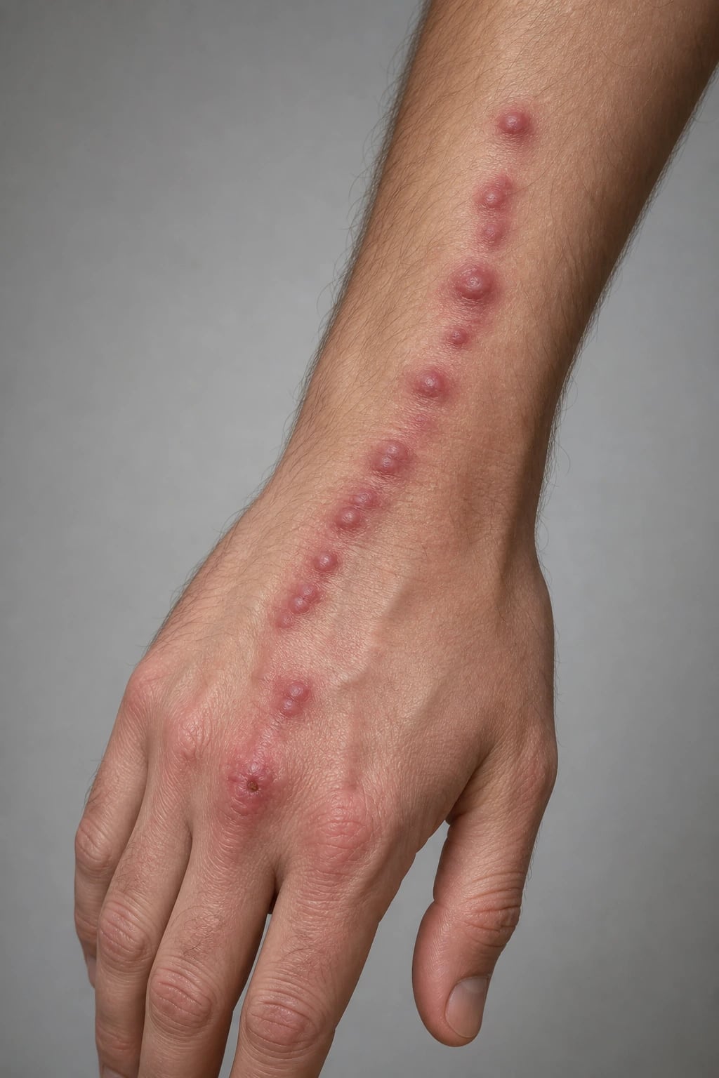
Clinical and histological overview of atypical mycobacterial infections: M. marinum fish tank granuloma on finger, M. ulcerans Buruli ulcer with undermined edges, M. abscessus post-procedure abscesses
FigureAtypical (nontuberculous) mycobacteria: environmental organisms causing cutaneous infection after trauma, aquatic exposure, or procedures. Key species: M. marinum (commonest), M. ulcerans (Buruli ulcer), M. abscessus (post-procedure). (AI-generated educational illustration.)

Classification

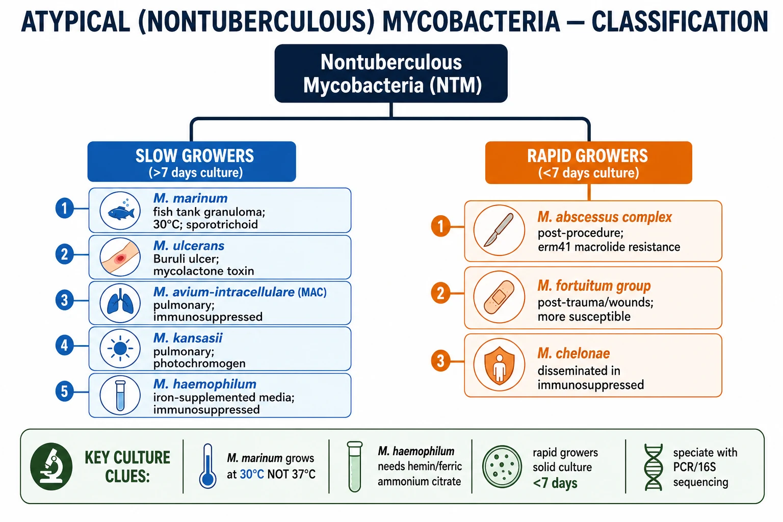
NTM classification tree: slow growers (M. marinum, M. ulcerans, MAC, M. kansasii, M. haemophilum) vs rapid growers (M. abscessus, M. fortuitum, M. chelonae) with growth temperatures and key features
FigureNTM classified by growth rate: SLOW growers ( width={1536} height={1024}>7 days culture) include M. marinum (30°C), M. ulcerans (mycolactone), MAC, M. kansasii, M. haemophilum (needs iron). RAPID growers ( less than 7 days) include M. abscessus complex, M. fortuitum, M. chelonae. (AI-generated educational figure.)
[1]

Atypical (nontuberculous) mycobacteria (NTM) are classified by growth rate in culture — the most practical division for dermatology:[1][2][9]

Slow growers (>7 days to culture positivity)

  • Mycobacterium marinum — commonest cutaneous NTM; grows at 30-32°C (not 37°C); aquatic exposure.
  • Mycobacterium ulcerans — Buruli ulcer; produces mycolactone toxin.
  • Mycobacterium avium-intracellulare complex (MAC) — usually pulmonary; cutaneous in immunosuppressed.
  • Mycobacterium kansasii — photochromogen; pulmonary; rarely cutaneous.
  • Mycobacterium haemophilum — requires iron-supplemented media (hemin/ferric ammonium citrate); immunosuppressed. [1]

Rapid growers ( less than 7 days; often 3-7 days)

  • Mycobacterium abscessus complex (subsp. abscessus, massiliense, bolletii) — post-procedure; intrinsic macrolide resistance (erm41 gene).
  • Mycobacterium fortuitum group — post-trauma/wounds; more antibiotic-susceptible.
  • Mycobacterium chelonae — disseminated cutaneous disease in immunosuppressed (corticosteroids).[6][8]

Pathophysiology

NTM are ubiquitous in the environment (water, soil, dust, biofilms in plumbing). Infection occurs via:[1][2]

  • Direct inoculation through broken skin (minor trauma, abrasions, bites, tattoos, injections, surgery).
  • Aquatic exposure — fish tanks, swimming pools, marine/brackish water (M. marinum).
  • Iatrogenic — contaminated instruments, injectable solutions, mesotherapy, liposuction, catheter sites (M. abscessus, M. fortuitum, M. chelonae).[6]
  • Disseminated disease in immunosuppressed (HIV, transplant, anti-TNF) — MAC, M. haemophilum, M. chelonae.[2]

The immune response produces suppurative and/or granulomatous inflammation (caseating or non-caseating). M. ulcerans is unique — its mycolactone toxin causes direct tissue necrosis and immune suppression (explains the painless, destructive ulcer).[2]

Atypical mycobacteria: environmental organisms causing skin disease via inoculation

Non-tuberculous mycobacteria (NTM) are environmental organisms found in water, soil, biofilms, and aquatic environments. Cutaneous infection occurs by direct inoculation through broken skin (minor trauma, fish tank exposure, contaminated surgery, injection). The two clinical clues are: (1) atypical mycobacterial culture at 25-32°C (M. marinum does not grow at 37°C), and (2) sporotrichoid (lymphocutaneous) spread for M. marinum/M. ulcerans. M. ulcerans (Buruli ulcer) is unique — its mycolactone toxin causes painless necrotising ulcers with undermined edges, particularly in West Africa and Australia. M. abscessus has intrinsic macrolide resistance (erm41 gene), making it difficult to treat.

[1]

Quick numbers for the examiner

30-32°C
Optimal culture temperature for M. marinum
Cultures at 37°C may be FALSE NEGATIVE
25-50%
Sporotrichoid (lymphocutaneous) spread in M. marinum
Linear chain of nodules along lymphatic drainage
10-25%
M. ulcerans patients with permanent disability
Buruli ulcer; can lead to contractures, amputation
2-3 weeks
Incubation period for M. marinum
Range 1-8 weeks
3-4 months
Treatment duration for M. marinum
Clarithromycin + ethambutol; surgical excision for deep
8 weeks
WHO-recommended antibiotic course for M. ulcerans
Rifampicin + clarithromycin (formerly streptomycin-based)

Clinical Features by Species [1]

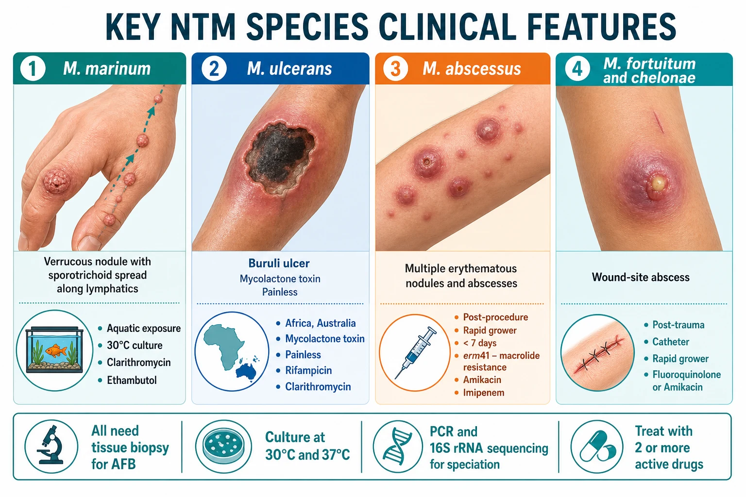
Four-panel comparative infographic: M. marinum (sporotrichoid nodules, fish tank), M. ulcerans (undermined ulcer, Buruli), M. abscessus (post-procedure abscesses), M. fortuitum/chelonae (wound infection)
FigureKey species compared: M. marinum (aquatic, sporotrichoid), M. ulcerans (Buruli, mycolactone, undermined), M. abscessus (post-procedure, rapid grower, erm41), M. fortuitum/chelonae (post-trauma, more susceptible). (AI-generated educational figure.)

Mycobacterium marinum ("fish tank / swimming pool granuloma")

  • Commonest cutaneous NTM. Aquatic exposure (fish tanks, aquariums, swimming pools, marine/brackish water, fish handling) + minor trauma.[3][5]
  • Incubation: 2-3 weeks (range 1-8 weeks).
  • Presentation: solitary verrucous nodule or plaque on a finger/hand (dominant hand); may ulcerate. Sporotrichoid (lymphocutaneous) spread — linear chain of nodules along lymphatic drainage up the arm (25-50% of cases).[3]
  • Deep infection: tenosynovitis, osteomyelitis, arthritis (if deep inoculation).
  • Culture grows at 30-32°C (NOT 37°C — crucial laboratory clue; cultures at 37°C may be negative).[3]
  • Treatment: clarithromycin + ethambutol for 3-4 months (first-line); alternatives: doxycycline, minocycline, rifampicin + ethambutol, TMP-SMX. Surgical excision/debridement for deep tenosynovitis/osteomyelitis.[4][5]

Mycobacterium ulcerans (Buruli / Bairnsdale / Daintree ulcer)

  • Endemic: West Africa (Benin, Ghana, Côte d'Ivoire), Australia (Victoria), Papua New Guinea, parts of SE Asia.[2]
  • Pathogenesis: mycolactone — a polyketide-derived macrolide toxin that causes tissue necrosis and suppresses local immune responses (macrophage apoptosis, cytokine suppression).[2]
  • Presentation: starts as a painless subcutaneous nodule, papule, plaque, or oedema → progresses to a large ulcer with characteristic UNDERMINED edges (necrosis extends subcutaneously, larger than the surface opening). Ulcers can be very large and destructive (skin, fat, even bone). Systemic symptoms absent.[2]
  • Stages: (1) pre-ulcerative nodule/plaque/oedema; (2) ulcerative (undermined edges); (3) fibrosis/contracture/scarring (healing, but with disability/contracture).
  • Treatment: WHO first-line: rifampicin + clarithromycin (or rifampicin + streptomycin/amikacin) for 8 weeks; surgery (debridement, skin grafting) for advanced/fibrotic lesions. Antibiotic therapy has replaced surgery as primary treatment.[2]
  • BCG gives partial protection (not a treatment; epidemiological observation).

Mycobacterium abscessus complex

  • Rapid grower (culture positive in 3-7 days). Post-procedural — injections, mesotherapy, liposuction, surgery, tattoos, piercing, catheter-related.[6][8]
  • Presentation: multiple erythematous nodules, papules, pustules, abscesses or draining sinus tracts weeks to months after the procedure. Often clustered along the procedural track.[6]
  • Intrinsic antibiotic resistance: the erm(41) gene confers inducible macrolide resistance in M. abscessus subsp. abscessus (but NOT in subsp. massiliense, which has a non-functional erm41). Subspecies identification is essential for predicting macrolide susceptibility.[8]
  • Treatment: amikacin + imipenem or cefoxitin + macrolide (if susceptible based on erm41/ subspecies); prolonged (6+ months); surgical excision often needed for localised disease.[6][8]
  • Pulmonary disease: significant pathogen in cystic fibrosis and chronic lung disease.[7][8]

Mycobacterium fortuitum group

  • Rapid grower. Post-trauma (wounds, lacerations), catheter-related, postsurgical, tattoo-related.[6]
  • Similar to M. abscessus but typically smaller abscesses and more antibiotic-susceptible.
  • Treatment: amikacin + fluoroquinolones (ciprofloxacin, moxifloxacin) or doxycycline or TMP-SMX for 4-6 months.[6]

Mycobacterium chelonae

  • Rapid grower. Post-procedure or disseminated cutaneous disease in immunosuppressed (especially chronic corticosteroid use).[6]
  • Multiple violaceous nodules/abscesses on extremities.
  • Treatment: tobramycin + clarithromycin + imipenem or linezolid; surgical excision for localised lesions.[6]

Mycobacterium haemophilum

  • Requires iron-supplemented media (hemin or ferric ammonium citrate) and 30°C incubation — easily missed if not specifically requested.[1]
  • Immunosuppressed (transplant, HIV, anti-TNF): tender violaceous papules/nodules/ulcers on extremities.

Mycobacterium avium-intracellulare complex (MAC)

  • Primarily pulmonary (chronic cough). Cutaneous disease rare; occurs in immunosuppressed (advanced HIV with CD4 less than 50; transplant).[2]
  • Prophylaxis in HIV: azithromycin when CD4 less than 50.[2]

Histopathology

  • Mixed suppurative and granulomatous inflammation — neutrophils + epithelioid granulomas ± necrosis (caseating or non-caseating).[2][10]
  • AFB stain (Ziehl-Neelsen / Fite) may reveal organisms, but sensitivity is variable (organisms may be scant). M. ulcerans lesions show extensive necrosis with clumps of extracellular AFB in the fat.[2]
  • Histology alone cannot speciate NTM — requires culture and/or molecular methods.

Investigations and Diagnosis

  • Tissue biopsy (deep, including subcutaneous fat) for:
    • AFB stain (Ziehl-Neelsen or Fite-Faraco).
    • Culture at multiple temperatures (30°C AND 37°C) on Lowenstein-Jensen/Middlebrook agar; specify request for NTM — slow growers >7 days, rapid growers less than 7 days.[1][3]
    • Molecular speciation: PCR, line probe assay (GenoType Mycobacterium), or 16S rRNA gene sequencing (gold standard for definitive speciation).[2]
    • Histopathology.
  • M. marinum cultures must be incubated at 30-32°C (37°C is usually negative).
  • M. haemophilum requires iron-supplemented media (hemin/ferric ammonium citrate) and 30°C.
  • M. abscessus subspecies identification (abscessus vs massiliense vs bolletii) via erm(41) gene sequencing — determines macrolide susceptibility.[8]
  • Antibiotic susceptibility testing — essential for all NTM; resistance patterns differ markedly from M. tuberculosis and between NTM species.

Differential Diagnosis

  • Sporotrichosis (sporotrichoid spread — identical pattern; differentiate by culture/histology).
  • Cutaneous tuberculosis (lupus vulgaris, scrofuloderma, warty TB — caseating granulomas; AFB stain; positive Mantoux/IGRA; response to RHZE).
  • Leishmaniasis (amastigotes on Giemsa; travel history).
  • Staphylococcal/streptococcal abscesses (acute onset; Gram stain).
  • Foreign-body granuloma (suture, silica, tattoo ink — polarised light).
  • Sarcoidosis (non-caseating "naked" granulomas; systemic features).[10]
  • Pyoderma gangrenosum (undermined ulcer; rapidly progressive; associated systemic disease).

Management
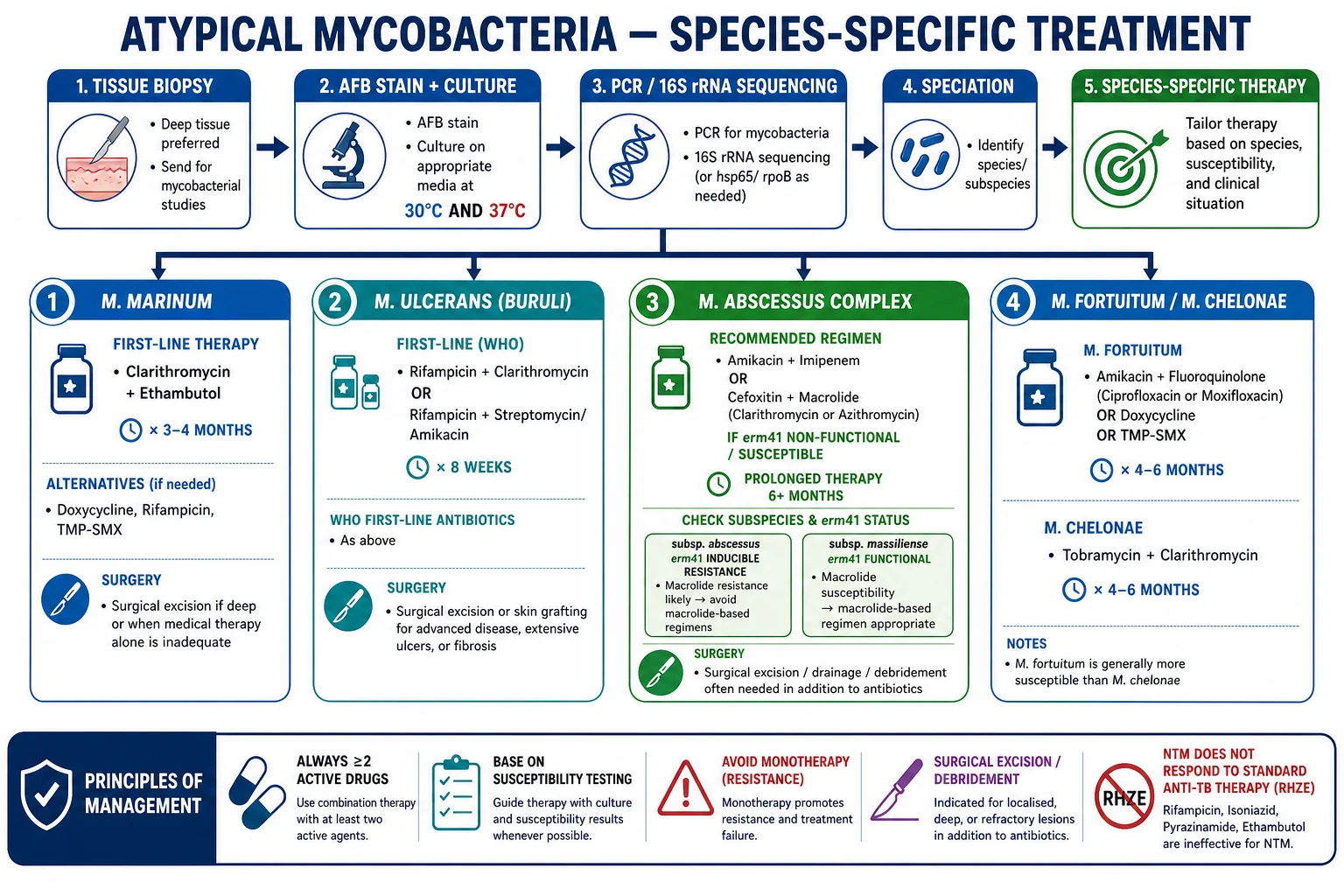
Species-specific treatment algorithm: M. marinum (clarithromycin + ethambutol 3-4mo), M. ulcerans (rifampicin + clarithromycin 8wk WHO), M. abscessus (amikacin + imipenem/cefoxitin + macrolide if susceptible), M. fortuitum/chelonae (amikacin + fluoroquinolone/TMP-SMX 4-6mo). Principles: width={1536} height={1024}>=2 drugs, susceptibility-guided, no monotherapy, surgery for deep lesions.
FigureSpecies-specific treatment: M. marinum (clarithromycin + ethambutol), M. ulcerans (rifampicin + clarithromycin per WHO), M. abscessus (amikacin + imipenem/cefoxitin ± macrolide; check erm41), M. fortuitum/chelonae (fluoroquinolones/amikacin). Always ≥2 drugs, susceptibility-guided. (AI-generated educational figure.)
[1]

Principles

  • Species-specific — always identify the organism (culture/PCR/16S sequencing).[1]
  • Susceptibility-guided — antibiotic choices based on in vitro susceptibility (NTM resistance patterns differ from TB and between species).[7]
  • Combination therapy (≥2 active drugs) to prevent resistance — monotherapy is never acceptable.[7]
  • Duration: typically 3-6+ months depending on species, severity, and immune status.
  • Surgical excision/debridement for localised, deep, or treatment-resistant lesions (tenosynovitis, osteomyelitis).
  • NTM does NOT respond to standard anti-TB therapy (RHZE) — a common exam and clinical pitfall.[1][7]

Species-specific regimens

SpeciesFirst-line regimenDuration
M. marinumClarithromycin + ethambutol (alternatives: doxycycline, minocycline, rifampicin + ethambutol, TMP-SMX)3-4 months[4][5]
M. ulceransRifampicin + clarithromycin (or rifampicin + streptomycin/amikacin); WHO first-line8 weeks[2]
M. abscessus complexAmikacin + imipenem or cefoxitin + macrolide (if erm41 non-functional / susceptible); surgery often needed6+ months[6][8]
M. fortuitumAmikacin + fluoroquinolone (cipro/moxi) or doxycycline or TMP-SMX4-6 months[6]
M. chelonaeTobramycin + clarithromycin + imipenem or linezolid; surgery for localised4-6 months[6]
M. haemophilumClarithromycin/azithromycin + rifampicin/ethambutol + amikacin/ciprofloxacinVariable
MACAzithromycin/clarithromycin + rifampicin + ethambutol ± amikacin12 months[7]

Prognosis

  • Most cutaneous NTM infections respond well to appropriate therapy (if species is correctly identified and treatment is susceptibility-guided).
  • M. abscessus is the most difficult to treat (intrinsic resistance; prolonged therapy; surgical excision often required).[8]
  • M. ulcerans can cause significant morbidity (contracture, disability) if treatment is delayed.
  • Disseminated disease (immunosuppressed) carries worse prognosis.[2]

Exam Pearls

High-yield points for fellowship exams

  1. M. marinum = commonest cutaneous NTM; aquatic exposure (fish tank); sporotrichoid spread; grows at 30-32°C (not 37°C); treat with clarithromycin + ethambutol.
  2. M. ulcerans = Buruli ulcer; mycolactone toxin (tissue necrosis + immune suppression); painless ulcer with undermined edges; WHO first-line: rifampicin + clarithromycin 8 weeks.
  3. Rapid growers ( less than 7 days culture): M. abscessus, M. fortuitum, M. chelonae — usually post-procedure/post-trauma.
  4. M. abscessus = erm(41) gene → inducible macrolide resistance (in subsp. abscessus but NOT massiliense); subspecies ID is essential.
  5. Culture at 30°C AND 37°C — M. marinum at 30°C, M. haemophilum needs iron-supplemented media at 30°C.
  6. PCR / 16S rRNA sequencing — gold standard for speciation.
  7. NTM does NOT respond to standard anti-TB therapy (RHZE) — common exam trap.
  8. Always ≥2 active drugs based on susceptibility testing — monotherapy causes resistance.
  9. Sporotrichoid spread on hand/arm — differentials: M. marinum vs Sporothrix schenckii (both need culture).
  10. M. chelonae = disseminated cutaneous in corticosteroid-immunosuppressed patients.
[1]

NTM are environmental saprophytes; treatment is 6-12 months minimum and guided by species susceptibility

Non-tuberculous mycobacteria (NTM) are environmental organisms distributed widely in water (mains, swimming pools, fish tanks, hospital water systems — including biofilms), soil, dust, and animals. Approximately 200 NTM species are recognised; only a fraction cause human disease. M. marinum (fish tank granuloma) is the commonest cutaneous pathogen in immunocompetent hosts; M. ulcerans (Buruli ulcer) is the third most common mycobacterial disease worldwide after TB and leprosy; rapid-growers (M. abscessus, M. fortuitum, M. chelonae) cause post-traumatic and post-surgical infections; MAC (M. avium complex) is associated with disseminated disease in HIV. Treatment of NTM cutaneous disease is prolonged (6-12 months minimum for most species), guided by in-vitro drug susceptibility testing (CLSI breakpoints), and often requires combination of 2-3 active agents (macrolide backbone with rifamycin, ethambutol, tetracycline, or fluoroquinolone). Surgery is adjunctive for extensive disease, abscesses, sinus tracts, and Buruli ulcer debridement. [1]

Diagnostic approach. The cornerstone of diagnosis is culture from biopsy or tissue aspirate on appropriate media at the correct temperature. M. marinum grows best at 30–32 degrees C, so cultures incubated only at 37 degrees C may be falsely negative. Rapid growers (M. abscessus, M. fortuitum, M. chelonae) grow on routine media within 3–5 days, while slow growers (M. marinum, M. kansasii, MAC, M. ulcerans) require 2–4 weeks. Histopathology shows caseating or non-caseating granulomas, lymphocytic infiltrate, and acid-fast bacilli (AFB) with Ziehl-Neelsen or Fite stains. Tissue PCR and rpoB gene sequencing can identify species rapidly when culture is negative. For M. ulcerans, PCR of swab or tissue detecting the IS2606 and IS2404 insertion sequences is diagnostic. [1]

Species-specific treatment regimens. M. marinum: first-line is clarithromycin 500 mg BD plus either rifampicin 600 mg daily or ethambutol 15 mg/kg daily for 3–4 months after clinical resolution (typically 6 months total). Severe/tenosynovial disease may require debridement. M. ulcerans: WHO recommends 8 weeks of rifampicin 10 mg/kg (max 600 mg) daily plus clarithromycin 7.5 mg/kg BD; surgery is indicated for large lesions, contractures, or paradoxical reactions. M. kansasii: rifampicin 600 mg, ethambutol 15 mg/kg, isoniazid 5 mg/kg, and clarithromycin or azithromycin for 12 months. MAC: clarithromycin 500 mg BD or azithromycin 250 mg daily plus ethambutol 15 mg/kg and rifampicin 600 mg for 12 months in disseminated disease; localised skin disease may respond to 6 months. Rapid growers: M. fortuitum is usually susceptible to amikacin, cefoxitin, ciprofloxacin, doxycycline, sulfonamides; M. abscessus is the most refractory due to inducible macrolide resistance (erm41 gene), requiring combination therapy with amikacin, cefoxitin, tigecycline, imipenem, and azithromycin/clarithromycin guided by susceptibility; treatment often exceeds 12 months. [1]

Immunosuppressed and special populations. In HIV, CD4 count less than 50 is associated with disseminated MAC; immune reconstitution inflammatory syndrome (IRIS) can paradoxically worsen pre-existing NTM infection. Solid-organ and stem-cell transplant recipients may develop cutaneous M. chelonae or M. abscessus at catheter sites or surgical wounds. Tumour necrosis factor-alpha inhibitors increase risk of NTM. Children may develop cervical lymphadenitis from M. avium complex, often treated with surgical excision plus clarithromycin-based therapy. In pregnancy, avoid aminoglycosides and fluoroquinolones; rifampicin and ethambutol are generally acceptable. [1]

Specific clinical syndromes and images. Bazin disease (erythema induratum of Bazin) is a panniculitis associated with M. tuberculosis but can be caused by M. avium complex and other NTM; it presents as tender violaceous nodules on the calves with ulceration and scars, classically in young women. Fish tank granuloma appears as a painless violaceous papule or nodule at the site of an aquatic injury, often with proximal lymphatic spread (sporotrichoid pattern) along the extremity. Buruli ulcer begins as a painless pre-ulcerative nodule or plaque, then progresses to a large undermined ulcer with white-yellow necrotic base; healing is slow and leaves extensive scarring. Swimming pool granuloma is a variant of M. marinum infection from inadequately chlorinated pools or hot tubs. Post-surgical NTM abscess presents weeks after cosmetic procedures, liposuction, or mesotherapy with erythematous, fluctuant nodules that fail routine antibiotics; M. abscessus and M. fortuitum are the usual culprits. Disseminated cutaneous NTM in immunosuppression produces multiple crusted papules, nodules, pustules, or ulcers; biopsy and blood culture are essential. [1]

Antimicrobial susceptibility and monitoring. Susceptibility testing follows CLSI M24 and M62 standards. For M. marinum, routine testing should include clarithromycin, rifampicin, ethambutol, and doxycycline. For M. abscessus, testing includes amikacin, cefoxitin, imipenem, tigecycline, clarithromycin (induced and non-induced macrolide testing), linezolid, and cotrimoxazole. Ethambutol optic neuritis requires baseline and monthly visual acuity testing; aminoglycosides need renal function and audiovestibular monitoring; macrolides require QTc and liver function monitoring; rifampicin induces CYP enzymes and interacts with many drugs including antiretrovirals, warfarin, and oral contraceptives. Treatment should be individualised and discussed with a specialist infectious diseases or clinical microbiology unit. [1]

NTM versus M. tuberculosis: clinical differentiation. Unlike tuberculosis, NTM disease is usually not transmitted person-to-person, is often associated with environmental exposure (water, soil, animals), and affects different risk groups (immunocompetent with aquatic or traumatic exposure, immunocompromised with disseminated disease). Cutaneous TB includes lupus vulgaris (plaque, apple-jelly nodules on diascopy), scrofuloderma (sinus tracts from underlying lymph nodes), tuberculosis verrucosa cutis (warty plaque in autoinoculation), and miliary TB. NTM lesions are more often sporotrichoid, nodular, ulcerative, or post-surgical abscesses. Histology can overlap; culture and molecular speciation are essential. Anti-TB first-line regimen (rifampicin, isoniazid, pyrazinamide, ethambutol) is generally inadequate for NTM because many species are innately resistant to isoniazid and pyrazinamide; incorrect therapy is a common pitfall leading to treatment failure. [1]

Genotypic markers and emerging resistance. M. abscessus subspecies include M. abscessus subsp. abscessus, subsp. bolletii, and subsp. massiliense. The erm(41) gene confers inducible macrolide resistance; subsp. massiliense has a non-functional erm(41) and is therefore macrolide-susceptible, a distinction that changes management. Mutations in the 23S rRNA rrl gene can cause constitutive macrolide resistance. Rifamycin resistance is conferred by rpoB mutations. Whole-genome sequencing and matrix-assisted laser desorption/ionisation time-of-flight mass spectrometry (MALDI-TOF MS) are increasingly used for rapid species identification in reference laboratories. Susceptibility testing should be repeated for relapse isolates because resistance can emerge during therapy. [1]

Surgical and procedural management. Surgery is not curative alone but is essential adjunctive therapy for many NTM cutaneous infections. For Buruli ulcer, wide local excision or debulking with skin grafting is indicated for large lesions, recurrent disease, fixed contractures, or severe paradoxical reactions. M. marinum tenosynovitis often requires synovectomy and debridement after or alongside antibiotic therapy. Post-surgical NTM abscesses need incision and drainage with culture-directed antibiotics. M. abscessus disease frequently requires repeated debridement and delayed reconstruction. For cervical lymphadenitis due to MAC in children, complete surgical excision of the affected lymph node can be curative without antibiotics in selected cases. Wound care, physiotherapy to prevent contracture, and reconstructive surgery for extensive scarring are integral to long-term outcomes. [1]

Prognosis and follow-up. M. marinum and M. ulcerans have excellent outcomes when treated appropriately, though Buruli ulcer healing can take months and scarring is often permanent. M. fortuitum usually responds well to susceptible antibiotics and surgery. M. abscessus is the most challenging, with cure rates of 50-70 percent even with prolonged combination therapy and surgery; relapse is common. All patients require regular clinical review, adverse-event monitoring, and repeat cultures where feasible. Imaging (MRI for soft tissue and bone, CT for pulmonary involvement, ultrasound for lymph nodes) guides response assessment. Treatment should be continued for at least 3-6 months after clinical resolution and until susceptibility-guided endpoints are met. [1]

NTM in the context of immunomodulatory therapy. Tumour necrosis factor-alpha inhibitors, rituximab, methotrexate, systemic corticosteroids, Janus kinase inhibitors, and calcineurin inhibitors all increase susceptibility to NTM. Patients starting biologics should be screened for latent TB and risk factors for NTM; active NTM infection is a relative contraindication to initiating anti-TNF therapy. Disseminated MAC is an AIDS-defining illness in HIV with CD4 under 50 cells per microlitre; prophylaxis with azithromycin 1200 mg weekly is indicated when CD4 is below 50 and no active MAC is present. In solid-organ transplant recipients, cutaneous NTM should prompt evaluation for disseminated disease and review of immunosuppression intensity. [1]

Key differential diagnoses. The sporotrichoid pattern of M. marinum can mimic sporotrichosis (Sporothrix schenckii, often cat-related thorn injury), cutaneous leishmaniasis, nocardiosis, atypical fungal infection, or mycetoma. Buruli ulcer must be distinguished from tropical ulcer, cutaneous leishmaniasis, squamous cell carcinoma, deep fungal infection, and pyoderma gangrenosum. Post-surgical abscesses should raise suspicion for MRSA, other bacterial pathogens, and rapidly growing NTM. Lymphocutaneous nodules in an aquarium worker or swimmer strongly suggest M. marinum. A non-healing ulcer in a returned traveller from West Africa or rural Australia should trigger Buruli PCR testing. Biopsy with culture and molecular testing is the only reliable way to distinguish these entities. [1]

Prevention and public health. Buruli ulcer prevention focuses on avoiding contact with stagnant or slow-flowing water in endemic regions, wearing protective clothing, and prompt wound cleaning. M. marinum prevention in aquarium workers involves gloves and prompt disinfection of skin breaks. In hospitals, strict water supply monitoring and biofilm control reduce M. fortuitum and M. abscessus outbreaks. Antenatal screening for syphilis does not detect NTM, but recognition of congenital TB vs NTM lymphadenitis requires careful microbiology. [1]

Non-tuberculous mycobacteria (NTM) are environmental organisms distributed widely in water (mains, swimming pools, fish tanks, hospital water systems — including biofilms), soil, dust, and animals. Approximately 200 NTM species are recognised; only a fraction cause human disease. M. marinum (fish tank granuloma) is the commonest cutaneous pathogen in immunocompetent hosts; M. ulcerans (Buruli ulcer) is the third most common mycobacterial disease worldwide after TB and leprosy; rapid-growers (M. abscessus, M. fortuitum, M. chelonae) cause post-traumatic and post-surgical infections; MAC (M. avium complex) is associated with disseminated disease in HIV. Treatment of NTM cutaneous disease is prolonged (6-12 months minimum for most species), guided by in-vitro drug susceptibility testing (CLSI breakpoints), and often requires combination of 2-3 active agents (macrolide backbone with rifamycin, ethambutol, tetracycline, or fluoroquinolone). Surgery is adjunctive for extensive disease, abscesses, sinus tracts, and Buruli ulcer debridement.

[1]

NTM treatment doses quick numbers

100-200 mg
Rifampicin daily (oral) - 6-9 months for M. marinum
Plus clarithromycin 500 mg BD; synergy for rifampicin-resistant strains
500 mg
Clarithromycin BD - 3-6 months for M. marinum
Macrolide backbone; substitute azithromycin 250-500 mg daily
15-25 mg/kg
Ethambutol daily - adjunctive for M. kansasii, MAC
Monitor for optic neuritis; monthly visual acuity
300 mg
Rifampicin monthly supervised (WHO Buruli regimen)
Plus clarithromycin 7.5 mg/kg BD; 8 weeks; may continue to 12 weeks
10-15 mg/kg
Amikacin IV daily (severe M. abscessus)
Monitor renal function; combination with cefoxitin or tigecycline
5 mg/kg
Streptomycin IM (Buruli alternative regimen)
Ototoxic; nephrotoxic; replace with clarithromycin in modern regimens
[1]

FISH-MAC - five rapid vs slow NTM pathogens

F Fortuitum (rapid grower, 3-7 days culture)

M. fortuitum - post-trauma, post-surgical, post-injection; no prior immune compromise; susceptible to amikacin, cefoxitin, fluoroquinolones, doxycycline

I Immunocompromised specialists - M. abscessus and M. chelonae

Rapid growers; M. abscessus has intrinsic macrolide resistance via erm41 gene; M. massiliense lacks erm41 and is macrolide-susceptible

S Slow growers (weeks-culture) - M. marinum, MAC, M. kansasii

M. marinum grows at 30-32 degrees C (not 37); MAC needs liquid medium; M. kansasii standard 4-drug regimen

H Healed by scarring (M. ulcerans - mycolactone toxin)

Buruli ulcer painless undermined edges; mycolactone tissue necrosis; standard WHO 8-week rifampicin + clarithromycin

M M. marinum - fish tank granuloma (most common cutaneous)

Aquatic exposure; sporotrichoid spread; clarithromycin + ethambutol 3-4 months; surgical excision for deep disease

A Atypical = non-tuberculous = environmental (water/soil/dust)

Distinguishes from M. tuberculosis; anti-TB RHZE not effective; treatment guided by species and susceptibility

C Clarithromycin is the macrolide backbone (with exceptions)

M. abscessus erm41+ resistant; M. massiliense susceptible; substitute azithromycin 250-500 mg daily

[1]

Red Flags

Exam application bank (NEET-PG / INICET)

One-line answer

Atypical (nontuberculous, NTM) mycobacteria are environmental organisms (water, soil) causing cutaneous and soft-tissue infections — usually after trauma, aquatic exposure, or iatrogenic procedures. Classified by growth rate: slow growers (M. marinum — 'fish tank granuloma', grows at 30°C, sporotrichoid spread; M. ulcerans — Buruli ulcer, mycolactone toxin, painless undermined ulcers) and rapid growers (M. abscessus complex — post-procedure, erm41 macrolide resistance; M. fortuitum group; M. chelonae). M. marinum is the commonest cutaneous NTM (aquatic exposure, clarithromycin + ethambutol). M. ulcerans causes Buruli ulcer (mycolactone toxin, WHO first-line rifampicin + clarithromycin). Diagnosis: tissue biopsy for AFB + culture at 30°C AND 37°C + PCR/16S sequencing for speciation. Treatment is species-specific, based on susceptibility testing, with ≥2 active drugs (never monotherapy — r

Worked stems (answer without another resource)

Stem 1 — Classic presentation. Map symptoms to mechanism; name the first investigation and first treatment step with dose/route if drug therapy is standard. [1]

Stem 2 — Unstable / complicated. List red flags that force immediate resuscitation, theatre, ICU, antidote, or reperfusion — and what you do in the first 15 minutes. [1]

Stem 3 — Atypical group. Elderly, pregnancy, child, or immunocompromised: how presentation and thresholds change. [1]

Stem 4 — Differential trap. Name the three closest mimics and one discriminator for each. [1]

Stem 5 — Disposition. Who goes home with safety-netting, who is admitted, who needs HDU/ICU/theatre, and what follow-up is mandatory. [1]

Rapid viva checklist

  1. Definition + classification
  2. Pathophysiology chain
  3. Bedside signs / criteria
  4. Score with exact components (if any)
  5. Emergency bundle
  6. Definitive therapy with doses
  7. Complications of disease and of treatment
  8. Special populations
  9. Guideline/trial name if classic
  10. Three exam traps

Coverage self-check

If you cannot answer any stem above from this page alone, re-read the matching section — the page is intended to be self-sufficient for final-prof and NEET-PG/INICET questions on Atypical (nontuberculous) mycobacterial infections.

Expanded exam teaching (depth pass)

Clinical reasoning

For Atypical (nontuberculous) mycobacterial infections, examiners test whether you can prioritise life threats, choose the right first test, and give specific therapy (agent, dose, route, timing). Generic phrases without numbers score poorly.

Mechanism → feature map

Build a short chain: cause → pathophysiologic intermediate → clinical feature → complication. Every major symptom in the classic vignette should sit on that chain.

Investigation strategy

  • Bedside/first-line tests that change immediate management
  • Confirmatory or staging tests
  • What a normal result does not exclude
  • When not to delay treatment for imaging (unstable patient)

Management ladder

  1. Resuscitation / ABC / sepsis or haemorrhage bundle as relevant
  2. Specific antidote / procedure / antimicrobial / reperfusion / surgery
  3. Supportive care and monitoring targets
  4. Definitive long-term therapy and secondary prevention
  5. Disposition and safety-net advice

Special populations

Always prepare one line each for children, pregnancy, elderly, renal/hepatic impairment, and immunocompromised patients when the topic allows.

Pitfalls that fail candidates

  • Treating the number not the patient
  • Missing pregnancy status when relevant
  • Imaging before stabilisation
  • Wrong empiric cover or wrong antidote timing
  • Incomplete counselling on recurrence, adherence, or red-flag return

Atypical (nontuberculous, NTM) mycobacteria are environmental organisms (water, soil) causing cutaneous and soft-tissue infections — usually after trauma, aquatic exposure, or iatrogenic procedures. Classified by growth rate: slow growers (M. marinum — 'fish tank granuloma', grows at 30°C, sporotrichoid spread; M. ulcerans — Buruli ulcer, mycolactone toxin, painless undermined ulcers) and rapid growers (M. abscessus complex — post-procedure, erm41 macrolide resistance; M. fortuitum group; M. che [1]

When NTM infections are serious

  • Large painless ulcer with undermined edges — M. ulcerans (Buruli); needs urgent antibiotics + surgical assessment to prevent contracture/disability.
  • Sporotrichoid nodules + deep infection (tenosynovitis/osteomyelitis) — M. marinum can invade deep structures; surgical debridement may be needed.
  • Post-procedure abscesses that fail standard antibiotics — think rapid-growing NTM (M. abscessus, M. fortuitum); biopsy for AFB + culture.
  • Treating NTM with anti-TB RHZE — most NTM are resistant; treatment failure inevitable.
  • Immunosuppressed patient with new cutaneous nodules — disseminated NTM (MAC, M. haemophilum, M. chelonae); assess for systemic disease.
[1]

NTM: exam-ready one-liners

  • M. marinum: fish tank exposure, sporotrichoid nodules, treat with clarithromycin + rifampicin/ethambutol 3-6 months, culture at 30-32 degrees C.
  • M. ulcerans: Buruli ulcer in West Africa/Australia, painless undermined ulcer, mycolactone toxin, rifampicin + clarithromycin 8 weeks.
  • M. abscessus: rapid grower, post-surgical, erm41 macrolide resistance, requires prolonged combination therapy and surgery.
  • Do NOT treat NTM with standard anti-TB RHZE regimens; species-specific susceptibility testing is essential.
[1]

References

  1. [1]Gonzalez-Santiago TM, Drage LA. Nontuberculous Mycobacteria: Skin and Soft Tissue Infections Dermatol Clin, 2015.PMID 26143432
  2. [2]Franco-Paredes C, Marcos LA, Henao-Martínez AF, et al. Cutaneous Mycobacterial Infections Clin Microbiol Rev, 2018.PMID 30429139
  3. [3]Aubry A, Mougari F, Reibel F, et al. Mycobacterium marinum Microbiol Spectr, 2017.PMID 28387180
  4. [4]Medel-Plaza M, Esteban J. Current treatment options for Mycobacterium marinum cutaneous infections Expert Opin Pharmacother, 2023.PMID 37145964
  5. [5]Tsiolakkis G, Liontos A, Filippas-Ntekouan S, et al. Mycobacterium marinum: A Case-Based Narrative Review of Diagnosis and Management Microorganisms, 2023.PMID 37512971
  6. [6]Sepulcri C, Vena A, Bassetti M. Skin and soft tissue infections due to rapidly growing mycobacteria Curr Opin Infect Dis, 2023.PMID 36718980
  7. [7]Lange C, Böttger EC, Cambau E, et al. Consensus management recommendations for less common non-tuberculous mycobacterial pulmonary diseases Lancet Infect Dis, 2022.PMID 35090639
  8. [8]Cristancho-Rojas C, Varley CD, Lara SC, et al. Epidemiology of Mycobacterium abscessus Clin Microbiol Infect, 2024.PMID 37778416
  9. [9]Emmerich K, Fabri M. [Nontuberculous mycobacteria] Hautarzt, 2017.PMID 28331949
  10. [10]Terziroli Beretta-Piccoli B, Mainetti C, Peeters MA, et al. Cutaneous Granulomatosis: a Comprehensive Review Clin Rev Allergy Immunol, 2018.PMID 29352388