Dermatology · Medicine
Becker's naevus
Also known as Becker's naevus · Becker melanosis · Pigmented hairy epidermal naevus · Nevoid hypertrichosis
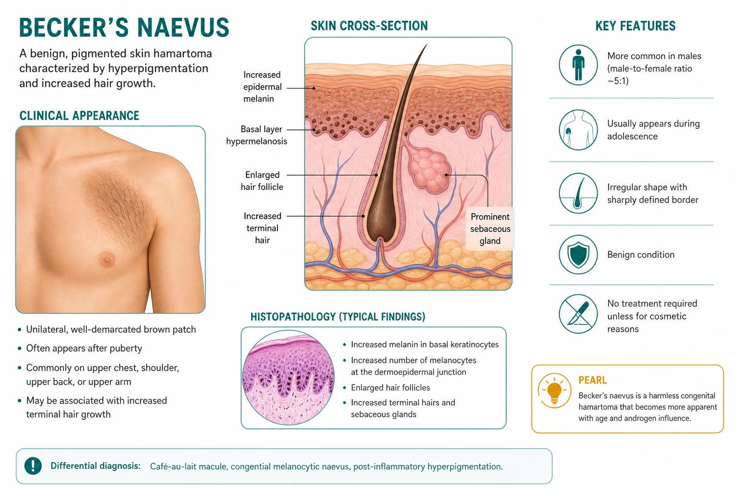
Becker's naevus is a benign, androgen-dependent hamartoma of skin presenting as a large, unilateral, light-brown to dark-brown patch with overlying hypertrichosis on the shoulder, upper chest, or upper back, first appearing at puberty. Histology shows increased basal-layer melanin, epidermal acanthosis and papillomatosis, and arrector pili (smooth muscle) hyperplasia — there are no naevus cells. The course is benign with no malignant potential; management is reassurance, with Q-switched laser for pigmentation and long-pulsed laser for hair if cosmesis is desired. Becker's naevus syndrome is the rare association with ipsilateral breast hypoplasia, skeletal anomalies and limb asymmetry.
On this page & tools
Your progress
Saved locally on this device.
Exam tags
Red flags
Overview and definition
Becker's naevus (best termed Becker melanosis) is a benign, acquired, androgen-dependent hamartoma of the skin that declares itself during the peripubertal years as a large, unilateral, light-brown to dark-brown, irregularly bordered patch on which coarse terminal hair (hypertrichosis) develops after puberty. The shoulder girdle, anterior chest and upper back are the classical territories, but the lesion can declare anywhere along the lines of Blaschko. It is benign, lifelong and without malignant potential.[1][6]
Two points of terminology are worth fixing early. First, although it carries the word "naevus", Becker's naevus is not a melanocytic naevus: it contains no naevus cells, and the brown colour arises from increased melanin in the basal layer rather than from a proliferated naevus-cell population — hence the more accurate name Becker melanosis. Second, it is properly understood as a cutaneous hamartoma — a disorganised but mature overgrowth of structures native to the area (epidermis, hair follicle and arrector pili smooth muscle) — rather than a tumour or a naevus in the cellular sense.[1]
The condition was first described by Samuel William Becker, an American dermatologist, in 1949, when he reported two young men with an acquired pigmented and hairy patch on the upper trunk. Since then Becker's naevus has been recognised as one of the canonical mosaic lesions of skin, taking its place alongside naevus sebaceous and linear epidermal naevus in any discussion of cutaneous hamartomas and paradominant inheritance.[1][2]
Mosaic lesions and the lines of Blaschko
Becker's naevus is a paradigmatic mosaic skin disorder. Mosaic lesions follow the lines of Blaschko — the S-shaped, wave-like developmental pathways along which clones of embryonic epidermal cells migrate — rather than the dermatomes or Langer's lines. A Becker's naevus therefore traces a single Blaschko field, which is why it is unilateral, sharply demarcated and often geographic rather than following a nerve distribution. This concept unifies Becker's naevus with other mosaic hamartomas — naevus sebaceous, linear epidermal naevus, linear porokeratosis and incontinentia pigmenti — all of which express somatic clonal mosaicism along Blaschko's lines. Examiners reward this framing because it links a common lesion to the broader principle of cutaneous mosaicism and paradominant inheritance. [1]
Definition checklist
Epidemiology and risk factors
Becker's naevus is common but under-reported, because most lesions are asymptomatic and never reach a clinician. Robust prevalence figures come from military and student screening surveys. [1]
The lesion is more frequent in males, with reported ratios between 2:1 and 5:1, reflecting its androgen dependence; the male sex hormone surge at puberty is the dominant driver of pigmentation and hair growth within the lesion.[1][3] Although most cases are sporadic, familial aggregation is well described, with vertical transmission compatible with paradominant inheritance — a pattern in which heterozygous carriers are phenotypically normal and the mutation only declares itself when a postzygotic somatic event (loss of the wild-type allele in a skin progenitor) unmasks it as a localised, mosaic lesion (see Pathophysiology).[2][4]
Risk factors / associations worth carrying into the exam: [1]
- Male sex — androgen-driven phenotype.
- Peripubertal hormonal surge — lesion darkens and grows hair as androgens rise.
- Family history (rare) — paradominant or autosomal-dominant-like inheritance with mosaic expression.
- Becker's naevus syndrome — a subset of patients (probably more often female) with ipsilateral developmental anomalies (see Specific subtypes).[2][5]
There is no convincing link to sun exposure, skin phototype or ethnicity; the lesion is described worldwide, although visible terminal hair makes it more conspicuous and more frequently reported in populations with dark, coarse body hair. [1]
The oft-quoted prevalence of approximately 0.25% derives from screening studies of military recruits and university students, in whom trunk examination is routine; clinic-based estimates are far lower because the asymptomatic majority never present. True prevalence is therefore probably higher than reported, particularly in females, in whom the lesion is both less common and more often overlooked. No racial predilection is established, though the hypertrichosis is more conspicuous and more likely to drive presentation in individuals with dark, coarse body hair. [1]
Pathophysiology
Becker's naevus is best understood as the convergence of two ideas: a localised cutaneous hamartoma and androgen-dependent clonal expansion.[1][6]
A hamartoma of three tissue elements
Histologically and conceptually, the lesion is an overgrowth of the normal skin structures native to the territory it occupies: [1]
- Epidermis — acanthosis (thickening), papillomatosis and elongation of rete ridges, with increased melanin in the basal keratinocytes but no increase in melanocyte number. This basal hyperpigmentation gives the brown colour.
- Hair follicles — enlarged, anatomically normal follicles producing coarse terminal hair, hence the hypertrichosis.
- Arrector pili smooth muscle — hyperplasia and increased dermal smooth-muscle bundles, sometimes palpable as subtle induration within the patch and confirmable with smooth-muscle actin (SMA) immunostain. [1]
Why it appears at puberty: androgen dependence
The defining biological clue is the pubertal onset. The hamartomatous clone is present from early life (congenital or early-childhood hypopigmentation is occasionally noted in retrospect) but remains clinically inconspicuous until the androgen surge of puberty activates androgen-responsive cells within it. Immunohistochemical studies by Grande Sarpa and colleagues demonstrated up-regulated androgen receptor expression in the basal keratinocytes and outer-root-sheath cells of follicles within Becker's naevus compared with surrounding normal skin, providing the molecular basis for the lesion's androgen responsiveness.[3]
This androgen dependence elegantly explains the four clinical signatures of the disease at the viva table: [1]
- Pubertal onset — lesions appear or declare at 10 to 15 years as androgens rise.
- Male predominance — higher androgen milieu drives a more florid phenotype.
- Hypertrichosis — androgens convert fine vellus follicles in the lesion into coarse terminal follicles.
- Intralesional acne — androgen-sensitive pilosebaceous units within the patch develop acneiform papules and pustules, sometimes the presenting complaint.[9]
Mosaicism and paradominant inheritance
The unilateral, geometric, sharply demarcated appearance is the signature of cutaneous mosaicism. Happle proposed that Becker's naevus arises from a postzygotic somatic mutation in a skin progenitor cell; the daughter clone expands along a developmental field and produces the localised lesion. In most cases the mutational event is sporadic, but in familial kindreds the pattern is paradominant: a heterozygous germline mutation is silently transmitted because carriers are phenotypically normal, and the lesion only emerges in a descendant when a second, somatic "loss of heterozygosity" event deletes the wild-type allele in a regional skin progenitor, producing the localised hamartoma.[2][4]
This model explains three otherwise puzzling observations: [1]
- Why the lesion is unilateral and sharply bounded (a single clonal field).
- Why familial cases are rare but vertical transmission is seen (the silent germline allele).
- Why Becker's naevus syndrome shows a spectrum of ipsilateral anomalies (the same clone affects nearby developmental fields — breast bud, rib, vertebra, limb). [1]
Happle has further speculated that some cases may relate to lethal somatic mutations survived by mosaicism — variants (notably in the beta-actin gene, ACTB) that would be lethal in the germline but are tolerated when confined to a mosaic skin clone.[10]
Clinical presentation
Becker's naevus tells its story through tempo, topography and texture. A careful history and examination usually make the diagnosis without investigation. [1]
Onset and evolution
The lesion classically appears in peripuberty (10-15 years), often as a faint, light-brown macule that the patient or parent first mistakes for a bruise, a "dirty patch" or a birthmark that "suddenly appeared". Over the following months to few years it undergoes a characteristic three-phase evolution: [1]
After the growth phase the lesion stabilises in early adulthood and persists, unchanged, for life. It does not regress. Some patients notice mild darkening during pregnancy or with exogenous androgens, consistent with hormone responsiveness.[1]
Morphology
- Colour: light brown to dark brown; occasionally with darker, mottled peripheral stippling.
- Border: irregular, geographic, sharply demarcated — never the smooth oval of a café-au-lait macule.
- Size: variable, from a few centimetres to large palm-sized patches and, rarely, extensive half-body involvement.
- Surface: flat to slightly thickened; the underlying smooth-muscle hyperplasia may give a subtly indurated or "rubbery" feel on palpation, and piloerection within the patch can produce a transient "goose-bump" quality.
- Hair: coarse, dark terminal hairs confined to the pigmented patch — the clinical hallmark.
- Intralesional acne: androgen-sensitive pilosebaceous units within the patch may develop comedones, papules and pustules — occasionally the presenting feature in an adolescent.[9]
Distribution
Becker's naevus is characteristically unilateral and respects the lines of Blaschko. Bilateral or multiple lesions are rare and should prompt consideration of a paradominant kindred or Becker's naevus syndrome. [1]
Although the shoulder girdle dominates the teaching image, the lesion can declare at any site. Alhusayen and colleagues catalogued Becker's naevus on the lower limb, reminding exam candidates that an atypical site does not exclude the diagnosis but warrants biopsy to exclude mimics.[8]
Symptoms
Becker's naevus is usually asymptomatic. Mild pruritus is occasionally reported, and intralesional acne may be sore or inflamed. The dominant complaint is cosmetic — the prominent hair and large pigmented patch on an exposed shoulder cause significant embarrassment in adolescents, and this psychological burden, rather than any physical danger, is the main reason patients seek care. [1]
Atypical presentations
Although the classical case is unmistakable, several atypical presentations test the candidate: [1]
- Onset in adulthood — rare; a Becker's naevus that first declares in the third decade or later should prompt biopsy to exclude a pigmented malignancy and reconsider the diagnosis.
- Bilateral or multiple lesions — uncommon; raises the possibility of a paradominant kindred or Becker's naevus syndrome and warrants examination of relatives.
- Large, extensive lesions — a half-body or segmental distribution increases the likelihood of an underlying smooth-muscle hamartoma and of the syndrome; examine the breast, spine and limbs.
- Lesion without hypertrichosis — early in the evolution (before the androgen surge has driven terminal hair) or in a female, the patch may be purely macular and easily confused with a café-au-lait macule; follow-up reveals the hair and confirms the diagnosis.
- Intralesional acne as the presenting complaint — the patient may not mention the pigmentation, focusing instead on the spots or the hair; recognition of the underlying naevus avoids misdiagnosis as ordinary acne.[9]
Differential diagnosis
The combination of pubertal onset, unilateral distribution, geographic border and hypertrichosis is distinctive, but several pigmented lesions must be excluded — particularly when the presentation is atypical (early onset, unusual site, absence of hair). [1]
| Differential | Key distinguishing features from Becker's naevus |
|---|---|
| Congenital melanocytic naevus (CMN) | Present at birth (not puberty); darker brown-black; often nodular or cerebriform surface; contains naevus cells on histology; may show neurocutaneous melanosis if giant |
| Congenital smooth-muscle hamartoma | Often present at/near birth; "Michelin-tyre" folds;firm, skin-coloured to brown plaque; prominent smooth-muscle bundles on histology with less epidermal change; may be a related entity |
| Café-au-lait macule (CALM) | Flat, uniform light-brown macule with smooth oval border; no hypertrichosis; present from early childhood; multiple CALMs suggest neurofibromatosis type 1 |
| Post-inflammatory hyperpigmentation | History of preceding rash, burn or trauma; no hypertrichosis; fades over months; confetted rather than geographic |
| Melasma | Bilateral, symmetrical facial patches; female predilection; hormonal trigger (pregnancy, OCP); no hair; central face distribution |
| Linear epidermal naevus | Verrucous/papillomatous surface (not smooth); follows Blaschko's lines; present at or near birth; purely epidermal on histology |
| Naevus of Ota / Ito | Blue-grey dermal melanocytosis; trigeminal (Ota) or scapular/shoulder (Ito) distribution; no hypertrichosis; slate-grey colour; present from childhood |
| Confluent and reticulated papillomatosis (CARP) | Reticulated brown papules coalescing centrally with reticulate periphery; truncal; retinoid/minocycline responsive |
The two most frequently tested distinctions are Becker's naevus versus café-au-lait macule (CALM is flat, smooth-bordered and hairless, and multiple CALMs point to NF1) and Becker's naevus versus congenital melanocytic naevus (CMN is present at birth, darker, may be nodular and contains naevus cells).[1]
Congenital smooth-muscle hamartoma: a related entity
Congenital smooth-muscle hamartoma (CSMH) is the most important and most debated relation of Becker's naevus. CSMH typically presents at or near birth as a firm, skin-coloured to lightly pigmented plaque, often on the trunk, with overlying vellus or terminal hair and prominent, sometimes "Michelin-tyre"-like skin folds. Histologically it shows mature smooth-muscle bundles in the dermis with minimal epidermal change. Many dermatopathologists regard Becker's naevus and CSMH as ends of a single spectrum of hamartomatous overgrowth, distinguished by which element dominates: in Becker's naevus the epidermal pigmentation and hair dominate with a smooth-muscle component, whereas in CSMH the smooth muscle dominates with little pigmentation. A positive pseudo-Darier sign can be elicited in both, supporting this relationship. [1]
Clinical and bedside assessment
The diagnosis of Becker's naevus is clinical and is made at the bedside. A focused examination establishes the diagnosis and screens for the syndrome. [1]
Focused skin examination
- Inspect the lesion for colour, border, size and the confined, coarse terminal hair; note that the hair lies within the pigmented patch (a key discriminator from CALM).
- Palpate the patch for the subtle induration of underlying smooth-muscle hyperplasia; light rubbing may induce piloerection confined to the lesion.
- Examine in good light and assess the surface for intralesional acne or comedones.
- Examine the whole integument for additional lesions (rare; suggests paradominant kindred or syndrome). [1]
Wood's lamp
A Wood's lamp (long-wave ultraviolet A) accentuates the pigmentation of Becker's naevus, confirming its largely epidermal (melanin) origin — dermal pigmentation (e.g. naevus of Ota) appears unchanged or accentuated less. This is a useful bedside discriminator but is not required for the diagnosis. [1]
Named signs and bedside manoeuvres
- Pseudo-Darier sign — firm rubbing of the lesion induces transient induration, perifollicular papules or piloerection confined to the patch, as the hypertrophied arrector pili smooth muscle contracts. This is the bedside signature of the smooth-muscle hamartoma component of Becker's naevus. It is mechanistically distinct from the true Darier sign of mastocytosis (which reflects mast-cell degranulation with urtication and erythema), although both are "rub and watch" manoeuvres.
- Confined piloerection — cold or stroking produces gooseflesh limited to the lesion, again reflecting the smooth-muscle hyperplasia.
- Hair confined to the patch — the single most reliable clinical discriminator from café-au-lait macule and most other pigmented patches: the coarse terminal hair lies strictly within the brown area. [1]
Screening for Becker's naevus syndrome
Whenever a Becker's naevus is large, involves the chest wall, or sits in a female patient, perform a targeted examination for the associated ipsilateral anomalies:[2][5]
- Breasts — inspect and palpate for ipsilateral breast hypoplasia or asymmetry (the most common associated feature, especially in females).
- Spine — inspect for scoliosis and vertebral anomalies; palpate the spinous processes.
- Chest wall — look for rib/pectoral hypoplasia and asymmetry.
- Limbs — compare limb length and girth; look for limb hypoplasia or asymmetry. [1]
Investigations
Becker's naevus is a clinical diagnosis; investigation is the exception, reserved for atypical presentation or diagnostic doubt. [1]
Skin biopsy
Biopsy is not routine. It is indicated when: [1]
- The presentation is atypical (unusual site such as face or lower limb; absent hypertrichosis; onset in adulthood; bilateral or multiple lesions).
- A changing or nodular component within a patch raises concern for melanoma (vanishingly rare, but any change in a pigmented lesion demands dermoscopy and, if needed, biopsy).
- There is genuine diagnostic uncertainty after clinical and Wood's-lamp assessment. [1]
Histopathology shows the hamartomatous triad:[1][4]
- Epidermis — acanthosis, papillomatosis, elongation of rete ridges, and increased melanin in the basal layer without an increase in melanocyte number.
- Hair follicles — enlarged, mature, terminal-hair-producing follicles.
- Dermis — increased mature smooth muscle (arrector pili), confirmed by smooth-muscle actin (SMA) immunohistochemical stain; androgen receptors are up-regulated in basal keratinocytes and follicular outer root sheath.[3]
The crucial negative finding is the absence of naevus cells — distinguishing Becker's naevus from a melanocytic naevus and from melanoma. [1]
Dermoscopy
Dermoscopy is supportive rather than diagnostic. It typically shows a regular network with thickened network lines, perifollicular hypopigmentation and the prominent follicular openings that accommodate the coarse terminal hairs. No melanoma-specific features are present. [1]
Detailed dermoscopic features include a thickened, regular pigment network with widened network lines, prominent and widened follicular openings containing terminal hair shafts, and perifollicular hypopigmentation. Papillomatous surface change may produce a cobblestone quality. The absence of regression structures (peppering, scar-like areas), atypical network, blue-white veil or crystalline structures distinguishes it from melanoma. Reflectance confocal microscopy, where available, shows a regular honeycomb epidermal architecture with bright basal keratinocytes and enlarged hair follicles, again without atypical melanocytes. [1]
Imaging
Imaging is not required for uncomplicated Becker's naevus. When Becker's naevus syndrome is suspected (clinical breast hypoplasia, spinal or limb asymmetry), request: [1]
- Chest / breast imaging — mammography, ultrasound or MRI to characterise breast hypoplasia and exclude other breast pathology.
- Spinal imaging — plain radiographs or MRI for scoliosis and vertebral anomalies.
- Limb imaging — long-film radiographs for limb-length discrepancy. [1]
Management — initial approach and reassurance
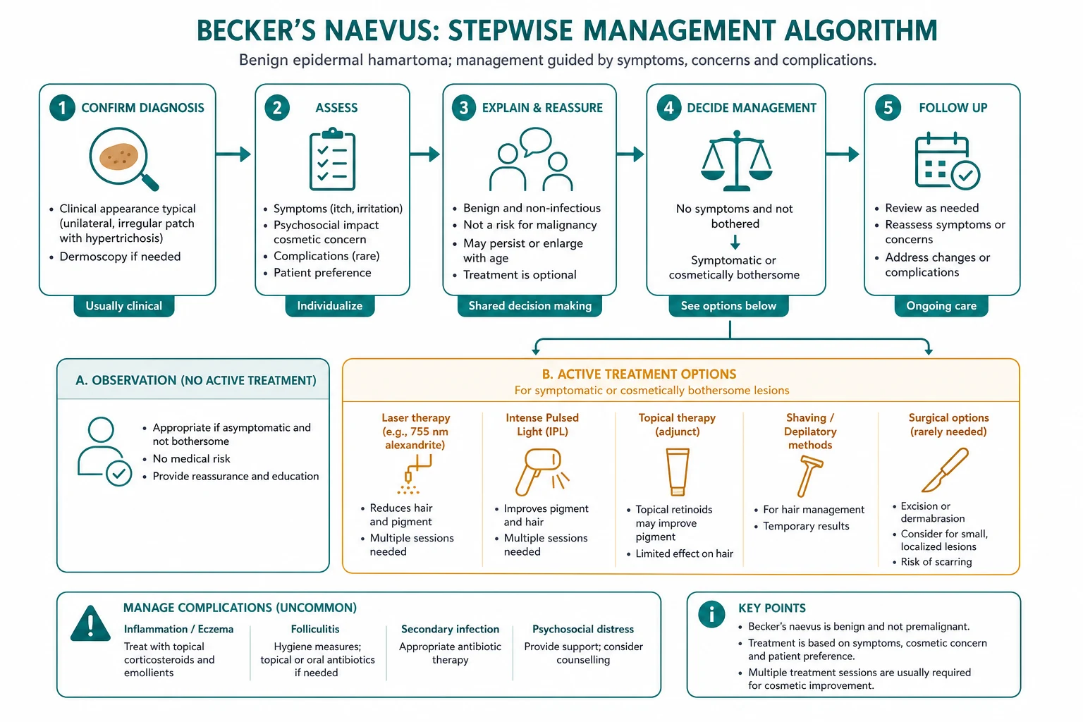
There is no resuscitative component to Becker's naevus; the lesion is benign and stable. The cornerstone of initial management is confident diagnosis and reassurance, which is itself therapeutic for the anxious adolescent. [1]
First-line: reassurance and observation
- Explain the diagnosis in plain language: a common, benign, hormone-responsive birthmark-like patch that "switches on" at puberty; it is not a mole, not cancer, and will not turn into cancer.
- Reassure about the natural history: the lesion will stabilise after the pubertal growth phase, will persist unchanged for life, and carries no malignant potential.[1]
- Address the cosmetic concern directly — for most adolescents the visible hair and the large brown patch are the entire problem; offer the cosmetic ladder (below) if distress is significant.
- Screen for the syndrome and arrange imaging if any ipsilateral anomaly is found.
Treatments with no role
Which commonly-tried treatments have NO role in Becker's naevus?
Topical bleaching agents (hydroquinone), topical retinoids and camouflage alone are ineffective against the pigmentation — the excess melanin is driven by androgen-responsive basal keratinocytes and recurs. There is also no role for cryotherapy or electrocautery; these leave scarring without improving the lesion.
Management — definitive and stepwise (cosmetic ladder)
When cosmetic concern drives the request for treatment, management is stepwise and multimodal, targeting the two cosmetically relevant components separately — the pigmentation and the hair. No single modality addresses both well, so combination therapy is the rule.[1][7]
Step 1 — Reassurance and observation
For the majority of patients, explanation and reassurance are sufficient. No active treatment is needed, and the risks and costs of laser (multiple sessions, variable response, recurrence) outweigh the benefit when the lesion is tolerated. [1]
Step 2 — Camouflage
Camouflage cosmetics (e.g. colour-matched paramedical camouflage make-up) provide a non-invasive, reversible option for patients who wish to conceal the pigmentation for social occasions without committing to laser. This is particularly useful in adolescents who are not yet ready for a procedural commitment. [1]
Step 3 — Laser for pigmentation
Pigment-targeting Q-switched lasers are the mainstay for the brown colour. The targets are the melanin granules in the basal layer. [1]
Response is variable and frequently incomplete: many patients achieve only partial lightening, and the pigmentation often recurs after treatment because the androgen-driven basal keratinocytes continue to overproduce melanin. Post-inflammatory hyperpigmentation is a risk, especially in darker phototypes, and patients must be warned of this and counselled about realistic outcomes before consenting.[6][7]
Step 4 — Laser for hypertrichosis
The coarse terminal hair responds better than the pigmentation to laser, because the hair follicle is a discrete, pigmented target. [1]
Long-pulsed alexandrite, diode and Nd:YAG lasers deliver sustained hair reduction over a course of sessions spaced to coincide with the hair-growth cycle. Hair reduction is generally more satisfactory and more durable than pigment reduction, and is often the intervention that most improves patient satisfaction. [1]
Step 5 — Combination and adjunctive therapy
The current consensus, summarised in the 2022 update by Zhou and colleagues, is that combination protocols outperform single-modality treatment: a Q-switched pigment laser plus a long-pulsed hair laser, sometimes with ablative fractional laser resurfacing as an adjunct to improve the slightly thickened texture and to enhance pigment-laser penetration. Treatment is individualised, outcomes are partial, and setting patient expectations is the single most important determinant of satisfaction.[6][7]
Step 6 — Surgical excision (rare)
Surgical excision is rarely used and reserved for small, discrete lesions in a location where the scar would be acceptable and the patient is deeply troubled. For the typical large shoulder/chest patch, the surgical scar is cosmetically worse than the lesion, so excision is generally inappropriate. There is no role for wide excision for "prevention" — the lesion is benign. [1]
Pitfalls of treatment
- Recurrence of pigmentation is common; warn the patient before consenting.
- Post-inflammatory hyperpigmentation after laser is a real risk in darker phototypes.
- Incomplete hair removal — multiple sessions are required and maintenance may be needed.
- Scarring from excision or overly aggressive laser is unacceptable in a benign, purely cosmetic indication. [1]
Specific subtypes and scenarios
Becker's naevus syndrome
Becker's naevus syndrome is the rare association of Becker's naevus with ipsilateral developmental anomalies of underlying structures. Happle, who named and defined the syndrome, emphasised that the cutaneous hamartoma and the associated defects reflect the same mosaic clonal field affecting adjacent developmental territories.[2][5]
The syndrome is probably under-recognised because the breast and skeletal anomalies may be subtle and are not always sought. Any Becker's naevus on the anterior chest wall, especially in a female, should trigger deliberate examination of the ipsilateral breast, spine and limbs, with imaging if an anomaly is suspected. Management is multidisciplinary — dermatology for the naevus, breast/endocrine or plastic surgery for hypoplasia, and orthopaedics for skeletal anomalies. [1]
Management of the syndrome
The cutaneous component is managed as for any Becker's naevus (reassurance plus laser if desired), but the associated anomalies require active, age-appropriate intervention: [1]
- Ipsilateral breast hypoplasia — the most psychologically significant defect in females. Definitive management is surgical reconstruction (tissue-expander and implant-based, or autologous flap reconstruction), deferred until breast development is complete, with psychological support through adolescence.
- Scoliosis and vertebral anomalies — orthopaedic assessment and monitoring through the growth years; bracing or surgical correction if the curve is progressive.
- Limb asymmetry — orthopaedic assessment for limb-length discrepancy; epiphysiodesis or lengthening procedures in selected cases.
- Chest-wall hypoplasia — plastic or thoracic input for significant pectus or rib anomalies. [1]
A named multidisciplinary "Becker's naevus syndrome" pathway — dermatology, plastic surgery, orthopaedics, breast or endocrine medicine, and psychology — is the appropriate model for these patients, who are otherwise at risk of fragmented, skin-only care that misses the developmental defects.[5]
Becker's naevus at atypical sites
Although the shoulder and upper chest dominate, Becker's naevus may occur on the lower back, thigh, face and neck. Alhusayen and colleagues reviewed lower-limb cases and confirmed that the clinical and histological features are identical to classical lesions, but an atypical site should lower the threshold for biopsy to exclude mimics such as linear epidermal naevus and dermal melanocytosis.[8]
Familial and paradominant Becker's naevus
Rare kindreds show vertical transmission of Becker's naevus, compatible with paradominant inheritance: carriers are silent and the lesion declares itself only after a somatic second hit. A family history of similar lesions should prompt examination of relatives and counselling about the (low) risk of transmission.[4]
Becker's naevus in females
Although less common, Becker's naevus in females carries particular significance because of the ipsilateral breast hypoplasia association; even a cosmetically tolerated patch on the chest may herald underdevelopment of the ipsilateral breast, which is far more distressing than the pigmentation itself and warrants careful evaluation.[5]
Intralesional acne
Acne confined to a Becker's naevus is a recognised, and occasionally the presenting, phenomenon — androgen-responsive pilosebaceous units within the patch develop comedones and inflammatory papules. Juhl and colleagues reported this in a 14-year-old girl, and it is a useful teaching point: the lesion is, by definition, androgen-responsive.[9]
Complications and pitfalls
Becker's naevus causes little physical harm; its complications are cosmetic, psychological and diagnostic. [1]
Cosmetic and psychological
The dominant morbidity is the psychological distress of a large, hairy, pigmented patch on an exposed area in a self-conscious adolescent. Anxiety, social withdrawal and avoidance of swimming or changing-room situations are common and are the principal driver of treatment-seeking. Taking this seriously — and offering the cosmetic ladder — is part of good care, even though the lesion is "only benign". [1]
Underlying smooth-muscle hamartoma
Many Becker's naevi have an underlying smooth-muscle (arrector pili) hamartoma, which accounts for the subtle induration and the occasionally reported pruritus or piloerection within the patch. This is part of the same hamartomatous process, not a separate complication. [1]
Becker's naevus syndrome
In extensive or chest-wall lesions, the principal "complication" is the unrecognised syndrome — ipsilateral breast hypoplasia or skeletal anomaly that is missed because the examiner stops at the skin. The cardinal pitfall is failing to look beyond the patch.[2]
Diagnostic pitfalls
- Mistaking Becker's naevus for a melanocytic naevus or melanoma — driven by the name and by the pigmentation. Remember: no naevus cells, geographic border, hypertrichosis confined to the patch.
- Mistaking it for a café-au-lait macule when the patient is seen before the hypertrichosis has developed — the CALM is flat, smooth-bordered and hairless.
- Over-investigating or over-treating a benign lesion — biopsy only if atypical; treat only if the patient wants treatment.
- Missing the syndrome by not examining the breast, spine and limbs. [1]
Malignant transformation
No case of malignant transformation of a Becker's naevus has been reported. Any nodular change, rapid darkening or ulceration within a patch should be biopsied — but on the principle that any changing pigmented lesion is assessed, not because Becker's naevus carries intrinsic malignant risk. [1]
Prognosis and disposition
The prognosis is excellent. The lesion enlarges through the pubertal growth phase, then stabilises in early adulthood and persists, unchanged, for life. It does not regress spontaneously, and it carries no malignant potential and no impact on lifespan or general health.[1]
Disposition is primary care or dermatology outpatient. Referral to dermatology is appropriate for diagnostic confirmation, for access to the cosmetic laser ladder, or when Becker's naevus syndrome is suspected and a multidisciplinary assessment (breast, orthopaedic, plastic) is needed. Follow-up is not required for an uncomplicated, stable lesion; the patient is discharged with advice to return only if the lesion changes or if cosmetic treatment is desired. [1]
Patient satisfaction after treatment is determined less by the objective degree of lightening or hair reduction and more by realistic pre-treatment counselling: those who understand that improvement will be partial and that maintenance is required are consistently more satisfied than those expecting cure.[6]
Special populations
Females
Although less common, Becker's naevus in females is clinically and psychologically significant because of the ipsilateral breast hypoplasia association. A patch over the breast bud region in a peripubertal girl must prompt careful evaluation of breast development, and plastic or breast surgical input may be needed for the hypoplasia, which is often more distressing than the skin lesion itself.[5]
Children
The lesion typically declares at 10 to 15 years, but a faint patch may be visible earlier. Parents of a younger child with a suspected Becker's naevus need reassurance about the benign course and realistic counselling that the lesion will darken and grow hair at puberty. Biopsy is avoided in children unless the diagnosis is genuinely in doubt. [1]
Pregnancy
Pregnancy, with its hormonal surge, may darken a Becker's naevus, consistent with its androgen and oestrogen responsiveness. No specific intervention is required; the change is cosmetic and typically partial. [1]
Becker's naevus syndrome in paediatrics
Children with Becker's naevus syndrome need developmental and skeletal surveillance — monitoring of scoliosis, limb-length discrepancy and breast development through the growth years, with timely orthopaedic and plastic surgical input. This is the one setting in which Becker's naevus is not a "leave alone" diagnosis. [1]
Evidence, guidelines and regional differences
There are no formal international guidelines for the management of Becker's naevus. Practice is built on case series, narrative reviews and expert consensus, because the condition is benign, uncommon in the clinic and not a research priority. [1]
What the evidence shows
The largest bodies of evidence concern laser treatment. Systematic and narrative reviews converge on several robust conclusions:[1][6][7]
- Q-switched pigment lasers (Nd:YAG 1064 nm, ruby 694 nm, alexandrite 755 nm) produce variable and often partial lightening, with frequent recurrence; pigment clearance is the harder target.
- Long-pulsed hair lasers (alexandrite, diode, Nd:YAG) produce more reliable hair reduction, which is usually the more satisfying outcome for patients.
- Combination protocols (pigment + hair laser, with or without fractional resurfacing) outperform single-modal approaches.
- Post-inflammatory hyperpigmentation is the principal adverse event, particularly in darker phototypes. [1]
Genetics
The paradominant inheritance model and the proposed role of postzygotic somatic mutations (including speculation on lethal beta-actin variants survived by mosaicism) remain the prevailing frameworks, but the precise molecular lesion has not been definitively characterised in most cases.[4][10]
Why no randomised trials exist
The absence of randomised controlled trial evidence is unsurprising and is not a weakness of practice. Becker's naevus is benign, asymptomatic in most, and the cosmetic outcome of any intervention is subjective and slow to declare. Recruitment to a sham-controlled laser trial would be difficult, and no commercial or public sponsor has prioritised the condition. Clinicians therefore rely on case series, on narrative reviews that pool these series, and on extrapolation from well-established laser principles for pigmented lesions and hair removal. The two most-cited recent syntheses — the 2015 overview by Patel and colleagues and the 2022 update by Zhou and colleagues — reach concordant conclusions: combination protocols outperform single modalities, hair responds better than pigment, and post-inflammatory hyperpigmentation is the principal adverse event. These conclusions are robust enough to guide practice even though they sit below the randomised-trial apex of the evidence pyramid.[1][6]
Regional deltas
No region-specific guidelines exist. Access to laser services, cost and number of funded sessions vary widely between health systems. In publicly funded systems, Becker's naevus laser treatment is usually classified as cosmetic and self-funded, while in some private systems partial insurance coverage may be available where there is documented psychological morbidity. Counselling about cost and the need for multiple sessions is part of consent everywhere.
Controversies
- Ablative fractional resurfacing as an adjunct to pigment laser — increasingly used, of unclear long-term benefit, with a scarring risk that must be weighed against a benign indication.
- Timing of laser — whether to treat during the active pubertal growth phase (when the lesion is still darkening and enlarging) or wait until stabilisation is debated; most practitioners wait until the lesion has stabilised to avoid treating a moving target.
- Whether Becker's naevus and congenital smooth-muscle hamartoma are ends of a spectrum or distinct entities remains unsettled. [1]
Exam pearls
[1]Exam stem: 'A 16-year-old boy has a 12 cm unilateral brown patch with coarse dark hair on his left shoulder, first noticed at age 13. What is the diagnosis, and what is the single most appropriate management?'
Diagnosis: Becker's naevus — classical triad of pubertal onset, unilateral geographic brown patch and confined hypertrichosis on the shoulder. Management: reassurance — the lesion is benign with no malignant potential; treatment is cosmetic only and at the patient's request. Examine the ipsilateral breast, spine and limbs to exclude Becker's naevus syndrome.
Red flags
Exam application bank (NEET-PG / INICET)
One-line answer
Becker's naevus is a benign, androgen-dependent hamartoma of skin presenting as a large, unilateral, light-brown to dark-brown patch with overlying hypertrichosis on the shoulder, upper chest, or upper back, first appearing at puberty. Histology shows increased basal-layer melanin, epidermal acanthosis and papillomatosis, and arrector pili (smooth muscle) hyperplasia — there are no naevus cells. The course is benign with no malignant potential; management is reassurance, with Q-switched laser for pigmentation and long-pulsed laser for hair if cosmesis is desired. Becker's naevus syndrome is the rare association with ipsilateral breast hypoplasia, skeletal anomalies and limb asymmetry.
Worked stems (answer without another resource)
Stem 1 — Classic presentation. Map symptoms to mechanism; name the first investigation and first treatment step with dose/route if drug therapy is standard. [1]
Stem 2 — Unstable / complicated. List red flags that force immediate resuscitation, theatre, ICU, antidote, or reperfusion — and what you do in the first 15 minutes. [1]
Stem 3 — Atypical group. Elderly, pregnancy, child, or immunocompromised: how presentation and thresholds change. [1]
Stem 4 — Differential trap. Name the three closest mimics and one discriminator for each. [1]
Stem 5 — Disposition. Who goes home with safety-netting, who is admitted, who needs HDU/ICU/theatre, and what follow-up is mandatory. [1]
Rapid viva checklist
- Definition + classification
- Pathophysiology chain
- Bedside signs / criteria
- Score with exact components (if any)
- Emergency bundle
- Definitive therapy with doses
- Complications of disease and of treatment
- Special populations
- Guideline/trial name if classic
- Three exam traps
Coverage self-check
If you cannot answer any stem above from this page alone, re-read the matching section — the page is intended to be self-sufficient for final-prof and NEET-PG/INICET questions on Becker's naevus.
BECKER
Summary
Becker's naevus is a benign, androgen-dependent cutaneous hamartoma of epidermis, hair follicle and arrector pili smooth muscle that appears at puberty as a large, unilateral, brown, geographic patch with hypertrichosis on the shoulder, chest or back. The diagnosis is clinical; histology shows basal hyperpigmentation, acanthosis and smooth-muscle hyperplasia without naevus cells. The lesion stabilises after puberty and carries no malignant potential. Reassurance is first-line; the cosmetic ladder — camouflage, Q-switched laser for pigment, long-pulsed laser for hair, occasionally excision of small lesions — is reserved for those who request it. Becker's naevus syndrome — ipsilateral breast hypoplasia and skeletal anomalies — must be sought in any chest-wall lesion, particularly in females. The molecular basis is a postzygotic somatic (paradominant) mutation producing a localised androgen-responsive clone, which is why the lesion declares at puberty and respects the lines of Blaschko. [1]
References
- [1]Patel P, et al. Sebaceus and Becker's Nevus: Overview of Their Presentation, Pathogenesis, Associations, and Treatment Am J Clin Dermatol, 2015.PMID 25782676
- [2]Happle R. Becker nevus syndrome Am J Med Genet, 1997.PMID 9024572
- [3]Grande Sarpa H, et al. Androgen receptor expression patterns in Becker's nevi: an immunohistochemical study J Am Acad Dermatol, 2008.PMID 19119099
- [4]Urbani CE, et al. Paradominant inheritance, supernumerary nipples and Becker's nevus: once again! Eur J Dermatol, 2001.PMID 11701421
- [5]Danarti R, et al. Becker's nevus syndrome revisited J Am Acad Dermatol, 2004.PMID 15583590
- [6]Zhou YJ, et al. An update on Becker's nevus: Pathogenesis and treatment Dermatol Ther, 2022.PMID 35502558
- [7]Zhong C, et al. Lasers for Becker's nevus Lasers Med Sci, 2019.PMID 30762191
- [8]Alhusayen S, et al. Becker nevus on the lower limb: case report and review of the literature J Cutan Med Surg, 2008.PMID 18258146
- [9]Juhl M, et al. Acne isolated within a Becker nevus of a 14 year-old girl Dermatol Online J, 2015.PMID 26437169
- [10]Happle R. Becker's Nevus and Lethal Beta-Actin Mutations J Invest Dermatol, 2017.PMID 28625464