Dermatology · Medicine
Behçet's disease
Also known as Behçet's disease · Behçet's syndrome · Adamantiades-Behçet disease · Silk Road disease
Behçet's disease = a relapsing-remitting multisystem VASCULITIS (affecting both veins AND arteries of all sizes — a 'variable-vessel' vasculitis) classically presenting with recurrent oral aphthous ulcers plus genital ulcers plus ocular inflammation (uveitis/retinal vasculitis) plus skin lesions. Demographic: Silk Road populations (Turkey, Japan, Korea, Mediterranean, Middle East); HLA-B51 association; young adults 20-40, more severe in young males. ICBD 2014 criteria (score 4 or above): oral aphthosis (2 pts), genital aphthosis (2 pts), ocular (2 pts), skin (1), neurological (1), vascular (1), pathergy optional (+1). Pathergy positive. Complications: blindness, neuro-Behçet, major-vessel thrombosis, pulmonary artery aneurysm (haemoptysis, life-threatening, leading cause of death). Management: colchicine first-line for mucocutaneous; azathioprine + corticosteroids for ocular; infliximab/adalimumab (anti-TNF) for sight-threatening or refractory disease; AVOID ciclosporin in neuro-Behçet.
On this page & tools
Your progress
Saved locally on this device.
Exam tags
Red flags
Behçet's disease rewards the candidate who understands three things at once: it is a vasculitis (not just an ulcer disease), it is geographically and genetically patterned, and its danger lies in the organ systems far from the mouth — the eye, the lung, and the brain. First described by the Turkish dermatologist Hulusi Behçet in 1937 as a triad of oral ulcers, genital ulcers and hypopyon uveitis, it is now recognised as a "variable-vessel" vasculitis capable of inflaming venules, veins and arteries of any size.[6] This single fact — that both the venous and arterial trees are at risk — explains its protean manifestations and its most feared complications, from deep-vein thrombosis to fatal pulmonary artery aneurysm rupture.
Overview & Definition
Behçet's disease is an immune-mediated, relapsing-remitting, multisystem vasculitis of unknown cause affecting vessels of all sizes and both types (venous and arterial).[3][6] In the 2012 revised International Chapel Hill Consensus Conference (CHCC) nomenclature, it is the prototype of the "variable-vessel vasculitis" category — meaning no single vessel type or size is preferentially spared, which distinguishes it from large-vessel diseases (giant-cell arteritis, Takayasu), medium-vessel disease (polyarteritis nodosa) and the ANCA-associated small-vessel vasculitides.[6]
The clinical hallmark is recurrent oral aphthous ulceration accompanied, over time, by a variable combination of genital ulceration, ocular inflammation (anterior/posterior uveitis, retinal vasculitis), skin lesions (erythema nodosum-like nodules, acneiform and papulopustular eruptions), a positive pathergy reaction, arthritis, vascular thrombosis or aneurysm, gastrointestinal ulceration and neurological disease.[3]
[1]It is not a classic autoimmune disease: there is no disease-specific autoantibody (ANA, ANCA, rheumatoid factor are typically negative), and the dominant histological finding is a neutrophilic and lymphocytic vasculitis rather than immune-complex deposition.[5] This is why it sits in the family of neutrophilic dermatoses alongside Sweet syndrome and pyoderma gangrenosum. The 2012 revised Chapel Hill Consensus Conference (CHCC) nomenclature places Behçet's in its own category — variable-vessel vasculitis — because, uniquely among the named vasculitides, it can affect any vessel type or size: small venules, capillaries, small and medium arteries, and large veins and arteries alike. This is a high-yield distinction from its neighbours in the vasculitis taxonomy: giant-cell arteritis and Takayasu arteritis are large-vessel diseases; polyarteritis nodosa is a medium-vessel disease; granulomatosis with polyangiitis, microscopic polyangiitis and eosinophilic granulomatosis with polyangiitis are small-vessel ANCA-associated diseases. Behçet's crosses all of these boundaries, which is why one patient may present with a superficial thrombophlebitis and another with a pulmonary artery aneurysm or a cerebral venous sinus thrombosis.[6]
Clinically, Behçet's is best thought of not as a single disease but as a constellation of phenotypes sharing a common vasculitic mechanism: a predominantly mucocutaneous phenotype (best prognosis), an ocular phenotype (threatens sight), a vascular phenotype (threatens life through aneurysm and thrombosis), a neurological phenotype (threatens independence), and a gastrointestinal phenotype (mimics Crohn disease). Treatment intensity tracks the phenotype, not the label — a principle candidates must articulate at viva. [1]
Epidemiology & Risk Factors
Behçet's disease has one of the most striking geographic distributions in all of medicine, tracking the ancient Silk Road trade route from the Mediterranean through the Middle East and Central Asia to East Asia.[3]
The strongest genetic association is HLA-B51 (encoded by the HLA-B*51 allele on chromosome 6). It is found in 50 to 80 percent of patients from endemic populations versus a much lower background rate, and confers roughly a six-fold increased risk of disease.[3] However, HLA-B51 is neither necessary nor sufficient for disease — it is present in many healthy carriers and absent in a sizeable minority of patients — so it is supportive, not diagnostic. Additional susceptibility loci (ERAP1, IL10, IL23R, CCR1) reinforce a picture of innate-immune and IL-23/Th-17 axis dysregulation.[6]
Male sex and young age at onset are the two most consistent predictors of severity: men are more likely to develop sight-threatening eye disease, vascular involvement and neuro-Behçet, and consequently have higher morbidity and mortality. Disease onset is typically in the third and fourth decades, but paediatric-onset (under 16) and late-onset forms are recognised. The pathergy test is positive in over 60 percent of patients in Turkey and Japan but in fewer than 20 percent of Northern Europeans — a geography-dependent feature that affects how criteria perform.[5] The disease was first described in 1937 by Hulusi Behçet, a Turkish dermatologist, who reported three patients with the triad of recurrent oral aphthous ulcers, genital ulcers and hypopyon uveitis. The same constellation was independently noted by Benediktos Adamantiades, a Greek ophthalmologist, in 1930 — hence the alternative name Adamantiades-Behçet disease.[3] The eponym "Silk Road disease" reflects its geographic predilection along the ancient trade routes linking East Asia to the Mediterranean, a distribution that aligns remarkably with the population frequency of HLA-B51.
Although Behçet's is rare in Northern Europe and North America, it is a major cause of morbidity in endemic regions: in Turkey it is among the leading causes of non-traumatic blindness in young men, and pulmonary artery aneurysm remains a significant cause of death in young adults across the Middle East and East Asia. The annual incidence of eye disease and new vascular events is highest in the first five years after onset, which is why aggressive early treatment — particularly for ocular disease — is emphasised in every guideline.[6]
Pathophysiology
The mechanism is best understood as a two-hit model: genetic susceptibility lowers the threshold for an exaggerated innate-immune response, which is then triggered by an environmental stimulus.[6]
The cascade proceeds in five linked steps. Genetic susceptibility — chiefly HLA-B51, with contributions from ERAP1 (which shapes peptide presentation), and IL10/IL23R variants — sets up a hyper-reactive immune system. An environmental trigger (bacterial antigens such as Streptococcus sanguinis, viral exposure, or cross-reactive heat-shock proteins) then activates innate immunity. The result is neutrophil hyperactivity: Behçet neutrophils show exaggerated chemotaxis and superoxide production, and innate lymphoid cells are activated. This drives a skewed adaptive response dominated by Th1 and Th17 cells (releasing interferon-gamma and IL-17) together with polyclonal B-cell activation and immune-complex formation. The end-organ result is neutrophilic and lymphocytic vasculitis of arteries and veins, with endothelial dysfunction, in-situ thrombosis and aneurysm formation.[5][6]
Pathergy — the formation of a papule or pustule 24 to 48 hours after a sterile needle prick — is simply the cutaneous expression of this same innate hyper-reactivity: an exaggerated response to minor injury, not an allergy. [1]
Why both thrombosis and aneurysm? Because the vessel wall itself is inflamed. Endothelial injury activates coagulation and impairs fibrinolysis, while neutrophil-mediated destruction of the media weakens the wall and permits aneurysm formation. This is why thrombosis in Behçet's is fundamentally an inflammatory phenomenon rather than a primary clotting disorder — a point that directly determines management (immunosuppression is central; anticoagulation alone is inadequate and sometimes dangerous). [1]
Genetic susceptibility
HLA-B51 plus ERAP1/IL10/IL23R variants lower the immune threshold.
Environmental trigger
Microbial antigens and heat-shock proteins activate innate immunity.
Innate hyperactivation
Neutrophil hyper-reactivity (chemotaxis, superoxide) and innate lymphoid-cell activation.
Adaptive skew
Th1/Th17 polarisation, IL-17 and IFN-gamma, polyclonal B-cell activation.
Variable-vessel vasculitis
Neutrophilic + lymphocytic vasculitis of arteries and veins → thrombosis and aneurysm.
A distinguishing pathological feature is the neutrophilic vascular reaction: on histology, early lesions show a dense perivascular neutrophil infiltrate with leukocytoclasia and fibrinoid necrosis in vessel walls, while older lesions acquire a lymphocytic-mononuclear infiltrate.[5] This dual pattern — neutrophilic early, lymphocytic late — places Behçet's within the family of neutrophilic dermatoses and explains its kinship with Sweet syndrome and pyoderma gangrenosum, with which it shares pathergy-like hyper-reactivity.
At the cytokine level, the dominant signature is Th1/Th17 polarisation: raised interleukin-17, interferon-gamma, tumour necrosis factor-alpha and interleukin-8 (a potent neutrophil chemoattractant), along with elevated interleukin-1-beta and interleukin-6. This cytokine profile provides the rationale for several targeted agents — anti-TNF (infliximab, adalimumab) targets the TNF-alpha excess; gevokizumab and canakinumab (interleukin-1 blockers) and secukinumab (interleukin-17 blockade) have been explored in trials. There is no disease-specific autoantibody: ANA, ANCA, rheumatoid factor and complement are typically normal or negative, which is a useful negative discriminator from lupus and the ANCA-associated vasculitides.[6]
Classification & Diagnostic Criteria
Diagnosis is clinical and rests on the recognition of a characteristic pattern after exclusion of mimics. Two criteria sets are commonly examined, and candidates must know which is current. [1]
The older 1990 International Study Group (ISG) criteria made recurrent oral ulceration mandatory; patients without oral ulcers could not be classified as Behçet's even if every other feature was present. These were superseded by the International Criteria for Behçet's Disease (ICBD), 2014, developed by the International Team for the Revision of the ICBD across 27 countries.[7] The ICBD dropped the mandatory status of oral ulcers, increased the weighting of ocular and genital lesions, and added pathergy as an optional item. In the validation cohort the ICBD achieved a sensitivity of 94.8 percent and specificity of 90.5 percent, outperforming the 1990 ISG criteria on both.[7]
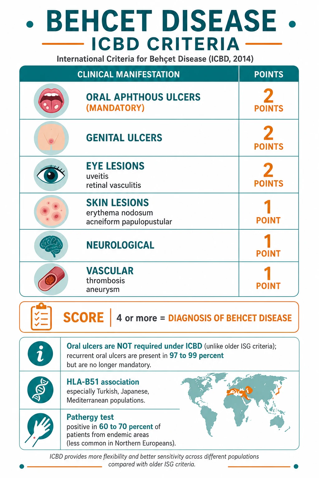
| ICBD 2014 feature | Points |
|---|---|
| Ocular lesions (uveitis, retinal vasculitis) | 2 |
| Genital aphthosis | 2 |
| Oral aphthosis | 2 |
| Skin lesions (erythema nodosum-like, acneiform, papulopustular) | 1 |
| Neurological manifestations | 1 |
| Vascular manifestations (thrombosis, aneurysm) | 1 |
| Positive pathergy test (optional) | 1 |
A score of 4 or above classifies the patient as having Behçet disease.[7]
[1]Clinical Presentation
The disease unfolds over years rather than days, and the tempo is relapsing-remitting. Oral ulcers are almost always the first manifestation; other features accumulate over time, sometimes separated by years.[3]
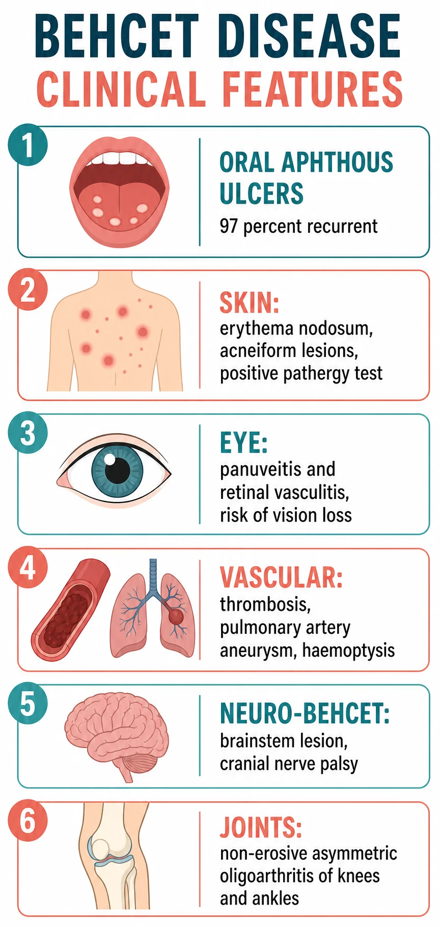
Mucocutaneous (present in nearly all patients)
Recurrent oral aphthous ulcers occur in 97 to 99 percent and are usually the presenting feature. They are painful, multiple, recurrent (classically three or more episodes per year), and found on the lips, buccal mucosa, tongue and palate. Minor aphthae heal in one to three weeks without scarring; major aphthae are larger, last longer and may scar.[4] They are clinically indistinguishable from ordinary recurrent aphthous stomatitis in isolation — it is the company they keep that defines Behçet's.
Genital ulcers occur in 80 to 90 percent and are painful, found on the scrotum in men and the vulva in women, and — critically — heal with scarring. This is a high-yield discriminator from oral aphthae.[6]
- 97–99% of patients
- Lips, buccal mucosa, tongue, palate
- Minor type heals WITHOUT scarring
- Usually the first manifestation
- 80–90% of patients
- Scrotum (men), vulva (women)
- Heal WITH scarring — key discriminator
- Painful, recurrent
Skin lesions affect 60 to 90 percent and take several forms: erythema nodosum-like tender red nodules on the lower legs (which may heal with residual pigmentation), acneiform and papulopustular eruptions on the trunk and face, and a positive pathergy reaction.[1][5] Histologically the nodules show a neutrophilic vascular reaction or septal panniculitis. Two skin phenomena are particularly characteristic and examiner-favoured. Erythema nodosum-like lesions are tender, subcutaneous, erythematous nodules on the anterior lower legs that histologically show a neutrophilic septal panniculitis (overlapping with true erythema nodosum); in Behçet's they tend to be more numerous and recurrent than in idiopathic erythema nodosum, and they may heal with post-inflammatory pigmentation.[1] Acneiform and papulopustular lesions on the trunk and face are so common that an adolescent-pattern acne in a young adult of Silk-Road origin should prompt consideration of Behçet's; histologically these are folliculitis with a neutrophilic infiltrate rather than typical acne. The diagnostic concept of pseudofolliculitis (folliculitis not attributable to acne or infection) is one of the ISG/ICBD skin criteria.[5]
Ocular involvement (about 50%)
On slit-lamp examination, anterior disease produces cells and flare in the anterior chamber, and a hypopyon (a settled layer of pus) — the latter was once the signature ocular sign of Behçet's and gave the original triad its name, though it is now uncommon because of earlier treatment. Posterior disease produces retinal vasculitis with venous and arterial sheathing, occlusion, cotton-wool spots and retinal haemorrhages; recurrent occlusive vasculitis causes retinal ischaemia, neovascularisation and disc/macular oedema, culminating in irreversible visual loss. A distinguishing feature from HLA-B27-associated acute anterior uveitis is that Behçet uveitis is typically bilateral, recurrent and panuveal, and frequently involves the posterior segment.[6]
The clinical corollary is that any new eye symptom — blurring, floaters, redness or pain — in a patient with known or suspected Behçet's is an emergency: permanent retinal damage accrues with each untreated attack, and early anti-TNF has been shown to reduce the rate of blindness substantially. [1]
Musculoskeletal (about 50%)
Arthritis is non-erosive, asymmetric, oligoarticular and affects the knees and ankles most often. It is usually self-limiting and responds to colchicine or NSAIDs. [1]
Vascular involvement (about 25%)
Behçet's is one of the few conditions that causes both venous thrombosis and arterial aneurysm. Venous disease predominates: deep-vein thrombosis, cerebral venous sinus thrombosis, and Budd-Chiari (hepatic vein) thrombosis. Arterial disease is less common but more dangerous — pulmonary artery aneurysm presents with haemoptysis and is a leading cause of death.[6] Vascular involvement in Behçet's has a paradox that examiners probe: venous thrombosis is common but pulmonary embolism is relatively rare. The reason is that Behçet thrombi are tightly adherent to an inflamed, thickened vessel wall and tend not to embolise — so a patient may have extensive lower-limb DVT with little breathlessness. When pulmonary symptoms do occur, the first thought should be in-situ pulmonary artery thrombosis or pulmonary artery aneurysm rather than embolic disease, and CT angiography is the definitive test. Superficial thrombophlebitis migrating up a limb is an early clue. Arterial disease — particularly pulmonary, femoral and aortic aneurysms — is less common but drives mortality; arterial lesions may present as a pulsatile mass, limb ischaemia, or haemoptysis, and carry a high risk of rupture.[6]
Neurological (5 to 10%)
Two further neuro-Behçet points are examiner-favoured. First, the brainstem is the classic site of parenchymal disease — a young man of Silk-Road origin with a brainstem stroke syndrome (crossed cranial-nerve and long-tract signs, ataxia, dysarthria) should prompt consideration of neuro-Behçet even before oral ulcers are volunteered. Second, a headache in a Behçet patient is never "just a headache" until cerebral venous sinus thrombosis and parenchymal disease have been excluded by MRI brain with venography. Cognitive and behavioural change may be the only clue to chronic parenchymal involvement. Optic neuropathy and peripheral nerve involvement are uncommon but described.[2] Neuro-Behçet takes two forms with very different prognoses. Parenchymal neuro-Behçet (brainstem and thalamocapsular lesions) presents with headache, cranial nerve palsies, hemiparesis, ataxia, dysarthria and behavioural change, and carries a poor prognosis. Cerebral venous sinus thrombosis presents with headache and papilloedema (an intracranial-pressure syndrome) and carries a better prognosis.[2]
Gastrointestinal (5 to 10%)
Ileocaecal ulceration mimics Crohn disease and may perforate or bleed. The histology (deep, punched-out ulcers rather than transmural granulomatous inflammation) and the distribution help distinguish Behçet enteropathy from inflammatory bowel disease.[6] Other less common manifestations complete the multisystem picture. Epididymitis occurs in a minority of men. Cardiac involvement (pericarditis, myocarditis, intracardiac thrombosis) and pulmonary parenchymal disease (pleurisy, infiltrates) are uncommon but serious. Renal disease is rare and is most often AA amyloidosis from chronic inflammation, presenting as nephrotic-range proteinuria; a Behçet-associated glomerulonephritis is also described.[6]
Atypical and delayed presentations
Examiners test the atypical deliberately. Paediatric-onset disease frequently masquerades as isolated recurrent aphthous stomatitis for years — sometimes a decade — before genital ulcers, eye disease or pathergy declare the full phenotype; a family history and HLA-B51 positivity should lower the threshold for specialist referral in a child with stubborn oral ulcers. Late-onset disease (after age 40) tends to run a milder, more mucocutaneous course with fewer sight-threatening or vascular events. A purely vascular or neurological presentation — a young man with an unexplained Budd-Chiari syndrome, cerebral venous sinus thrombosis, or pulmonary artery aneurysm, and no oral ulcers — is a recognised trap: the ICBD 2014 criteria (which do not require oral ulcers) exist precisely for such patients. Pregnancy can alter the pattern, with flares commonest in the postpartum period rather than during gestation.[3]
Differential Diagnosis
A confident differential is essential because several mimics share oral/genital ulceration, uveitis or vasculitis. The table below lists the principal contenders and the features that separate them. [1]
- Oral + genital ulcers + ileocaecal disease overlap
- Transmural granulomatous inflammation on histology
- Perianal disease, fistulae
- Distinguish on endoscopy + biopsy + GI Behçet has punched-out ulcers
- Triad: urethritis, conjunctivitis, asymmetric arthritis
- Circinate balanitis, keratoderma blennorrhagicum
- Post-STI or post-enteric trigger
- No oral aphthae pattern
- Painful papulonodular skin lesions with neutrophilic infiltrate
- May be paraneoplastic or drug-related
- Typically no genital ulceration or uveitis
- Distinguish on systemic distribution
- Behçet + relapsing polychondritis overlap
- Auricular and nasal cartilage inflammation
- Same treatment principles
- Recognised overlap variant
- Herpes simplex, syphilis, HIV cause oral/genital ulcers
- Exclude by serology and PCR
- Acute, monophasic pattern usually
- No vasculitic systemic syndrome
- Reactive arthritis, ankylosing spondylitis
- Acute anterior uveitis (not panuveitis/retinal vasculitis)
- No pathergy, no genital ulcer-scarring pattern
- HLA-B27, not HLA-B51
The single most commonly confused entity is Crohn disease, because both produce oral aphthae, genital ulcers (fissures) and ileocaecal disease. The discriminating features are the pattern of uveitis (Behçet causes panuveitis and retinal vasculitis; Crohn causes anterior uveitis), the presence of pathergy and pseudofolliculitis (Behçet), and the histology (granulomatous transmural inflammation in Crohn versus neutrophilic vasculitis in Behçet).[6]
Clinical & Bedside Assessment
The focused assessment is built around three questions: Is the eye involved? Is there large-vessel disease? Is the brain involved? [1]
History should establish the onset, frequency and healing of oral/genital ulcers (and whether genital ulcers scar), eye symptoms (blurring, floaters, redness, pain), skin lesions, any thrombotic events, neurological symptoms (headache, weakness, sensory change, seizures), abdominal pain and family origin along the Silk Road. A family history of Behçet's raises suspicion. [1]
Examination includes a careful inspection of the oral cavity (ulcers at all stages), the genitalia (active ulcers and scars), the skin (erythema-nodosum-like nodules, acneiform lesions, scars from previous pathergy testing), a slit-lamp examination of the eyes (mandatory — anterior chamber cells, hypopyon, retinal vasculitis), the joints, peripheral pulses, and a neurological examination looking for cranial nerve palsies, long-tract signs and ataxia.[3]
The pathergy test is performed by making a sterile oblique prick with a 20-gauge needle into the forearm skin and reading the site at 24 to 48 hours. The formation of a papule or pustule 2 mm or larger constitutes a positive result. Pathergy has high specificity (particularly in endemic populations) but variable, geography-dependent sensitivity (over 60 percent in Turkey and Japan, under 20 percent in Northern Europe), so a negative test never excludes the disease.[5]
Investigations
There is no confirmatory laboratory test for Behçet's. Investigations serve three purposes: to support the clinical pattern, to define the extent of organ involvement, and to exclude mimics. [1]
Bloods typically show raised inflammatory markers (ESR, CRP) during active disease and a mild anaemia of chronic disease; ANA, dsDNA and ANCA are usually negative (useful negative discriminators). HLA-B51 testing is supportive in the right ethnic context but is not diagnostic.[3]
Ocular assessment is non-negotiable in any suspected or confirmed case. Slit-lamp examination detects anterior chamber inflammation and hypopyon; fundoscopy (ideally with fluorescein angiography) detects retinal vasculitis, vascular sheathing, occlusion and leakage; optical coherence tomography (OCT) quantifies macular oedema.[6]
Imaging is directed by symptoms: CT or MR angiography for suspected pulmonary artery aneurysm or large-vessel arterial disease; Doppler ultrasound for deep-vein thrombosis; and MRI brain (with venography) for suspected neuro-Behçet — parenchymal lesions typically involve the brainstem and thalamocapsular region, while MR venography defines cerebral venous sinus thrombosis.[2]
Biopsy of a skin or genital ulcer shows a neutrophilic vascular reaction or lymphocytic vasculitis — supportive but non-specific. A gastrointestinal biopsy distinguishes Behçet enteropathy (punched-out ulcers) from Crohn disease (granulomatous transmural inflammation).[6] Because the diagnosis is one of exclusion as much as pattern recognition, a mimics panel is part of every work-up: viral swabs and PCR for herpes simplex, serology for syphilis (treponemal) and HIV, and — where the ileocaecal picture dominates — faecal calprotectin, colonoscopy with biopsy and inflammatory-bowel serology to separate Behçet enteropathy from Crohn disease. A thrombophilia screen is reasonable when thrombosis is the presenting feature, recognising that Behçet's is itself the explanation in most cases.[6]
Ophthalmologists grade ocular inflammation using the Standardization of Uveitis Nomenclature (SUN) working-group criteria — anterior chamber cells, flare and hypopyon graded 0 to 4+ — and monitor disease course with the Behçet's Disease Ocular Attack Score 24 (BOS24), which quantifies the frequency and severity of uveitic attacks over 24 months. These tools are how retinal vasculitis severity and treatment response are tracked objectively.[6]
| Investigation | Finding in Behçet's | Role |
|---|---|---|
| Slit-lamp + fundoscopy | Cells, hypopyon, retinal vasculitis | Mandatory at baseline and every flare |
| Fluorescein angiography | Vascular leakage, occlusion, neovascularisation | Defines retinal vasculitis |
| OCT | Macular oedema | Tracks structural damage |
| CT/MR angiography | Pulmonary arterial aneurysm, large-vessel disease | Suspected vascular involvement |
| MRI brain + venography | Brainstem/thalamocapsular lesions; sinus thrombosis | Suspected neuro-Behçet |
| Doppler ultrasound | DVT | Symptomatic limb |
| ESR/CRP | Raised in active disease | Activity marker (non-specific) |
| ANA, ANCA, RF | Typically negative | Negative discriminators |
| HLA-B51 | Positive in 50–80% endemic | Supportive, not diagnostic |
| Pathergy test | Papule/pustule at 24–48h | High specificity, variable sensitivity |
Management — Resuscitation
Most Behçet presentations are subacute, but four scenarios are time-critical and demand immediate action before definitive organ-based therapy is planned. [1]
[1]The unifying principle is that acute severe inflammation is treated with corticosteroids first, while the slower-acting steroid-sparing agent (azathioprine, anti-TNF) is layered on to maintain remission and taper the steroid.[8]
Management — Definitive & Stepwise
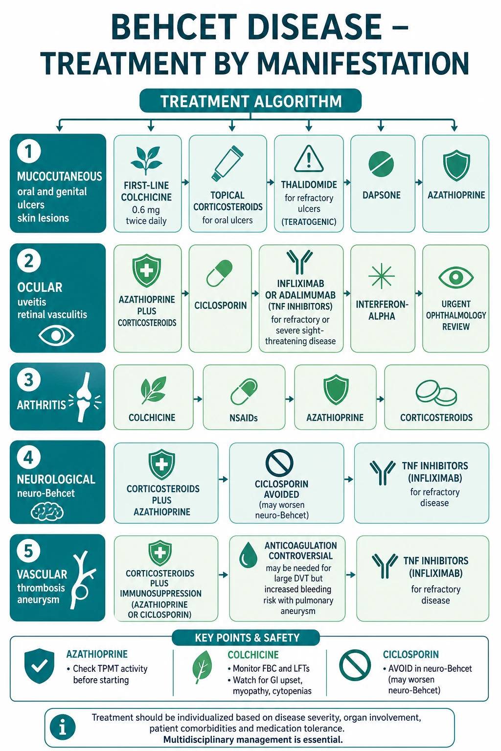
Long-term treatment is organ-based — therapy is chosen by the dominant manifestation, not applied uniformly. The 2018 EULAR recommendations formalise this approach.[8]
Mucocutaneous disease
Colchicine is first-line for recurrent oral and genital ulcers and erythema-nodosum-like lesions, typically 0.5 mg twice daily (1 mg/day), with efficacy supported by randomised trial evidence. Topical corticosteroids (triamcinolone dental paste) and topical anaesthetics provide symptomatic relief for individual oral ulcers.[4] For refractory mucocutaneous disease, options include apremilast (a phosphodiesterase-4 inhibitor, 30 mg twice daily, demonstrated effective for oral ulcers in a phase 2 trial),[9] thalidomide (50–150 mg/day; effective but teratogenic and neurotoxic, requiring strict pregnancy-prevention and monitoring for neuropathy), dapsone, and azathioprine.[3]
Ocular disease
Because eye disease threatens sight, it is treated aggressively. Azathioprine plus corticosteroids is the backbone for uveitis and retinal vasculitis (check TPMT activity before starting azathioprine). For sight-threatening posterior segment disease or disease refractory to first-line therapy, an anti-TNF agent — infliximab (5 mg/kg infusions) or adalimumab (40 mg subcutaneous) — is the treatment of choice and has transformed the ocular prognosis.[8] Interferon-alpha and ciclosporin are alternatives for ocular disease, but ciclosporin must be avoided when there is any neurological involvement (see below). Intravitreal corticosteroid or anti-VEGF therapy may supplement systemic treatment for macular oedema.
Articular disease
Colchicine and NSAIDs are first-line for the non-erosive arthritis. Persistent or recurrent arthritis escalates to azathioprine, interferon-alpha or anti-TNF. [1]
Neuro-Behçet
High-dose corticosteroids (IV methylprednisolone pulse followed by oral prednisolone) plus a steroid-sparing agent — azathioprine for ongoing suppression, with infliximab for refractory disease — is the standard approach.[2] Ciclosporin must be avoided: it is neurotoxic and has been associated with precipitation or worsening of central nervous system disease.[2] Cyclophosphamide is used for severe disease. Cerebral venous sinus thrombosis is managed with anticoagulation (once haemorrhage excluded) plus corticosteroids.
Vascular disease
Because thrombosis is driven by vessel-wall inflammation, immunosuppression is central — corticosteroids plus azathioprine (or cyclophosphamide for severe large-vessel or pulmonary artery aneurysm disease).[6] The role of anticoagulation is controversial: it may be considered for extensive venous thrombosis, but it carries a dangerous bleeding risk when a pulmonary artery aneurysm is present, and thrombi in Behçet's are often adherent and rarely embolise. Anti-TNF is an option for refractory vascular disease. Surgical or endovascular intervention for aneurysms is high-risk (perioperative inflammatory flare) and is usually combined with intensive immunosuppression.
Gastrointestinal disease
Corticosteroids plus 5-aminosalicylic acid agents or azathioprine are first-line; surgery is reserved for perforation or refractory bleeding; anti-TNF for refractory disease.[6]
Colchicine
Dose
0.5 mg PO twice daily (1 mg/day)
Infliximab (anti-TNF)
Dose
5 mg/kg IV infusion at weeks 0, 2, 6, then 8-weekly
Treatment escalation principles [1]
Three principles govern escalation across every organ system. First, corticosteroids control the flare; immunosuppression prevents relapse — a short course of high-dose corticosteroid (oral prednisolone 0.5–1 mg/kg/day, or IV methylprednisolone 0.5–1 g daily for 3 days for severe flares) is layered onto a slower-acting steroid-sparing agent (azathioprine, ciclosporin or anti-TNF), and the steroid is tapered as the steroid-sparing agent takes effect. Second, escalate early for sight- or life-threatening disease — do not wait for failure of first-line therapy in posterior uveitis, retinal vasculitis, pulmonary artery aneurysm or parenchymal neuro-Behçet; move to anti-TNF or cyclophosphamide promptly. Third, avoid ciclosporin whenever the nervous system is or may become involved, because of its well-described neurotoxicity.[2][8]
Pre-treatment screening and monitoring
Before starting any immunosuppressant, screen for latent tuberculosis (interferon-gamma release assay or chest X-ray), hepatitis B and C, and ensure vaccinations (inactivated influenza, pneumococcal, and where indicated, hepatitis B) are up to date — live vaccines are contraindicated once immunosuppression begins. Azathioprine requires TPMT (and, in high-risk ancestries, NUDT15) genotyping before starting, plus baseline and regular full blood count and liver function tests to detect myelosuppression and hepatotoxicity. Colchicine monitoring is full blood count and liver function tests, with dose reduction in renal or hepatic impairment and caution with strong CYP3A4 or P-glycoprotein inhibitors. Ciclosporin requires blood pressure, renal function and drug-level monitoring. Anti-TNF therapy demands TB and hepatitis screening, vigilance for serious infection, and — for adalimumab and infliximab in some regions — monitoring for antidrug antibodies. Thalidomide demands a rigorous pregnancy-prevention programme and baseline plus serial nerve conduction or symptom surveillance for peripheral neuropathy.[8]
Specific Subtypes & Scenarios
Several subtypes are examined in depth because they carry distinct prognoses and management traps. [1]
Ocular Behçet is the prototype of sight-threatening disease. The key teaching point is that posterior segment involvement (retinal vasculitis, macular oedema) drives permanent visual loss, and early anti-TNF has reduced the historical blindness rate. Anterior uveitis with hypopyon, once the signature sign, is now less common with earlier treatment.[6]
Vascular Behçet is dominated by the pulmonary artery aneurysm subtype, which is the single most feared vascular lesion: it presents with haemoptysis, may rupture catastrophically, and is a leading cause of death. Treatment is IV corticosteroids plus cyclophosphamide, with embolisation or surgery for high-rupture-risk lesions.[6]
Neuro-Behçet divides into two patterns with sharply different outcomes, and candidates must distinguish them:[2]
- Brainstem and thalamocapsular lesions on MRI
- Cranial nerve palsy, hemiparesis, ataxia, dysarthria, behavioural change
- Poor prognosis; permanent deficit and cognitive decline
- Treated with high-dose corticosteroids + azathioprine/anti-TNF; AVOID ciclosporin
- Intracranial-pressure syndrome: headache, papilloedema
- Seen on MR venography
- Better prognosis than parenchymal form
- Treated with anticoagulation (once haemorrhage excluded) + corticosteroids
Paediatric Behçet (onset under 16) often begins with recurrent oral ulcers alone, with the full phenotype emerging only years later; a family history and HLA-B51 positivity raise the index of suspicion. MAGIC syndrome (Mouth And Genital ulcers with Inflamed Cartilage) is the recognised overlap of Behçet's with relapsing polychondritis.[3]
Complications & Pitfalls
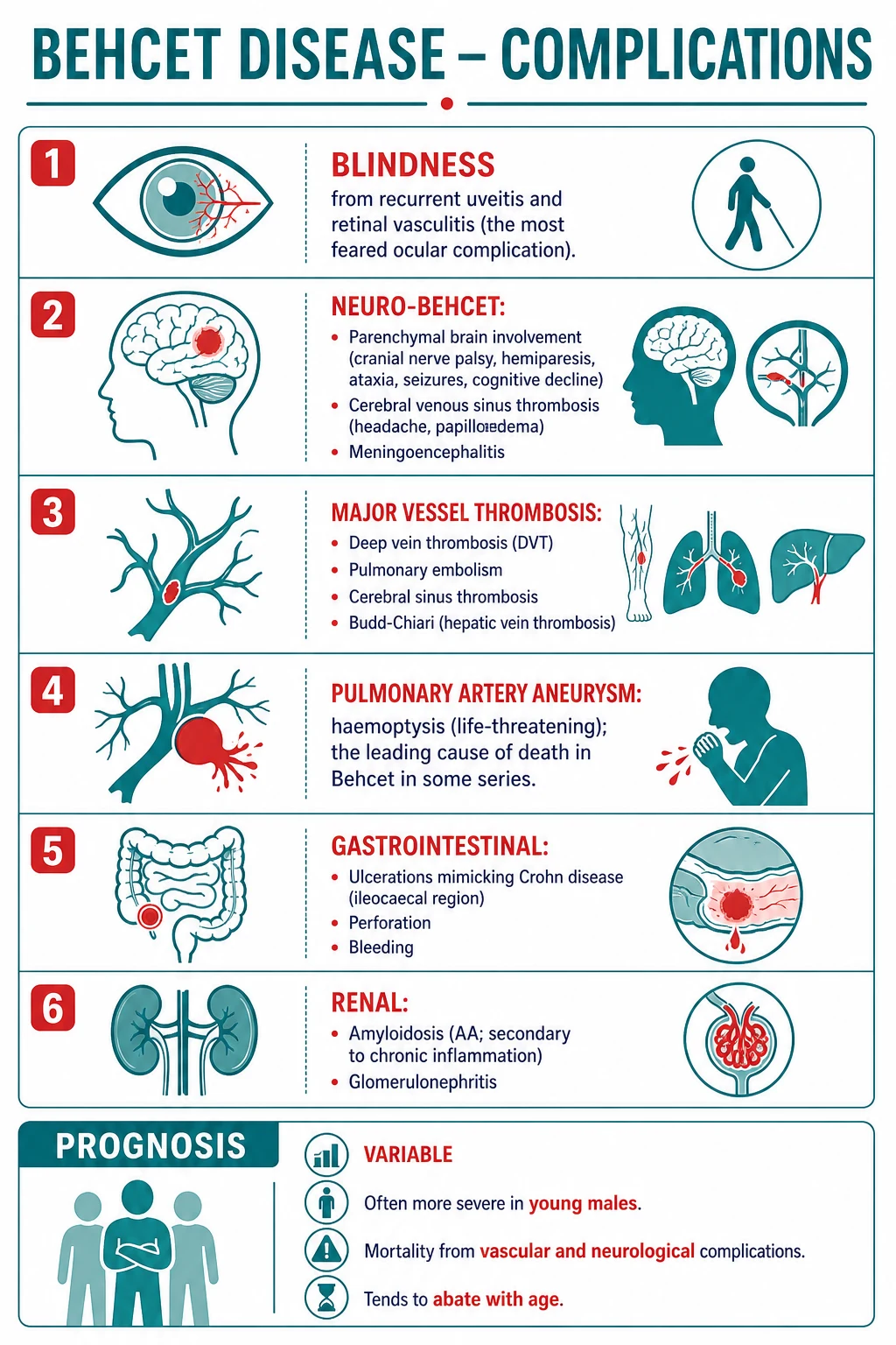
The complications span every system involved, but the high-yield ones cluster around vision, the lung vasculature and the brain. [1]
Blindness results from recurrent posterior uveitis and retinal vasculitis causing retinal ischaemia, neovascularisation and atrophy; early anti-TNF has reduced its incidence. Pulmonary artery aneurysm causes massive haemoptysis and fatal haemorrhage and remains a leading cause of death. Neuro-Behçet (parenchymal type) leaves permanent neurological and cognitive deficit. Major-vessel thrombosis produces DVT, Budd-Chiari syndrome, cerebral sinus thrombosis and arterial occlusion. Gastrointestinal ulceration may perforate or bleed. Renal AA amyloidosis (secondary to chronic inflammation) and glomerulonephritis are uncommon but recognised.[6]
- Treating Behçet thrombosis with anticoagulation alone
- Using ciclosporin in a patient with neuro involvement
- Missing a pulmonary artery aneurysm in a patient with haemoptysis
- Failing to arrange slit-lamp examination in an asymptomatic eye
- Over-calling Behçet's on oral ulcers alone without systemic pattern
- Add immunosuppression — the clot is inflammation-driven
- Switch to azathioprine or anti-TNF; ciclosporin is neurotoxic
- Urgent CT angiography; treat as vascular emergency
- Refer ophthalmology regardless of symptoms
- Apply ICBD criteria and exclude mimics (IBD, infection, reactive arthritis)
Treatment-related complications matter too: corticosteroid toxicity (diabetes, osteoporosis, infection), ciclosporin nephro- and neurotoxicity, colchicine myelosuppression and myopathy, anti-TNF infection and TB reactivation (screen before starting), and thalidomide peripheral neuropathy and teratogenicity. A key teaching point on mortality: although the overall standardised mortality ratio has improved with modern immunosuppression, young men with vascular or neurological involvement retain a measurably reduced life expectancy, driven principally by pulmonary artery aneurysm rupture, fatal neuro-Behçet, and catastrophic large-vessel arterial disease. By contrast, isolated mucocutaneous disease carries near-normal life expectancy.[6] The corollary is that prognosis is determined less by the disease label than by which organs are involved — a Behçet patient with oral ulcers alone and a Behçet patient with a pulmonary artery aneurysm are prognostically different diseases.
Prognosis & Disposition
The course is relapsing-remitting, and — a much-tested pearl — disease activity often abates with age. Young men have the worst prognosis because they are disproportionately affected by sight-threatening ocular disease, vascular involvement and neuro-Behçet. Overall mortality ranges from near-zero to around 8 percent over decades depending on phenotype, with pulmonary artery aneurysm, neuro-Behçet and large-vessel arterial disease as the principal causes of death.[6]
Management is lifelong and multidisciplinary, typically coordinating rheumatology (lead), ophthalmology (essential and frequent review), dermatology, neurology, gastroenterology and vascular surgery as the phenotype demands. Patients should be educated to recognise flares — particularly eye symptoms, headache and haemoptysis — and to seek early review, because prompt treatment of ocular and pulmonary vascular events prevents irreversible damage. [1]
Special Populations
Pregnancy requires coordinated obstetric–rheumatology care. The EULAR position is that colchicine may be continued in pregnancy where the benefit justifies it; azathioprine is acceptable; thalidomide, methotrexate and ciclosporin carry specific concerns and are generally avoided. Disease may flare in the postpartum period.[8]
Paediatric disease often smoulders as isolated oral aphthosis for years before the full phenotype declares itself, so a high index of suspicion and longitudinal follow-up are needed; colchicine is dosed by weight. [1]
Late-onset disease (over 40) tends to run a milder course with fewer severe ocular and vascular complications. [1]
Geographic variation affects both presentation and diagnosis: pathergy positivity is high in Turkey and Japan and low in Northern Europe, so the criteria behave differently across populations — a patient in London may meet ICBD without a positive pathergy test that would be expected in Istanbul.[5]
Evidence, Guidelines & Regional Differences
The two landmark evidence pillars are the ICBD 2014 criteria and the EULAR 2018 management recommendations. [1]
The ICBD 2014 study, a collaboration across 27 countries, validated a points-based score with a threshold of 4, achieving sensitivity of 94.8 percent and specificity of 90.5 percent and replacing the 1990 ISG criteria — chiefly by dropping the mandatory status of oral ulceration and up-weighting ocular and genital lesions.[7]
EULAR 2018 recommendations (Hatemi et al., Ann Rheum Dis 2018)
Key finding
Updated organ-based treatment algorithm for Behçet syndrome — colchicine for mucocutaneous and articular disease; azathioprine plus corticosteroids for eye, neuro, vascular involvement; anti-TNF for refractory/sight-threatening disease; ciclosporin avoided in neuro disease.
Practice change
Established the organ-based treatment standard now used worldwide.
Apremilast for oral ulcers (Hatemi et al., NEJM 2015, phase 2)
Key finding
Apremilast significantly reduced the number of oral ulcers in Behçet syndrome versus placebo.
Practice change
Introduced a targeted oral small-molecule option for refractory mucocutaneous disease; approved in some regions (e.g. Japan) for Behçet oral ulcers.
- Colchicine dosing: most international and EULAR guidance uses 1–1.2 mg/day (0.5 mg twice daily); some US sources round to 1.2 mg/day (0.6 mg twice daily using the US tablet size).
- Anticoagulation in vascular Behçet: Japanese practice favours immunosuppression alone for most thrombosis (reflecting the adherent, non-embolic nature of Behçet clots and the bleeding risk of pulmonary aneurysm); some European centres add anticoagulation for extensive DVT after excluding aneurysmal disease. A pulmonary artery aneurysm is a relative contraindication to anticoagulation.
- Apremilast approval: licensed for Behçet oral ulcers in Japan; used off-label elsewhere.
The evidence base is moderate rather than large — Behçet's rarity outside endemic regions means most treatment data come from cohort studies, registry data and a modest number of randomised trials (notably colchicine, azathioprine and apremilast). Anti-TNF evidence for sight-threatening disease is particularly strong despite resting largely on uncontrolled cohorts.[8]
Where the evidence is weak and controversies live
Several questions remain genuinely unresolved. Anticoagulation for Behçet thrombosis is the longest-standing controversy: because the thrombus is inflammation-driven and adherent, and because pulmonary artery aneurysm creates a bleeding hazard, there is no randomised evidence to guide the decision, and practice splits between immunosuppression alone (favoured in Japan) and immunosuppression plus anticoagulation for extensive venous thrombosis (commoner in Europe).[6] Interleukin-1 and interleukin-17 blockade (canakinumab, secukinumab, gevokizumab) shows promise but has not displaced anti-TNF for ocular disease. JAK inhibitors (tofacitinib, baricitinib) are emerging for refractory mucocutaneous and gastrointestinal disease but lack guideline endorsement. Whether colchicine should be continued in pregnancy is settled by EULAR as acceptable when benefit justifies risk, but thalidomide and methotrexate are clearly prohibited.[8]
Prognostic determinants at a glance
Exam Pearls
BEHÇET
Exam application bank (NEET-PG / INICET)
One-line answer
Behçet's disease = a relapsing-remitting multisystem VASCULITIS (affecting both veins AND arteries of all sizes — a 'variable-vessel' vasculitis) classically presenting with recurrent oral aphthous ulcers plus genital ulcers plus ocular inflammation (uveitis/retinal vasculitis) plus skin lesions. Demographic: Silk Road populations (Turkey, Japan, Korea, Mediterranean, Middle East); HLA-B51 association; young adults 20-40, more severe in young males. ICBD 2014 criteria (score 4 or above): oral aphthosis (2 pts), genital aphthosis (2 pts), ocular (2 pts), skin (1), neurological (1), vascular (1), pathergy optional (+1). Pathergy positive. Complications: blindness, neuro-Behçet, major-vessel thrombosis, pulmonary artery aneurysm (haemoptysis, life-threatening, leading cause of death). Management: colchicine first-line for mucocutaneous; azathioprine + corticosteroids for ocular; infliximab/
Worked stems (answer without another resource)
Stem 1 — Classic presentation. Map symptoms to mechanism; name the first investigation and first treatment step with dose/route if drug therapy is standard. [1]
Stem 2 — Unstable / complicated. List red flags that force immediate resuscitation, theatre, ICU, antidote, or reperfusion — and what you do in the first 15 minutes. [1]
Stem 3 — Atypical group. Elderly, pregnancy, child, or immunocompromised: how presentation and thresholds change. [1]
Stem 4 — Differential trap. Name the three closest mimics and one discriminator for each. [1]
Stem 5 — Disposition. Who goes home with safety-netting, who is admitted, who needs HDU/ICU/theatre, and what follow-up is mandatory. [1]
Rapid viva checklist
- Definition + classification
- Pathophysiology chain
- Bedside signs / criteria
- Score with exact components (if any)
- Emergency bundle
- Definitive therapy with doses
- Complications of disease and of treatment
- Special populations
- Guideline/trial name if classic
- Three exam traps
Coverage self-check
If you cannot answer any stem above from this page alone, re-read the matching section — the page is intended to be self-sufficient for final-prof and NEET-PG/INICET questions on Behçet's disease.
Self-test: which neuro-Behçet subtype carries the worse prognosis?
The parenchymal form (brainstem and thalamocapsular lesions) carries the worse prognosis, with permanent neurological deficit and cognitive decline. Cerebral venous sinus thrombosis presents with headache and papilloedema and carries a better prognosis — it is treated with anticoagulation (after excluding haemorrhage) plus corticosteroids.
References
- [1]Pérez-Garza DM, Chavez-Alvarez S, Ocampo-Candiani J, et al. Erythema Nodosum: A Practical Approach and Diagnostic Algorithm Am J Clin Dermatol, 2021.PMID 33683567
- [2]Belfeki N, Ghriss N, Fourati M, et al. Neuro-Behçet's disease: A review Rev Med Interne, 2024.PMID 38937151
- [3]Fazaa A, Makhlouf Y, Ben Massoud F, et al. Behçet disease: epidemiology, classification criteria and treatment modalities Expert Rev Clin Immunol, 2024.PMID 39101633
- [4]Manfredini M, Guida S, Giovani M, et al. Recurrent Aphthous Stomatitis: Treatment and Management Dermatol Pract Concept, 2021.PMID 34631263
- [5]Nelson CA, Stephen S, Ashchyan HJ, et al. Neutrophilic dermatoses: Pathogenesis, Sweet syndrome, neutrophilic eccrine hidradenitis, and Behçet disease J Am Acad Dermatol, 2018.PMID 29653210
- [6]Lavalle S, Caruso S, Foti R, et al. Behçet's Disease, Pathogenesis, Clinical Features, and Treatment Approaches: A Comprehensive Review Medicina (Kaunas), 2024.PMID 38674208
- [7]International Team for the Revision of the International Criteria for Behçet's Disease (ITR-ICBD). The International Criteria for Behçet's Disease (ICBD): a collaborative study of 27 countries on the sensitivity and specificity of the new criteria J Eur Acad Dermatol Venereol, 2014.PMID 23441863
- [8]Hatemi G, Christensen R, Bang D, et al. 2018 update of the EULAR recommendations for the management of Behçet's syndrome Ann Rheum Dis, 2018.PMID 29625968
- [9]Hatemi G, Melikoglu M, Tunc R, et al. Apremilast for Behçet's syndrome--a phase 2, placebo-controlled study N Engl J Med, 2015.PMID 25875256