Dermatology · General Medicine
Benign Skin Lesions
Also known as Benign skin lesions · Seborrhoeic keratosis · Melanocytic naevus · Mole · Pyogenic granuloma · Dermatofibroma · Epidermoid cyst · Lipoma · Xanthelasma
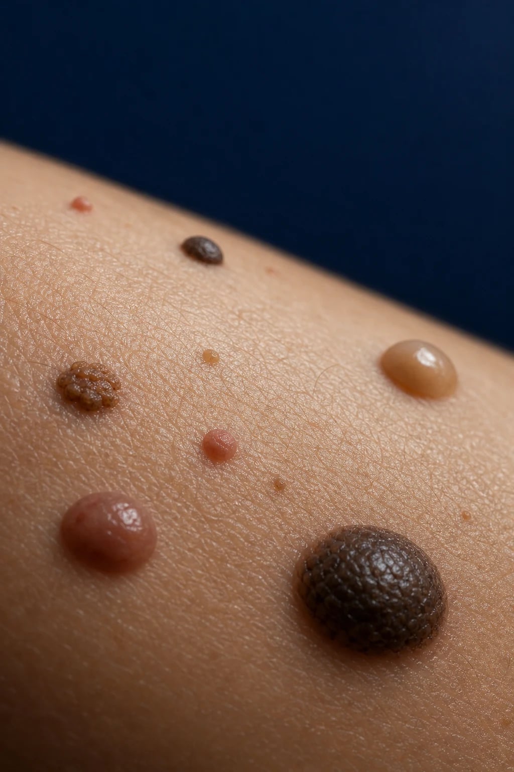
Benign skin lesions are the most common tumours in medicine. The high-yield catalogue: seborrhoeic keratosis ('stuck-on' waxy brown plaques, older adults, most common benign tumour of all — comedo-like horn cysts on dermoscopy), melanocytic naevi (moles — junctional, compound, intradermal; plus Spitz, halo, blue, dysplastic variants), pyogenic granuloma (lobular capillary haemangioma — friable bleeding papule after trauma), dermatofibroma (firm brown leg papule, dimple sign), sebaceous hyperplasia (yellow umbilicated facial papule), epidermoid and pilar cysts (firm subcutaneous nodule with punctum — keratin not sebum), lipoma (soft doughy mobile mass), milia, syringoma, cherry angioma, neurofibroma (buttonhole sign, NF1) and xanthelasma (yellow eyelid plaque — check lipids). The cardinal skill: distinguish benign from malignant with dermoscopy and the ABCDE/ugly-duckling screen, and biopsy any changing or atypical lesion before destructive treatment.
On this page & tools
Your progress
Saved locally on this device.
Exam tags
Red flags
Overview & Definition
A benign skin lesion is any non-malignant tumour or hamartomatous proliferation of the skin's component cells — keratinocytes, melanocytes, fibrohistiocytic cells, adnexal (hair follicle, sweat gland, sebaceous) epithelium, vessels, fat or peripheral nerve. They are, collectively, the most common tumours encountered in clinical practice: almost every adult carries seborrhoeic keratoses, melanocytic naevi, cherry angiomas and at least one epidermoid cyst, and most are noticed by the patient rather than the doctor. The job of the clinician is not to remove them all, but to triage: recognise the benign pattern, reassure the patient, and — most importantly — identify the lesion that is not benign and biopsy it before any destructive treatment.[1]
Two principles govern the whole topic. First, a disease is global; its treatment and thresholds are regional — but benign lesions themselves are managed similarly worldwide, with only cosmetic technique and statin thresholds (for xanthelasma) differing by guideline. Second, dermoscopy is the single most useful bedside tool in distinguishing benign from malignant pigmented and non-pigmented lesions; adding it to naked-eye examination improves sensitivity for melanoma and keratinocyte cancers and reduces unnecessary excisions. The Triage Amalgamated Dermoscopic Algorithm (TADA) formalises this: teach the common benign patterns first, then flag everything else for expert review.[2][9]
The cardinal screen for any pigmented lesion is the ABCDE criteria — Asymmetry, Border irregularity, Colour variation, Diameter over 6 mm, and Evolving (changing in size, shape, colour, elevation, or new symptoms such as itch or bleeding) — supplemented by the ugly duckling sign: a mole that looks different from the patient's other moles is the suspicious one. Any pigmented lesion meeting these criteria, or any rapidly growing or bleeding lesion regardless of colour, needs excisional biopsy with a 2 mm margin rather than destructive treatment.[1]
Classification
Benign lesions are best classified by the tissue of origin, because each lineage produces a recognisable clinical and dermoscopic pattern. The catalogue below is the core MBBS knowledge — examiners reward the ability to name the lesion, its lineage, and its distinguishing clinical sign in one breath. [1]
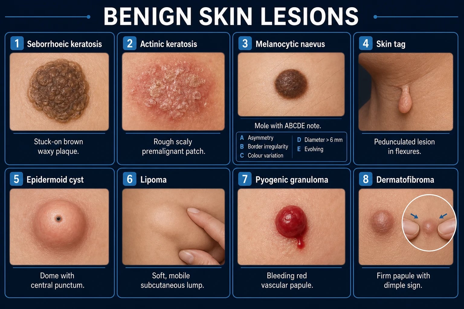
Seborrhoeic keratosis
Keratinocyte (basaloid)
- Most common benign tumour of all
- 'Stuck-on', waxy, verrucous brown-black plaque
- Older adults; trunk, face; multiple
- Dermoscopy: comedo-like horn cysts, milia-like cysts
- Leave, or cryotherapy/curettage/shave if symptomatic
Melanocytic naevus
Melanocyte nest
- Junctional (flat) / compound (raised) / intradermal (dome, flesh-coloured)
- Special types: Spitz, halo (Sutton), blue, dysplastic
- Observe unless ABCDE/ugly duckling
- Excisional biopsy 2 mm margin if suspicious
Pyogenic granuloma
Vascular (capillary haemangioma)
- Lobular capillary haemangioma — NOT pyogenic, NOT granuloma
- Rapidly growing friable bleeding red papule after trauma
- Shave excision + electrocautery of base; always send histology
- Recurrence up to 40% if feeder vessel untreated
Dermatofibroma
Fibrohistiocytic
- Firm brown dermal papule/nodule, lower leg
- Dimple (Fitzpatrick) sign on lateral compression
- Often follows insect bite / minor trauma
- Benign — leave alone unless symptomatic
Epidermoid / pilar cyst
Adnexal (follicular infundibulum)
- Firm subcutaneous nodule + central punctum
- 'Sebaceous cyst' is a misnomer — content is keratin
- Pilar (trichilemmal): scalp, no punctum, hereditary
- Excise entire wall + punctum when quiescent; never squeeze
Lipoma
Adipose
- Soft, doughy, mobile, lobulated subcutaneous mass
- Neck, shoulders, back, proximal limbs
- Multiple: familial multiple lipomatosis, Dercum, Madelung
- Excise only if symptomatic/cosmetic/growing/suspicious
Sebaceous hyperplasia
Adnexal (sebaceous gland)
- Yellow/cream 2–5 mm papule, forehead/face of elderly
- Central umbilication (duct opening)
- Dermoscopy: crown of radial vessels around central lobule
- Leave, or laser/cautery/isotretinoin if cosmetic
Cherry angioma
Vascular
- Bright red dome papule, trunk; multiple; age over 30
- Campbell de Morgan spots
- Dermoscopy: red-purple lacunae
- Leave, or laser/electrocautery if cosmetic
Neurofibroma
Peripheral nerve sheath
- Soft, fleshy, skin-coloured pedunculated papule
- Buttonhole sign (invaginates through dermal defect)
- Solitary, or part of NF1
- Excise if symptomatic; plexiform — MPNST risk
Xanthelasma
Lipid-laden macrophage
- Yellow/orange soft plaque, eyelids (medial canthus)
- 50% have dyslipidaemia — check fasting lipids
- Marker of atherosclerotic cardiovascular risk
- Treat lipids first; excision/laser/trichloroacetic acid
Milia (tiny white epidermal keratin cysts on the eyelids/cheeks), syringoma (flesh-coloured papules from eccrine ducts on the lower eyelids), skin tags (acrochordons — pedunculated fleshy papules in flexures) and acral naevi complete the catalogue. The high-yield exam point is that each lesion has a single defining clinical sign or dermoscopic feature that, once seen, makes the diagnosis instant. [1]
Epidemiology & Risk Factors
Benign skin lesions — the numbers that matter
The epidemiology follows the lesion. Seborrhoeic keratosis is essentially universal in the elderly — prevalence climbs from a few per cent in the third decade to near-total by the eighth. Acquired melanocytic naevi accumulate through childhood, peak at 20–40 years (an average of 10–40 moles), and then regress; a high naevus count (over 50, or any naevus over 5 mm in diameter) is the strongest phenotypic risk factor for melanoma. Pyogenic granuloma is most common in children, young adults, and pregnant women (the 'pregnancy tumour' or granuloma gravidarum, often on the gingiva). Dermatofibroma predominates in young-to-middle-aged women, classically on the lower leg, often with a recalled insect bite. Epidermoid cysts and lipomas are most common in mid-adult life; lipomas are the most common soft-tissue tumour overall. Cherry angiomas, sebaceous hyperplasia and xanthelasma are diseases of ageing. Neurofibroma, when multiple, points to neurofibromatosis type 1 (NF1), which affects roughly 1 in 3000.[3][8]
Risk factors worth naming in an exam answer: chronic ultraviolet exposure (drives seborrhoeic keratosis and, separately, the premalignant actinic keratosis), fair skin type, a family history of dysplastic naevi or NF1, immunosuppression (raises the burden of both benign and malignant keratinocyte lesions), and — for xanthelasma — dyslipidaemia, diabetes, obesity and hypothyroidism.[1]
Pathophysiology
The mechanism is best understood lineage by lineage, because the cell of origin dictates the morphology. The diagram figure summarises the cascade. [1]
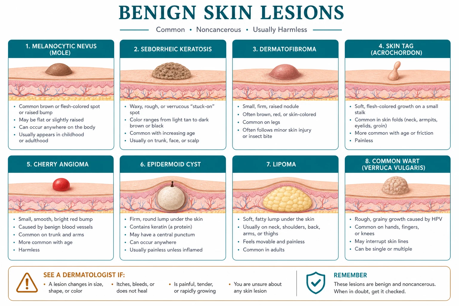
Seborrhoeic keratosis is a clonal benign proliferation of basaloid keratinocytes confined to the epidermis (a basaloid acanthoma). The tumour grows above the skin surface in a raised plaque and traps keratin within the lesion to form the pathognomonic comedo-like horn cysts and milia-like cysts seen on dermoscopy. It carries somatic mutations in FGFR3 and PIK3CA (the same pathways as many cancers, but here indolent), which explains its frequency but also its benign course.[3]
Melanocytic naevi arise from melanocyte precursors (nevomelanocytes) that fail to complete their migration from the neural crest to the epidermis and instead arrest in nests. A junctional naevus sits at the dermo-epidermal junction (flat, pigmented, macular — typical of youth); as nests drop into the dermis it becomes a compound naevus (raised, brown, papular); with complete dermal migration and epidermal junctional clearing it becomes an intradermal naevus (dome-shaped or pedunculated, often flesh-coloured, typical of the face in older adults). This 'maturation' is why a mole becomes less pigmented and more fleshy with age — a benign trajectory the examiner wants articulated. Dysplastic (atypical, Clark) naevi show cytological atypia and architectural disorder and mark increased melanoma risk; Spitz naevi are composed of spindle and epithelioid melanocytes (a pink papule in a child that histologically mimics melanoma); halo naevi show a T-cell autoimmune response against the naevus melanocytes, producing a depigmented ring; blue naevi harbour dendritic dermal melanocytes whose deep position scatters blue light (Tyndall effect) to give the lesion its blue-black colour.[4]
Pyogenic granuloma is a lobular capillary haemangioma: a benign, exuberant proliferation of capillary-sized blood vessels arranged in lobules, embedded in a loose oedematous stroma, often eroding through a thinned epidermis so that the lesion bleeds on the slightest touch. The name is a double misnomer — it is neither pyogenic (not caused by infection) nor granulomatous (not a granuloma). Trauma, pregnancy hormones and retinoid therapy are recognised triggers.[5]
Dermatofibroma (benign fibrous histiocytoma) is a reactive fibrohistiocytic proliferation, most often a response to minor trauma such as an insect bite; the tethering of the lesion to the overlying epidermis produces the dimple sign.[6] Epidermoid and pilar cysts arise when epidermal (or, in pilar cysts, outer-root-sheath trichilemmal) epithelium is implanted or retained in the dermis, forming a stratified squamous wall that continually sheds keratin into a slowly expanding cavity — the cheesy, malodorous material is keratin, not sebum, which is why 'sebaceous cyst' is a misnomer. Lipoma is a encapsulated tumour of mature white adipocytes; neurofibroma is a benign tumour of peripheral nerve sheath (Schwann cell plus perineural fibroblast) cells within the dermis, explaining its soft, fleshy feel and the buttonhole sign. Xanthelasma deposits lipid-laden macrophages (foam cells) in the superficial dermis of the eyelid, the yellow colour reflecting intracellular lipid.[7]
Clinical Presentation
Each benign lesion has a pathognomonic story that an examiner can build a vignette around. The following accounts are written as the clinician should describe them at the bedside — site, morphology, feel, and the single defining sign. [1]
Seborrhoeic keratosis presents as a well-defined, oval, 'stuck-on' plaque with a waxy or verrucous surface, tan to dark brown or black, ranging from a few millimetres to several centimetres. The lesion looks as though it has been glued onto the skin and could be flicked off. It is usually multiple, distributed on the trunk, face and extremities, and appears from middle age onward. Itch is common. Variants include dermatosis papulosa nigra (many small dark papules on the malar face of dark-skinned individuals) and stucco keratosis (grey-white rough papules on the dorsum of feet and ankles). A sudden, explosive eruption of many pruritic seborrhoeic keratoses is the Sign of Leser-Trélat and demands investigation for an internal malignancy, classically a gastric adenocarcinoma.[3]
Melanocytic naevi are classified clinically by their architecture. A junctional naevus is a flat, evenly pigmented brown macule with a regular border, usually under 6 mm, on the trunk or extremities. A compound naevus is raised, dome-shaped or papular, evenly brown, and may show central hair. An intradermal naevus is a dome-shaped or pedunculated, flesh-coloured or lightly pigmented papule, typically on the face of an older adult, often with a central hair. The special-type naevi are high-yield: a Spitz naevus is a pink, hairless, firm, rapidly appearing papule or nodule in a child (classically on the cheek or leg); a halo naevus (Sutton naevus) is a pigmented naevus surrounded by a ring of depigmentation in an adolescent, often multiple, benign, with an association with vitiligo; a blue naevus is a solitary blue-black firm papule on the dorsum of the hand or foot, or the buttock, present since youth; a dysplastic (atypical) naevus is irregular in outline and colour, larger than 5 mm, with a 'fried-egg' pebbly surface, and may be sporadic or part of the dysplastic naevus syndrome / familial atypical multiple mole melanoma (FAMM) syndrome; a congenital melanocytic naevus is present at birth, ranges from small to giant (garment-type, over 20 cm), and carries a melanoma risk proportional to size.[4]
Pyogenic granuloma presents as a bright red, glistening, friable, pedunculated or sessile papule or nodule, a few millimetres to a centimetre, that appears and grows over days to weeks, often at the site of recent minor trauma. It bleeds easily and copiously with the slightest touch — the patient's main complaint. Typical sites are the fingers, lips, oral mucosa (especially the gingiva in pregnancy — the 'pregnancy tumour'), face and trunk. Dermatofibroma is a firm, dimpled, brown dermal papule or nodule, 3–10 mm, almost always on a lower extremity (often the shin), often with a history of an insect bite weeks earlier. It is tethered to the overlying skin but freely mobile over deeper structures.[5][6]
Sebaceous hyperplasia is a soft, yellow or skin-coloured papule, 2–5 mm, on the forehead, nose or cheeks of an elderly person, with a characteristic central umbilication marking the sebaceous duct opening; lesions are usually solitary or few. Epidermoid cysts present as a firm, round, mobile, skin-coloured subcutaneous nodule, often with a visible central punctum, on the face, neck, scalp, trunk or scrotum; pilar (trichilemmal) cysts are firmer nodules on the scalp (90%) without a punctum, often multiple and inherited. Rupture produces acute inflammation, pain and erythema that mimics an abscess. Lipoma presents as a soft, doughy, lobulated, mobile subcutaneous mass that slips under the examining finger, nontender, on the neck, shoulders, back or proximal limbs; it grows slowly over years. Milia are tiny 1–2 mm white or yellow keratin cysts on the eyelids and cheeks; syringoma are 1–3 mm flesh-coloured papules in bilateral symmetrical crops on the lower eyelids of young women; cherry angiomas (Campbell de Morgan spots) are bright red, dome-shaped, 1–5 mm papules on the trunk, appearing from the third decade and multiplying with age.[1]
Neurofibroma presents as a soft, flesh-coloured, buttonhole-shaped or pedunculated papule or nodule that can be invaginated into the dermis through a defect on firm pressure (the buttonhole sign); when multiple it prompts assessment for NF1, whose revised 2021 diagnostic criteria require two or more of: six or more café-au-lait macules, skinfold freckling, two or more neurofibromas or one plexiform neurofibroma, optic pathway glioma, two or more Lisch nodules, a characteristic bony lesion, a pathogenic NF1 variant, or an affected first-degree relative. Xanthelasma presents as soft, yellow-orange, slightly raised plaques on the eyelids, most often the medial aspect of the upper lid; they are often bilateral and are a cutaneous marker of dyslipidaemia in half of patients.[7][8]
Differential Diagnosis
The pitfall in benign lesions is the mimic — a malignancy wearing a benign face. The differential is therefore organised by the presenting morphology, and the cardinal rule is: a single atypical feature is enough to biopsy. [1]
| Morphology | Benign (the common answer) | Must-not-miss mimic | The feature that separates them |
|---|---|---|---|
| Stuck-on brown plaque | Seborrhoeic keratosis | Nodular / pigmented melanoma | SK is stuck-on, waxy, uniform; melanoma is infiltrative, growing, ABCDE-positive — biopsy if any doubt |
| Pigmented papule | Junctional/compound naevus | Melanoma, pigmented BCC | Naevus is symmetric, even colour, regular border; melanoma is ABCDE-positive |
| Vascular red nodule | Pyogenic granuloma | Amelanotic melanoma, SCC | PG bleeds easily, grows over days; biopsy mandatory before ablation |
| Red papule, trunk | Cherry angioma | Kaposi sarcoma, angiosarcoma | Cherry angioma is small, dome-shaped, stable; KS is patch/plaque, violaceous |
| Yellow facial papule | Sebaceous hyperplasia | Nodular basal cell carcinoma | BCC has a rolled pearly border, telangiectasia, central ulceration |
| Yellow eyelid plaque | Xanthelasma | — (no malignant mimic) | But it signals systemic dyslipidaemia and CV risk |
| Firm leg nodule | Dermatofibroma | Dermatofibrosarcoma protuberans (DFSP), melanoma | Dimple sign + stability favours dermatofibroma; biopsy if growing or over 1 cm |
| Subcutaneous mass | Lipoma | Liposarcoma | Liposarcoma is large, deep, firm, fixed, growing — MRI and biopsy |
| Scalp nodule | Pilar cyst | Pilar tumour (proliferating trichilemmal tumour) | Solid, growing, ulcerated scalp mass in elderly woman — biopsy |
| Pedunculated fleshy papule | Skin tag, neurofibroma | Pedunculated melanoma | Asymmetry, colour change, bleeding — biopsy |
The non-malignant mimics matter too: a sebaceous hyperplasia papule is mistaken for an early BCC; a dermatofibroma for a melanoma (until the dimple sign and dermoscopy reassure); a cherry angioma for a petechial spot (it does not blanch, unlike a petechia — actually cherry angioma also does not blanch, distinguishing it from a spider naevus which does). A sudden crop of seborrhoeic keratoses with pruritus is not a benign event but the Sign of Leser-Trélat, an internal-malignancy marker.[1][9]
Clinical & Bedside Assessment
Diagnosis of a benign lesion is clinical, made on morphology and distribution, and refined at the bedside by a small set of named manoeuvres and by dermoscopy. No blood test diagnoses a benign lesion — but some demand a blood test for what they imply systemically (xanthelasma, multiple lipomas, NF1). [1]
The named signs are the high-yield bedside toolkit: [1]
[1]Dermoscopy is mandatory for any pigmented lesion and invaluable for non-pigmented ones. Each benign lesion carries a reproducible pattern. A seborrhoeic keratosis shows comedo-like openings (horn cysts), milia-like cysts, and a gyrated 'brain-like' surface with sharp demarcation — and crucially no pigment network, which separates it from a melanocytic lesion. A junctional naevus shows a regular pigment network that thins at the periphery; a compound/intradermal naevus shows a globular or homogeneous pattern with central globules and cobblestoning. A blue naevus shows a homogeneous steel-blue pigmentation with no network. A dermatofibroma shows a central white scar-like patch with a delicate peripheral pigment network. A sebaceous hyperplasia shows crown vessels — radial, arborising telangiectasias surrounding a central yellow-white lobule. A pyogenic granuloma shows reddish homogeneous areas with white collarette and rail-like vessels at the periphery. A cherry angioma shows red-purple lacunae (well-defined roundish red structures). The TADA algorithm teaches these benign patterns first, then flags anything that deviates.[2][9]
The focused examination also includes a total body skin examination (especially relevant when assessing a naevus, since the ugly duckling sign requires comparison with the patient's whole mole set) and, for xanthelasma, a search for other xanthomas (tuberous, tendinous, eruptive) and for stigmata of dyslipidaemia (corneal arcus, tendon xanthomata). [1]
Investigations
Most benign lesions require no investigation at all — the diagnosis is clinical and the lesion is either left alone or treated. Investigation becomes necessary in three situations: to exclude malignancy before destructive treatment; to characterise a deep lesion; and to assess systemic implications of a cutaneous marker. [1]
To exclude malignancy, the definitive investigation is excisional biopsy with a 2 mm clinical margin for any pigmented lesion that is changing, atypical, or fails the ugly-duckling test, and shave or punch biopsy with histology for any rapidly growing or bleeding non-pigmented lesion (pyogenic granuloma must always be sent for histology to exclude amelanotic melanoma). For a lesion being removed cosmetically by curettage or shave (e.g., a seborrhoeic keratosis), the tissue should still be submitted for histology whenever the diagnosis is not certain. Destructive treatment without biopsy is never acceptable when there is any diagnostic doubt — a nodular melanoma destroyed by cryotherapy is a catastrophic and avoidable error.[1]
To characterise a deep lesion, a large (over 5 cm), deep, firm, fixed or rapidly growing subcutaneous mass that might be a liposarcoma warrants an MRI and a needle or incisional biopsy before excision; liposarcoma cannot be reliably distinguished from lipoma clinically. Ultrasonography can confirm the cystic nature of an epidermoid cyst and the fatty nature of a lipoma at the bedside. [1]
To assess systemic implications: xanthelasma mandates a fasting lipid panel (total cholesterol, LDL, HDL, triglycerides) and a cardiovascular risk assessment, because roughly half of patients have a lipid disorder and the plaques are a marker of atherosclerotic risk independent of lipid levels. Multiple lipomas prompt consideration of familial multiple lipomatosis, Dercum's disease (adiposis dolorosa) and Madelung's benign symmetric lipomatosis, the last associated with alcohol misuse. Multiple neurofibromas prompt a full NF1 work-up against the revised 2021 criteria, including slit-lamp examination for Lisch nodules, ophthalmology review for optic glioma, and — for a plexiform or rapidly painful neurofibroma — MRI and consideration of malignant peripheral nerve sheath tumour (MPNST).[7][8]
Which benign lesion always mandates histology even though the diagnosis seems obvious?
Pyogenic granuloma. An amelanotic melanoma can present as an identical pink, friable, rapidly growing, bleeding nodule, and the two cannot be reliably separated clinically. Every pyogenic granuloma removed — by shave, curettage or excision — must be sent for histopathology, and the feeding vessel at the base electrocauterised to prevent the ~40% recurrence. Dermatofibroma, by contrast, is left alone when the dimple sign and dermoscopy are typical; biopsy is reserved for atypical or growing lesions.
Management — Resuscitation
Benign skin lesions are not, in themselves, time-critical — there is no resuscitation bundle for a lipoma. The two urgent scenarios are an acutely bleeding pyogenic granuloma (controlled with firm direct pressure for 10 minutes, then definitive ablation of the feeding vessel) and an acutely inflamed, infected epidermoid cyst (managed as an abscess: incision and drainage, a swab for culture, oral antibiotics such as flucloxacillin 500 mg four times daily for seven days if there is surrounding cellulitis, and deferral of definitive excision until the inflammation has settled — operating on an inflamed cyst risks incomplete wall removal and recurrence). The most important 'urgent' decision, however, is the diagnostic one: a lesion that might be melanoma should not be frozen, cauterised or lasered — it should be biopsied. The only true emergency in this topic is the mistake of destroying a lesion that should have been biopsied.[1]
Management — Definitive & Stepwise
Definitive treatment follows the principle: leave it if it is benign and asymptomatic; remove it if it is symptomatic, cosmetic, or there is diagnostic doubt; and never destroy a lesion you have not first characterised. The ladder is organised by lesion. [1]
Seborrhoeic keratosis — reassure the patient that the lesion is benign and leave it; if it is itchy, catches on clothing, or is cosmetically unacceptable, treat with liquid nitrogen cryotherapy (two freeze-thaw cycles), curettage with electrocautery of the base, shave excision, or ablative laser (CO2). Histology is sent whenever the diagnosis is not certain.[3]
Melanocytic naevus — observe with the ABCDE and ugly-duckling screen; advise self-examination and sun protection. Excise only if atypical or changing, with an excisional biopsy and a 2 mm clinical margin, orienting the ellipse along the relaxed skin tension lines. Never shave or destroy a suspicious pigmented lesion — it compromises histological staging and may upstage a melanoma. Congenital melanocytic naevi are observed; small lesions need no excision, but giant congenital naevi (over 20 cm projected adult diameter) carry a significant melanoma risk and warrant specialist surveillance and consideration of prophylactic excision, plus MRI screening for neurocutaneous melanosis in infancy.[4]
Pyogenic granuloma — shave excision of the friable tissue followed by electrocautery of the base (the feeder vessel) is the standard and reduces recurrence. Alternatives include curettage and cautery, cryotherapy (less effective for raised lesions), and pulsed-dye or Nd:YAG laser. Always send the tissue for histology. Recurrence occurs in up to 40% if the feeder vessel is not ablated; recurrent lesions warrant full surgical excision. In pregnancy, defer definitive treatment to the postpartum period if possible, as lesions may regress.[5]
Dermatofibroma — leave alone; it is benign and stable. Excise only if symptomatic (catches, itches) or if there is diagnostic doubt, with a narrow ellipse including the tethered overlying skin. Recurrence after excision is uncommon.[6]
Epidermoid and pilar cyst — the definitive treatment is complete surgical excision of the cyst wall together with the punctum through a small elliptical incision, performed when the cyst is quiescent. An acutely inflamed or infected cyst is incised and drained first, with antibiotics if cellulitis is present, and the definitive excision deferred for four to six weeks until the inflammation settles. Never squeeze a cyst — it ruptures the wall into the dermis, causes intense inflammation and scarring, and makes later complete excision harder. Pilar cysts are excised similarly; milia are simply nicked with a needle and the keratin extruded.[1]
Lipoma — observe; excise only if symptomatic, cosmetic, growing, or suspicious. Excision is through a small overlying incision, enucleating the encapsulated tumour; liposuction is an alternative for large or multiple lesions. A lesion that is large (over 5 cm), deep, firm, fixed or rapidly growing is biopsied before excision to exclude liposarcoma.[1]
Sebaceous hyperplasia, cherry angioma, syringoma, milia, skin tags — leave, or treat cosmetically: electrocautery, cryotherapy, CO2 laser, or pulsed-dye laser for vascular lesions; needle extrusion for milia; snip excision for skin tags. Neurofibroma — excise if symptomatic or cosmetic; a rapidly growing or painful lesion, or a plexiform neurofibroma, is referred for specialist assessment of MPNST.[8]
Xanthelasma — address the dyslipidaemia and cardiovascular risk first (statin therapy per regional guidelines, glycaemic and blood pressure control, smoking cessation), because lesion recurrence is common unless lipids are controlled and because the systemic risk outweighs the cosmetic one. Local treatment options include surgical excision (considering eyelid laxity and the risk of ectropion), CO2 or erbium:YAG laser ablation, trichloroacetic acid chemical peeling, and ** topical imiquimod**; recurrence occurs in up to 40% regardless of modality.[7]
[1]Specific Subtypes & Scenarios
The melanocytic naevus family
The naevus family is the most examined corner of the topic. Spitz naevus — a pink, firm, hairless, rapidly growing papule in a child (cheek or leg), histologically composed of spindle and epithelioid melanocytes — is a diagnostic trap because it mimics both pyogenic granuloma (clinically) and melanoma (histologically). 'Spitzoid tumours of uncertain malignant potential' (STUMP) are managed jointly by dermatology and dermatopathology, often with complete excision. Halo naevus (Sutton naevus) — a pigmented naevus surrounded by a depigmented halo in an adolescent, often multiple — is benign and requires only reassurance and observation (the naevus usually regresses over years); check for coexisting vitiligo but do not excise. Blue naevus — a firm blue-black dermal papule on the dorsum of the hand or foot, or the buttock — is benign; excise only if changing or symptomatic. Dysplastic (atypical, Clark) naevus — irregular, larger than 5 mm, 'fried-egg' surface — is a marker of melanoma risk; management is observation and total-body skin examination, with excision only of lesions that change. Patients with the dysplastic naevus syndrome (FAMM), defined by over 100 naevi plus a family history of melanoma, enter a surveillance programme with six-to-twelve-monthly total-body skin examination and dermoscopy.[4]
The cyst family
Epidermoid (infundibular) cyst — face, neck, trunk, scrotum; central punctum; keratinaceous content; arises from the follicular infundibulum, often after implantation or from a blocked pilosebaceous unit. Pilar (trichilemmal) cyst — scalp (90%), no punctum, firmer, contents more solid, inherited autosomal dominant; the wall is outer-root-sheath (trichilemmal) keratinisation without a granular layer. Milia — tiny superficial epidermoid cysts, 1–2 mm, eyelids and cheeks; primary or secondary to blistering/trauma/dermabrasion. Steatocystoma multiplex — multiple yellow chest cysts containing oily fluid, from sebaceous glands. Dermoid cyst — a congenital sequestration cyst along embryonic fusion lines, most often the lateral eyebrow in a child; may extend deeply and requires imaging before excision.[1]
The lipoma syndromes
A solitary lipoma needs no further thought. Multiple lipomas prompt the syndromic differential: familial multiple lipomatosis (autosomal dominant, multiple discrete painless lipomas on the forearms and thighs, beginning in adulthood); Dercum's disease (adiposis dolorosa) — multiple painful lipomas in postmenopausal women, with asthenia and psychiatric features; and Madelung's disease (benign symmetric lipomatosis) — large, symmetric, non-encapsulated fat deposits around the neck and upper trunk in middle-aged men, strongly associated with alcohol misuse and metabolic syndrome. A rapidly growing, deep, firm or fixed lipomatous mass is liposarcoma until proven otherwise — MRI and biopsy.[1]
Neurofibroma and the NF1 work-up
A solitary neurofibroma is benign and excised if symptomatic. Multiple neurofibromas mandate assessment for neurofibromatosis type 1, now diagnosed against the revised 2021 international consensus criteria: two or more of (1) six or more café-au-lait macules over 5 mm in prepubertal or 15 mm in postpubertal individuals; (2) axillary or inguinal freckling; (3) two or more neurofibromas of any type or one plexiform neurofibroma; (4) optic pathway glioma; (5) two or more Lisch nodules; (6) a characteristic bony lesion (sphenoid wing dysplasia, bowing of a long bone with or without pseudarthrosis); (7) a pathogenic NF1 variant; or (8) an affected first-degree relative by the above. The criteria were revised to add the genetic criterion and to distinguish NF1 from Legius syndrome (café-au-lait macules and freckling only, from a SPRY1/SPRED1 variant, without neurofibromas). A plexiform neurofibroma — a diffuse, bag-of-worms mass along a nerve — carries a lifetime risk of around 10% of malignant peripheral nerve sheath tumour (MPNST); new pain, rapid growth or neurological deficit triggers urgent MRI and PET-CT.[8]
Xanthelasma and the lipid work-up
Xanthelasma is the most common of the cutaneous xanthomas and the one most strongly linked to dyslipidaemia. Roughly half of patients have a fasting lipid abnormality (most often raised LDL or apolipoprotein B). Even when lipids are normal, xanthelasma is an independent marker of atherosclerotic cardiovascular disease — patients carry a higher ischaemic heart disease risk than controls — so the work-up includes a fasting lipid panel, a cardiovascular risk assessment, and consideration of statin therapy on cardiovascular grounds alone, independent of the cosmetic lesion.[7]
Complications & Pitfalls
The complications are mostly iatrogenic and avoidable. The cardinal pitfalls: [1]
The five avoidable errors in benign skin lesions — and how to dodge them
Procedure-related complications deserve naming: cryotherapy causes hypopigmentation (a major concern in dark skin), blistering and occasionally scarring; curettage without histology forfeits diagnosis; lipoma excision risks haematoma and injury to nearby nerves (especially in the proximal limbs and neck); xanthelasma excision risks ectropion if too much eyelid skin is removed — laser or trichloroacetic acid is preferred for large or multiple lesions; and pigmented-lesion excision must be oriented along skin tension lines and the specimen marked to guide any re-excision. [1]
A final disease-related complication: a ruptured epidermoid cyst mimics an abscess, and incision and drainage in the acute phase is correct — but the definitive excision must follow once settled, or the lesion will recur with a vengeance. [1]
Prognosis & Disposition
For a truly benign lesion the prognosis is excellent — the lesion is harmless, and treatment, when needed, is cosmetic and curative. Three prognostic nuances matter for the exam. First, dysplastic naevi are not premalignant in themselves but are markers of melanoma risk: a patient with the dysplastic naevus syndrome has a substantially elevated lifetime melanoma risk and enters a surveillance programme of six-to-twelve-monthly total-body skin examination with dermoscopy. Second, giant congenital melanocytic naevi carry a real melanoma risk (several per cent) and, in large axial lesions, a risk of neurocutaneous melanosis; they warrant specialist surveillance and often prophylactic excision in infancy. Third, xanthelasma is a prognostic marker of cardiovascular disease independent of lipid levels — the disposition is to a cardiovascular risk assessment and lipid management, not just to a cosmetic clinic.[4][7]
Most benign lesions are managed in primary care or dermatology outpatients; surgical excision of a cyst or lipoma is a day-case procedure. Patients with suspected NF1, dysplastic naevus syndrome, giant congenital naevi, or a suspected liposarcoma or MPNST are referred to specialist services. The safety-net for every patient sent home with a benign lesion is clear advice: return if the lesion changes in size, shape, colour or sensation, or bleeds. [1]
Special Populations
Children
Weight-based & developmental
- Pyogenic granuloma and Spitz naevus are common; congenital naevi present at birth
- Avoid painful procedures where possible; favour curettage over deep excision
- Giant congenital naevus — specialist surveillance and MRI for neurocutaneous melanosis
- Any drug dose is weight-based (e.g., flucloxacillin for infected cyst)
Pregnancy
Hormone-driven change
- Pyogenic granuloma (granuloma gravidarum / pregnancy tumour) on the gingiva
- Moles may darken and enlarge; do not over-investigate — observe
- Defer elective excision to the postpartum period where possible
Elderly
High benign burden, high cancer risk
- High burden of seborrhoeic keratoses, cherry angiomas, sebaceous hyperplasia, xanthelasma
- Coexisting actinic keratoses (premalignant) — surveillance
- Xerosis exacerbates itch; emollients help
Immunosuppressed
Transplant / HIV
- High burden of actinic keratoses and SCC risk — aggressive field therapy
- Refractory or atypical warts; aggressive seborrhoeic keratoses
- Regular total-body skin examination; high index of suspicion for malignancy
Dark skin
Pigmentation & keloid risk
- Dermatosis papulosa nigra on the malar face
- Hypopigmentation from cryotherapy a major concern — use with caution
- Keloid scarring after excision — counsel and consider
Two further groups: the anticoagulated patient bleeds more with any excision or shave (plan cautery and pressure dressings, and do not stop anticoagulants for minor skin surgery); and the patient with multiple lipomas (assess for familial multiple lipomatosis, Dercum, Madelung) or multiple neurofibromas (assess for NF1 against the 2021 criteria). [1]
Evidence, Guidelines & Regional Differences
The evidence base for benign lesion recognition is anchored on dermoscopy. The 2018 Cochrane review by Dinnes and colleagues evaluated visual inspection and dermoscopy for diagnosing keratinocyte skin cancers and showed that dermoscopy added to naked-eye examination improves sensitivity for both melanoma and keratinocyte cancers; the 2019 Seiverling TADA study showed that teaching clinicians the common benign dermoscopic patterns first improves triage sensitivity for malignancy — the rationale for the benign-first approach taken in this topic.[2][9]
Lesion-specific evidence: the 2023 Barthelmann review of seborrhoeic keratosis summarises the molecular (FGFR3, PIK3CA) and clinical features and treatment; the 2021 Jahnke review in Pediatrics gives management recommendations for congenital melanocytic naevi; the 2022 NEJM clinical image of pyogenic granuloma and the 2016 Bandyopadhyay review of atypical dermatofibroma clarify those lesions; the 2018 Laftah review covers xanthelasma treatment modalities; and the 2021 Legius international consensus revises the NF1 diagnostic criteria, adding the genetic criterion and separating Legius syndrome.[3][4][5][6][7][8]
[1] [1]The revised 2021 NF1 diagnostic criteria and the dermoscopic TADA algorithm are applied worldwide. Cryotherapy, curettage and surgical excision are universally available; laser and photodynamic modalities vary by setting. The core principle — biopsy any changing or atypical lesion, and never destroy what you have not characterised — is universal.
Where the evidence is weak: the optimal management of a Spitz naevus in an adult remains debated (excise vs observe), reflecting the difficulty of distinguishing a benign Spitz naevus from a spitzoid melanoma; and recurrence rates after xanthelasma treatment are high (up to 40%) with every modality, so no single technique is clearly superior — the systemic lipid management is the more important intervention. [1]
Exam Pearls
Benign lesion catalogue — the CATALOGUE memory hook
CATALOGUE
Campbell de Morgan spots — red dome papules, trunk, age over 30
Skin tag — pedunculated fleshy papule, neck/axillae/groin
Scalp, no punctum, hereditary; excise wall
Lobular capillary haemangioma — bleed easily, send histology
Soft doughy mobile mass; exclude liposarcoma if large/deep/fixed
Eyelid plaque — check lipids, CV risk
Congenital — melanoma + neurocutaneous melanosis risk
The mole that looks different — biopsy it
Punctum, keratin (not sebum); excise entire wall
The high-yield one-liners an examiner rewards: [1]
Frequently-misremembered facts, correctly stated: a junctional naevus is flat and a intradermal naevus is raised and flesh-coloured (not the reverse — the naevus matures from junctional to intradermal as it becomes more dermal and less pigmented); pyogenic granuloma contains no pus despite its name; the dimple sign is positive in dermatofibroma and negative in melanoma; and cryotherapy causes hypopigmentation in dark skin, not hyperpigmentation. [1]
Exam application bank (NEET-PG / INICET)
One-line answer
Benign skin lesions are the most common tumours in medicine. The high-yield catalogue: seborrhoeic keratosis ('stuck-on' waxy brown plaques, older adults, most common benign tumour of all — comedo-like horn cysts on dermoscopy), melanocytic naevi (moles — junctional, compound, intradermal; plus Spitz, halo, blue, dysplastic variants), pyogenic granuloma (lobular capillary haemangioma — friable bleeding papule after trauma), dermatofibroma (firm brown leg papule, dimple sign), sebaceous hyperplasia (yellow umbilicated facial papule), epidermoid and pilar cysts (firm subcutaneous nodule with punctum — keratin not sebum), lipoma (soft doughy mobile mass), milia, syringoma, cherry angioma, neurofibroma (buttonhole sign, NF1) and xanthelasma (yellow eyelid plaque — check lipids). The cardinal skill: distinguish benign from malignant with dermoscopy and the ABCDE/ugly-duckling screen, and biop
Worked stems (answer without another resource)
Stem 1 — Classic presentation. Map symptoms to mechanism; name the first investigation and first treatment step with dose/route if drug therapy is standard. [1]
Stem 2 — Unstable / complicated. List red flags that force immediate resuscitation, theatre, ICU, antidote, or reperfusion — and what you do in the first 15 minutes. [1]
Stem 3 — Atypical group. Elderly, pregnancy, child, or immunocompromised: how presentation and thresholds change. [1]
Stem 4 — Differential trap. Name the three closest mimics and one discriminator for each. [1]
Stem 5 — Disposition. Who goes home with safety-netting, who is admitted, who needs HDU/ICU/theatre, and what follow-up is mandatory. [1]
Rapid viva checklist
- Definition + classification
- Pathophysiology chain
- Bedside signs / criteria
- Score with exact components (if any)
- Emergency bundle
- Definitive therapy with doses
- Complications of disease and of treatment
- Special populations
- Guideline/trial name if classic
- Three exam traps
Coverage self-check
If you cannot answer any stem above from this page alone, re-read the matching section — the page is intended to be self-sufficient for final-prof and NEET-PG/INICET questions on Benign Skin Lesions.
“”
Short-answer question — a bleeding nodule on the finger
10 minutes · 10 marks
A 23-year-old carpenter presents with a red, friable nodule on his index finger that appeared two weeks after a minor injury and bleeds every time he knocks it. It is 8 mm, pedunculated, and glistening red. (a) What is the most likely diagnosis, and what is its histological nature? (b) Give the definitive management, naming two technical steps that reduce recurrence. (c) What is the single most important safety step and why?
References
- [1]Brodsky J. Management of benign skin lesions commonly affecting the face: actinic keratosis, seborrheic keratosis, and rosacea Curr Opin Otolaryngol Head Neck Surg, 2009.PMID 19465852
- [2]Dinnes J, Deeks JJ, Chuchu N, et al. Visual inspection and dermoscopy, alone or in combination, for diagnosing keratinocyte skin cancers in adults Cochrane Database Syst Rev, 2018.PMID 30521688
- [3]Barthelmann S, Butsch F, Lang BM, et al. Seborrheic keratosis J Dtsch Dermatol Ges, 2023.PMID 36892019
- [4]Jahnke MN, O'Haver J, Gupta D, et al. Care of Congenital Melanocytic Nevi in Newborns and Infants: Review and Management Recommendations Pediatrics, 2021.PMID 34845496
- [5]Komakech D, Ssenkumba B. Pyogenic Granuloma N Engl J Med, 2022.PMID 36416770
- [6]Bandyopadhyay MR, Besra M, Dutta S, et al. Dermatofibroma: Atypical Presentations Indian J Dermatol, 2016.PMID 26955137
- [7]Laftah Z, Al-Niaimi F. Xanthelasma: An Update on Treatment Modalities J Cutan Aesthet Surg, 2018.PMID 29731585
- [8]Legius E, Messiaen L, Wolkenstein P, et al. Revised diagnostic criteria for neurofibromatosis type 1 and Legius syndrome: an international consensus recommendation Genet Med, 2021.PMID 34012067
- [9]Seiverling EV, Ahrns HT, Greene A, et al. Teaching Benign Skin Lesions as a Strategy to Improve the Triage Amalgamated Dermoscopic Algorithm (TADA) J Am Board Fam Med, 2019.PMID 30610147