Dermatology · Medicine
Blue naevus and dermal melanocytosis
Also known as Blue naevus · Common blue naevus · Cellular blue naevus · Combined blue naevus · Dermal melanocytosis · Naevus of Ota (oculodermal melanocytosis) · Naevus of Ito · Mongolian spot (congenital dermal melanocytosis)
A blue naevus is a benign melanocytic lesion built from heavily pigmented, dendritic and spindled DERMAL melanocytes in the reticular dermis, whose blue colour is an optical effect (Tyndall scattering) of the deep pigment location rather than blue pigment. The common blue naevus is a small, stable, blue-black papule of the extremities; the cellular blue naevus is a larger, deeper lesion of the buttock, sacrum or scalp with a small malignant potential; and dermal melanocytoses (Mongolian spot, naevus of Ota, naevus of Ito) are persistent ectopic dermal melanocytes distributed by site. Stable classic lesions are observed; any changing or atypical blue lesion is biopsied or excised to exclude melanoma.
On this page & tools
Your progress
Saved locally on this device.
Exam tags
Red flags
Overview and definition
A blue naevus is a benign melanocytic lesion formed by a localised proliferation of heavily pigmented, dendritic and spindled melanocytes that live in the dermis, not in the epidermis. Because the melanin sits deep — in the reticular dermis, beneath a layer of collagen and overlying skin — the light returning to the examiner's eye has been selectively scattered so that the lesion appears blue, slate-grey or blue-black rather than brown. This is the Tyndall effect: the same optics that make superficial veins look blue. There is no blue pigment in a blue naevus; the colour is an optical consequence of where the melanin sits.[1][3]
Two facts follow from this definition and govern the whole topic. First, a blue naevus is built from a proliferation of dermal melanocytes — a small, discrete tumour — which distinguishes it from the dermal melanocytoses (Mongolian spot, naevus of Ota, naevus of Ito), where scattered dendritic melanocytes simply persist in the dermis without forming a tumour, producing a flat patch rather than a papule.[3] Second, because the colour depends on depth and pigment density rather than biology, a long list of unrelated lesions — melanoma, a traumatic tattoo, a venous lake, a dermatofibroma, a pigmented basal cell carcinoma — can also look blue. The defining clinical skill in this topic is therefore not recognising the textbook blue naevus, but deciding which blue lesion to leave alone and which to biopsy.[1][2]
The natural history is benign and stable in the overwhelming majority, which is why a common blue naevus is managed by reassurance. The exceptions that carry real risk — the cellular blue naevus with its small potential for malignant transformation, naevus of Ota with its ocular melanoma and glaucoma risk, and the multiple blue naevi of Carney complex — are the conditions that earn the topic its examination weight.[6][7]
[1]Classification
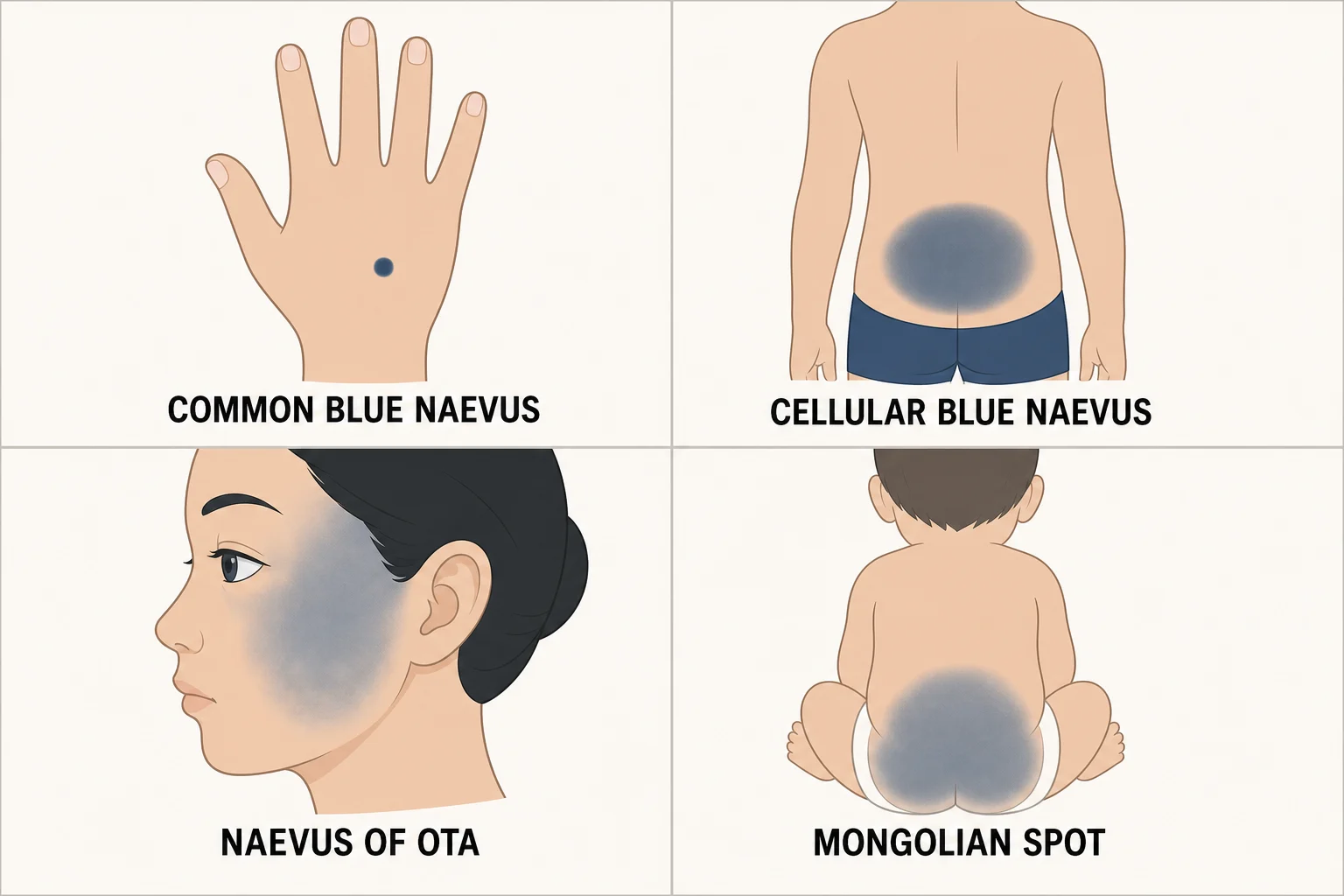
Blue naevi and the dermal melanocytoses are best understood as one biological family — ectopic dermal melanocytes — split by whether the cells proliferate into a tumour (the naevi) or simply sit in the dermis as a patch (the melanocytoses), and then by their characteristic anatomical territory.[3]
The common and cellular blue naevi are the two canonical types an examiner expects a candidate to separate cleanly: the common type is the everyday stable papule of the extremities with no malignant potential, while the cellular type is the larger, deeper, often congenital lesion of the buttock and scalp that carries a small but real risk of malignant blue naevus and is therefore excised.[1][3]
Epidemiology and risk factors
The common blue naevus is one of the commonest acquired melanocytic lesions, present in roughly 1 to 3 percent of the general population. Most appear in childhood, adolescence or early adulthood, persist unchanged thereafter, and show a slight female predilection in some series. The great majority are solitary, but multiple lesions are well recognised and — when numerous — should prompt consideration of a syndrome (see Special subtypes).[1]
The cellular blue naevus is rare, far less common than the common type. It affects children and young adults, has a female predominance, and is notable in that around half are present at birth or arise in early childhood — a congenital blue nodule of the scalp or buttock is a classical presentation. Its epidemiology matters precisely because it is the variant with a recognised, if small, risk of malignant transformation.[1][3]
The dermal melanocytoses are strongly ethnically patterned. The Mongolian spot is present in over 80 to 90 percent of Asian and Black neonates but in fewer than 10 percent of White neonates; it is so common in its characteristic populations that it is regarded as a normal variant. The naevus of Ota is most frequent in East Asian and Black women (around 0.02 to 0.8 percent prevalence), is nearly always unilateral, and is permanent — unlike the Mongolian spot it does not fade.[7][8]
The principal risk factors for clinical concern are not risk factors for getting a blue naevus but for a blue naevus behaving badly: a cellular subtype, large size (over 1 cm), congenital onset, a changing lesion, and — at the syndrome level — Carney complex, in which multiple blue naevi cluster with lentigines, cardiac myxomas and endocrine overactivity.[6]
Pathophysiology
Embryological origin
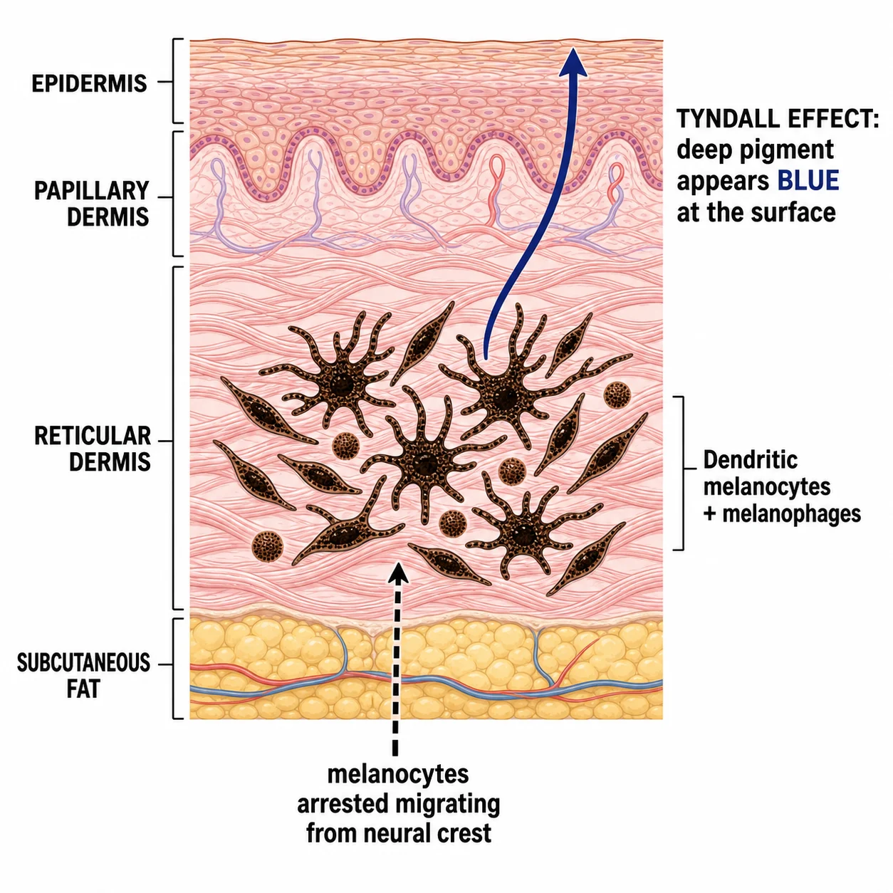
Melanocytes originate in the neural crest and migrate along a defined pathway to take up residence in the basal layer of the epidermis during the first trimester. In blue naevi and dermal melanocytoses a clone of melanocytes is arrested in, or fails to complete, this migration and persists and proliferates within the dermis. The same developmental logic explains the characteristic sites: the lumbosacral Mongolian spot sits over the last region of the embryo to be melanocyte-colonised, and naevus of Ota and naevus of Ito follow the trigeminal and cervical nerve distributions along which the migrating cells travelled.[3]
The Tyndall effect — why a brown pigment looks blue
The colour is purely optical. Melanin is brown-black, but when it lies in the deep dermis the overlying collagen and epidermis act as a scattering medium. Shorter (blue) wavelengths are preferentially scattered back to the eye while longer (red/brown) wavelengths are absorbed or pass deeper. The depth and density of the pigment determine the shade: a superficial dermal deposit looks steel-blue, a deeper and denser deposit looks slate-blue to blue-black. This is the same physics that makes superficial veins appear blue through the skin.[1]
Molecular driver
Most blue naevi and dermal melanocytoses carry activating mutations in the G-protein signalling pathway — most commonly GNAQ (the Q209 hotspot), and less often GNA11, PLCB4 or CYSLTR2. These mutations drive the MAPK pathway constitutively, which is why a blue naevus sits on the same molecular spectrum as uveal melanoma (which shares the GNAQ/GNA11 driver). This shared biology explains why blue naevi only very rarely progress to melanoma but do so through the same pathway, and why atypical cellular blue naevus and melanoma can be histologically very difficult to separate.[1][2]
The biphasic histology of cellular blue naevus
The cellular blue naevus earns its name from a characteristic two-component architecture: a deep common-blue-naevus component (densely pigmented dendritic and spindled melanocytes with melanophages, hugging collagen bundles) plus a larger, more superficial nodular component of tightly packed fascicles and nests of plump spindle/ovoid cells that often extends into the subcutis in a wedge or dumbbell shape following skin appendages. The cells are low in atypia and rarely mitotic — and it is the loss of these benign features (atypia, mitoses, necrosis, deep infiltration) that defines malignant blue naevus.[3]
Clinical presentation
Common blue naevus
The textbook lesion is a solitary, asymptomatic, well-circumscribed, dome-shaped blue-black to steel-blue papule, 2 to 10 mm across, firm to the touch, with the overlying skin markings preserved. It most often sits on the dorsum of the hands and feet, then the scalp, face, wrists and forearms. Once it appears it is stable for years to life, which is the single most reassuring clinical feature: a blue papule that has not changed in a decade is a common blue naevus until proven otherwise.[1]
Cellular blue naevus
The cellular type is larger (1 to 3 cm) and deeper, presenting as a bluish-black nodule or plaque that may be multinodular. Its classical territory is the buttock and sacrococcygeal area (the same developmental field as the Mongolian spot) and the scalp, and around half are congenital — a blue-black plaque present since birth over the sacrum or a blue nodule on the scalp of a child is the canonical stem. Because of its depth it may feel less sharply defined than the common type, and because of its recognised malignant potential any change in it is a red flag.[1][3]
Dermal melanocytoses
Mongolian spot is a blue-grey patch over the lumbosacral area and buttocks present at birth, most common in Asian and Black infants, that fades over the first two to four years of life as the melanocytes disperse and the pigment is cleared. Naevus of Ota is a unilateral blue-grey/brown macular pigmentation of the skin of the first and second trigeminal dermatomes — temple, cheek, periorbital skin — almost always with scleral (and sometimes conjunctival and uveal) pigmentation; it is permanent. Naevus of Ito is the same process in the shoulder girdle, scapula and lateral neck distribution, without ocular involvement.[7][8]
Atypical and less common presentations
Examiners deliberately probe the edges of the distribution. A combined blue naevus presents as a blue papule within or beside a brown conventional naevus — a clinical melanoma mimic. An epithelioid (animal-type) blue naevus is a heavily pigmented plaque on a histological spectrum with pigmented epithelioid melanocytoma. An amelanotic blue naevus lacks the blue colour entirely and presents as a skin-coloured dermal nodule, mimicking a neurofibroma or appendage tumour. Subungual and periungual blue naevi produce a blue band or nodule under the nail and must be distinguished from subungual melanoma.[5] Mucosal blue naevi occur in the oral mucosa, conjunctiva and sinonasal tract, and a congenital giant cellular blue naevus of the scalp in a bathing-suit distribution may be associated with neurocutaneous melanosis and warrants neuroimaging.[3]
Differential diagnosis
The differential of a blue or blue-black cutaneous lesion is long, because depth of pigment — from any source — produces the same optical effect. The task is not to list every mimic but to separate the benign stable lesion from melanoma, and to recognise the handful of specific mimics that have their own management.[2][3]
For naevus of Ota specifically, the facial differential includes melasma, Hori's naevus (acquired bilateral naevus of Ota-like macules), post-inflammatory hyperpigmentation, and physiological scleral pigmentation. In infants, a persistent or ectopic Mongolian spot must be distinguished from a bruise — a distinction with safeguarding implications, since a Mongolian spot is present at birth, sits in the classic lumbosacral area, does not evolve through the colour stages of a bruise, and is far commoner in pigmented infants.[7]
Clinical and bedside assessment
The focused assessment of any blue-black lesion is built around a single question: is this lesion behaving like a benign blue naevus, or is it changing? History establishes the age of onset, duration, and — critically — any change in size, shape, colour, elevation, or any new symptom (itch, pain, bleeding, ulceration). A stable lesion present and unchanged for years in a typical site is the classic benign pattern; a new, enlarging or symptomatic blue lesion reopens the question of melanoma in every case.[1][2]
Examination confirms the morphology (papule vs plaque vs patch), site, colour homogeneity, and the presence or absence of the ABCDE features of melanoma (Asymmetry, Border irregularity, Colour variation, Diameter over 6 mm, Evolution). Dermoscopy is the decisive bedside tool: a classic common blue naevus shows structureless, homogeneous, steel-blue to blue-black pigmentation with no pigment network, no globules, no regression structures and no atypical vessels — the absence of melanoma features is as important as the presence of the blue field.[4] A few named bedside manoeuvres resolve the common mimics at the chairside. Diascopy (pressing a glass slide over the lesion) empties a venous lake — a compressible vascular papule on the lower lip or ear of an older adult blanches to nothing, whereas a blue naevus does not change. The dimple sign (the lesion dimples inward on lateral pinch between finger and thumb) marks a dermatofibroma, a firm dermal papule of the lower leg with a central white scar-like patch and peripheral pigment network on dermoscopy. Cold sensitivity with paroxysmal pain points to a glomus tumour, classically a tender bluish subungual nodule. These three tests cost nothing and sharpen the differential before any instrument reaches the skin.[3]
A whole-skin examination is mandatory for two reasons: to look for multiple blue naevi (which flag Carney complex), and, in any blue lesion of concern, to search for a primary melanoma elsewhere. For naevus of Ota, the examination extends to the eyes — document the scleral pigmentation and arrange tonometry for glaucoma and a dilated fundus examination for uveal melanoma.[6][7]
Investigations
Clinical diagnosis
A typical common blue naevus is a clinical diagnosis, confirmed by dermoscopy. No biopsy, blood test or imaging is required for a stable, classic, asymptomatic lesion in a typical location — investigation in that setting is over-investigation.[1]
Dermoscopy
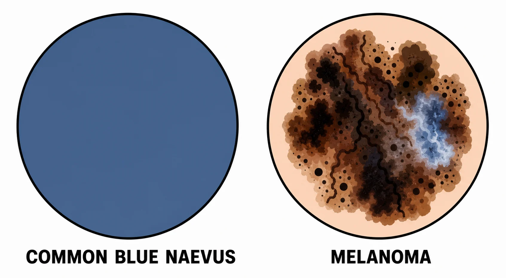
The dermoscopic signature of a common blue naevus is a structureless, homogeneous, steel-blue to blue-black area occupying the whole lesion, with no pigment network, no globules, no streaks, no regression (scar-like) areas and no atypical vessels. The cellular blue naevus may add a central structureless blue area with peripheral regular globules or streaks. Occasional variants show polychromatic structureless areas or, rarely, white rosettes under polarised dermoscopy, but the key point for the candidate is the negative feature: the absence of melanoma structures is what permits observation.[4]
Reflectance confocal microscopy and sequential monitoring
In specialist pigmented-lesion centres, two non-invasive techniques refine the decision to biopsy an equivocal blue lesion. Reflectance confocal microscopy images the dermo-epidermal junction and superficial dermis at cellular resolution in vivo: a blue naevus shows dermal dendritic cells and bright stellate melanophages without the pagetoid spread or atypical nests of melanoma, allowing a lesion of uncertain nature to be characterised without excision in selected cases. Sequential digital dermoscopy and total-body photography document stability over time — a lesion whose homogeneous blue pattern and dimensions are unchanged at three-month and then six-month review can be safely observed, whereas any architectural change converts observation to excision. These tools are adjuncts to, not substitutes for, the cardinal rule: a clinically changing blue lesion is excised regardless of its confocal or dermoscopic appearance.[4]
Indications for biopsy or excision
Biopsy or excision is mandated whenever the lesion departs from the reassuring template: [1]
- any change — growth, colour change, shape change, elevation, or new symptoms;
- an atypical dermoscopic pattern (asymmetry, atypical network, blue-white veil, regression, irregular vessels);
- a new blue lesion in an adult, especially if enlarging;
- a lesion over 1 cm, a cellular blue naevus, or any diagnostic uncertainty;
- a lesion in a site that cannot be reliably monitored (e.g. scalp, mucosa, subungual).[1][2]
Where biopsy is indicated the preferred technique is a complete excisional biopsy with a narrow margin of normal skin — it provides the whole lesion for histology, which is essential because the distinction between atypical cellular blue naevus and melanoma can require examination of the entire architecture and the deep margin. A partial or shave biopsy that transects a cellular blue naevus risks an inadequate sample and recurrence. [1]
Histopathology
The common blue naevus shows a poorly demarcated dermal lesion of densely pigmented, elongated dendritic and spindled melanocytes lying between and within collagen bundles of the reticular dermis, accompanied by numerous melanophages and variable dermal fibrosis. There is no junctional activity (no naevus cells at the dermo-epidermal junction — this distinguishes it from a conventional compound naevus), no atypia and no mitoses.[3]
The cellular blue naevus shows the characteristic biphasic pattern: a deep common-blue-naevus component plus nests and short fascicles of plump spindle or ovoid cells, often extending in a wedge shape into the subcutis along appendages. Cellularity is greater than the common type but atypia is mild and mitoses are rare. The melanocytes are immunoreactive for S-100, SOX10, HMB-45 and Melan-A.[3]
The combined blue naevus shows the two populations side by side (a blue naevus component and a conventional junctional/compound naevus component) — a useful histological clue when the clinical and dermoscopic picture is mixed. Malignant blue naevus is defined by cytological atypia, mitoses (especially atypical), necrosis, infiltrative growth and loss of the orderly biphasic architecture, arising either within a long-standing cellular blue naevus or de novo.[2]
Management — resuscitation (the safety step)
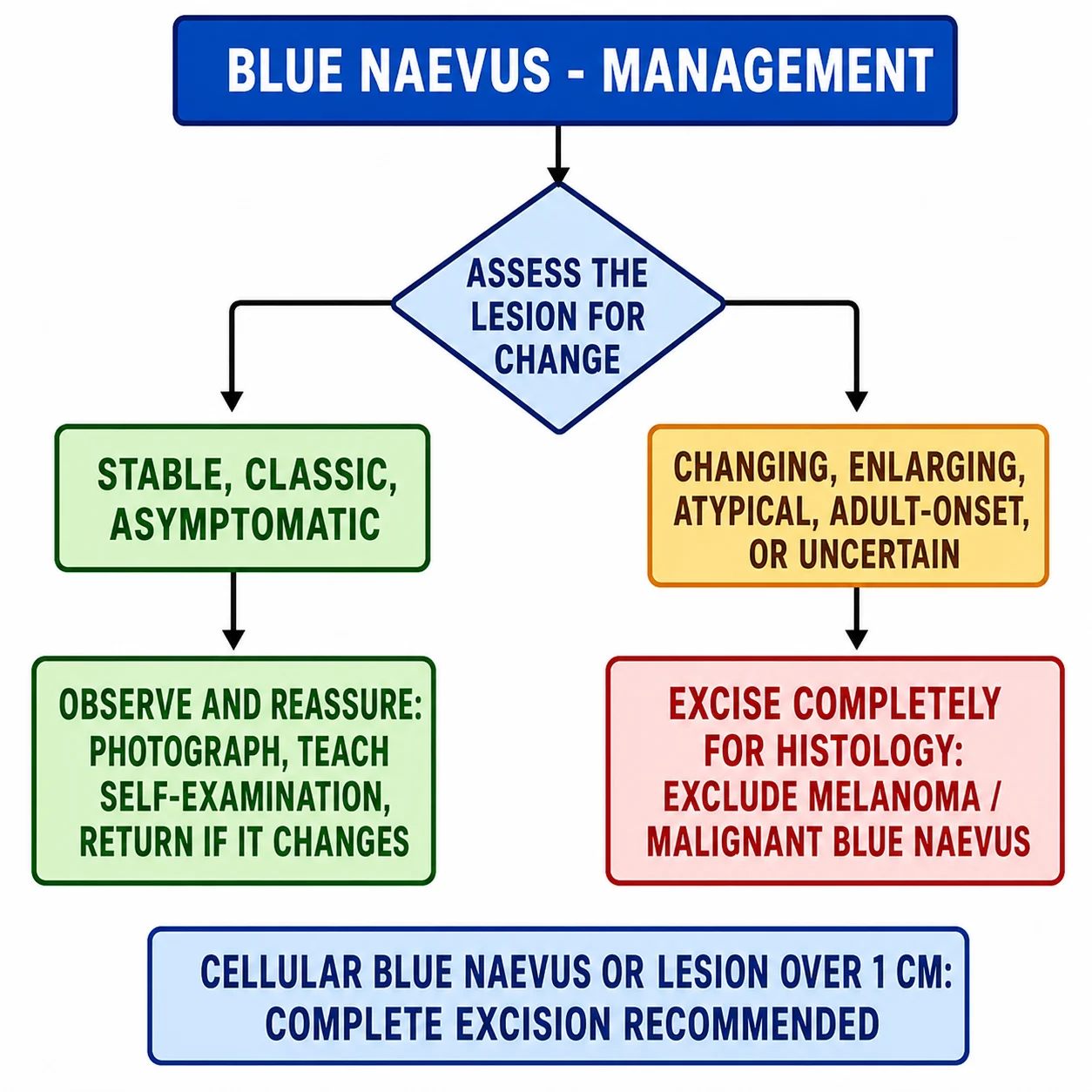
Blue naevus is not a time-critical emergency, but every blue lesion must be put through a diagnostic safety step: exclude melanoma before reassuring. This means a focused history for change, a dermoscopic examination for atypical features, and a low threshold for biopsy. A new, rapidly enlarging blue-black nodule in any age group, or any blue lesion that bleeds or ulcerates, is treated as possible melanoma and undergoes prompt complete excisional biopsy.[1][2]
The corollary, equally important, is that multiple new blue lesions in an adult should prompt a search for the syndrome or the occult melanoma, not blanket reassurance — multiple blue naevi raise Carney complex, and a blue-naevus-like lesion may on rare occasion be a melanoma mimicking a blue naevus.[6]
Management — definitive and stepwise
The definitive management follows the lesion's behaviour and subtype, not its colour: [1]
Step 1 — the stable classic common blue naevus: observe and reassure. Document the lesion with a photograph and dermoscopy, explain the benign natural history, teach the patient self-examination, and advise them to return if it changes. No treatment is required unless the patient requests removal for cosmetic reasons. This is the correct answer to the common examination stem of a stable blue papule on the hand of a young adult.[1]
Step 2 — cosmetic or definitive removal when desired: complete surgical excision. Excision is both the definitive treatment and a source of tissue for histology, and is the preferred option whenever removal is contemplated. Q-switched lasers (ruby 694 nm, alexandrite 755 nm, Nd:YAG 1064 nm) can lighten the dermal pigment of naevus of Ota and dermal melanocytoses over multiple sessions, but are less reliable for nodular blue naevi and crucially provide no tissue diagnosis — a laser must never be applied to a lesion not first confirmed benign.[8]
Step 3 — the cellular blue naevus and any atypical or uncertain lesion: complete surgical excision. Complete excision with a margin of normal skin is recommended for the cellular blue naevus and for any lesion with atypical features, change, or diagnostic uncertainty. This is both diagnostic (the whole lesion is available for histology, which is essential to separate atypical cellular blue naevus from melanoma) and therapeutic. Submit the specimen in full.[1][3]
Step 4 — malignant blue naevus: treat as melanoma. Malignant blue naevus is managed within the melanoma pathway: wide local excision with margins guided by Breslow thickness, sentinel lymph node biopsy where appropriate to thickness, staging imaging, and multidisciplinary-team management, exactly as for a cutaneous melanoma of equivalent thickness.[2]
Dermal melanocytoses. The Mongolian spot requires only reassurance — it fades. The naevus of Ota is permanent: Q-switched laser (ruby, alexandrite or Nd:YAG) is the treatment of choice for the cutaneous pigmentation, typically over multiple sessions with variable but often substantial lightening, and the patient requires lifelong annual ophthalmology review for glaucoma (intraocular pressure measurement) and uveal/ocular melanoma (dilated fundus examination).[7][8]
Self-test: a 9-year-old has a 3 mm blue-black papule on the back of the hand, unchanged for three years. What next, and what would change your plan?
The lesion is a classic common blue naevus — stable, small, in a typical site, with (one assumes) a homogeneous structureless blue dermoscopic pattern. The correct management is reassurance, photographic documentation and self-examination advice; no biopsy is needed. The plan changes — to complete excisional biopsy — the moment the lesion shows any change (growth, colour, shape, elevation), any symptom (itch, pain, bleeding), any atypical dermoscopic feature, or any diagnostic uncertainty. The cardinal rule, repeated for emphasis: never reassure a blue lesion that is changing.[1][2]
SAQ — A changing blue lesion on the scalp (10 marks, 10 minutes)
10 minutes · 10 marks
“”
Specific subtypes and scenarios
Cellular blue naevus
The cellular blue naevus deserves individual attention because it is the variant with malignant potential. It is larger (1 to 3 cm), deeper, and often congenital, occupies the buttock, sacrum and scalp, and shows the biphasic histology described above. Its management is complete surgical excision because (a) the whole lesion must be examined histologically to exclude atypia, and (b) it carries a small risk of malignant blue naevus. Recurrence after incomplete excision is well described, and the recurrence may be histologically atypical — a further reason to excise completely at the first operation.[1][3]
Combined blue naevus and deep penetrating naevus
A combined blue naevus is the coexistence of a blue naevus and a conventional acquired melanocytic naevus in one lesion. Clinically and dermoscopically it can mimic melanoma (a blue area within a brown naevus sets off alarm bells), and the diagnosis rests on histology showing the two distinct melanocyte populations. The deep penetrating naevus is a related lesion with a wedge-shaped deep extension and a plexiform growth pattern, again on the benign side of a spectrum that includes melanoma; complete excision is the safe course when the diagnosis is not straightforward.[3]
Epithelioid (animal-type) blue naevus and malignant blue naevus
The epithelioid (animal-type) blue naevus is built from heavily pigmented epithelioid melanocytes and sits on a histological spectrum with pigmented epithelioid melanocytoma; it is one of the most diagnostically difficult melanocytic lesions and demands expert pathology. Malignant blue naevus is a melanoma arising in a long-standing blue or cellular blue naevus (or de novo), presenting as change in a known blue lesion and defined histologically by atypia, mitoses, necrosis and infiltration. It is managed as melanoma of equivalent thickness.[2]
Naevus of Ota, naevus of Ito and Mongolian spot
Naevus of Ota is permanent and carries the ocular risk that defines its follow-up: roughly 10 percent of patients develop glaucoma (raised intraocular pressure, which can present at any age and may be years after the skin lesion), and a smaller but significant minority develop uveal/ocular melanoma. Both mandate lifelong annual ophthalmology review — tonometry for glaucoma and dilated funduscopy for melanoma. The cutaneous pigmentation is treated with Q-switched laser for cosmetic reasons.[7][8] Naevus of Ito is the same lesion in the shoulder-girdle distribution and carries no ocular risk. The Mongolian spot fades by age two to four and needs only reassurance, with the safeguarding caveat that an atypical or ectopic persistent patch should be reconsidered.
Carney complex (NAME / LAMB syndrome)
Multiple blue naevi are the cutaneous clue to Carney complex, an autosomal-dominant multiple-neoplasia syndrome (PRKAR1A). Its eponyms encode the features: NAME (Naevi, Atrial myxoma, Myxoid tumours, Ephelides) and LAMB (Lentigines, Atrial myxoma, Mucocutaneous myxomas, Blue naevi). The life-threatening component is the cardiac myxoma, so a patient with multiple blue naevi must be screened with an echocardiogram, and the endocrine overactivity — most characteristically Cushing syndrome from primary pigmented nodular adrenocortical disease — demands a targeted endocrine workup. Recognising the cutaneous sign can prevent sudden death from an atrial myxoma.[6]
LAMB
Complications and pitfalls
Disease-related
The common blue naevus has effectively no complications: it is benign, stable, and does not transform. The cellular blue naevus carries the small risk of malignant transformation into malignant blue naevus. Naevus of Ota causes glaucoma and uveal/ocular melanoma, the principal ocular complications, and significant cosmetic and psychological distress from the facial pigmentation. Carney complex, signalled by multiple blue naevi, brings the sudden-death risk of atrial myxoma and the morbidity of endocrine overactivity.[6][7]
Treatment-related
Surgical excision carries the usual minor risks of scarring, dyspigmentation, infection and bleeding, and on the face these matter cosmetically. Incomplete excision of a cellular blue naevus may recur, and the recurrence can be histologically atypical, complicating later diagnosis. Q-switched laser treatment of dermal melanocytoses can produce post-inflammatory hyper- or hypopigmentation and gives variable, often only partial, lightening — it must never be used on a lesion not first confirmed benign.[8]
Classic pitfalls
Five pitfalls account for most examination and clinical errors in this topic: [1]
- The melanoma pitfall — reassuring a changing blue lesion. Any change reopens melanoma; biopsy.
- The amelanotic pitfall — missing a blue naevus that lacks the blue colour; the diagnosis is histological.
- The laser-without-histology pitfall — lasering a lesion that turns out to be melanoma; never laser an unbiopsied pigmented lesion.
- The Ota pitfall — failing to arrange ophthalmology review for glaucoma and ocular melanoma in naevus of Ota.
- The multiple-blue-naevi pitfall — missing Carney complex and not screening for an atrial myxoma.[1][6]
Prognosis and disposition
The common blue naevus has an excellent prognosis: it is benign, persists unchanged for life, does not regress spontaneously, and has effectively no malignant potential. The cellular blue naevus is benign in the great majority but carries a small, recognised risk of malignant blue naevus, which is why it is excised and followed. Malignant blue naevus has the prognosis of a melanoma of equivalent thickness, governed by Breslow thickness, ulceration and nodal status, and is generally guarded. Naevus of Ota requires lifelong ophthalmology surveillance; the Mongolian spot fades with an excellent prognosis.[1][7]
Disposition follows the lesion. A typical common blue naevus is managed in primary care or general dermatology with reassurance. A cellular blue naevus, any changing or atypical lesion, naevus of Ota, or multiple blue naevi should be referred to a dermatology or specialist pigmented-lesion service. The safety-net for every patient is the same advice: return if the lesion changes. [1]
Special populations
Children and neonates. The Mongolian spot is a normal variant in Asian and Black infants and fades by age two to four; parents need reassurance and the lesion should be documented. A congenital cellular blue naevus of the scalp or buttock merits excision. A congenital giant cellular blue naevus in a bathing-suit distribution (scalp, posterior neck, spine) demands evaluation for neurocutaneous melanosis with an MRI of the brain and spine, because melanin-producing cells in the leptomeninges can cause hydrocephalus, seizures and a high risk of leptomeningeal melanoma.[3]
Pregnancy. Blue naevi and dermal melanocytoses (including naevus of Ota) may darken under hormonal influence during pregnancy. The approach is reassurance and dermoscopy; excision is deferred to the postpartum period unless the lesion is changing, in which case it is biopsied on its own merits. [1]
Darker phototypes (Fitzpatrick IV–VI). Blue naevi and dermal melanocytoses are more common and more visually striking, and the cosmetic impact — especially of facial naevus of Ota — is greater. Laser results are variable and the risk of post-inflammatory dyspigmentation higher, so expectations must be carefully set.[8]
Naevus of Ota (all ages). Lifelong annual ophthalmology review (tonometry for glaucoma, dilated funduscopy for uveal melanoma) is the single most important follow-up; laser is offered for cutaneous pigment.[7]
Multiple blue naevi. Screen for Carney complex — echocardiogram for atrial myxoma and an endocrine workup (Cushing evaluation) — and offer genetic counselling if confirmed.[6]
Immunocompromised. No specific increase in blue naevi, but any new or changing pigmented lesion must be examined by dermoscopy to exclude melanoma, the diagnosis of which is delayed in this group. Anticoagulated patients. Blue naevi do not bleed spontaneously, but any decision to excise an atypical blue lesion in a patient on warfarin or a direct oral anticoagulant must balance the small risk of a delayed melanoma diagnosis against perioperative bleeding. Excision should not be deferred for anticoagulation: minor cutaneous surgery is routinely performed without interrupting warfarin provided the INR is in range, and direct oral anticoagulants can be managed per local protocol. The principle holds — a changing blue lesion is excised on its merits; the anticoagulation is managed around it, not used as a reason to observe a worrying lesion. [1]
Evidence, guidelines, and regional differences
The conceptual framework for the blue naevus family — the classical types, their histology and their differential diagnosis — was laid down in the 1994 differential-diagnostic review of González-Cámpora and colleagues, which remains a useful single reference for the histological separation of common, cellular and related entities.[3] The 2016 concise review by Sugianto, Ralston and Metcalf is the best modern single overview of common, cellular and malignant blue naevus, and the 2017 comprehensive review by Borgenvik and colleagues of blue-naevus-like and blue-naevus-associated melanoma underpins the malignant-transformation and biopsy rules.[1][2] The dermoscopic characterisation of blue naevi, including the structureless homogeneous blue pattern and the rare polarised-light rosettes, is captured in recent dermoscopy literature.[4] The naevus of Ota literature defines the glaucoma and ocular-melanoma risk and the role of Q-switched lasers.[7][8]
The guideline deltas across regions are modest, because the biology is the same everywhere — what varies is the threshold for biopsy and the intensity of screening. [1]
[1] [1]In Australia and New Zealand (RANZCD / Cancer Council), the emphasis is on melanoma exclusion given the very high background burden of melanoma; dermoscopy is universal in primary care, and a low threshold for excision of any changing blue lesion is the norm. Naevus of Ota follow-up is through ophthalmology.
Universally: a stable, classic, asymptomatic common blue naevus is observed; any changing, atypical, cellular, or uncertain blue lesion is excised for histology; and naevus of Ota carries a lifelong ophthalmology commitment for glaucoma and ocular melanoma. The cardinal biopsy rule does not vary by region.
Exam pearls
[1]Exam application bank (NEET-PG / INICET)
One-line answer
A blue naevus is a benign melanocytic lesion built from heavily pigmented, dendritic and spindled DERMAL melanocytes in the reticular dermis, whose blue colour is an optical effect (Tyndall scattering) of the deep pigment location rather than blue pigment. The common blue naevus is a small, stable, blue-black papule of the extremities; the cellular blue naevus is a larger, deeper lesion of the buttock, sacrum or scalp with a small malignant potential; and dermal melanocytoses (Mongolian spot, naevus of Ota, naevus of Ito) are persistent ectopic dermal melanocytes distributed by site. Stable classic lesions are observed; any changing or atypical blue lesion is biopsied or excised to exclude melanoma.
Worked stems (answer without another resource)
Stem 1 — Classic presentation. Map symptoms to mechanism; name the first investigation and first treatment step with dose/route if drug therapy is standard. [1]
Stem 2 — Unstable / complicated. List red flags that force immediate resuscitation, theatre, ICU, antidote, or reperfusion — and what you do in the first 15 minutes. [1]
Stem 3 — Atypical group. Elderly, pregnancy, child, or immunocompromised: how presentation and thresholds change. [1]
Stem 4 — Differential trap. Name the three closest mimics and one discriminator for each. [1]
Stem 5 — Disposition. Who goes home with safety-netting, who is admitted, who needs HDU/ICU/theatre, and what follow-up is mandatory. [1]
Rapid viva checklist
- Definition + classification
- Pathophysiology chain
- Bedside signs / criteria
- Score with exact components (if any)
- Emergency bundle
- Definitive therapy with doses
- Complications of disease and of treatment
- Special populations
- Guideline/trial name if classic
- Three exam traps
Coverage self-check
If you cannot answer any stem above from this page alone, re-read the matching section — the page is intended to be self-sufficient for final-prof and NEET-PG/INICET questions on Blue naevus and dermal melanocytosis.
In summary
Blue naevus is a topic that rewards the candidate who holds two ideas at once: the blue colour is an optical consequence of deep dermal melanin (Tyndall effect), and colour alone never confirms biology. A stable, classic, asymptomatic common blue naevus is observed and the patient reassured; a cellular blue naevus, any changing or atypical lesion, and any diagnostic uncertainty are excised completely for histology; and the systemic and ocular syndromes — Carney complex signalled by multiple blue naevi, and naevus of Ota's lifelong glaucoma and ocular-melanoma risk — are the conditions that earn the topic its weight. Hold the dermoscopic pattern (a structureless homogeneous blue field with no melanoma features), the cardinal biopsy rule (never reassure a changing blue lesion), and the Carney-complex and naevus-of-Ota follow-up commitments, and the topic is yours.[1][6][7]
References
- [1]Sugianto JZ, Ralston JS, Metcalf JS Blue nevus and malignant blue nevus: A concise review Semin Diagn Pathol, 2016.PMID 27199078
- [2]Borgenvik TL, Karlsvik TM, Ray S Blue nevus-like and blue nevus-associated melanoma: a comprehensive review of the literature ANZ J Surg, 2017.PMID 28318130
- [3]González-Cámpora R, Galera-Davidson H, Vázquez-Ramírez FJ Blue nevus: classical types and new related entities. A differential diagnostic review Pathol Res Pract, 1994.PMID 7984522
- [4]Salas-Callo CI, Riera-Monroig J, Podlipnik S Blue Nevus With Rosettes on Polarized Light Dermoscopy Dermatol Pract Concept, 2020.PMID 31921504
- [5]Satolli F, Gandolfi M, Rovesti M Blue nevus of the nail: A case report and review Dermatol Ther, 2020.PMID 32500667
- [6]Bertherat J Carney complex (CNC) Orphanet J Rare Dis, 2006.PMID 16756677
- [7]Williams NM, Gurnani P, Labib A Melanoma in the setting of nevus of Ota: a review for dermatologists Int J Dermatol, 2021.PMID 32808287
- [8]Shah VV, Bray FN, Aldahan AS Lasers and nevus of Ota: a comprehensive review Lasers Med Sci, 2016.PMID 26563954