Dermatology · General Medicine
Burns & Thermal Injury
Also known as Burns · Thermal injury · Burn wound · Scalds · Electrical burns · Chemical burns
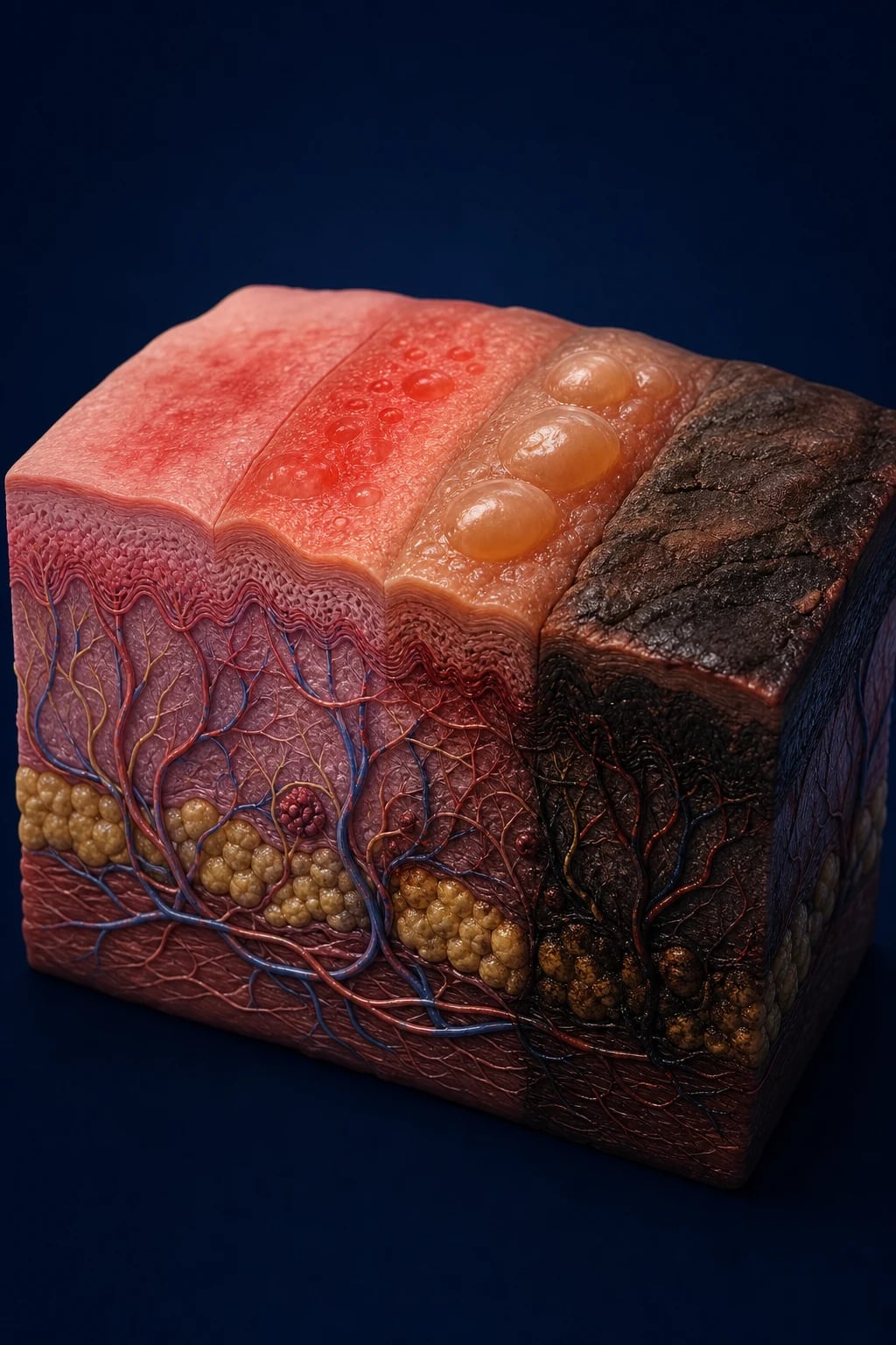
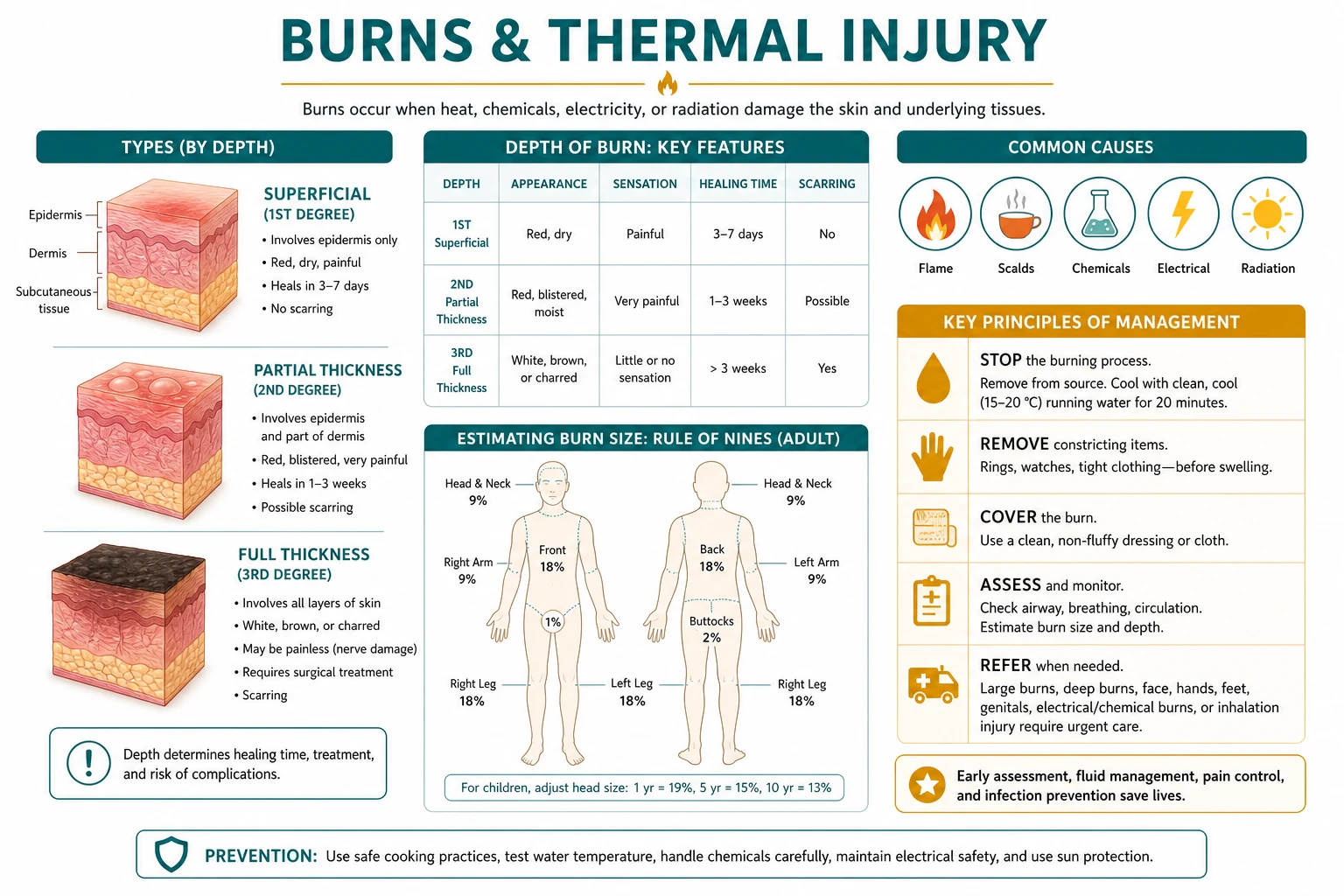
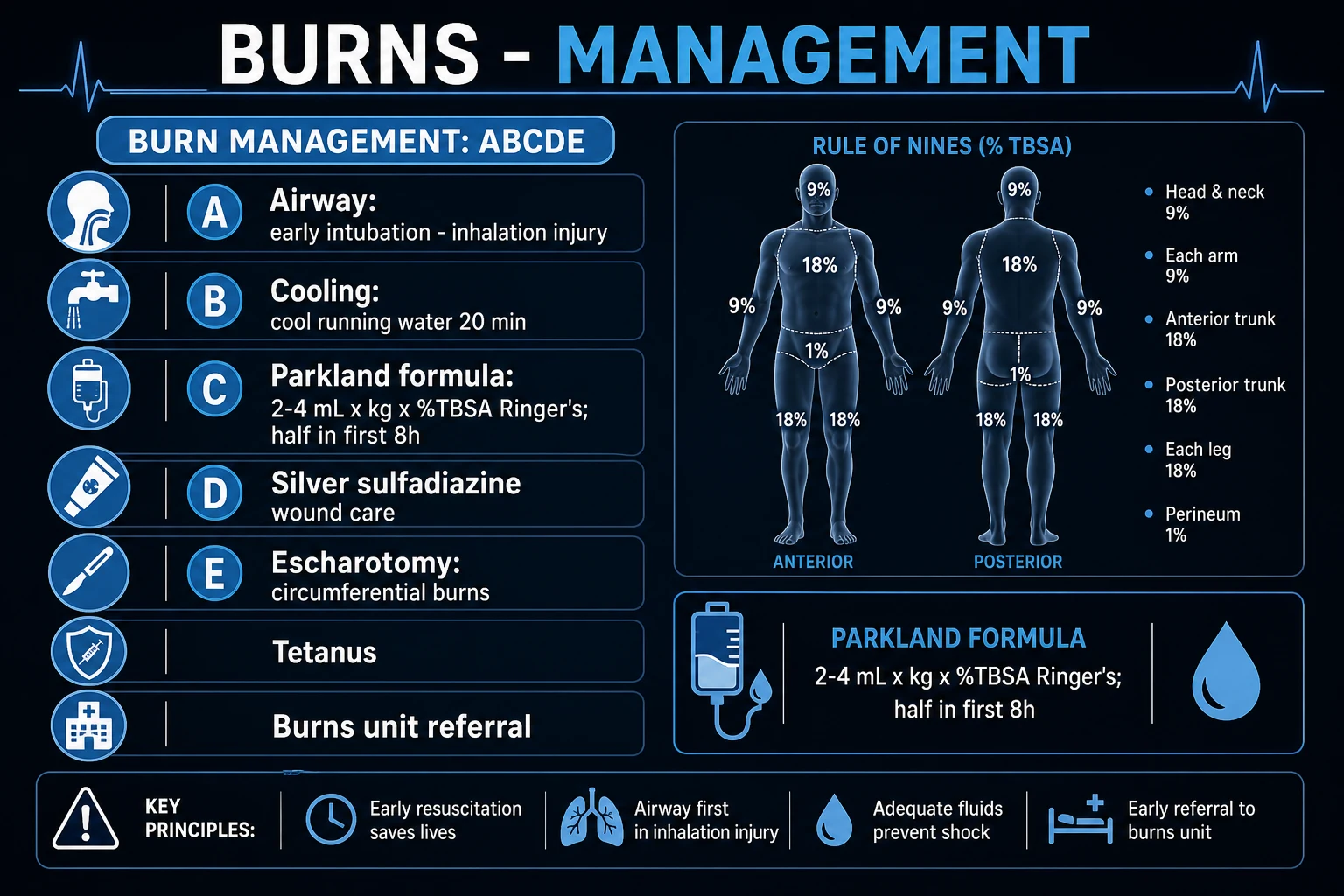
Burns are skin and deeper-tissue injuries caused by thermal, chemical, electrical or radiation energy, classified by depth (first-degree / epidermal — erythema, pain, no blisters; superficial second-degree / superficial partial-thickness — blisters, moist, painful, blanches; deep second-degree / deep dermal — dry, pale, slow capillary refill; third-degree / full-thickness — dry, leathery, insensate, no blanching; fourth-degree — muscle, tendon, bone) and by total body surface area (TBSA) using the Rule of Nines (adults), the Lund-Browder chart (children) or the palm method (patient palm equals 1 percent TBSA). Major burn criteria: over 10 percent TBSA in adults, over 5 percent in children or elderly, any full-thickness burn over 5 percent, burns to face, hands, feet, genitals, perineum or major joints, inhalation injury, electrical or chemical burns, or burns with comorbidity. Management is ABCDE with early airway control, cooling with running water for 20 minutes within 3 hours, Parkland formula resuscitation (4 mL Ringer lactate x kg x percent TBSA, half in first 8 hours), wound care (silver sulfadiazine, hydrocolloids, biological membranes), escharotomy for circumferential full-thickness burns, tetanus prophylaxis, early enteral nutrition and referral to a burns unit.
On this page & tools
Your progress
Saved locally on this device.
Exam tags
Red flags
Overview & Definition
A burn is coagulative destruction of the skin and, in deeper injuries, of subcutaneous fat, fascia, muscle and bone, produced by the transfer of thermal, chemical, electrical or radiant energy at a rate that exceeds the tissue's capacity to dissipate it. The dermatological and plastics perspective is what distinguishes the modern management of burns from general resuscitation: the wound itself is the disease, and every decision — depth, TBSA, dressing choice, timing of excision, grafting and rehabilitation — is taken to minimise scarring, preserve function in the hands, face and over joints, and prevent the late disfigurement that is the principal long-term burden of a burn survivor.[2][9]
Burns are the most devastating yet most preventable of traumas. The World Health Organization estimates that burns cause over 180,000 deaths each year, the overwhelming majority in low- and middle-income countries where open-flame cooking, kerosene heaters, unsafe electrical wiring and lack of smoke alarms converge. The skill of assessing a burn rests on three pillars: accurate assessment of depth and surface area, protection of the airway (because inhalation injury kills by oedema and obstruction — intubate early, before the airway is lost), and fluid resuscitation guided by the Parkland formula and titrated to urine output (under-resuscitation causes burn shock, over-resuscitation causes oedema, compartment syndrome and abdominal compartment syndrome). Circumferential full-thickness burns of a limb or the chest demand escharotomy. Electrical burns cause deep tissue injury far disproportionate to the small visible entry wound — they need cardiac monitoring and a search for myoglobinuria. Chemical burns demand copious irrigation; hydrofluoric acid in particular requires calcium gluconate gel.[1][2]
Definition & Classification
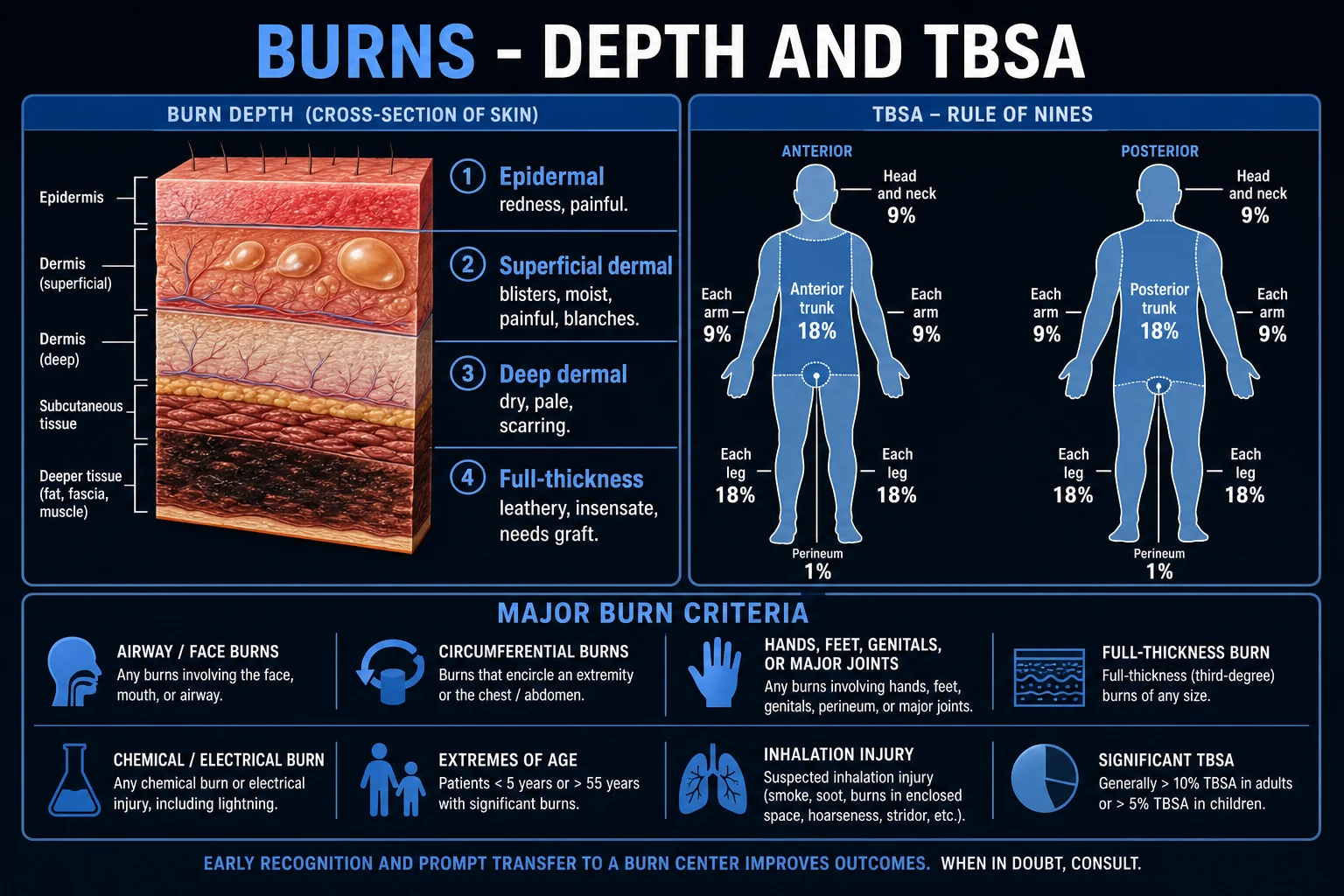
Burn classification has two independent axes — depth and total body surface area (TBSA) — and every burn must be described on both, because depth governs healing potential and the need for surgery while TBSA governs the systemic (fluid) response. First-degree (epidermal) burns are excluded from the TBSA calculation because they do not produce the capillary leak that drives burn shock. [1]
Depth classification
The depth of a burn is the single most important wound-level decision the dermatologist or plastic surgeon makes, because it predicts whether the wound will heal spontaneously from epithelial appendages within two to three weeks (no scar), heal slowly with scarring (may need graft), or require excision and grafting (will not re-epithelialise). The clinical signs that discriminate depth are blanching, capillary refill, pinprick sensation, moisture and the appearance of the wound surface.[2][6]
The dermatological pearl is that depth can evolve over the first 48 to 72 hours: a burn that initially looks superficial dermal can deepen into a deep dermal or full-thickness burn if the zone of stasis (see Pathophysiology) is lost to under-resuscitation, infection or desiccation. This is why serial re-assessment of depth at 24 and 48 hours, and the use of ** moist occlusive dressings** or biological membranes to keep the wound environment optimised, is standard plastics practice.[2][9]
Surface area classification — Rule of Nines, Lund-Browder, palm method
Only count partial-thickness (2nd degree) and deeper burns in the TBSA. Superficial epidermal (1st degree) erythema is excluded. A common MCQ trap is to include sunburn-like erythema in the Parkland calculation — it must not be.[5][6]
[1]The Lund-Browder chart corrects the Rule of Nines for children, whose head is proportionally much larger (up to 19 percent in an infant) and whose legs are smaller. Using the adult Rule of Nines in a young child underestimates the head burn and overestimates the leg burn, leading to under-resuscitation of the most dangerous region (the head) — a classic and dangerous error.[4]
Epidemiology & Risk Factors
Burns are overwhelmingly a disease of poverty and domesticity. The systematic review by Smolle and colleagues found that, globally, the highest incidence and mortality are in the WHO South-East Asian and African regions, with flame burns dominating in adults and scalds (hot liquids) predominating in children under five. Risk is concentrated in the home kitchen, around open fires, kerosene stoves and hot bathwater; occupational electrical and chemical burns cluster in construction, electrical and chemical industries.[1]
In children, a non-accidental injury must be suspected whenever the history is inconsistent with the injury, presentation is delayed, the burn has a stocking-glove or doughnut (central sparing) distribution suggestive of forced immersion, or there are associated signs of neglect. A dermatologist asked to assess a burned child is also safeguarding that child.[4]
Pathophysiology
The Jackson burn model — three concentric zones
The local response to a burn is described by the Jackson zones, three concentric zones visible within and around the burn wound at the moment of injury. Understanding this model is what separates burns surgery from simple wound care, because the central zone is dead but the middle zone is salvageable — and good resuscitation, dressings and infection control are precisely the measures that rescue it.[2]
The therapeutic implication is enormous: the zone of stasis is the tissue the resuscitation is fought for. Under-resuscitation, desiccation, infection or a continued inflammatory cascade converts the zone of stasis into the zone of coagulation — the burn deepens, more tissue is lost, and the eventual scar and graft burden are worse. Every action in the first 48 hours is in part an attempt to save the zone of stasis. [1]
Systemic pathophysiology — capillary leak, burn shock and the hypermetabolic state
A burn that exceeds roughly 20 percent TBSA in adults (10 percent in children) produces a generalised increase in capillary permeability throughout the body — not just at the burn site. Inflammatory mediators (histamine, bradykinin, serotonin, prostaglandins, leukotrienes, thromboxane, cytokines such as interleukin-1, interleukin-6 and tumour necrosis factor-alpha) are released and circulate. The result is a massive transcapillary leak of fluid, electrolytes and protein into the interstitium, producing the oedema of burn shock and a dramatic fall in intravascular volume, cardiac output and urine output within the first 8 to 12 hours.[2][3]
This is the rationale for the Parkland formula: crystalloid given in a calculated volume in the first 24 hours to replace the leak. After 24 hours the capillary leak partly resolves and protein begins to be needed (colloid). If the burn is not adequately resuscitated, the patient develops hypovolaemic (burn) shock with progressive multi-organ dysfunction. If over-resuscitated, fluid tracks into the soft tissues and may precipitate compartment syndrome of a limb or abdominal compartment syndrome — the syndrome called fluid creep.[3][5]
After the acute phase a profound hypermetabolic response sets in: resting energy expenditure can rise to 1.5 to 2 times baseline in burns over 40 percent TBSA, driven by catecholamine, cortisol and glucagon release, with fever, tachycardia, muscle catabolism, immune suppression and impaired wound healing. This is why early enteral nutrition (begun within 24 to 48 hours) is a treatment, not a comfort measure: it blunts the catabolic drive and protects the gut mucosa, reducing bacterial translocation and Curling's ulcer risk.[2]
Clinical Presentation
The clinical presentation of a burn is dominated by three questions answered at the bedside: how deep, how much, and where. [1]
Depth signs. A superficial dermal burn is red, moist, blistering and exquisitely painful, with brisk blanching and refill (capillaries are alive). A deep dermal burn is drier, paler or mottled, with sluggish capillary refill and reduced pinprick sensation. A full-thickness burn is dry, leathery and insensate (the nerves are destroyed), often white, grey or charred, with no blanching. A painless burn is a deep burn — one of the most important clinical rules in the specialty.[6][9]
Surface area. TBSA is estimated by the Rule of Nines, Lund-Browder chart or palm method. Always exclude first-degree erythema from the calculation. [1]
Special sites. Burns of the face (inhalation injury, eye injury, cosmetic), hands (function — position of safety), feet (mobility), perineum and genitals (infection risk, micturition), and over major joints (contracture) each carry specific threats and lower the threshold for burns-unit referral and surgical management. [1]
Inhalation injury presents with soot in the mouth or sputum, singed nasal and facial hairs, hoarseness, stridor, a history of exposure in an enclosed space, and altered consciousness from carbon monoxide poisoning. Hoarseness or stridor is a pre-emergency warning — the airway will close as oedema worsens, so the patient must be intubated early, before the airway is lost.[11]
Differential Diagnosis
Not every blistering or desquamating skin eruption is a burn. The dermatologist must distinguish a true thermal burn from its mimics, several of which are life-threatening in their own right. [1]
The key bedside discriminator is the history: a thermal burn has a clear heat-source history, whereas TEN/SJS has a drug exposure, SSSS an infectious prodrome in a child, and phytophotodermatitis plant-plus-sun exposure. The distribution also differs: thermal burns follow a splash, scald or flame pattern, whereas TEN is symmetrical and mucosal.[2]
Clinical & Bedside Assessment
The ABCDE primary survey is performed before any burn assessment, because an obstructed airway or a tension pneumothorax from associated trauma will kill the patient before the burn does. [1]
The focused burn assessment
- History — mechanism (flame, scald, electrical, chemical), time of burn (for the Parkland clock), enclosed space (inhalation), first aid already given, tetanus status, comorbidities, drugs, and — for children — the safeguarding question of whether the history fits the injury.
- Airway and breathing — signs of inhalation injury (above); assess oxygenation.
- Depth assessment — examine for blanching (press with a glass slide or finger), capillary refill, pinprick sensation, moisture and appearance. Document depth on a body chart.
- TBSA — Rule of Nines or Lund-Browder; confirm with the palm method for small or patchy burns.
- Special sites — face and eyes (fluorescein staining for corneal burns), ears (cartilage — mafenide acetate), hands (neurovascular, position of safety), perineum, joints.
- Circumferential burns — look for and document any circumferential full-thickness burn of a limb or the chest; check distal perfusion and chest expansion.
- Neurovascular distal to the burn — pulses, capillary refill, sensation — to detect early compartment syndrome.[6][9]
The burn assessment sequence — 'DABS'
Investigations
Burns are a clinical diagnosis; investigations serve the resuscitation, the search for complications and the assessment of inhalation and electrical injury rather than the diagnosis itself. [1]
Inhalation injury is diagnosed clinically and confirmed bronchoscopically: the finding of carbonaceous material, oedema, erythema or mucosal necrosis in the airway is diagnostic.[11] Carbon monoxide poisoning is detected by an elevated carboxyhaemoglobin and treated with 100 percent oxygen (hyperbaric oxygen is considered in selected severe cases, though evidence is debated).
Management — Resuscitation
First aid and pre-hospital care
Pre-hospital and emergency department first aid for a burn
STOP the burning process — remove from source, extinguish flames, remove hot/wet clothing and jewellery (before oedema makes this impossible), but do NOT peel off adherent clothing
COOL with running water at 15 to 25 degrees Celsius for 20 minutes — effective if started within 3 hours of the burn. Do NOT use ice (causes vasoconstriction that deepens the zone of stasis and risks hypothermia)
COVER with cling film (does not stick, allows inspection) or a clean dry dressing; keep the patient warm to prevent hypothermia (especially children)
CALL for help; transport to hospital; major burns to a burns unit per referral criteria
ABC — assess and secure airway (early intubation for inhalation injury); oxygen; IV access; begin Parkland resuscitation for major burns
Cooling with running water for 20 minutes, ideally begun within three hours of the burn, reduces pain, limits depth progression and improves outcome. Ice must not be used — it causes vasoconstriction that worsens the zone of stasis and can produce frostbite on top of any thermal injury, and it risks systemic hypothermia, especially in children.[2]
Airway and breathing
In any suspected inhalation injury, secure the airway early by endotracheal intubation before oedema makes it impossible. Indications include hoarseness, stridor, significant facial burns with soot or singed hairs, and altered consciousness. Give 100 percent oxygen via a non-rebreather mask pending intubation; this treats carbon monoxide poisoning (CO half-life falls from around 320 minutes on room air to around 80 minutes on 100 percent oxygen). Arrange bronchoscopy to grade inhalation injury.[11]
Fluid resuscitation — the Parkland (Baxter) formula
For major burns (over 10 percent TBSA in adults, over 5 percent in children), calculate the fluid requirement for the first 24 hours with the Parkland formula.[3][5]
[1]Children require maintenance fluid (containing glucose) in addition to the calculated resuscitation volume, because their glycogen stores are limited. From 24 hours onwards, capillary leak has largely resolved and colloid (albumin) is introduced, with the rate reduced; oral/enteral intake is encouraged as the patient tolerates.[3][4]
- Parkland (Baxter) — 4 mL/kg/percent TBSA, crystalloid: the most widely taught and used formula globally (US ABA, India ICMR/NABI, much of Europe). It tends to deliver a larger crystalloid volume.
- Modified Brooke — 2 mL/kg/percent TBSA, crystalloid: developed to counter 'fluid creep'; favoured by many military and ANZBA-influenced units. Delivers less crystalloid, lowering oedema and compartment-syndrome risk.
- Rule: whatever the formula, resuscitation is titrated to urine output (0.5 mL/kg/h adult, 1 mL/kg/h child) and clinical end-points. The formula is only the starting point.[3][5]
Analgesia, tetanus and the escharotomy
Analgesia: burns are extremely painful. Give intravenous morphine 0.1 mg/kg (titrated, repeated as needed) or fentanyl; paracetamol as baseline. Never give intramuscular analgesia in a major burn (absorption is unreliable in shock). Cover the wound to reduce air-current pain. [1]
Tetanus prophylaxis: all burns are tetanus-prone wounds. Check immunisation status and give tetanus toxoid (and immunoglobulin if the wound is contaminated and the patient is not immune) per local protocol. [1]
Escharotomy: a circumferential full-thickness burn of a limb or the chest forms an inelastic eschar. As oedema accumulates beneath it, distal perfusion is compromised (limb) or chest expansion is restricted (thorax). A surgical escharotomy — incisions through the eschar down to subcutaneous fat along the lateral and medial aspects of the limb or the anterior chest — relieves the pressure. It is a bedside procedure performed under sedation; the incision should be bloodless (full-thickness burn is insensate). Indications include a cool, pulseless or painful distal limb, falling pulse-oximetry, paraesthesia, and reduced chest expansion. A fasciotomy (deeper, through fascia) is needed for electrical burns and deep thermal injury with muscle involvement.[2][9]
Management — Definitive & Stepwise
Minor (outpatient) burns
Minor burns — small partial-thickness burns under 10 percent TBSA in adults (under 5 percent in children), not involving special sites, not circumferential — are managed as outpatients.[6]
Outpatient management of a minor burn
Cool, then clean with saline or chlorhexidine
Debride ruptured blisters and loose skin (leave intact blisters over 24 h unless on a joint or palm where they interfere with function)
Apply a topical antimicrobial: SILVER SULFADIAZINE 1 percent cream once or twice daily (the standard); MAFENIDE ACETATE 10 percent for ear/cartilage burns (penetrates cartilage, no silver staining)
Cover with a non-adherent dressing and a hydrocolloid or foam secondary layer; or a biological membrane (Biobrane, Suprathel) for superficial partial-thickness burns
Analgesia (paracetamol +/- codeine), tetanus prophylaxis, review at 24-48 h and weekly until healed
Advise sun protection and scar/massage once healed; refer if not healed by 2 weeks (likely deep dermal)
Dressing choice depends on depth and exudate. Hydrocolloids (occlusive, autolytic) suit clean superficial partial-thickness burns. Silver sulfadiazine 1 percent remains the topical antimicrobial standard — broad-spectrum (gram-positive including some MRSA, gram-negative including Pseudomonas, and some fungal), painless on application, but it is sulfa-based (contraindicated in sulfa allergy and pregnancy near term) and causes a transient leucopenia in roughly 5 to 15 percent of patients. Mafenide acetate 10 percent penetrates cartilage and eschar and is preferred for ear burns (to prevent suppurative chondritis) but it is painful on application and can cause a metabolic acidosis (carbonic anhydrase inhibition). Biological and biosynthetic membranes (Biobrane — a nylon-collagen-silicone bilayer; Suprathel — a copolymer) act as a temporary skin substitute for clean superficial partial-thickness burns, reducing pain, accelerating re-epithelialisation and allowing outpatient management.[12]
Major burns — definitive surgical management
Definitive management of a major burn is surgical and is undertaken in a burns unit. The principle of modern burns surgery is early tangential excision and split-thickness skin grafting (STSG) of deep dermal and full-thickness burns, ideally within the first 3 to 5 days, to remove the necrotic eschar (a culture medium for infection), reduce the inflammatory and hypermetabolic burden, shorten hospital stay and improve survival. Early excision changed burns mortality in the late 20th century and is now standard.[2][9]
Definitive surgical management of a major burn
Resuscitate (Parkland) and stabilise; secure airway; nutrition begun early (enteral within 24-48 h)
Tangential excision of deep dermal and full-thickness eschar (typically day 3 to 5), in stages if the TBSA is large
Cover the excised wound: split-thickness skin graft (STSG) from unburned donor sites; meshed for large areas; sheet (unmeshed) grafts for face, hands, joints (better cosmesis and function)
For very large burns: temporary cover with cadaver allograft or biological membranes to allow donor sites to re-epithelialise for re-harvest; cultured epithelial autografts (CEA) for massive burns
Positioning and splinting — the 'position of safety' for hands and joints to prevent contracture
Rehabilitation: physiotherapy, pressure garments, scar massage, silicone gel sheeting for hypertrophic scars; psychological support
Antibiotics are NOT given prophylactically in burns — they do not prevent wound infection, select resistant organisms, and are reserved for documented infection (cellulitis, bacteraemia, sepsis) guided by culture. The single most important infection-prevention measure is early excision and closure of the wound.[2]
The 'position of safety' for the burned hand
The hand is the single most important functional unit in burn rehabilitation, and a burned hand held in the wrong position for two weeks will contract into a useless claw. The position of safety (anti-deformity position) splints the hand in: wrist extended 20 to 30 degrees, metacarpophalangeal joints flexed 70 to 90 degrees, interphalangeal joints fully extended, thumb abducted and opposed. This position lengthens the collateral ligaments and the palmar skin so that, when oedema and scarring contract, they do not pull the hand into the classic contracture (wrist flexed, MCPs extended, IPs flexed, thumb adducted).[9]
Specific Subtypes & Scenarios
Electrical burns
Electrical injury is deceptively dangerous because the skin wound is small but the deep tissue injury is large — current follows the path of least resistance through nerves, blood vessels and muscle, sparing the high-resistance skin. The entry wound may be a tiny charred puncture; the exit wound may be similarly small; in between, muscle, nerve and vessel may be destroyed. Vigilance for deep injury is everything.[2]
[1]Chemical burns
Chemical burns differ from thermal burns because the injuring agent keeps damaging tissue until it is removed or neutralised. The first principle is copious, prolonged irrigation with water (most agents) for at least 20 minutes — and longer for alkalis.[8]
[1]Hydrofluoric acid is the most feared chemical burn because the fluoride ion chelates calcium and magnesium, producing deep, excruciating pain disproportionate to the visible injury, progressive tissue necrosis, life-threatening hypocalcaemia and hypomagnesaemia, and fatal ventricular arrhythmia. The specific antidote is calcium gluconate: a 10 percent gel massaged into the affected skin, and intradermal or intra-arterial calcium gluconate for severe or digital involvement.[8]
Special sites
[1]Complications & Pitfalls
[1]Curling's ulcer deserves a note: it is an acute stress ulceration of the duodenum (and sometimes stomach) occurring in the setting of severe burns (or, by analogous pathophysiology, head injury — Cushing's ulcer). Modern prophylaxis — early enteral feeding and acid suppression in major burns — has made it uncommon, but bleeding or perforation in a major burn is still Curling's until proven otherwise.[7]
Prognosis & Disposition
Prognosis in burns is governed by age, TBSA, inhalation injury and comorbidity. The classic Baux score (age + percent TBSA) gives a rough mortality estimate: a score over 100 historically implied almost certain mortality, though modern burns care has substantially improved survival beyond this. The revised Baux score adds inhalation injury (age + percent TBSA + 17 if inhalation injury present) and predicts in-hospital mortality more accurately.[10]
Disposition. Minor burns (small partial-thickness, not special sites, not circumferential) are managed as outpatients. Major burns and all special-site, electrical, chemical, inhalation and circumferential burns are referred to a burns unit.[6]
Refer to a specialist burns unit any patient with:
- Partial-thickness burn over 10 percent TBSA (adults) or over 5 percent (children/elderly)
- Any full-thickness burn over 5 percent TBSA
- Burns to special sites: face, hands, feet, genitalia, perineum, major joints
- Electrical, chemical, or inhalation injury
- Circumferential burns of any limb or the chest
- Burns at the extremes of age (under 5 or over 65), or in patients with significant comorbidity
- Burns with associated trauma or non-accidental injury (children)
Special Populations
Children. The TBSA threshold for a major burn is lower (over 5 percent). Children have a larger surface-area-to-mass ratio, so fluid needs per kg are higher and they desiccate and hypotherm faster. The Lund-Browder chart must be used (the adult Rule of Nines underestimates the head and overestimates the legs). Maintenance fluid containing glucose is added to the Parkland resuscitation. Non-accidental injury must be considered whenever the history is inconsistent, presentation is delayed, or the pattern is a stocking-glove, doughnut (central sparing, from forced immersion in hot water) or contact pattern suggesting a heated object.[4]
The elderly. Skin is thinner, so the same insult produces a deeper burn. Comorbidities (cardiac, respiratory, diabetes) limit reserve, and mortality at any given TBSA is higher. Polypharmacy (beta-blockers, anticoagulants) complicates resuscitation and surgery. [1]
Electrical injury. Cardiac monitoring, rhabdomyolysis and deep-tissue surveillance as above. [1]
Pregnancy. The fetus is vulnerable to maternal hypovolaemia and hypoxia; resuscitation must be aggressive, the patient positioned to avoid aortocaval compression, and fetal monitoring instituted for viable pregnancies. [1]
Evidence, Guidelines & Regional Differences
The Parkland (Baxter) formula (Baxter, 1968) — 4 mL/kg/percent TBSA of crystalloid, half in the first 8 hours — remains the most widely taught resuscitation formula. Its simplicity is its strength, but it has been criticised for the syndrome of fluid creep (over-resuscitation leading to oedema, compartment syndrome and abdominal compartment syndrome) seen since the 2000s. The Modified Brooke formula (2 mL/kg/percent TBSA) was developed in response, delivering less crystalloid, and many modern units use a goal-directed (urine-output-titrated) approach regardless of starting formula.[3][5]
- American Burn Association (ABA) / US: Parkland 4 mL/kg/percent TBSA crystalloid; colloid after 24 h; early excision and grafting standard; ABA referral criteria as above.
- UK / NHS / British Burns Association: similar; National Burn Care Review referral pathways; Parkland widely taught; modified Brooke increasingly used.
- ANZBA (Australia/NZ): Modified Brooke (2 mL) preferred in many units; ANZBA Bi-National Disaster Plan for mass-casualty burns.
- India (ICMR / National Academy of Burns India, NABI): Parkland formula standard teaching; mortality higher in India due to delayed presentation, open-flame cooking, and limited burns-unit access; WHO-supported prevention programmes (safe cooking, smoke alarms) emphasised.
- Controversies: colloid timing (some advocate earlier colloid), the role of high-dose vitamin C (ascorbic acid) in reducing fluid needs, and the role of hyperbaric oxygen in inhalation injury and CO poisoning — all debated with mixed evidence.
Prevention
Burns are largely preventable, and prevention is the most cost-effective intervention at a population level. WHO and national programmes target: smoke alarms and home fire escape plans; lowering hot-water thermostat settings (to under 50 degrees Celsius) to prevent scalds; child-resistant cigarette lighters; safe cooking stoves (away from children, stable, no open flame at floor level in LMIC); fireworks regulation; electrical safety in occupational and domestic settings; chemical safety training and personal protective equipment; first-aid education ('cool with running water for 20 minutes'). At the individual level, the dermatologist and plastics team contribute by participating in school and community education and in the identification and safeguarding of children with non-accidental burns.[1]
Exam Pearls
Burns — the high-yield exam one-liners ('BURNED')
Exam application bank (NEET-PG / INICET)
One-line answer
Burns are skin and deeper-tissue injuries caused by thermal, chemical, electrical or radiation energy, classified by depth (first-degree / epidermal — erythema, pain, no blisters; superficial second-degree / superficial partial-thickness — blisters, moist, painful, blanches; deep second-degree / deep dermal — dry, pale, slow capillary refill; third-degree / full-thickness — dry, leathery, insensate, no blanching; fourth-degree — muscle, tendon, bone) and by total body surface area (TBSA) using the Rule of Nines (adults), the Lund-Browder chart (children) or the palm method (patient palm equals 1 percent TBSA). Major burn criteria: over 10 percent TBSA in adults, over 5 percent in children or elderly, any full-thickness burn over 5 percent, burns to face, hands, feet, genitals, perineum or major joints, inhalation injury, electrical or chemical burns, or burns with comorbidity. Management
Worked stems (answer without another resource)
Stem 1 — Classic presentation. Map symptoms to mechanism; name the first investigation and first treatment step with dose/route if drug therapy is standard. [1]
Stem 2 — Unstable / complicated. List red flags that force immediate resuscitation, theatre, ICU, antidote, or reperfusion — and what you do in the first 15 minutes. [1]
Stem 3 — Atypical group. Elderly, pregnancy, child, or immunocompromised: how presentation and thresholds change. [1]
Stem 4 — Differential trap. Name the three closest mimics and one discriminator for each. [1]
Stem 5 — Disposition. Who goes home with safety-netting, who is admitted, who needs HDU/ICU/theatre, and what follow-up is mandatory. [1]
Rapid viva checklist
- Definition + classification
- Pathophysiology chain
- Bedside signs / criteria
- Score with exact components (if any)
- Emergency bundle
- Definitive therapy with doses
- Complications of disease and of treatment
- Special populations
- Guideline/trial name if classic
- Three exam traps
Coverage self-check
If you cannot answer any stem above from this page alone, re-read the matching section — the page is intended to be self-sufficient for final-prof and NEET-PG/INICET questions on Burns & Thermal Injury.
References
- [1]Smolle C, Cambiaso-Daniel J, Forbes AA, et al. Recent trends in burn epidemiology worldwide: A systematic review Burns, 2017.PMID 27600982
- [2]Jeschke MG, van Baar ME, Choudhry MM, et al. Burn injury Nat Rev Dis Primers, 2020.PMID 32054846
- [3]Guilabert P, Usúa G, Martín N, et al. Fluid resuscitation management in patients with burns: update Br J Anaesth, 2016.PMID 27543523
- [4]Jeschke MG, Herndon DN. Burns in children: standard and new treatments Lancet, 2014.PMID 24034453
- [5]Alvarado R, Chung KK, Cancio LC, Wolf SE. Burn resuscitation Burns, 2009.PMID 18539396
- [6]Kiwan O, Al-Nammari N, Karoo R, et al. What You Need to Know About: Assessment of Burns and Initial Management Br J Hosp Med (Lond), 2025.PMID 41134176
- [7]Kanchan T, Krishan K, Mishra A. Curling's ulcer - have these stress ulcers gone extinct? Burns, 2015.PMID 25440842
- [8]Pan CS, Jiang RS, Wang H, et al. Assessing the efficacy and safety of calcium gluconate soaking as a treatment modality for hydrofluoric acid burns Burns, 2025.PMID 40088690
- [9]Tredget EE. Management of the acutely burned upper extremity Hand Clin, 2000.PMID 10791166
- [10]Belayneh ES, Alemayehu WA, Tadesse TA. Accuracy of the revised Baux score for predicting in-hospital mortality of patients with burns: A retrospective cohort study from Ethiopia J Plast Reconstr Aesthet Surg, 2026.PMID 42399141
- [11]Dries DJ, Endorf FW, Gamelli RL. Inhalation injury: epidemiology, pathology, treatment strategies Scand J Trauma Resusc Emerg Med, 2013.PMID 23597126
- [12]Tan H, He J, Wang X, et al. Effective use of Biobrane as a temporary wound dressing prior to definitive split-skin graft in the treatment of severe burn: A retrospective analysis Burns, 2015.PMID 25767062