Dermatology · Medicine
Café-au-lait macules and neurofibromatosis type 1
Also known as Café-au-lait macules (CALMs) · Neurofibromatosis type 1 (NF1) · Von Recklinghausen disease · Segmental neurofibromatosis
Neurofibromatosis type 1 (NF1) is an autosomal dominant neurocutaneous disorder caused by mutations in the NF1 gene on chromosome 17, characterised by cafe-au-lait macules, neurofibromas, Lisch nodules, and systemic manifestations including optic pathway glioma, skeletal dysplasia, and learning disability. Diagnosis requires 2 or more NIH criteria.
On this page & tools
Your progress
Saved locally on this device.
Exam tags
Red flags
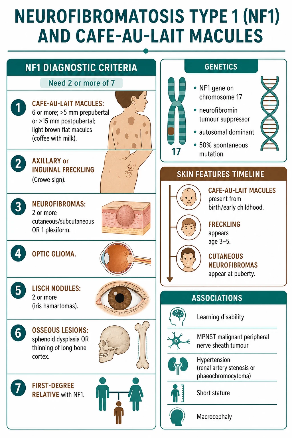
Café-au-Lait Macules (CALMs)
- Light brown ('coffee with milk'), flat, oval macules — evenly pigmented; smooth borders ('coast of California' — smooth, unlike McCune-Albright's irregular 'coast of Maine').[3]
- Present from birth or early childhood; increase in number and size during childhood.
- Distribution: trunk, extremities (not mucosa).
- Up to 3 CALMs can be found in ~10% of the normal population.
- ≥6 CALMs (>5 mm prepubertal or >15 mm postpubertal) = diagnostic criterion for NF1.[2]
NF1 Diagnostic Criteria (NIH — Revised 2021)
| # | Criterion | Description |
|---|---|---|
| 1 | ≥6 café-au-lait macules | >5 mm prepubertal; >15 mm postpubertal |
| 2 | Axillary/inguinal freckling (Crowe sign) | Small 1-3 mm pigmented macules in axillae, groin, base of neck; pathognomonic for NF1; appears age 3-5 |
| 3 | ≥2 neurofibromas (any type) or 1 plexiform | Cutaneous: soft, fleshy, pedunculated (buttonhole sign); plexiform: diffuse, congenital, bag of worms |
| 4 | Optic glioma | Optic pathway glioma (juvenile pilocytic astrocytoma); risk highest in young children |
| 5 | ≥2 Lisch nodules | Iris hamartomas (asymptomatic; detectable on slit-lamp) |
| 6 | Osseous lesion | Sphenoid wing dysplasia OR thinning of long bone cortex (congenital tibial pseudarthrosis) |
| 7 | First-degree relative with NF1 (by above criteria) | Parent, sibling, child |
Cutaneous Manifestations of NF1
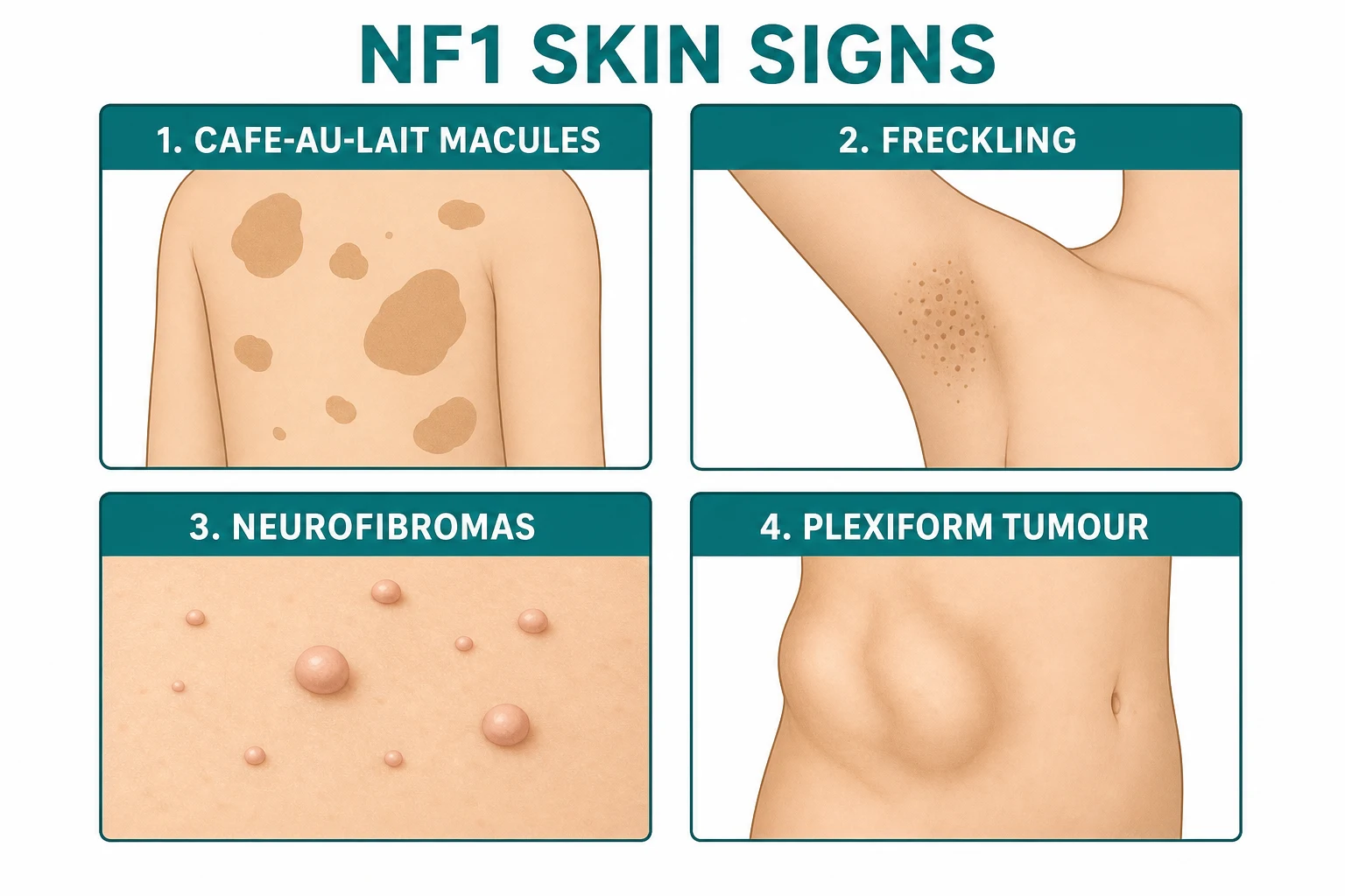
Café-au-lait macules
- Light brown, flat, oval, evenly pigmented; smooth borders (coast of California).
- Present from birth/early childhood.
Axillary/inguinal freckling (Crowe sign)
- Small (1-3 mm) pigmented macules in axillae, groin, base of neck, submammary.[3]
- Pathognomonic for NF1 (not seen in Legius syndrome or other mimics).
- Appears age 3-5.
Cutaneous neurofibromas
- Soft, fleshy, pedunculated or dome-shaped pink-skin coloured papules/nodules.[3]
- Buttonhole sign — invaginate through a buttonhole when pressed (the tumour herniates through the dermal defect).[3]
- Appear at puberty; increase in number and size with age and pregnancy.
- Benign (no malignant potential for cutaneous neurofibromas).
Plexiform neurofibroma
- Large, diffuse, congenital tumour along a nerve.[4]
- Feels like a 'bag of worms' on palpation.
- May cause overlying skin hypertrophy and hyperpigmentation.
- Risk of malignant transformation to MPNST (malignant peripheral nerve sheath tumour) — ~10% lifetime risk.[4]
- Surgical excision difficult (infiltrative along nerve trunks).
- Selumetinib (MEK inhibitor) — FDA-approved 2020 for symptomatic unresectable plexiform neurofibromas in children.[4]
Genetics
- NF1 gene on chromosome 17; encodes neurofibromin (tumour suppressor; negative regulator of RAS).[1]
- Autosomal dominant (50% transmission from affected parent).[1]
- ~50% spontaneous mutations (no family history).[1]
- Variable expressivity — affected parent may have mild disease but child may have severe.[1]
- 100% penetrance (all individuals with the mutation show some features by adulthood).
Quick numbers for the examiner
Differential Diagnosis of Café-au-Lait Macules [1]
[1]Quick numbers for the examiner
NEUROFIBROMATOSIS — NF1 features
Cutaneous (buttonhole sign), subcutaneous (firm nodules), plexiform (bag of worms, congenital, 5-10% MPNST)
Lisch: iris hamartomas (slit-lamp); OPG in 15-20% children, screen to age 8
Sphenoid wing dysplasia, long-bone pseudarthrosis (esp. tibia), scoliosis
50% de novo mutations; 100% penetrance by adulthood; variable expressivity
Annual visual assessment until age 8; then 18-monthly; urgent MRI if visual loss, proptosis, abnormal VEP
Axillary/inguinal freckling is pathognomonic; appears age 3-5; 90% of NF1 patients
8-13% MPNST lifetime (from plexiform); JMML (juvenile myelomonocytic leukaemia); GIST; pheochromocytoma
Hypertension common; renal artery stenosis, coarctation, pheochromocytoma; annual BP
50% learning disability; ADHD; macrocephaly; seizures; comprehensive multidisciplinary care
CALMs present at birth or first year; axillary freckling by age 5; neurofibromas at puberty
Annual surveillance; MEK inhibitors (selumetinib FDA 2020) for symptomatic inoperable plexiform NF; surgery
NF1 gene on 17q11.2; neurofibromin is a tumour suppressor; loss leads to RAS-MAPK activation
Selumetinib, trametinib; also JAK inhibitors under investigation
Screen for pheo in adult NF1 with hypertension: plasma metanephrines, MRI
Annual dermatology, ophthalmology, orthopaedics, neurology, cardiology, oncology review
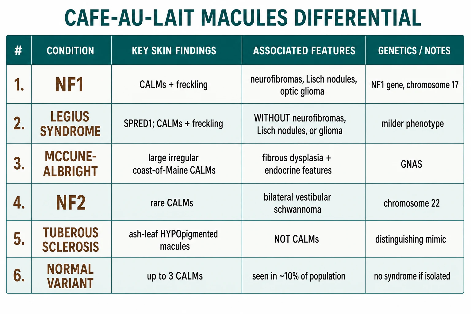
| Condition | Key distinguishing features |
|---|---|
| NF1 | CALMs + freckling + neurofibromas + Lisch nodules + optic glioma; NF1 gene chr 17 |
| Legius syndrome | CALMs + freckling WITHOUT neurofibromas, Lisch nodules, or optic glioma; SPRED1 gene; milder phenotype[2] |
| McCune-Albright syndrome | Large, unilateral CALMs with irregular borders ('coast of Maine'); polyostotic fibrous dysplasia; endocrine hyperfunction (precocious puberty); GNAS mosaic |
| NF2 | CALMs are rare (fewer and smaller than NF1); bilateral vestibular schwannoma is diagnostic; NF2 gene chr 22 (merlin) |
| Tuberous sclerosis | Ash-leaf macules are hypopigmented (not hyperpigmented like CALMs); TSC1/TSC2 |
| Normal | Up to 3 CALMs can be found in ~10% of normal individuals |
Systemic Features of NF1
NF1 is a multisystem disorder: almost every organ system is at risk and the systemic burden dominates long-term morbidity and mortality more than the cutaneous lesions do. Systemic features fall into seven domains, each requiring its own surveillance strategy.[5]
Neurological. Specific learning disability affects 40-60% of children with NF1 — the most common neurological morbidity. ADHD is present in ~30-40%, autism spectrum disorder in ~15-25%, and frank intellectual disability (IQ under 70) in ~5-8%. Macrocephaly (head circumference >97th centile) is seen in ~25%. Seizures occur in ~5-7%. Cerebral tumours include low-grade astrocytoma (especially optic pathway glioma, 15-20% of NF1 children) and, more rarely, high-grade glioma. Unidentified bright objects (UBOs) on T2/FLAIR MRI are seen in up to 70% of children — they are typically asymptomatic, may wax and wane, and do NOT require biopsy.[5]
Ophthalmological. Optic pathway glioma (OPG), usually a juvenile pilocytic astrocytoma, develops in 15-20% of NF1 children (vs 0.1% in the general population). It almost always presents before age 8 (median age 4-5 yr), is bilateral in ~50%, and is usually indolent; only ~30-50% of radiologically detected OPGs are symptomatic. Symptomatic OPG causes progressive visual loss, proptosis, optic atrophy, or precocious puberty (chiasmal involvement). Lisch nodules (iris hamartomas) are asymptomatic pigmented iris lesions on slit-lamp, present in >90% of adults with NF1 — useful discriminator from Legius syndrome. Other findings: optic atrophy, retinal hamartomas (rare).[3]
Cardiovascular. Hypertension affects 15-25% of NF1 patients — three principal mechanisms: (1) renal artery stenosis (fibromuscular dysplasia-like) is the most common paediatric cause; (2) phaeochromocytoma in 3-13% of adults; (3) coarctation of the aorta (rare). Cerebrovascular dysplasia (moya-moya-like arteriopathy) and spinal arteriovenous malformations may present with stroke or haemorrhage. Congenital heart disease (pulmonary stenosis, mitral valve prolapse) in 1-2%.[5]
Skeletal. Major orthopaedic manifestations: sphenoid wing dysplasia (a distinctive osseous criterion for NF1; may be associated with pulsatile exophthalmos); congenital tibial pseudarthrosis (1-4% of NF1 — bowed tibia at birth, often fractures); scoliosis (10-30% of NF1 patients; dystrophic type with rapid progression requiring fusion); spondylolisthesis; short stature (10-15%); macrocephaly; pectus excavatum; non-ossifying fibromas; bony overgrowth of a limb associated with plexiform NF.[5]
Endocrine. Precocious puberty (especially in OPG or hypothalamic involvement). Phaeochromocytoma — annual biochemical screen (plasma free metanephrines) in any adult NF1 with hypertension, episodic symptoms, or new-onset spells.[5]
Oncological — malignancy burden. MPNST (malignant peripheral nerve sheath tumour) is the leading cause of NF1-related death. Cumulative lifetime risk 8-13% in NF1 overall; 5-10% of plexiform neurofibromas undergo malignant transformation. Mean age at MPNST diagnosis is 26-30 years. Five-year survival is 35-50% (much worse than sporadic MPNST). JMML (juvenile myelomonocytic leukaemia) is rare but has a 200-500× increased incidence in NF1 children under 5 yr; presents with hepatosplenomegaly, monocytosis, thrombocytopenia. GIST (gastrointestinal stromal tumour) is increased 30-50× in NF1, typically multifocal small-bowel, KIT/PDGFRA-wildtype. Breast cancer risk is increased 3-5 fold in women under 50 with NF1. Rhabdomyosarcoma, neuroblastoma, phaeochromocytoma, and glomus tumours are also over-represented.[4][5]
Gastrointestinal. GIST, periampullary/peripancreatic neuroendocrine tumours, intestinal dysmotility from mesenteric plexiform NF.[5]
Management and Surveillance
Surveillance framework — what is done, when, and by whom
NF1 requires lifelong, multidisciplinary surveillance coordinated through a specialist NF1 clinic (dermatology, clinical genetics, paediatrics, neurology, ophthalmology, orthopaedics, oncology, psychology). The schedule below reflects the 2019 AAP Health Supervision for Children with Neurofibromatosis Type 1 statement and the 2021 international consensus criteria update.[7][2]
Children (birth to 18 yr) [1]
- At diagnosis: genetic counselling, baseline MRI brain/orbits with contrast, baseline ophthalmology, baseline audiology, developmental/behavioural assessment.[7]
- Annual: clinical examination including blood pressure (every visit, every age), height/weight/head circumference, full skin examination with documentation of plexiform neurofibromas, neurodevelopmental review.[3][7]
- Annual dermatology review: count and photograph CALMs, document size/number/distribution of neurofibromas, map plexiform NF burden.[3]
- Annual ophthalmology (visual acuity, colour vision, visual fields, fundoscopy) until age 8, then 18-monthly to age 18. This is the single most important OPG screening protocol. MRI brain/orbits is reserved for symptomatic patients (new visual loss, optic disc pallor, proptosis, abnormal visual-evoked potentials, new-onset nystagmus).[3][7]
- Orthopaedic review annually until skeletal maturity: clinical spine examination for scoliosis, tibial inspection for pseudarthrosis/dysplasia, sphenoid wing on imaging if symptomatic.[7]
- BP every visit with appropriate cuff size; if hypertensive — first-line imaging = renal artery Doppler/MR angiography + plasma free metanephrines to exclude phaeochromocytoma.[3][7]
- Developmental/behavioural screening at each well-child visit; formal psychometric testing at school entry and again at 11-12 yr.[5]
Adults (≥18 yr) [1]
- Annual: BP, full skin examination, brief neurological review, blood pressure and BMI, breast examination (women — NF1 raises breast cancer risk 3-5× under age 50; consider annual mammogram from age 30 with MRI adjunct).[5]
- Phaeochromocytoma screen (plasma free metanephrines or 24-h urine fractionated metanephrines): in any adult NF1 with hypertension, episodic spells, or unexplained tachycardia. Repeat every 1-3 years in asymptomatic patients with prior negative screen.[5]
- Plexiform neurofibroma surveillance: clinical examination every 6-12 months; MRI of any symptomatic or enlarging plexiform — new pain, rapid growth, change in texture, new neurological deficit = urgent MRI + PET-CT + biopsy to exclude MPNST.[4]
- Scoliosis/orthopaedic: as needed.
- Genetic counselling before pregnancy; prenatal diagnosis (chorionic villus sampling or non-invasive prenatal testing for the familial NF1 variant) is available; pre-implantation genetic diagnosis is an option.[1]
Plexiform neurofibroma and MPNST screening
Plexiform neurofibromas are present in 30-50% of NF1 patients, often congenital. They are infiltrative, often disfiguring, may compress adjacent organs (airway, spinal cord, bowel, bladder, orbit), and carry the lifetime malignant potential to MPNST. MPNST screening is therefore a core surveillance task:[4]
- Clinical red flags for malignant transformation: new or worsening pain (especially nocturnal or unremitting), rapid growth, new neurological deficit (motor, sensory, autonomic), change in consistency (firm-to-hard), new overlying skin ulceration or discolouration. Any one red flag = urgent MRI with contrast of the lesion + whole-body 18F-FDG PET-CT (SUVmax >3.5 raises MPNST suspicion) and image-guided core biopsy in an NF1 specialist centre.[4]
- Whole-body MRI (WB-MRI) is emerging as a surveillance tool for high-burden plexiform disease and asymptomatic MPNST detection in research protocols; not yet standard of care everywhere but increasingly used in tertiary NF1 centres.[4]
- Treatment of inoperable plexiform NF: selumetinib (Koselugo; MEK1/2 inhibitor) — the SPRINT phase II trial (Gross et al., NEJM 2020) showed 70% of children had ≥20% volume reduction and durable responses; FDA-approved April 2020 for symptomatic inoperable plexiform NF in children ≥2 yr; dose 25 mg/m² orally twice daily (about 50 mg/m²/day), continuously in 28-day cycles until progression or unacceptable toxicity. Common toxicities: acneiform rash, diarrhoea, nausea, CK elevation, asymptomatic LVEF reduction.[6][8] Trametinib, cobimetinib, and binimetinib are alternative MEK inhibitors under investigation; selumetinib is the only FDA-approved agent.[4][8]
- Surgery: complete excision of a plexiform is rarely feasible (infiltrative along nerve trunks); debulking or partial excision may relieve mass effect; amputation is reserved for severe limb plexiforms.[4]
- MPNST itself is treated with wide surgical excision + adjuvant radiotherapy; doxorubicin + ifosfamide is the standard first-line chemotherapy for unresectable/metastatic disease; overall 5-year survival 35-50% — early detection is the strongest prognostic factor.[4]
Pregnancy in NF1
Pregnancy may accelerate growth of cutaneous and plexiform neurofibromas (hormonal effect). Specific risks: rapid growth of pre-existing plexiform, new hypertension (including phaeochromocytoma unmasked), cerebrovascular complications, increased caesarean rate due to pelvic/sacral neurofibromas. Pre-conception genetic counselling; multidisciplinary antenatal care; post-partum re-evaluation of neurofibromas.[5]
Subtypes of NF1 and Related RASopathies
NF1 is part of the RASopathy family (germline disorders of the RAS-MAPK pathway). Recognition of subtypes informs prognosis, surveillance, and reproductive counselling.[1][2]
Generalised NF1
The classic phenotype described above: bilateral CALMs + freckling + cutaneous/subcutaneous/plexiform neurofibromas + Lisch nodules +/− optic glioma +/− bone dysplasia. Autosomal dominant NF1 mutation in every cell; 50% de novo, 50% inherited.
Segmental NF1 (mosaic NF1)
A post-zygotic NF1 mutation in one embryonic lineage produces disease confined to a body region (often Blaschkoid/dermatomal distribution). Clinical features (CALMs, freckling, neurofibromas) are restricted to that region; contralateral side and systemic involvement may be absent or minimal. MPNST risk appears lower than generalised NF1 but is not zero. Gonadal mosaicism is the critical reproductive concern — a segmental NF1 patient can still transmit a full generalised NF1 to offspring if the gonad harbours the mutation; pre-conception genetic counselling is mandatory.[3]
Spinal NF1
Dumbbell neurofibromas arising from spinal nerve roots within the spinal canal +/− paraspinal extension. May present with radiculopathy, myelopathy, or progressive scoliosis. MRI of the entire neuraxis is required in NF1 patients with scoliosis, focal neurological deficit, or unexplained pain; surgical decompression may be required for symptomatic lesions.[5]
Optic pathway glioma (OPG)
Already detailed above — see Systemic Features. 15-20% of NF1 children; symptomatic in 30-50% of those. Treatment of symptomatic/progressive OPG is first-line carboplatin + vincristine (or vinblastine monotherapy); second-line thioguanine + procarbazine + CCNU/lomustine (TPC) or selumetinib. Avoid radiotherapy in children under 5 yr because of secondary tumour risk.[5]
MPNST (malignant peripheral nerve sheath tumour)
The principal lethal tumour in NF1. 8-13% lifetime risk; 5-10% of plexiform neurofibromas transform. Five-year survival 35-50%. Aggressive; presents with new pain, rapid growth, neurological deficit. Wide surgical excision + adjuvant radiotherapy ± doxorubicin/ifosfamide chemotherapy is standard.[4]
GIST (gastrointestinal stromal tumour)
30-50× increased incidence in NF1. Usually multifocal, small-bowel, KIT/PDGFRA-wildtype (imatinib-resistant). Presents with GI bleeding, anaemia, or abdominal pain; resection is curative for localised disease.[5]
JMML (juvenile myelomonocytic leukaemia)
200-500× increased risk in NF1 children, especially under 5 yr. Fatal without allogeneic HSCT (5-year survival ~50% post-HSCT). Presents with hepatosplenomegaly, monocytosis, thrombocytopenia, elevated HbF. Spontaneous resolution in ~10% (Noonan-syndrome-type cases more often than NF1).[5]
Phaeochromocytoma / paraganglioma
3-13% of adult NF1 patients. Screen annually with plasma free metanephrines in any NF1 adult with hypertension or symptoms; image with MRI abdomen / MIBG if positive.[5]
Learning disability and neurodevelopmental profile
Specific learning disability (esp. visuospatial, executive function) in 40-60%; ADHD 30-40%; autism spectrum disorder 15-25%; IQ generally 5-10 points below siblings. Multidisciplinary support with educational psychology, speech therapy, occupational therapy, and behavioural intervention is the mainstay.[5]
Scoliosis
10-30% of NF1 patients. Two patterns: (1) idiopathic-like (most common, mild, adolescent onset, brace-responsive); (2) dystrophic (early-onset, sharp angulation, rapid progression, often associated with underlying plexiform NF or vertebral dysplasia; requires posterior spinal fusion ± growing-rod construct).[5]
Congenital pseudarthrosis of the tibia (CPT)
Bowed tibia at birth, often fractures within the first year. Strong NF1 association (40-80% of CPT cases have NF1). Treatment: intramedullary rodding ± bone grafting ± BMP; amputation for refractory cases. Sphenoid wing dysplasia and long-bone pseudarthrosis are the distinctive osseous criteria in the NIH list.[5]
Related RASopathies (for completeness)
- Legius syndrome (SPRED1): CALMs +/− freckling; NO neurofibromas, Lisch, OPG; benign course.[2]
- Noonan syndrome (PTPN11, SOS1, RAF1, KRAS): short stature, webbed neck, pulmonary stenosis, bleeding diathesis.
- Cardiofaciocutaneous syndrome (BRAF, MAP2K1, MAP2K2, KRAS): facial dysmorphism, ectodermal abnormalities, cardiac defects.
- Costello syndrome (HRAS): coarse facies, papillomata, cardiac hypertrophy, malignancy risk.
- Watson syndrome: NF1 + pulmonary stenosis + café-au-lait; historically considered allelic.
Diagnostic Criteria Detail — 2021 International Consensus Update
The 2021 international consensus recommendation (Legius E et al., Genetics in Medicine 2021) retained the NIH 1988 framework but clarified and modified several criteria. The key changes are:[2]
- CALMs remain criterion 1. Pre-pubertal cut-off is now diameter >5 mm in greatest measurement; post-pubertal >15 mm. The number criterion remains ≥6 macules. The previous text "pre-pubertal ≥5 mm" was re-emphasised to discourage under-call in children with smaller but numerous macules (e.g. 6 macules of 4 mm still do NOT qualify).
- Axillary and inguinal freckling (Crowe sign) is confirmed as criterion 2. The 2021 panel clarified that freckling must be bilateral OR — if unilateral — associated with another diagnostic feature to count as criterion 2.
- Neurofibromas (criterion 3) include cutaneous, subcutaneous, and plexiform; ≥2 of any type OR one plexiform suffices. The 2021 panel specifically notes that "one plexiform neurofibroma" alone is sufficient — this was a frequent source of misclassification.
- Optic pathway glioma (criterion 4) must be confirmed on MRI or by an ophthalmologist; asymptomatic lesions picked up on screening imaging count if formally documented.
- Lisch nodules (criterion 5): ≥2 confirmed on slit-lamp examination by an ophthalmologist. The 2021 panel removed the older alternative of ≥2 choroidal abnormalities detected on infrared imaging — these are not yet a formal criterion.
- Distinctive osseous lesion (criterion 6): sphenoid wing dysplasia, long-bone pseudarthrosis (tibial), or thinning of long-bone cortex. Plain radiographs or CT are sufficient; MRI not required.
- First-degree relative with NF1 (criterion 7): parent, sibling, or child with NF1 by the same criteria. [1]
Legius syndrome (SPRED1) is included in the 2021 differential for the first time — patients with CALMs + freckling but without neurofibromas, Lisch nodules, or optic glioma should be tested for SPRED1 mutation before concluding NF1, because Legius syndrome carries a much better prognosis (no MPNST risk) and a different surveillance pathway.[2]
A parent with NF1 confirmed by these criteria can establish criterion 7 in a child without further work-up; conversely, a child of an unaffected parent needs ≥1 criterion of their own plus a second feature to make the diagnosis. By age 8, ~97% of children who will meet NF1 criteria already do so; by age 20, virtually 100%. Under 5% of NF1 patients are mosaic and may not meet full criteria — molecular genetic testing is then required.[2]
Genotype-phenotype correlations: large 17q11.2 microdeletions (encompassing the NF1 gene plus neighbouring genes) are associated with a more severe phenotype — earlier and more numerous neurofibromas, higher MPNST risk, learning disability, dysmorphic features, and cardiovascular anomalies. Specific missense variants (e.g. p.Arg1809Cys, p.Met992del) produce milder, CALM-predominant phenotypes. NF1 genetic testing is offered when clinical diagnosis is uncertain, for prenatal/pre-implantation diagnosis, or to confirm mosaicism. Testing includes sequencing (≥95% sensitivity for point mutations) and copy-number analysis (MLPA or chromosomal microarray for the 5% who have whole-gene deletions).[1][2]
Pathophysiology — RAS-MAPK Pathway and Therapeutic Targeting

Neurofibromin is a large (~2,818 amino acid) cytoplasmic protein encoded by the NF1 gene (chromosome 17q11.2). Its central domain acts as a RAS-GTPase-activating protein (RAS-GAP) — it accelerates conversion of active RAS-GTP to inactive RAS-GDP. Loss-of-function mutations (most are truncating; >3,000 different NF1 variants have been catalogued) leave RAS stuck in the active GTP-bound state, producing constitutive activation of the RAF-MEK-ERK (MAPK) and PI3K-AKT-mTOR pathways.[1]
Cellular consequences. Sustained RAS-MAPK signalling drives proliferation of neural-crest-derived cells in particular: Schwann cells (neurofibromas, MPNST), melanocytes (CALMs, axillary freckling), and certain neuronal/glial precursors. Mast cells, recruited by stem cell factor (SCF) signalling, infiltrate neurofibromas and contribute to tumour growth and pruritus. Second-hit (Knudson) somatic loss of the remaining NF1 allele is required for tumour formation in NF1 haploinsufficient cells — this explains the focal, multifocal nature of neurofibromas.[1][4]
Therapeutic translation. The MAPK pathway is druggable: MEK inhibitors (selumetinib, trametinib, cobimetinib, binimetinib) suppress ERK phosphorylation downstream of RAS and shrink plexiform neurofibromas. Selumetinib is the only FDA/EMA-approved agent for inoperable plexiform NF in children. mTOR inhibitors (sirolimus, everolimus) reduce subcutaneous neurofibroma burden in preclinical models but clinical efficacy is modest. Cabozantinib (multi-kinase TKI) is under investigation for MPNST. MEK + mTOR combination trials are underway to address resistance.[4][8]

MPNST RED FLAGS — investigate urgently
New, worsening, nocturnal, or unremitting pain in a known plexiform neurofibroma — the most sensitive single feature of malignant transformation. Urgent MRI + PET-CT.
Rapid enlargement of a previously stable plexiform — measure dimensions and compare to prior imaging. ≥20% volume increase in 6-12 months raises suspicion.
New motor, sensory, or autonomic deficit in the territory of a plexiform — suggests nerve infiltration by MPNST. MRI of the plexus + chest/abdominal imaging.
Plexiform becomes firm or hard (previously soft/rubbery); fixed to deep structures rather than mobile. Palpate serially at every clinic visit.
New overlying skin discolouration, ulceration, telangiectasia, or warmth. Inflammation may reflect tumour necrosis or invasion.
Weight loss, night sweats, persistent low-grade fever, rising inflammatory markers (CRP, ESR). Exclude MPNST and JMML in children.
Exam Pearls
MPNST RED FLAGS — investigate urgently (acronym: P-A-N-C-E-U)
PANCE-U
New, worsening, nocturnal, or unremitting pain in a known plexiform neurofibroma — the most sensitive single feature of malignant transformation. Urgent MRI + PET-CT.
Rapid enlargement of a previously stable plexiform — measure dimensions and compare to prior imaging. ≥20% volume increase in 6-12 months raises suspicion.
New motor, sensory, or autonomic deficit in the territory of a plexiform — suggests nerve infiltration by MPNST. MRI of the plexus + chest/abdominal imaging.
Plexiform becomes firm or hard (previously soft/rubbery); fixed to deep structures rather than mobile. Palpate serially at every clinic visit.
New overlying skin discolouration, ulceration, telangiectasia, or warmth. Inflammation may reflect tumour necrosis or invasion.
Weight loss, night sweats, persistent low-grade fever, rising inflammatory markers (CRP, ESR). Exclude MPNST and JMML in children.
Exam application bank (NEET-PG / INICET)
One-line answer
Neurofibromatosis type 1 (NF1) is an autosomal dominant neurocutaneous disorder caused by mutations in the NF1 gene on chromosome 17, characterised by cafe-au-lait macules, neurofibromas, Lisch nodules, and systemic manifestations including optic pathway glioma, skeletal dysplasia, and learning disability. Diagnosis requires 2 or more NIH criteria.
Worked stems (answer without another resource)
Stem 1 — Classic presentation. Map symptoms to mechanism; name the first investigation and first treatment step with dose/route if drug therapy is standard. [1]
Stem 2 — Unstable / complicated. List red flags that force immediate resuscitation, theatre, ICU, antidote, or reperfusion — and what you do in the first 15 minutes. [1]
Stem 3 — Atypical group. Elderly, pregnancy, child, or immunocompromised: how presentation and thresholds change. [1]
Stem 4 — Differential trap. Name the three closest mimics and one discriminator for each. [1]
Stem 5 — Disposition. Who goes home with safety-netting, who is admitted, who needs HDU/ICU/theatre, and what follow-up is mandatory. [1]
Rapid viva checklist
- Definition + classification
- Pathophysiology chain
- Bedside signs / criteria
- Score with exact components (if any)
- Emergency bundle
- Definitive therapy with doses
- Complications of disease and of treatment
- Special populations
- Guideline/trial name if classic
- Three exam traps
Coverage self-check
If you cannot answer any stem above from this page alone, re-read the matching section — the page is intended to be self-sufficient for final-prof and NEET-PG/INICET questions on Café-au-lait macules and neurofibromatosis type 1.
References
- [1]Tamura R. Current Understanding of Neurofibromatosis Type 1, 2, and Schwannomatosis Int J Mol Sci, 2021.PMID 34072574
- [2]Legius E, Messiaen L, Wolkenstein P, et al. Revised diagnostic criteria for neurofibromatosis type 1 and Legius syndrome: an international consensus recommendation Genet Med, 2021.PMID 34012067
- [3]Ly KI, Blakeley JO. The Diagnosis and Management of Neurofibromatosis Type 1 Med Clin North Am, 2019.PMID 31582003
- [4]Fisher MJ, Blakeley JO, Weiss BD, et al. Management of neurofibromatosis type 1-associated plexiform neurofibromas Neuro Oncol, 2022.PMID 35657359
- [5]Saleh M, Dib A, Beaini S, et al. Neurofibromatosis type 1 system-based manifestations and treatments: a review Neurol Sci, 2023.PMID 36826455
- [6]Gross AM, Wolters PL, Dombi E, et al. Selumetinib in Children with Inoperable Plexiform Neurofibromas N Engl J Med, 2020.PMID 32187457
- [7]Miller DT; Committee on Genetics. Health Supervision for Children With Neurofibromatosis Type 1 Pediatrics, 2019.PMID 31010905
- [8]Anderson MK, Gjerris M, Tarp CB, et al. A Review of Selumetinib in the Treatment of Neurofibromatosis Type 1-Related Plexiform Neurofibromas Ann Pharmacother, 2022.PMID 34541874